Biatrial Inflammatory and Fibrotic Remodeling in Severe Aortic Stenosis Compared with CABG Controls


 ,
,  , , , and
Highlights
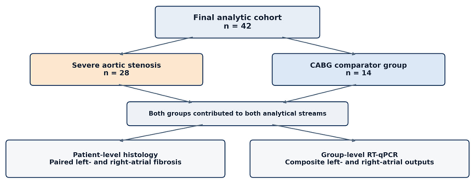
- Building on extensive evidence that aortic stenosis is a whole-heart fibro-remodeling disease, this study provides complementary direct human biatrial tissue evidence: severe aortic stenosis was associated with greater patient-level left- and right-atrial fibrosis than CABG controls without significant valvular disease.
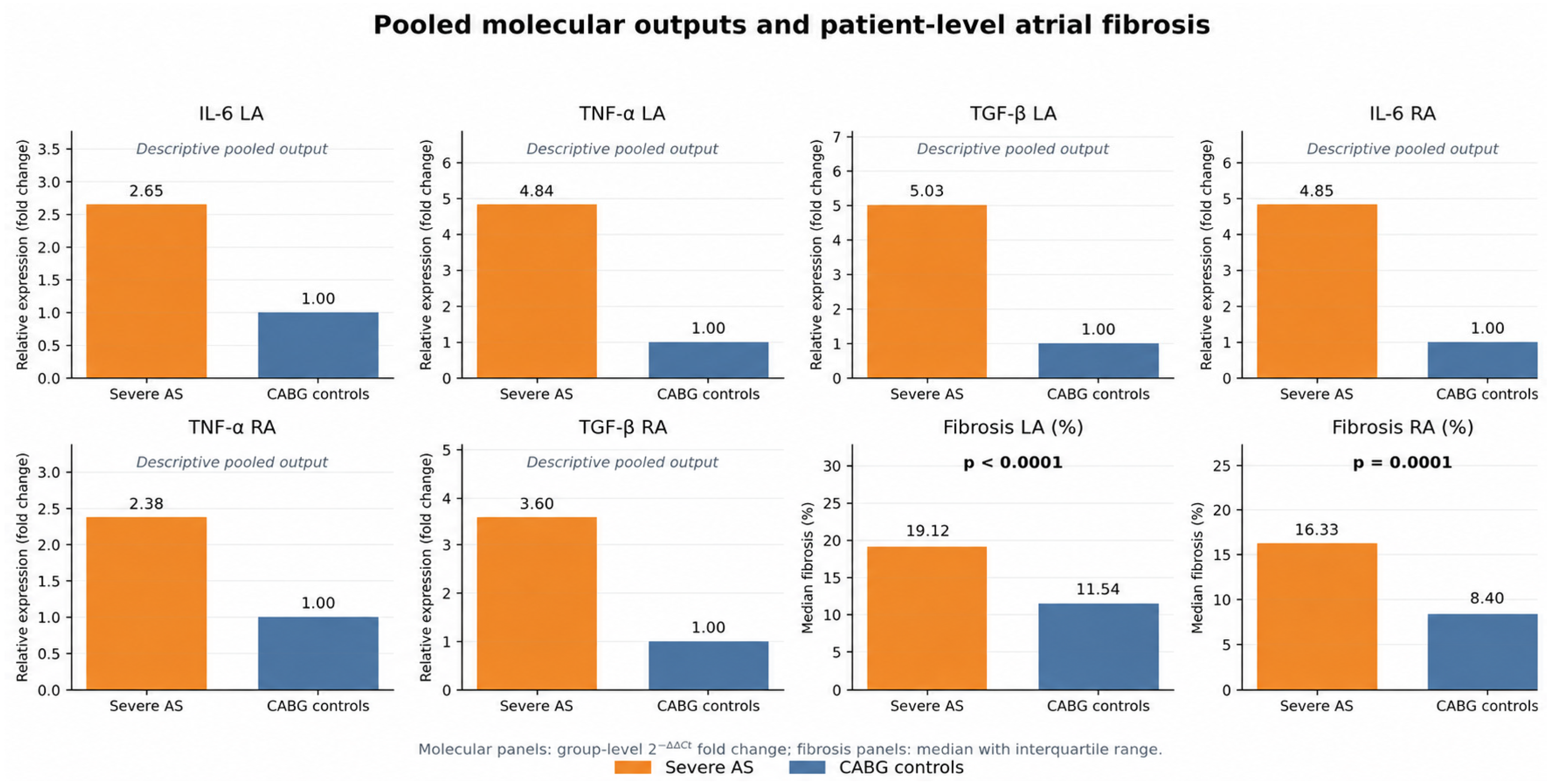
- Pooled RT-qPCR outputs descriptively suggested a biatrial inflammatory/profibrotic profile involving IL-6, TNF-alpha, and TGF-beta; no inferential p values are reported for these group-level molecular endpoints.
- The findings complement the established ventricular fibrosis literature by extending direct tissue characterization to both atria rather than proposing fibrosis as a newly recognized aortic stenosis mechanism.
- Patient-level molecular analyses, formal laboratory reproducibility studies, and advanced atrial functional imaging are required to quantify individual mechanistic relationships.
Abstract
1. Introduction
2. Materials and Methods
2.1. Study Design and Patient Population
2.2. Tissue Sampling and Processing
2.3. RT-qPCR Analysis
2.4. Histology and Fibrosis Quantification
2.5. Clinical, Laboratory, and Echocardiographic Assessment
2.6. Ethics Statement
2.7. Statistical Analysis
3. Results
3.1. Clinical Characteristics and Treatment Profile
3.2. Echocardiographic Findings
3.3. Tissue Biomarker Expression and Atrial Fibrosis
3.4. Correlation Analysis
3.5. Exploratory Regression Analysis
4. Discussion
5. Conclusions
Author Contributions
Funding
Institutional Review Board Statement
Informed Consent Statement
Data Availability Statement
Acknowledgments
Conflicts of Interest
References
- Vahanian, A.; Beyersdorf, F.; Praz, F.; Milojevic, M.; Baldus, S.; Bauersachs, J.; Capodanno, D.; Conradi, L.; De Bonis, M.; De Paulis, R.; et al. 2021 ESC/EACTS Guidelines for the management of valvular heart disease. Eur. Heart J. 2022, 43, 561–632. [Google Scholar] [CrossRef] [Scilit] [PubMed]
- Rana, M. Aortic Valve Stenosis: Diagnostic Approaches and Recommendations of the 2021 ESC/EACTS Guidelines for the Management of Valvular Heart Disease—A Review of the Literature. Cardiol. Cardiovasc. Med. 2022, 6, 315–324. [Google Scholar] [CrossRef] [Scilit] [PubMed]
- Otto, C.M.; Prendergast, B. Aortic-valve stenosis—From patients at risk to severe valve obstruction. N. Engl. J. Med. 2014, 371, 744–756. [Google Scholar] [CrossRef] [Scilit] [PubMed]
- Dweck, M.R.; Boon, N.A.; Newby, D.E. Calcific aortic stenosis: A disease of the valve and the myocardium. J. Am. Coll. Cardiol. 2012, 60, 1854–1863. [Google Scholar] [CrossRef] [Scilit] [PubMed]
- Lindman, B.R.; Clavel, M.A.; Mathieu, P.; Iung, B.; Lancellotti, P.; Otto, C.M.; Pibarot, P. Calcific aortic stenosis. Nat. Rev. Dis. Prim. 2016, 2, 16006. [Google Scholar] [CrossRef] [Scilit] [PubMed]
- Pawade, T.A.; Newby, D.E.; Dweck, M.R. Calcification in aortic stenosis: The skeleton key. J. Am. Coll. Cardiol. 2015, 66, 561–577. [Google Scholar] [CrossRef] [Scilit] [PubMed]
- O’Brien, K.D. Pathogenesis of calcific aortic valve disease: A disease process comes of age (and a good deal more). Arterioscler. Thromb. Vasc. Biol. 2006, 26, 1721–1728. [Google Scholar] [CrossRef] [Scilit] [PubMed]
- Rajamannan, N.M.; Evans, F.J.; Aikawa, E.; Grande-Allen, K.J.; Demer, L.L.; Heistad, D.D.; Simmons, C.A.; Masters, K.S.; Mathieu, P.; O’BRien, K.D.; et al. Calcific aortic valve disease: Not simply a degenerative process: A review and agenda for research from the National Heart, Lung, and Blood Institute Aortic Stenosis Working Group. Circulation 2011, 124, 1783–1791. [Google Scholar] [CrossRef] [Scilit] [PubMed]
- Yutzey, K.E.; Demer, L.L.; Body, S.C.; Huggins, G.S.; Towler, D.A.; Giachelli, C.M.; Hofmann-Bowman, M.A.; Mortlock, D.P.; Rogers, M.B.; Sadeghi, M.M.; et al. Calcific aortic valve disease: A consensus summary from the Alliance of Investigators on Calcific Aortic Valve Disease. Arterioscler. Thromb. Vasc. Biol. 2014, 34, 2387–2393. [Google Scholar] [CrossRef] [Scilit] [PubMed]
- Everett, R.J.; Tastet, L.; Clavel, M.A.; Chin, C.W.; Capoulade, R.; Vassiliou, V.S.; Kwiecinski, J.; Gomez, M.; van Beek, E.J.; White, A.C.; et al. Progression of Hypertrophy and Myocardial Fibrosis in Aortic Stenosis: A Multicenter Cardiac Magnetic Resonance Study. Circ. Cardiovasc. Imaging 2018, 11, e007451. [Google Scholar] [CrossRef] [Scilit] [PubMed]
- Chin, C.W.L.; Everett, R.J.; Kwiecinski, J.; Vesey, A.T.; Yeung, E.; Esson, G.; Jenkins, W.; Koo, M.; Mirsadraee, S.; White, A.C.; et al. Myocardial Fibrosis and Cardiac Decompensation in Aortic Stenosis. JACC Cardiovasc. Imaging 2017, 10, 1320–1333. [Google Scholar] [CrossRef] [Scilit] [PubMed]
- Weidemann, F.; Herrmann, S.; Störk, S.; Niemann, M.; Frantz, S.; Lange, V.; Beer, M.; GattenlöhNer, S.; Voelker, W.; Ertl, G.; et al. Impact of myocardial fibrosis in patients with symptomatic severe aortic stenosis. Circulation 2009, 120, 577–584. [Google Scholar] [CrossRef] [Scilit] [PubMed]
- Azevedo, C.F.; Nigri, M.; Higuchi, M.L.; Pomerantzeff, P.M.; Spina, G.S.; Sampaio, R.O.; Tarasoutchi, F.; Grinberg, M.; Rochitte, C.E. Prognostic significance of myocardial fibrosis quantification by histopathology and magnetic resonance imaging in patients with severe aortic valve disease. J. Am. Coll. Cardiol. 2010, 56, 278–287. [Google Scholar] [CrossRef] [Scilit] [PubMed]
- Dweck, M.R.; Joshi, S.; Murigu, T.; Alpendurada, F.; Jabbour, A.; Melina, G.; Banya, W.; Gulati, A.; Roussin, I.; Raza, S.; et al. Midwall fibrosis is an independent predictor of mortality in patients with aortic stenosis. J. Am. Coll. Cardiol. 2011, 58, 1271–1279. [Google Scholar] [CrossRef] [Scilit] [PubMed]
- Milano, A.D.; Faggian, G.; Dodonov, M.; Golia, G.; Tomezzoli, A.; Bortolotti, U.; Mazzucco, A. Prognostic value of myocardial fibrosis in patients with severe aortic valve stenosis. J. Thorac. Cardiovasc. Surg. 2012, 144, 830–837. [Google Scholar] [CrossRef] [Scilit] [PubMed]
- Treibel, T.A.; Lopez, B.; Gonzalez, A.; Menacho, K.; Schofield, R.S.; Ravassa, S.; Fontana, M.; White, S.K.; DiSalvo, C.; Roberts, N.; et al. Reappraising myocardial fibrosis in severe aortic stenosis: An invasive and non-invasive study in 133 patients. Eur. Heart J. 2018, 39, 699–709. [Google Scholar] [CrossRef] [Scilit] [PubMed]
- Everett, R.J.; Treibel, T.A.; Fukui, M.; Lee, H.; Rigolli, M.; Singh, A.; Bijsterveld, P.; Tastet, L.; Al Musa, T.; Dobson, L.; et al. Extracellular Myocardial Volume in Patients With Aortic Stenosis. J. Am. Coll. Cardiol. 2020, 75, 304–316. [Google Scholar] [CrossRef] [Scilit] [PubMed]
- Bing, R.; Cavalcante, J.L.; Everett, R.J.; Clavel, M.A.; Newby, D.E.; Dweck, M.R. Imaging and Impact of Myocardial Fibrosis in Aortic Stenosis. JACC Cardiovasc. Imaging 2019, 12, 283–296. [Google Scholar] [CrossRef] [Scilit] [PubMed]
- Elmariah, S.; O’Brien, K.D.; Otto, C.M. Patterns of left ventricular remodeling in aortic stenosis: Therapeutic implications. Curr. Treat. Options Cardiovasc. Med. 2015, 17, 391. [Google Scholar] [CrossRef] [Scilit] [PubMed]
- Lisi, M.; Pastore, M.C.; Fiorio, A.; Cameli, M.; Mandoli, G.E.; Righini, F.M.; Cavigli, L.; D’Ascenzi, F.; Focardi, M.; Rubboli, A.; et al. Left Atrial Remodeling in Response to Aortic Valve Replacement: Pathophysiology and Myocardial Strain Analysis. Life 2022, 12, 2074. [Google Scholar] [CrossRef] [Scilit] [PubMed]
- Kristensen, C.B.; Jensen, J.S.; Søgaard, P.; Carstensen, H.G.; Mogelvang, R. Atrial fibrillation in aortic stenosis--echocardiographic assessment and prognostic importance. Cardiovasc. Ultrasound 2012, 10, 38. [Google Scholar] [CrossRef] [Scilit] [PubMed]
- Fragão-Marques, M.; Miranda, I.; Martins, D.; Barroso, I.; Mendes, C.; Pereira-Neves, A.; Falcão-Pires, I.; Leite-Moreira, A. Atrial matrix remodeling in atrial fibrillation patients with aortic stenosis. BMC Cardiovasc. Disord. 2020, 20, 468. [Google Scholar] [CrossRef] [Scilit] [PubMed]
- Saeed, S.; Rajani, R.; Tadic, M.; Parkin, D.; Chambers, J.B. Left atrial volume index predicts adverse events in asymptomatic moderate or severe aortic stenosis. Echocardiography 2021, 38, 1893–1899. [Google Scholar] [CrossRef] [Scilit] [PubMed]
- Burstein, B.; Nattel, S. Atrial fibrosis: Mechanisms and clinical relevance in atrial fibrillation. J. Am. Coll. Cardiol. 2008, 51, 802–809. [Google Scholar] [CrossRef] [Scilit] [PubMed]
- Nattel, S.; Burstein, B.; Dobrev, D. Atrial remodeling and atrial fibrillation: Mechanisms and implications. Circ. Arrhythm. Electrophysiol. 2008, 1, 62–73. [Google Scholar] [CrossRef] [Scilit] [PubMed]
- Dzeshka, M.S.; Lip, G.Y.H.; Snezhitskiy, V.; Shantsila, E. Cardiac Fibrosis in Patients With Atrial Fibrillation: Mechanisms and Clinical Implications. J. Am. Coll. Cardiol. 2015, 66, 943–959. [Google Scholar] [CrossRef] [Scilit] [PubMed]
- Hu, Y.F.; Chen, Y.J.; Lin, Y.J.; Chen, S.A. Inflammation and the pathogenesis of atrial fibrillation. Nat. Rev. Cardiol. 2015, 12, 230–243. [Google Scholar] [CrossRef] [Scilit] [PubMed]
- Nso, N.; Bookani, K.R.; Metzl, M.; Radparvar, F. Role of inflammation in atrial fibrillation: A comprehensive review of current knowledge. J. Arrhythm. 2021, 37, 1–10. [Google Scholar] [CrossRef] [Scilit] [PubMed]
- Ihara, K.; Sasano, T. Role of Inflammation in the Pathogenesis of Atrial Fibrillation. Front. Physiol. 2022, 13, 862164. [Google Scholar] [CrossRef] [Scilit] [PubMed]
- Rusinaru, D.; Bohbot, Y.; Kowalski, C.; Ringle, A.; Maréchaux, S.; Tribouilloy, C. Left Atrial Volume and Mortality in Patients With Aortic Stenosis. J. Am. Heart Assoc. 2017, 6, e006615. [Google Scholar] [CrossRef] [Scilit] [PubMed]
- Weber, J.; Bond, K.; Flanagan, J.; Passick, M.; Petillo, F.; Pollack, S.; Robinson, N.; Petrossian, G.; Cao, J.J.; Barasch, E. The Prognostic Value of Left Atrial Global Longitudinal Strain and Left Atrial Phasic Volumes in Patients Undergoing Transcatheter Valve Implantation for Severe Aortic Stenosis. Cardiology 2021, 146, 489–500. [Google Scholar] [CrossRef] [Scilit] [PubMed]
- Tan, E.S.J.; Jin, X.; Oon, Y.Y.; Chan, S.P.; Gong, L.; Lunaria, J.B.; Liew, O.-W.; Chong, J.P.-C.; Tay, E.L.; Soo, W.M.; et al. Prognostic Value of Left Atrial Strain in Aortic Stenosis: A Competing Risk Analysis. J. Am. Soc. Echocardiogr. 2023, 36, 29–37.e5. [Google Scholar] [CrossRef] [Scilit] [PubMed]
- Baumgartner, H.; Hung, J.; Bermejo, J.; Chambers, J.B.; Edvardsen, T.; Goldstein, S.; Lancellotti, P.; LeFevre, M.; Miller, F.; Otto, C.M. Recommendations on the echocardiographic assessment of aortic valve stenosis: A focused update from the European Association of Cardiovascular Imaging and the American Society of Echocardiography. Eur. Heart J. Cardiovasc. Imaging 2017, 18, 254–275. [Google Scholar] [CrossRef] [Scilit] [PubMed]
- Baumgartner, H.; Hung, J.; Bermejo, J.; Chambers, J.B.; Evangelista, A.; Griffin, B.P.; Iung, B.; Otto, C.M.; Pellikka, P.A.; Quiñones, M. Echocardiographic assessment of valve stenosis: EAE/ASE recommendations for clinical practice. J. Am. Soc. Echocardiogr. 2009, 22, 1–23. [Google Scholar] [CrossRef] [Scilit] [PubMed]
- Rosca, C.I.; Lighezan, D.F.; Georgescu, D.; Branea, H.S.; Kundnani, N.R.; Nicoras, A.V.; Bita, R.G.; Nisulescu, D.D. Triglyceride-to-HDL Cholesterol Ratio Is Associated with Ischemic Stroke Risk in Patients-With Paroxysmal Atrial Fibrillation. Metabolites 2026, 16, 110. [Google Scholar] [CrossRef] [Scilit] [PubMed]
- Scisciola, L.; Paolisso, P.; Belmonte, M.; Gallinoro, E.; Delrue, L.; Taktaz, F.; Fontanella, R.A.; Degrieck, I.; Pesapane, A.; Casselman, F.; et al. Myocardial sodium-glucose cotransporter 2 expression and cardiac remodelling in patients with severe aortic stenosis: The BIO-AS study. Eur. J. Heart Fail. 2024, 26, 471–482. [Google Scholar] [CrossRef] [Scilit] [PubMed]
- Sastre-Oliva, T.; Corbacho-Alonso, N.; Rodriguez-Sanchez, E.; Mercado-García, E.; Perales-Sanchez, I.; Hernandez-Fernandez, G.; Juarez-Alia, C.; Tejerina, T.; López-Almodóvar, L.F.; Padial, L.R.; et al. Albumin Redox Modifications Promote Cell Calcification Reflecting the Impact of Oxidative Status on Aortic Valve Disease and Atherosclerosis. Antioxidants 2024, 13, 108. [Google Scholar] [CrossRef] [Scilit] [PubMed]
- Koschutnik, M.; Brunner, C.; Nitsche, C.; Donà, C.; Dannenberg, V.; Halavina, K.; Koschatko, S.; Jantsch, C.; Mascherbauer, K.; Kronberger, C.; et al. Biomarkers of Inflammation and Association with Cardiovascular Magnetic Resonance Imaging for Risk Stratification and Outcome in Patients with Severe Aortic Stenosis. J. Clin. Med. 2025, 14, 2512. [Google Scholar] [CrossRef] [Scilit] [PubMed]
- Houessou, U.; Zamani, P.; Manikpurage, H.D.; Li, Z.; Gaudreault, N.; Dahmene, M.; Dagenais, F.; Couture, C.; Clavel, M.-A.; Pibarot, P.; et al. Transcriptomic Signatures of Calcific Aortic Valve Stenosis Severity in Human Tricuspid and Bicuspid Aortic Valves. JACC Basic Transl. Sci. 2025, 10, 724–741. [Google Scholar] [CrossRef] [Scilit] [PubMed]
- Small, A.M.; Yang, T.Y.; Itoh, S.; Thériault, S.; Dufresne, L.; Kurosawa, R.; Komuro, I.; Matsuda, K.; Vy, H.M.T.; Farber-Eger, E.H.; et al. Genomic and transcriptomic analyses of aortic stenosis enhance therapeutic target discovery and disease prediction. Nat. Genet. 2026, 58, 57–66. [Google Scholar] [CrossRef] [Scilit] [PubMed]



| Variable | Severe AS (n = 28) | Control (n = 14) | p Value |
|---|---|---|---|
| Age, years | 64.6 ± 8.7 | 62.4 ± 7.0 | 0.3812 |
| Male sex | 18 (64.3%) | 11 (78.6%) | 0.4852 |
| Urban residence | 15 (53.6%) | 7 (50.0%) | 1.0000 |
| Persistent atrial fibrillation | 4 (14.3%) | 1 (7.1%) | 0.6496 |
| Postoperative atrial fibrillation | 5 (17.9%) | 4 (28.6%) | 0.4508 |
| Mixed dyslipidemia | 19 (67.9%) | 13 (92.9%) | 0.1249 |
| Hypertension | 27 (96.4%) | 13 (92.9%) | 1.0000 |
| Hypertension grade 1 | 0 (0.0%) | 1 (7.1%) | 0.3333 |
| Hypertension grade 2 | 23 (82.1%) | 11 (78.6%) | 1.0000 |
| Hypertension grade 3 | 5 (17.9%) | 1 (7.1%) | 0.6448 |
| Chronic kidney disease | 1 (3.6%) | 0 (0.0%) | 1.0000 |
| Chronic obstructive pulmonary disease | 2 (7.1%) | 2 (14.3%) | 0.5902 |
| Diabetes mellitus | 0 (0.0%) | 0 (0.0%) | 1.0000 |
| Obesity | 1 (3.6%) | 0 (0.0%) | 1.0000 |
| Hyperuricemia | 1 (3.6%) | 0 (0.0%) | 1.0000 |
| Dilated cardiomyopathy | 6 (21.4%) | 5 (35.7%) | 0.4588 |
| Hypertensive/hypertrophic cardiomyopathy | 13 (46.4%) | 7 (50.0%) | 1.0000 |
| Preserved EF phenotype | 23 (82.1%) | 6 (42.9%) | 0.0097 |
| Reduced EF phenotype | 5 (17.9%) | 8 (57.1%) | 0.0148 |
| Previous myocardial infarction | 0 (0.0%) | 3 (21.4%) | 0.0317 |
| Anemia | 6 (21.4%) | 0 (0.0%) | 0.0831 |
| Vitamin K antagonist | 17 (60.7%) | 0 (0.0%) | 0.0006 |
| NOAC | 1 (3.6%) | 1 (7.1%) | 1.0000 |
| Beta-blocker | 27 (96.4%) | 14 (100.0%) | 1.0000 |
| Diuretic | 27 (96.4%) | 12 (85.7%) | 0.2537 |
| Spironolactone | 27 (96.4%) | 12 (85.7%) | 0.2537 |
| Statin | 21 (75.0%) | 13 (92.9%) | 0.2328 |
| Clopidogrel | 0 (0.0%) | 4 (28.6%) | 0.0089 |
| SGLT2 inhibitor | 0 (0.0%) | 3 (21.4%) | 0.0317 |
| Proton pump inhibitor | 27 (96.4%) | 13 (92.9%) | 1.0000 |
| Variable | Severe AS (n = 28) | Control (n = 14) | p Value |
|---|---|---|---|
| Left-atrial dilatation > 4.5 cm | 22 (78.6%) | 5 (35.7%) | 0.0168 |
| Atrial dilatation by estimated LA volume | 25 (89.3%) | 11 (78.6%) | 0.3825 |
| LVEDD > 5.7 cm | 4 (14.3%) | 5 (35.7%) | 0.1326 |
| LVEDV > 114.8 mL | 18 (64.3%) | 6 (42.9%) | 0.1478 |
| LVESV > 48.1 mL | 19 (67.9%) | 6 (42.9%) | 0.1622 |
| Diastolic dysfunction type I | 24 (85.7%) | 14 (100.0%) | 0.2829 |
| Diastolic dysfunction type II | 4 (14.3%) | 0 (0.0%) | 0.2829 |
| Severe aortic stenosis | 28 (100.0%) | 0 (0.0%) | <0.0001 |
| Aortic regurgitation grade II | 11 (39.3%) | 2 (14.3%) | 0.1587 |
| Tricuspid regurgitation grade I | 4 (14.3%) | 6 (42.9%) | 0.0592 |
| Mild pulmonary hypertension | 21 (75.0%) | 14 (100.0%) | 0.0752 |
| Moderate pulmonary hypertension | 6 (21.4%) | 0 (0.0%) | 0.0831 |
| Biological aortic prosthesis implanted at index surgery | 12 (42.9%) | 0 (0.0%) | 0.0034 |
| Mechanical aortic prosthesis implanted at index surgery | 16 (57.1%) | 0 (0.0%) | 0.0011 |
| Fibrotic aortic valves | 5 (17.9%) | 10 (71.4%) | 0.0021 |
| Variable | Severe AS (n = 28) | Control (n = 14) | p Value |
|---|---|---|---|
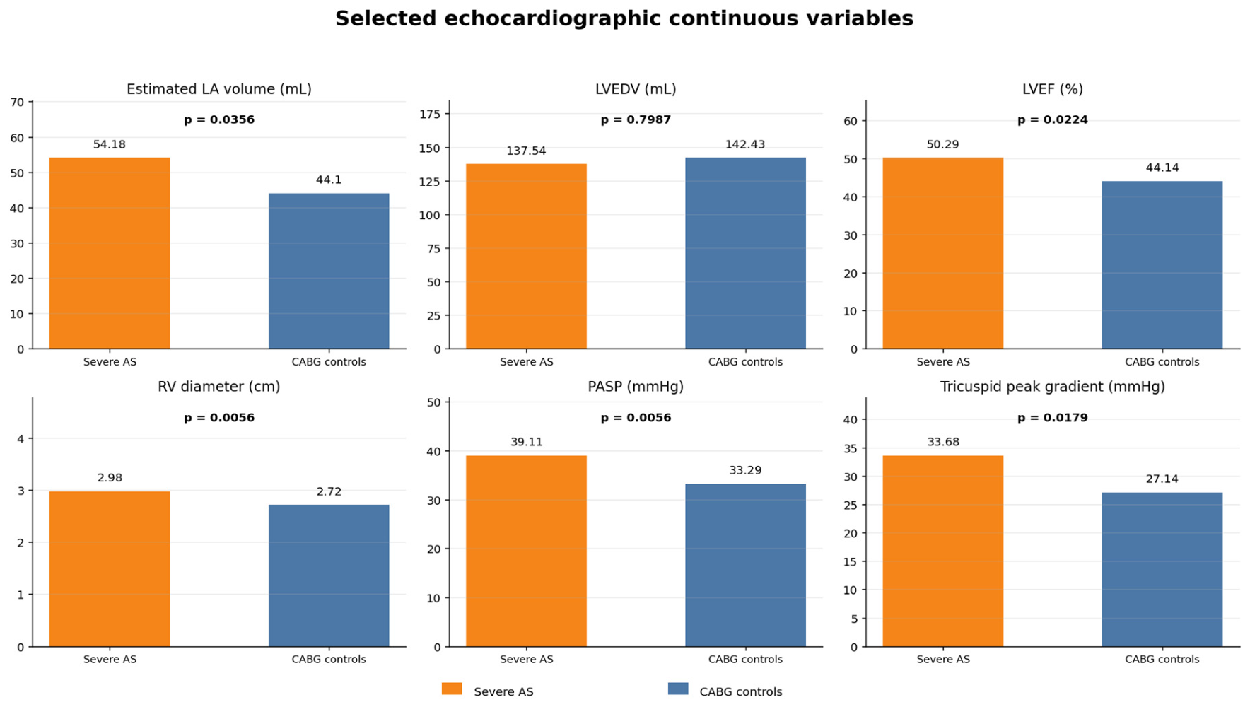
| Ascending aorta, cm | 3.87 ± 0.58 | 3.41 ± 0.41 | 0.0055 |
| LVEDD, cm | 5.00 ± 0.68 | 5.29 ± 0.78 | 0.2426 |
| LVEDV, mL | 137.54 ± 46.54 | 142.43 ± 62.75 | 0.7987 |
| LVESV, mL | 67.36 ± 26.27 | 82.00 ± 49.01 | 0.3106 |
| LVEF, % | 50.29 ± 5.48 | 44.14 ± 8.38 | 0.0224 |
| Mitral E wave, m/s | 0.75 ± 0.41 | 0.64 ± 0.21 | 0.2242 |
| Mitral A wave, m/s | 0.93 ± 0.25 | 0.95 ± 0.15 | 0.7294 |
| E/A ratio | 0.83 ± 0.44 | 0.66 ± 0.17 | 0.0881 |
| Aortic Vmax, m/s | 4.45 ± 0.99 | 1.15 ± 0.43 | <0.0001 |
| Aortic peak gradient, mmHg | 82.07 ± 31.64 | 5.64 ± 6.34 | <0.0001 |
| Aortic mean gradient, mmHg | 49.86 ± 20.66 | 2.93 ± 3.79 | <0.0001 |
| Tricuspid peak gradient, mmHg | 33.68 ± 6.92 | 27.14 ± 8.23 | 0.0179 |
| Pulmonary artery systolic pressure, mmHg | 39.11 ± 7.59 | 33.29 ± 5.08 | 0.0056 |
| IVS thickness, cm | 1.63 ± 0.28 | 1.23 ± 0.18 | <0.0001 |
| Posterior wall thickness, cm | 1.38 ± 0.17 | 1.18 ± 0.13 | 0.0003 |
| Right-ventricular diameter, cm | 2.98 ± 0.26 | 2.72 ± 0.26 | 0.0056 |
| Estimated left-atrial volume, mL | 54.18 ± 16.53 | 44.10 ± 12.65 | 0.0356 |
| Variable | Severe AS | CABG Controls | p Value |
|---|---|---|---|
| IL-6 LA | 2.65-fold | 1.00 (calibrator) | Not applicable |
| TNF-α LA | 4.84-fold | 1.00 (calibrator) | Not applicable |
| TGF-β LA | 5.03-fold | 1.00 (calibrator) | Not applicable |
| Fibrosis LA, % | 19.12 [16.12–23.12]; 20.77 ± 6.52 | 11.54 [9.53–14.29]; 12.17 ± 4.56 | <0.0001 |
| IL-6 RA | 4.85-fold | 1.00 (calibrator) | Not applicable |
| TNF-α RA | 2.38-fold | 1.00 (calibrator) | Not applicable |
| TGF-β RA | 3.60-fold | 1.00 (calibrator) | Not applicable |
| Fibrosis RA, % | 16.33 [15.07–19.13]; 16.47 ± 5.28 | 8.40 [5.75–11.27]; 9.01 ± 4.46 | 0.0001 |
| Variable 1 | Variable 2 | Interpretation | Spearman Rho | p Value |
|---|---|---|---|---|
| Fibrosis LA | Estimated LA volume | Left-atrial structural remodeling | 0.223 | 0.2545 |
| Fibrosis LA | IVS thickness | Pressure-overload hypertrophic remodeling | 0.130 | 0.5112 |
| Fibrosis LA | Posterior wall thickness | Concentric remodeling | 0.168 | 0.3928 |
| Fibrosis LA | LVEDV | Ventricular chamber remodeling | 0.147 | 0.4546 |
| Fibrosis LA | LVEF | Systolic phenotype | −0.090 | 0.6500 |
| Fibrosis LA | PASP | Pulmonary pressure burden | −0.191 | 0.3297 |
| Fibrosis LA | Aortic mean gradient | Valvular pressure-overload severity | −0.008 | 0.9690 |
| Fibrosis RA | PASP | Right-sided pressure burden | −0.028 | 0.8863 |
| Fibrosis RA | Tricuspid peak gradient | Right-heart hemodynamic load | 0.089 | 0.6542 |
| Fibrosis RA | RV diameter | Right-sided chamber remodeling | 0.012 | 0.9520 |
| Fibrosis RA | Estimated LA volume | Interatrial remodeling link | 0.395 | 0.0373 |
| Fibrosis RA | Ascending aorta | Aortic-root/ascending-aorta remodeling phenotype | −0.424 | 0.0246 |
| Fibrosis LA | Fibrosis RA | Interatrial histological fibrosis coupling | 0.296 | 0.1257 |
| Model | Cohort | Dependent Variable | Main Predictor/Adjustment | B Coefficient (95% CI) | p Value; R2 |
|---|---|---|---|---|---|
| Model 1 | Whole cohort (n = 42) | Fibrosis LA | Severe AS status; adjusted for age and sex | 8.548 (4.440 to 12.655) | p = 0.0001; R2 = 0.329 |
| Model 2 | Whole cohort (n = 42) | Fibrosis RA | Severe AS status; adjusted for age and sex | 7.746 (4.396 to 11.097) | p < 0.0001; R2 = 0.386 |
Disclaimer/Publisher’s Note: The statements, opinions and data contained in all publications are solely those of the individual author(s) and contributor(s) and not of MDPI and/or the editor(s). MDPI and/or the editor(s) disclaim responsibility for any injury to people or property resulting from any ideas, methods, instructions or products referred to in the content. |
© 2026 by the authors. Licensee MDPI, Basel, Switzerland. This article is an open access article distributed under the terms and conditions of the Creative Commons Attribution (CC BY) license.
Share and Cite
Merce, A.-G.; Nișulescu, D.D.; Hermenean, A.; Merce, A.-P.; Muntean, R.; Brie, D.-M.; Burciu, O.-M.; Găiță, D.; Ionac, A.; Crișan, S.; et al. Biatrial Inflammatory and Fibrotic Remodeling in Severe Aortic Stenosis Compared with CABG Controls. Metabolites 2026, 16, 574. https://doi.org/10.3390/metabo16080574
Merce A-G, Nișulescu DD, Hermenean A, Merce A-P, Muntean R, Brie D-M, Burciu O-M, Găiță D, Ionac A, Crișan S, et al. Biatrial Inflammatory and Fibrotic Remodeling in Severe Aortic Stenosis Compared with CABG Controls. Metabolites. 2026; 16(8):574. https://doi.org/10.3390/metabo16080574
Chicago/Turabian StyleMerce, Adrian-Grigore, Daniel Dumitru Nișulescu, Anca Hermenean, Adrian-Petru Merce, Raluca Muntean, Daniel-Miron Brie, Oana-Maria Burciu, Dan Găiță, Adina Ionac, Simina Crișan, and et al. 2026. "Biatrial Inflammatory and Fibrotic Remodeling in Severe Aortic Stenosis Compared with CABG Controls" Metabolites 16, no. 8: 574. https://doi.org/10.3390/metabo16080574
APA StyleMerce, A.-G., Nișulescu, D. D., Hermenean, A., Merce, A.-P., Muntean, R., Brie, D.-M., Burciu, O.-M., Găiță, D., Ionac, A., Crișan, S., & Mornoș, C. (2026). Biatrial Inflammatory and Fibrotic Remodeling in Severe Aortic Stenosis Compared with CABG Controls. Metabolites, 16(8), 574. https://doi.org/10.3390/metabo16080574