Article

Inhibitory Effect of Manassantin B Isolated from Saururus chinensis on Skin Heat Aging

1
Bio Convergence R&D Center, CoSeedBioPharm Corporation, Heungdeok-gu, Cheongju, Chungbuk 28161, Korea
2
Future Science Research Center, Coreana Cosmetics Corporation, Seobuk-gu, Cheonan, Chungnam 31041, Korea
*
Author to whom correspondence should be addressed.
Cosmetics 2020, 7(2), 47; https://doi.org/10.3390/cosmetics7020047
Submission received: 8 May 2020 / Revised: 5 June 2020 / Accepted: 12 June 2020 / Published: 16 June 2020

Abstract

:
Heat shock treatment-induced skin aging causes a thickened epidermis, increased matrix metalloproteinase (MMP)-1 expression, collagen degradation, and deep wrinkles. In this study, we investigated the effect of manassantin B in preventing heat shock treatment-induced aging. We first separated manassantin B (MB) from the roots of Saururus chinensis, and the structure was identified using 1H- and 13C-NMR spectroscopy. RT-PCR and western blotting were applied to investigate the anti-aging effect of manassantin B. Manassantin B decreased MMP-1 expression through transient receptor potential vanilloid (TRPV) 1 channel inhibition and significantly increased procollagen expression. In addition, manassantin B suppressed MAPK phosphorylation in a dose-dependent manner. Our results suggest that manassantin B, the active ingredient in S. chinensis, can be effectively used to inhibit heat shock treatment-induced skin aging.

1. Introduction

Human skin is exposed daily to sunlight, and in particular, to UV, visible, and IR radiation. UV radiation is known to induce skin aging, thereby causing wrinkles, freckles, dryness, and rough skin. In addition, several studies have shown that IR radiation can cause skin aging through IR-induced thermogenesis, leading to increased skin temperature from the normal 33 °C to more than 40 °C [1,2]. Heat exposure in human skin causes inflammation by inducing cytokines, such as transforming growth factor-β, interleukin-6, and interleukin-12 [3]. These cytokines are involved in the regulation of the levels of matrix metalloproteinases (MMPs) and extracellular matrix (ECM) proteins, such as collagen. Collagen is the major structural protein in the ECM, and collagen fibrils contribute to the resilience and strength of the skin [4]. MMPs degrade ECM proteins, such as collagen, elastin, and fibronectin [4,5]. Aged skin appears to have increased MMP expression and activity, resulting in a decreased collagen level. MMPs are involved in the treatment of conditions such as inflammation and wounding, as well as bone and vascular remodeling [6]. Heat induces MMP-1 and MMP-3 expression through the activation of extracellular signal-regulated kinase (ERK), c-Jun N-terminal kinase (JNK), and an autocrine interleukin-6 loop [7]. Besides, heat shock induces the activation of the transient receptor potential vanilloid 1 (TRPV1) channel in vitro and in vivo [8]. TRPV1 is a non-selective cation channel that is activated by various stimuli. These include vanilloids, capsaicin, heat, external acidic pH, and bioactive lipids such as lysophosphatidic acid [9,10,11]. Heat induces calcium influx via TRPV1 activation, leading to increased MMP-1 expression [12]. Furthermore, heat shock treatment-induced MMP-1 expression is mediated by TRPV1 via PKCα signaling in human epidermal keratinocytes [8].
Manassantin B (MB) is a neolignan constituent of the roots of Saururus chinensis, which is used in the treatment of gonorrhea, edema, and jaundice in Korean traditional medicine [13]. According to previous studies, MB exerts anti-tumor [14,15], anti-inflammatory [16,17], anti-melanogenic [18,19], anti-human immunodeficiency virus [20], and anti-obesity [21,22] activities. In addition, MB exhibits an anti-tumor effect by inhibiting cell aggregation via the inhibition of phorbol 12-myristate 13-acetate-induced intercellular cell adhesion molecule-1 expression in HL-60 cells [14]. Among lignans isolated from S. chinensis, MB exhibits the greatest inhibitory effects toward Epstein–Barr virus lytic replication [23]. However, the anti-aging effects of MB have not been reported yet. In this study, we investigated the effect of MB on heat shock treatment-induced aging, not the effect of aging by UV.

2. Materials and Methods

2.1. Materials

ERK, phospho-ERK, JNK, phospho-JNK, p38, and phospho-p38 antibodies were purchased from Cell Signaling Technology (Danvers, MA, USA). MMP-1 antibody was purchased from Santa Cruz Biotechnology (Santa Cruz, CA, USA). TRPV1 antibody was purchased from Abcam (Cambridge, MA, UK). β-Actin antibody was purchased from Sigma-Aldrich (St. Louis, MO, USA).

2.2. Isolation of Manassantin B

S. chinensis roots (SCR) were collected in October 2016 at Okcheon, Chungbuk, Korea. Dried SCR (1 kg) was ground to a particle size smaller than 0.5 mm and soaked in ethanol:water (7:3) for three days at room temperature. The supernatant was filtrated with filter paper and evaporated at 40 °C until we removed solvent. From the concentrate, an SCR extract (270 g) was obtained. The SCR extract was suspended in distilled water and successively partitioned with n-hexane and dichloromethane to yield n-hexane (8.1 g), dichloromethane (6.9 g), and water (92.2 g) fractions. The dichloromethane fraction was chromatographed on a silica gel column using an isocratic elution with n-hexane/ethyl acetate (1:1) to obtain six subfractions (SCR-M1–SCR-M6). MB (51.7 mg) was purified from subfraction SCR-M3 using prep-HPLC (C18 column, 0.1% TFA in ACN/0.1% TFA in water). The amorphous powder was obtained (Figure 1): [α]D 25−51.7o (c 0.33, CD3OD). The 1H- and 13C-NMR data were consistent with literature values [24]; ESI-MS m/z 739 [M+Na]+. Figure 2a shows the structure of the MB.

2.3. Cell Subculture

Human dermal fibroblasts, neonatal (HDFn) were purchased from ATCC (Manassas, VA, USA). The cells were cultured in F12:Dulbecco’s Modified Eagle Medium (Gibco, NY, USA) supplemented with 10% fetal bovine serum (FBS, Gibco, NY, USA), 100 U/mL penicillin, and 100 μg/mL streptomycin. The cells were subcultured every 2 or 3 days and maintained in the culture dish at 37 °C in a 5% CO2 incubator.

2.4. 3-(4,5-Dimethylthiazol-2-yl)-2,5-Diphenyltetrazolium Bromide (MTT) Assay

HDFn cells were seeded at 2.5 × 10³ cells/mL in 96-well culture plates and incubated for 24 h. Further, HDFn cells were treated with various concentrations of MB. After incubation for 24 h, the cells were treated with 50 μL of 5 mg/mL MTT reagent. After 2 h, the supernatants were removed, and the insoluble formazan crystals were dissolved completely with 100 μL of dimethyl sulfoxide. Absorbance was measured at 540 nm.

2.5. Reverse Transcription-Polymerase Chain Reaction (RT-PCR)

Total RNA was extracted using the TRIzol™ Reagent (Molecular Research Center, Cincinnati, OH, USA). Single-strand cDNA was synthesized from 2 μg total RNA. PCR was performed using the PCR premix (Bioneer, Daejeon, Korea), and the primer sequences used were as follows: procollagen-1 (forward, 5′-TGA CGA GAC CAA GAA CTG-3′, reverse, 5′-CCA TCC AAA CCA CTG AAA CC-3′), GAPDH (forward, 5′-TCC ATG ACA ACT TTG GTA TCG-3′, and reverse, 5′-TGT AGC CAA ATT CGT TGT CA-3′). PCR products were detected on ethidium bromide-stained agarose gels.

2.6. Western Blotting

HDFn cells were seeded at 5 × 10⁵ cells/well in 6 well plates. For heat treatment, a plate was sealed with parafilm and then immersed for 20 min in a circulating water bath at 43 °C [8]. The cells were harvested by a scraper and washed with PBS. Then, the cells were lysed in 100 µl of 1× cell lysis buffer (Cell Signaling Technology, Danvers, MA, USA). After centrifugation for 15 min at 12,000 rpm, the supernatants were used to prepare total protein extract. The protein concentration was calculated using a bicinchoninic acid assay (BioRad, Hercules, CA, USA). Equal amounts of protein were fractionated by 10% SDS-PAGE and electrophoresed at 95 V for 2 h. Then, the separated protein in the gel was electro-transferred to polyvinylidene difluoride membranes (Millipore, Billerica, MA, USA) at 100 V for 1.5 h. The membranes were blocked with 5% skim milk in Tween 20 plus tris-buffered saline (TTBS) for 1 h and then treated with primary antibodies in 5% bovine serum albumin (in TTBS) overnight at 4 °C. The bands were detected by enhanced chemiluminescence (Amersham Pharmacia Biotech, Piscataway, NJ, USA) and the intensities were calculated using the Image J 1.47 software.

2.7. Procollagen Synthesis Assay (ELISA)

HDFn cells were plated in 24 well plates at 1 × 10⁵ cells/well and incubated for 24 h. The culture medium was changed to an FBS-free medium, and the cells were treated with various concentrations of MB. After 24 h of incubation, the supernatants were collected and the procollagen content was measured using the procollagen type I c-peptide EIA kit (Takara, Otsu, Shiga, Japan).

2.8. Statistics

All data were expressed as the mean ± standard deviation (SD) of at least three separate experiments carried out in triplicates. Statistical analysis was performed using Microsoft Excel software 2016 (Student’s t-test, * p < 0.05).

3. Results and Discussion

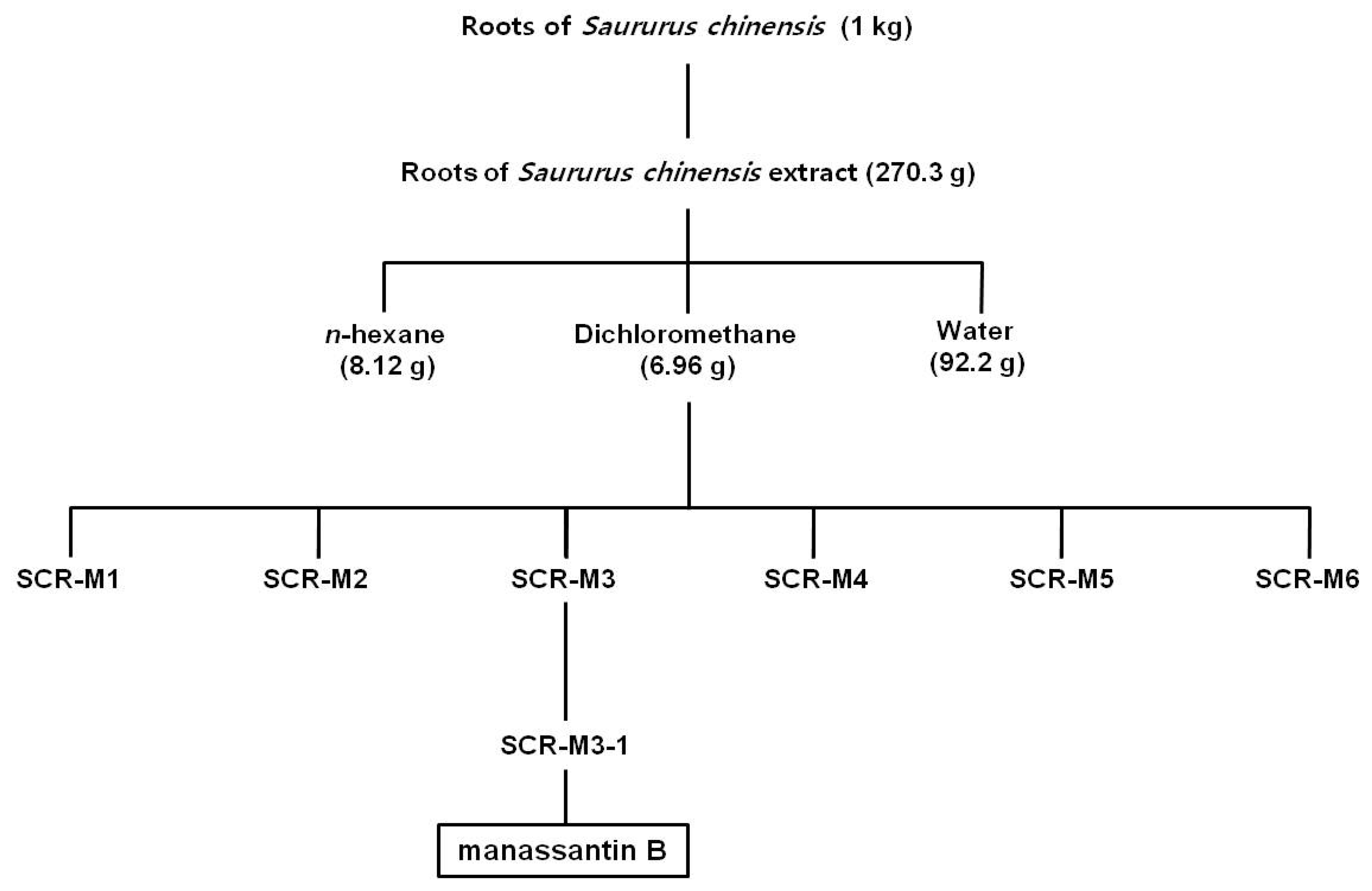
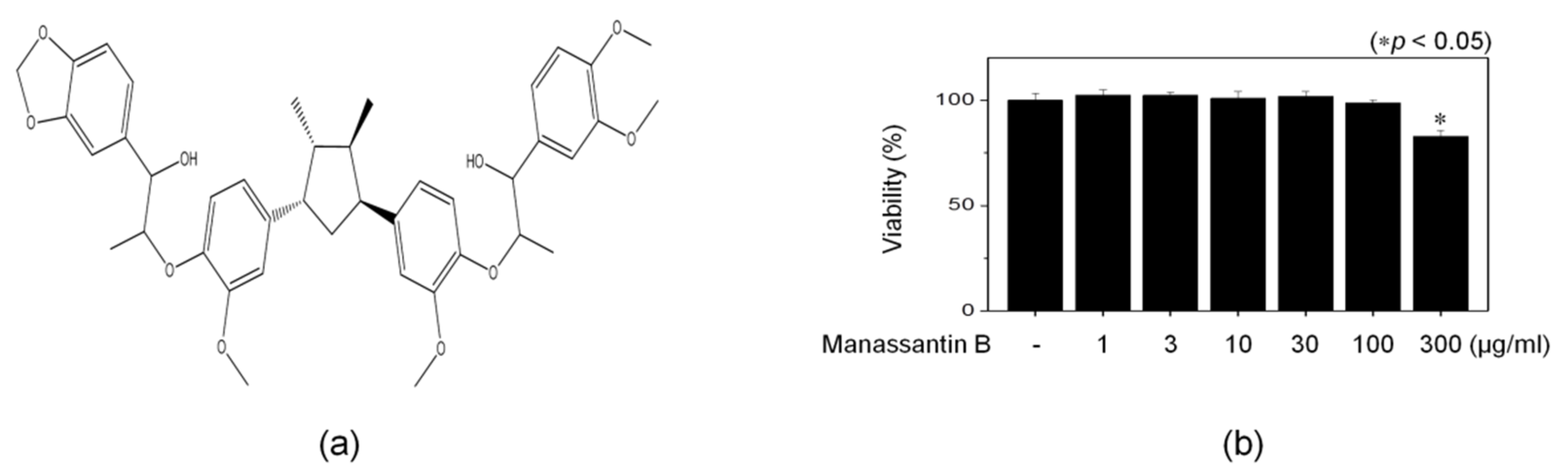
3.1. Isolation of MB

The 70% EtOH extract of the SCR was partitioned successfully with n-hexane, dichloromethane, and water. The dichloromethane fraction was subjected to silica gel column chromatography and prep-HPLC to purify a single compound from SCR-M3-1 subfraction. By comparing our 1H- and 13C-NMR spectra data with published values, this compound was characterized as MB (Figure 2a) [24].
1H-NMR (500 MHz, CDCl3, δ, ppm, J/Hz): 6.79–7.02 (12H, m, H-2, H-2′, H-3, H-3′, H-6, H-6′ and H-2″, H-2‴, H-3″, H-3‴, H-6″, H-6‴), 5.97 (2H, s, OCH2O), 5.48 (2H, d, J = 6.0, H-7 and H-7′), 4.66 (1H, d, J = 8.4, H-7″), 4.63 (1H, d, J = 8.4, H-7‴), 4.08 (2H, m, H-8″, H-8‴), 3.95 (3H, s, Ar-OMe), 3.94 (3H, s, Ar-OMe), 3.91 (3H, s, Ar-OMe), 3.90 (3H, s, Ar-OMe), 2.29 (2H, m, H-8 and H-8′), 1.18 (3H, d, J = 6.0, H-9‴), 1.15 (3H, d, J = 6.0, H-9′), 0.72(6H, d, J = 5.6, H-9 and H-9′).
13C-NMR (125 MHz, CDCl3, δ): 136.6 (C-1), 136.5 (C-1′), 132.5 (C-1″), 133.9 (C-1‴), 110.1(C-2 and C-2′), 110.8 (C-2″), 107.5 (C-2‴), 146.4 (C-3), 146.3 (C-3′), 148.8 (C-3″), 147.3 (C-3‴), 150.6 (C-4 and C-4′), 149.0 (C-4″), 147.7 (C-4‴), 118.7 (C-5 and C-5′), 118.7 (C-5″), 108.1 (C-5‴), 120.0 (C-6 and C-6′), 121.1 (C-6″), 118.9 (C-6‴), 83.3 (C-7 and C-7′), 84.0 (C-7″ and C-7‴), 44.2 (C-8 and C-8′), 78.4 (C-8″ and C-8‴), 14.9 (C-9 and C-9′), 17.0(C-9″), 16.9 (C-9‴), 101.0 (C-10), and 55.9 (OMe).

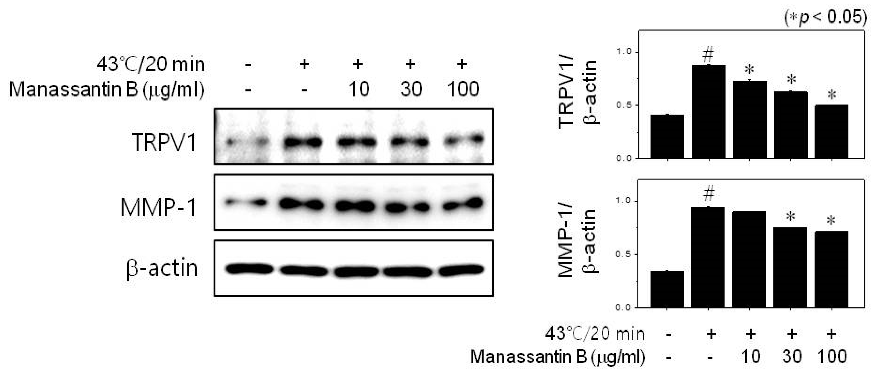
3.2. MB Inhibits MMP-1 Expression through TRPV1 Activation

We first investigated the effect of MB on the viability of HDFn cells using the MTT assay. HDFn cells were treated with various concentrations of MB for 24 h. There was no cytotoxicity at concentrations of less than 100 µg/mL, but cytotoxicity was shown at concentrations of 300 µg/mL (Figure 2b).
MMPs are multi-gene endoproteinases with a broad range of substrate specificities [5,25]. They are responsible for the degradation of ECM proteins, such as collagen, elastin, and fibronectin, inducing wrinkles [5]. In addition, MMPs are involved in multiple biological processes, including wound healing, cancer, inflammation, and tissue remodeling [4]. Among all MMPs, the expression of MMP-1, which plays critical roles in the aging processes, is induced in dermal fibroblasts and keratinocytes in response to UV radiation, cytokines, oxidative stress, and heat. To investigate the effect of MB on heat-induced MMP-1 expression, we treated the cells with heat shock (43 °C) for 20 min. Heat shock increased MMP-1 expression in HDFn cells, but the MB decreased this in a dose-dependent manner (Figure 3). Thus, MB may be effective in inhibiting heat shock treatment-induced aging.
According to previous studies, MMP-1 expression is mediated by TRPV1 activation [12]. To confirm the effect of MB on heat-induced TRPV1 activation, we performed western blot analysis. Heat shock significantly increased TRPV1 expression compared to untreated cells (Figure 3). However, MB decreased the heat-induced expression of TRPV1 in a dose-dependent manner (Figure 3). MB showed a more than 80% inhibitory effect on heat-induced TRPV1 expression at 100 μg/mL. These results indicate that MB inhibits MMP-1 by reducing TRPV1 expression.

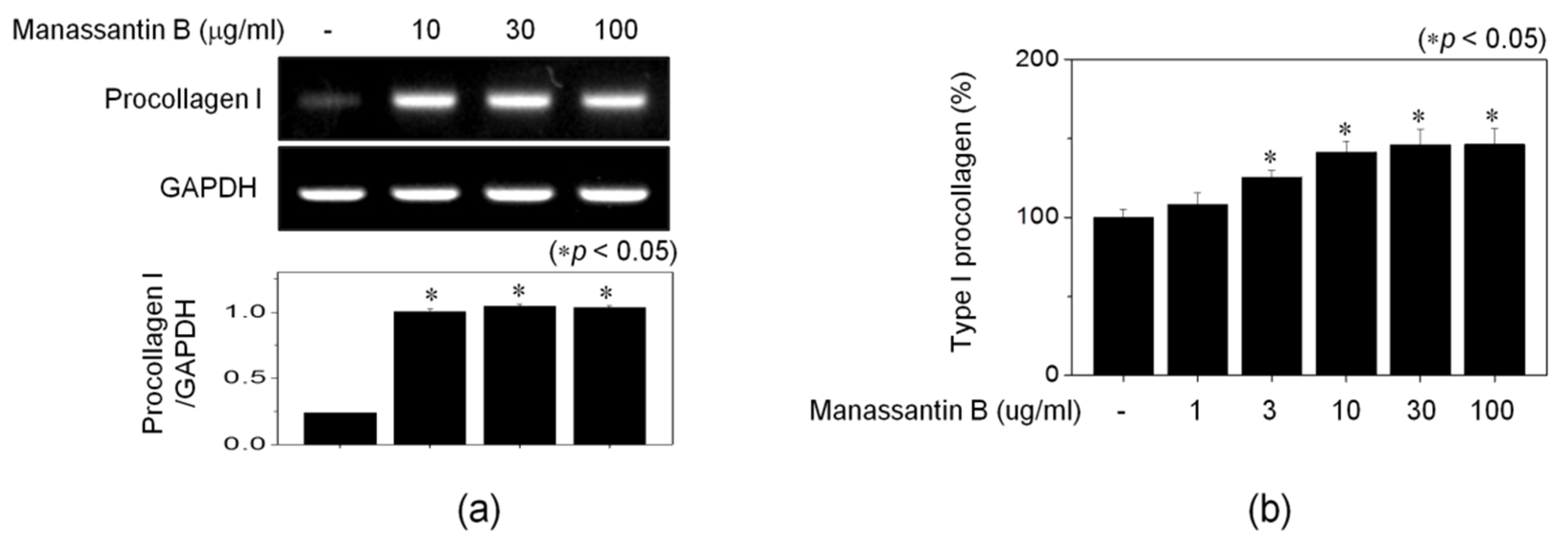
3.3. MB Increases Procollagen Expression

Heat induces MMP-1 and MMP-3 expression [7]. MMP-1 and MMP-3 can degrade type I and III collagen and elastin [26]. Collagen, the most abundant fibrous protein, is the main constituent of the ECM. It regulates the adhesion, tensile strength, and migration of cells [27,28]. Collagen is synthesized in the form of a precursor called procollagen, and the degree of synthesis of collagen can be determined by measuring the amount of procollagen. We investigated whether MB affects collagen protein and RNA levels. MB significantly increased collagen expression in RNA and protein levels (Figure 4). In particular, MB increased collagen expression by approximately 46% at a concentration of 100 μg/mL.

3.4. MB Inhibits Heat-Induced Mitogen-Activated Protein Kinase (MAPK) Activation

MMP-1 expression is stimulated by various extracellular stimuli, such as cytokines, mitogenic growth factors, and contact with the ECM [29,30]. Above all, the activation of activator protein-1 is the most important factor in MMP-1 expression and is induced by MAPKs [31,32]. MAPKs, which include ERK, p38, and JNK protein kinase, regulate cell migration, proliferation, differentiation, and MMP-1 expression [33,34]. To examine the effect of MB on heat-induced MAPK activation, we exposed the HDFn cells to heat shock (43 °C) for 20 min and treated them with various concentrations of MB for 15 min. The phosphorylation of ERK, JNK, and p38 was significantly increased owing to heat (Figure 5). However, MB decreased the heat-induced phosphorylation of ERK, JNK, and p38 in a dose-dependent manner (Figure 5). These results verified that MB suppressed MMP-1 expression by inhibiting MAPK activation.

4. Conclusions

In summary, we isolated MB as the active compound from the roots of S. chinensis. MB strongly increases procollagen expression and decreases MMP-1 and TRPV1 expression in human fibroblast cells by inhibiting MAPK activation. Although the exact target was not clear, we suggest that MB may be used to effectively inhibiting heat-induced aging.

Author Contributions

H.S.R. designed the experiments and performed the experiments; J.-Y.C. and K.-S.L. performed the structural experiments and contributed separation tools; H.S.R., J.-N.L., C.M.L. and S.-M.P. wrote the paper. All authors have read and agreed to the published version of the manuscript.

Funding

This research was financially supported by the Ministry of Trade, Industry and Energy (MOTIE), the Republic of Korea, under the “Business Cooperation Industry development program R&D” (reference number: R0004134) supervised by the Korea Institute for Advancement of Technology (KIAT).

Conflicts of Interest

The authors declare no conflict of interest.

References

  1. Cho, S.; Shin, M.H.; Kim, Y.K.; Seo, J.E.; Lee, Y.M.; Park, C.H.; Chung, J.H. Effects of infrared radiation and heat on human skin aging in vivo. J. Investig. Derm. Symp Proc. 2009, 14, 15–19. [Google Scholar] [CrossRef] [PubMed] [Green Version]
  2. Lee, H.S.; Lee, D.H.; Cho, S.; Chung, J.H. Minimal heating dose A novel biological unit to measure infrared irradiation. Photodermatol. Photoimmunol. Photomed. 2006, 22, 148–152. [Google Scholar] [CrossRef] [PubMed]
  3. Seo, J.Y.; Chung, J.H. Thermal aging: A new concept of skin aging. J. Dermatol. Sci. Suppl. 2006, 2, S13–S22. [Google Scholar] [CrossRef]
  4. Quan, T.; Qin, Z.; Xia, W.; Shao, Y.; Voorhees, J.J.; Fisher, G.J. Matrix-degrading metalloproteinases in photoaging. J. Investig. Derm. Symp. Proc. 2009, 14, 20–24. [Google Scholar] [CrossRef] [PubMed] [Green Version]
  5. Pittayapruek, P.; Meephansan, J.; Prapapan, O.; Komine, M.; Ohtsuki, M. Role of Matrix Metalloproteinases in Photoaging and Photocarcinogenesis. Int. J. Mol. Sci. 2016, 17, 868. [Google Scholar] [CrossRef] [PubMed] [Green Version]
  6. Page-McCaW, A.; Ewald, A.J.; Werb, Z. Matrix metalloproteinases and the regulation of tissue remodeling. Nat. Rev. Mol. Cell Biol 2007, 8, 221–233. [Google Scholar] [CrossRef]
  7. Park, C.H.; Lee, M.J.; Ahn, J.; Kim, S.; Kim, H.H.; Kim, K.H.; Eun, H.C.; Chung, J.H. Heat shock-induced matrix metalloproteinase (MMP)-1 and MMP-3 are mediated through ERK and JNK activation and via an autocrine interleukin-6 loop. J. Invest. Derm. 2004, 123, 1012–1019. [Google Scholar] [CrossRef]
  8. Lee, Y.M.; Li, W.H.; Kim, Y.K.; Kim, K.H.; Chung, J.H. Heat-induced MMP-1 expression is mediated by TRPV1 through PKCalpha signaling in HaCaT cells. Exp. Derm. 2008, 17, 864–870. [Google Scholar] [CrossRef]
  9. Zheng, J. Molecular mechanism of TRP channels. Compr. Physiol. 2013, 3, 221–242. [Google Scholar] [CrossRef] [Green Version]
  10. Klionsky, L.; Tamir, R.; Holzinger, B.; Bi, X.; Talvenheimo, J.; Kim, H.; Martin, F.; Louis, J.C.; Treanor, J.J.; Gavva, N.R. A polyclonal antibody to the prepore loop of transient receptor potential vanilloid type 1 blocks channel activation. J. Pharm. Exp. 2006, 319, 192–198. [Google Scholar] [CrossRef] [Green Version]
  11. Morales-Lazaro, S.L.; Liorente, I.; Sierra-Ramirez, F.; Lopez-Romero, A.E.; Oritiz-Renteria, M.; Serrano-Flores, B.; Simon, S.A.; Islas, L.D.; Rosenbaum, T. Inhibition of TRPV1 channels by a naturally occurring omega-9 fatty acid reduces pain and itch. Nat. Commun. 2016, 127, 2328–2335. [Google Scholar] [CrossRef] [PubMed] [Green Version]
  12. Li, W.H.; Lee, Y.M.; Kim, J.Y.; Kang, S.; Kim, S.; Kim, K.H.; Park, C.H.; Chung, J.H. Transient receptor potential vanilloid-1 mediates heat-shock-induced matrix metalloproteinase-1 expression in human epidermal keratinocytes. J. Investig. Derm. 2007, 127, 2328–2335. [Google Scholar] [CrossRef] [PubMed] [Green Version]
  13. Son, K.N.; Song, I.S.; Shin, Y.H.; Pai, T.K.; Chung, D.K.; Baek, N.I.; Lee, J.J.; Kim, J. Inhibition of NF-IL6 activity by manassantin B, a dilignan isolated from Saururus chinensis, in phorbol myristate acetate-stimulated U937 promonocytic cells. Mol. Cells 2005, 20, 105–111. [Google Scholar] [PubMed]
  14. Rho, M.C.; Kwon, O.E.; Kim, K.; Lee, S.W.; Chung, M.Y.; Kim, Y.H.; Hayashi, M.; Lee, H.S.; Kim, Y.K. Inhibitory effects of manassantin A and B isolated from the roots of Saururus chinensis on PMA-induced ICAM-1 expression. Planta Med. 2003, 69, 1147–1149. [Google Scholar] [CrossRef]
  15. Hodges, T.W.; Hossain, C.F.; Kim, Y.P.; Zhou, Y.D.; Nagle, D.G. Molecular-targeted antitumor agents the Saururus cernuus dineolignans manassantin B and 4-O-demethylmanassantin B are potent inhibitors of hypoxia-activated HIF-1. J. Nat. Prod. 2004, 67, 767–771. [Google Scholar] [CrossRef]
  16. Park, H.C.; Bae, H.B.; Jeong, C.W.; Lee, S.H.; Jeung, H.J.; Kwak, S.H. Effect of manassantin B, a lignan isolated from Saururus chinensis, on lipopolysaccharide-induced interleukin-1beta in RAW 264.7 cells. Korean J. Anesth. 2012, 62, 161–165. [Google Scholar] [CrossRef] [Green Version]
  17. Lu, Y.; Hwang, S.L.; Son, J.K.; Chang, H.W. Manassantin B isolated from Saururus chinensis inhibits cyclooxygenase-2-dependent prostaglandin D2 generation by blocking Fyn-mediated nuclear factor-kappaB and mitogen activated protein kinase pathways in bone marrow derived-mast cells. Biol. Pharm. Bull. 2013, 36, 1370–1374. [Google Scholar] [CrossRef] [Green Version]
  18. Seo, C.S.; Lee, W.H.; Chung, H.W.; Chang, E.J.; Lee, S.H.; Jahng, Y.; Hwang, B.Y.; Son, J.K.; Han, S.B.; Kim, Y. Manassantin A and B from Saururus chinensis inhibiting cellular melanin production. Phytother. Res. 2009, 23, 1531–1536. [Google Scholar] [CrossRef]
  19. Chang, H.; Choi, H.; Joo, K.M.; Kim, D.; Lee, T.R. Manassantin B inhibits melanosome transport in melanocytes by disrupting the melanophilin-myosin Va interaction. Pigment. Cell Melanoma Res. 2012, 25, 765–772. [Google Scholar] [CrossRef] [PubMed] [Green Version]
  20. Lee, J.; Huh, M.S.; Kim, Y.C.; Hattori, M.; Otake, T. Lignan, sesquilignans and dilignans, novel HIV-1 protease and cytopathic effect inhibitors purified from the rhizomes of Saururus chinensis. Antivir. Res. 2010, 85, 425–428. [Google Scholar] [CrossRef]
  21. Lee, W.S.; Lee, D.W.; Baek, Y.I.; An, S.; Cho, K.H.; Choi, Y.K.; Kim, H.C.; Park, H.Y.; Bae, K.H.; Jeong, T.S. Human ACAT-1 and -2 inhibitory activities of saucerneol B, manassantin A and B isolated from Saururus chinensis. Bioorg. Med. Chem. Lett. 2004, 14, 3109–3112. [Google Scholar] [CrossRef] [PubMed]
  22. Chen, L.; Lafond, J.; Pelletier, R.M. A novel technical approach for the measurement of individual ACAT-1 and ACAT-2 enzymatic activity in the testis. Methods Mol. Biol. 2009, 550, 169–177. [Google Scholar] [CrossRef] [PubMed]
  23. Cui, H.; Xu, B.; Wu, T.; Xu, J.; Yuan, Y.; Gu, Q. Potential antiviral lignans from the roots of Saururus chinensis with activity against Epstein-Barr virus lytic replication. J. Nat. Prod. 2014, 77, 100–110. [Google Scholar] [CrossRef] [PubMed]
  24. Wang, L.; Zhou, X.; Xu, T.; Yang, X.; Liu, Y. Lignans from Saururus chinensis. Chem. Nat. Compd. 2010, 46, 450–451. [Google Scholar] [CrossRef]
  25. He, X.; Dai, J.; Fan, Y.; Zhang, C.; Zhao, X. Regulation function of MMP-1 downregulated by siRNA on migration of heat-denatured dermal fibroblasts. Bioengineered 2017, 8, 686–692. [Google Scholar] [CrossRef] [Green Version]
  26. Li, D.Q.; Meller, D.; Liu, Y.; Tseng, S.C. Overexpression of MMP-1 and MMP-3 by cultured conjunctivochalasis fibroblasts. Invest. Ophthalmol Vis. Sci. 2000, 41, 404–410. [Google Scholar]
  27. Frantz, C.; Stewart, K.M.; Weaver, V.M. The extracellular matrix at a glance. J. Cell Sci. 2010, 123, 4195–4200. [Google Scholar] [CrossRef] [Green Version]
  28. Rozario, T.; DeSimone, D.W. The extracellular matrix in development and morphogenesis A dynamic view. Dev. Biol 2010, 341, 126–140. [Google Scholar] [CrossRef] [Green Version]
  29. Reunanen, N.; Li, S.P.; Ahonen, M.; Foschi, M.; Han, J.; Kahari, V.M. Activation of p38 alpha MAPK enhances collagenase-1 (matrix metalloproteinase (MMP)-1) and stromelysin-1 (MMP-3) expression by mRNA stabilization. J. Biol. Chem. 2002, 277, 32360–32368. [Google Scholar] [CrossRef] [Green Version]
  30. Westermarck, J.; Kahari, V.M. Regulation of matrix metalloproteinase expression in tumor invasion. Faseb J. 1999, 13, 781–792. [Google Scholar] [CrossRef] [Green Version]
  31. Lewis, T.S.; Shapiro, P.S.; Ahn, N.G. Signal transduction through MAP kinase cascades. Adv. Cancer Res. 1998, 74, 49–139. [Google Scholar] [CrossRef] [PubMed]
  32. Garrington, T.P.; Johnson, G.L. Organization and regulation of mitogen-activated protein kinase signaling pathways. Curr. Opin. Cell Biol. 1999, 11, 211–218. [Google Scholar] [CrossRef]
  33. Lam, E.; Kilani, R.T.; Li, Y.; Tredget, E.E.; Ghahary, A. Stratifin-induced matrix metalloproteinase-1 in fibroblast is mediated by c-fos and p38 mitogen-activated protein kinase activation. J. Invest. Derm. 2005, 125, 230–238. [Google Scholar] [CrossRef] [PubMed] [Green Version]
  34. Pearson, G.; Robinson, F.; Beers Gibson, T.; Xu, B.E.; Karandikar, M.; Berman, K.; Cobb, M.H. Mitogen-activated protein (MAP) kinase pathways: Regulation and physiological functions. Endocr. Rev. 2001, 22, 153–183. [Google Scholar] [CrossRef] [PubMed] [Green Version]
Figure 1. Isolation of manassantin B from S. chinensis roots dichloromethane fraction.
Cosmetics 07 00047 g001
Figure 2. The structure and cell viability of manassantin B. Manassantin B (a). HDFn cells were treated with various concentrations of MB for 24 h (b). Cell viability was determined in the MTT assay, and absorbance was measured by 540 nm. Significance was determined compared to the untreated cells (* p < 0.05). All data are expressed as mean ± SD of three separate experiments performed in triplicate.
Cosmetics 07 00047 g002
Figure 3. Effects of manassantin B on heat-induced MMP-1 and TRPV1 expression. HDFn cells were stimulated with heat shock (43 °C) for 20 min and treated with various concentrations of MB for 6 h. Total cell extracts were blotted with MMP-1, TRPV1, and β-actin antibodies. Band intensities were quantified using ImageJ 1.47 software and normalized to β-actin (* p < 0.05). All data are expressed as mean ± SD of three separate experiments performed in triplicate.
Cosmetics 07 00047 g003
Figure 4. Effects of manassantin B on procollagen expression. HDFn cells were treated with various concentrations of MB for 6–24 h. The mRNA levels of procollagen were measured using RT-PCR (a). Band intensities were estimated using ImageJ 1.47 software and normalized to GAPDH (* p < 0.05). The protein levels of procollagen were measured by ELISA (b). All data are expressed as mean ± SD of three separate experiments performed in triplicate.
Cosmetics 07 00047 g004
Figure 5. Effects of manassantin B on MAPK activation. HDFn cells were stimulated with heat shock (43 °C) for 20 min and treated with various concentrations of MB for 15 min. Total cell extracts were blotted with p-ERK, ERK, p-JNK, JNK, p-p38, and p38 antibodies. Band intensities were quantified using ImageJ 1.47 software and normalized to non-phosphorylated form (* p < 0.05). All data are expressed as mean ± SD of three separate experiments performed in triplicate.
Cosmetics 07 00047 g005

