Article

In Vitro and In Vivo Study on Humans of Natural Compound Synergy as a Multifunctional Approach to Cellulite-Derived Skin Imperfections

1
Complife Italia, Garbagnate Milanese (MI), 20024 Garbagnate Milanese (MI), Italy
2
Department of Pharmacy, Health and Nutritional Sciences at University of Calabria, 87036 Arcavacata di Rende (CS), Italy
*
Author to whom correspondence should be addressed.
Cosmetics 2020, 7(2), 48; https://doi.org/10.3390/cosmetics7020048
Submission received: 16 April 2020 / Revised: 11 June 2020 / Accepted: 12 June 2020 / Published: 16 June 2020
(This article belongs to the Special Issue Feature Papers in Cosmetics in 2020)

Abstract

:
Aim: The present study aimed to assess the efficacy of a nutraceutical ingredient, SelectSIEVE® Rainbow, based on botanical extracts, in ameliorating cellulite-derived skin imperfections and microcirculation. The nutritional supplement contained a mixture of Oryza sativa (L.), Citrus sinensis (L.) Osbeck, Ananas comosus (L.) Merr, and Actinidia chinensis Planch; all ingredients were botanicals that can be used in food supplements. Results: In vitro studies showed the high capacity of the supplement to have an anti-inflammatory, antioxidant, and hypolipidemic effect, accompanied by an interesting proteolytic activity. The randomized double-blind placebo-controlled clinical trial, carried out on 60 women during an 8-week treatment period, confirmed the in vitro study results. SelectSIEVE® Rainbow showed a whole-body shaping activity, with a reduction of the waist, hip, and tight circumference of 0.8, 0.65 and 0.72 cm, respectively. It also showed a reduction of subcutaneous fat mass of 1.24 mm and body weight, with an average of 0.7 kg and positive peaks of −2.9 kg. Skin health and appearance were also improved: +5.4% skin elasticity, +5.5% skin tonicity and +5.7% skin draining. Finally, the dermatological evaluation of the cellulite score and microcirculation showed an improvement in 57% and 60% of the subjects enrolled in the studies. Conclusions: This first study provides interesting inputs on the effectiveness of the nutraceutical complex standardized in polyphenols, anthocyanins and proteolytic enzymes to counteract cellulite blemishes and improve local microcirculation. The positive response encourages deeper studies and further investigation.

1. Introduction

Cellulite (also known as gynoid lipodystrophy, “orange peel” syndrome, or “cottage cheese”, like dimpling of the skin) is a complex and multifactorial disorder of the subcutaneous fat layer that can involve several mechanisms, such as local fat accumulation, microcirculation failure, and inflammatory processes [1,2]. It is caused by an uneven distribution of fat deposits and it is commonly found in the thighs, abdomen, and hips of women and men [3]. Although it is possible to reduce the appearance of cellulite by exercising regularly and eating a healthful diet, it will not completely disappear. Since cellulite is caused by the accumulation of fat deposits, the first step in reducing the appearance of cellulite is weight loss [4]. A healthy diet and cardiovascular exercise will help to lose weight and start reducing cellulite [5]. Aerobic exercises, such as running, cycling, and swimming, are excellent means of burning calories, increasing the metabolism, and improving weight loss [6].
Several herbal supplements can accelerate the rate of lipolysis, so they may help to achieve weight loss goals faster than diet and exercise alone [7,8,9,10]. Furthermore, a relevant effect in ameliorating the skin imperfections of cellulite, and so changing the cellulite severity grading, can be achieved by supplements containing botanic origin substances that, beside the lipolysis capacity, can act in draining liquids, eliminating toxins, modulating positively the capillary tonicity, inducing microcirculation, preventing and lowering the inflammatory process mediated by free radicals, and firming and toning the skin [11,12,13,14].
Due to their properties, all these actives can be classified as nutricosmetics. Born from the intersection of cosmeceuticals and nutraceuticals [15], these products started being developed with the objective of encouraging the draining of excess fluid to reduce the appearance of cellulite [16], which, due to its complex etiology, requires a multifaceted approach to effectively counteract its signs.
Black rice is well known for its high content of phenolic compounds, such as flavonoids, that are reported to have beneficial impacts on human health for their role as natural antioxidants [17,18,19,20], together with recognized effects on several diseases [21,22,23,24,25]. Anthocyanins are a flavonoid sub-class commonly known, among others, for their potential antidiabetic effect [26,27,28]. It is also well known that these compounds are effective on venous microcirculation and lymph drainage, and they can reduce capillary permeability and fragility, while also showing antiedemigenic properties [29,30,31]. Recent investigation efforts have focused on the novel health benefits of anthocyanins. The latest findings indicate a positive antiobesity effect. Obesity is often associated with adipocyte dysfunction. In this context, anthocyanins may act on adipocytes by modulating the expression levels of adipocytokines [32,33]. It was also found that anthocyanins can influence lipid absorption by exerting an inhibitory effect on pancreatic lipase [34]. Moreover, several studies have shown that they could directly affect lipid metabolism by attenuating the expressions of adipogenic genes and thus preventing lipogenesis hepatic lipid accumulation [35,36]. In addition, an enhancement of fibroblast proliferation and collagen synthesis is reported for fruits rich in anthocyanins and cereal extracts [37,38].
The fruit Citrus sinensis (L.) Osbeck is traditionally used as a natural antioxidant due to the presence of vitamin C. However, the active healing properties of this fruit are also related to other biologically active compounds, such as phytochemical antioxidants and fibers, with claimed benefits against several diseases and disorders [39,40,41]. Citrus flavonoids, especially naringin, naringenin, and hesperedin, have been reported to exert strong anti-inflammatory and antioxidative effects and they are considered as promising therapeutic agents for the treatment of metabolic dysregulation [42]. In general, citrus flavonoids are also noteworthy for their anti-inflammatory effect on keratinocytes [4,43] and their positive effect on glucose absorption and lipid metabolism [43,44,45,46,47,48]. Several studies reported the importance of extracts obtained from orange peel or juice for their ability to inhibit adipogenesis and increase lipolysis activity and thermogenesis [49,50].
The pineapple Ananas comosus (L.) Merr has been used as a medicinal plant in several cultures [51]. Its major active principle, bromelain, is a mixture of proteolytic enzymes from the stem of the pineapple plant and has demonstrated anti-inflammatory activity in a wide variety of diseases [52,53,54]. It induces the production of anti-inflammatory prostaglandins and reduces capillary permeability [55]. Several studies indicated that bromelain also shows antiedemigenic and coagulation inhibiting effects [56,57,58,59,60,61]. All these properties indicate that bromelain might be effective in reducing swelling [62].
It is well known that kiwifruit possesses antioxidant properties, mainly correlated to the high content of polyphenols, vitamin C, and dietary fibers [63,64]. It is reported that its ascorbic acid content is higher than the average of many other commercial fruits [65] and that vitamin C seems to be the major contributor to its total antioxidant activity [66]. Additionally, the polysaccharides contained in kiwifruit have been reported to have a potential use as actives. They showed stimulating activity on the proliferation of fibroblasts and keratinocytes, leading to an increase in metabolic activity and in the production of collagen in vitro, thus suggesting a possible use for dermatological skin treatment [67]. Among other positive effects on the cardiovascular system, kiwifruit may increase high-density lipoprotein cholesterol, and decrease triglycerides [68]. Due to its high level of antioxidants, it may protect the body from endogenous oxidative damage and can reduce oxidative stress [69]. Kiwifruit contains the cysteine protease actinidin, which is used as a dietary proteinase supplement to facilitate the digestion of proteins in the digestive tract [70].
The present study aimed to assess the efficacy of a nutraceutical ingredient, SelectSIEVE® Rainbow, based on botanical extracts, in ameliorating cellulite-derived skin. The nutritional supplement contains a mixture of Oryza sativa L. (black rice), Citrus sinensis (L.) Osbeck (orange), Ananas comosus (L.) Merr (pineapple), and Actinidia chinensis Planch. (kiwi) extracts; all ingredients are botanicals that can be used in food supplements.

2. Materials and Methods

2.1. Composition of the Nutraceutical Ingredient

The composition of the product SelectSIEVE® Rainbow (ROELMI HPC Srl, Italy) used in this study is reported in Table 1.

2.2. ABTS Assay

The antioxidant properties of the nutritional complex were evaluated in terms of scavenging activity on ABTS (2,2′-azinobis-(3-ethylbenzothiazoline-6-sulfonic acid)) radical. ABTS radical cation was generated by incubation of ABTS (38 mg) and potassium persulfate (5.6 mg) in 10 mL of distilled water in dark conditions and at room temperature for 12–16 h. The obtained solution was further diluted (1:36) with distilled water to an absorbance of 0.970 ± 0.020 measured at 734 nm. The scavenging activity of the nutritional complex toward ABTS radical was investigated according to the literature with slight modification [71] and the obtained results were expressed as IC50 (concentration providing 50% radical inhibition). Briefly, 5 mg of product was dissolved in 25 mL of methanol by sonication for 10 min. Then, aliquots of 200, 100, 75, 50, 35, 25, and 15 μL of the product were added to 2.0 mL of ABTS solution. After 5 min in dark conditions, the absorbance was measured colorimetrically at 734 nm using as the control a solution prepared in the same experimental conditions, but the product SelectSIEVE® Rainbow was replaced by methanol and so the solvent in which the product was dissolved. We compared the obtained results with those obtained using the negative control. The scavenging abilities of the product were evaluated in terms of the ABTS reduction and data were expressed as the percentage inhibition calculated according to Equation (1):
inhibition ( % ) = A 0   A 1 A 0 × 100 ,
where A0 is the absorbance of the control solution prepared in the same conditions of the product, and A1 is the absorbance of the product. Each measurement was carried out in triplicate and data were expressed as mean (±SE).

2.3. DPPH Assay

In order to evaluate the antioxidant properties of the nutraceutical ingredient, its scavenging properties toward DPPH (2,2′-diphenyl-1-picrylhydrazyl) radical were investigated according to the literature with slight modification [72] and the obtained results were expressed as IC50 (concentration providing 50% radical inhibition). Briefly, 5 mg of the product were dissolved in 25 mL of methanol by sonication for 10 min. Then, aliquots of 0.5, 1.0, 1.5, 2.0, 2.5, and 3.0 mL of the product were added to 4.0 mL of a methanolic DPPH solution (200 μM). Finally, methanol was added to bring each sample to a final volume of 10 mL. After 15 min in dark conditions, the absorbance was measured colorimetrically at 517 nm using as the control a solution prepared in the same experimental conditions, but the product SelectSIEVE® Rainbow was replaced by methanol and so the solvent in which the product was dissolved. We compared the obtained results with by using a negative control. The scavenging abilities of the tested items were evaluated in terms of DPPH reduction and data were expressed as the percentage inhibition calculated according to Equation (1). Each measurement was carried out in triplicate and data were expressed as means (±SE).

2.4. Total Polyphenols Content

The Folin–Ciocalteu assay was used as a conventional method for the quantification of the amount of disposable phenolic groups [73]. Briefly, 10 mg of the product were added to 1 mL of distilled water, 1 mL of the Folin–Ciocalteu reagent, and 1 mL of a sodium carbonate solution (7.5% w/v). After 2 h at room temperature in the dark, the absorbance was measured at 760 nm using a control prepared in the same reaction condition. The sample was diluted in distilled water (1:10) before the measurement of the absorbance. The experiment was performed in triplicate and the total polyphenols content was expressed as mg catechin equivalent per gram of sample (mg eq. CA/g) by using the equation obtained from the calibration curve of the antioxidant. This one was recorded by employing different catechin standard solutions. The absorbance of the solutions was measured at 760 nm to record the calibration curve, correlation coefficient (R2), slope, and intercept of the regression equation obtained by the least square method.

2.5. Total Anthocyanins Content

The total anthocyanins content was analyzed. Briefly, 20 mg of the tested item were added to 60 mL of a 2% hydrochloric acid-methanol solution (w/v). The product was incubated at 80 °C for 30 min and then cooled to room temperature and brought to a volume of 100 mL with the 2% hydrochloric acid-methanol solution (w/v) to obtain “solution A”. Then, 5 mL of the prepared solution A were withdrawn and brought to a final volume of 50 mL with the 2% hydrochloric acid-methanol solution (w/v) to obtain “solution B”. Finally, solution B was diluted 1:10 and the absorbance measured at 540 nm. The anthocyanins content was expressed as the percentage, using delphinidin as the reference compound, and calculated according to the following Equation (2):
%   = A   × 1 ×   f 1020 ×   m 2 × 100 ,
where A is the absorbance at 540 nm, f is the solution A to solution B dilution factor, and M2 is the amount of the product (g).

2.6. Proteolytic Activity

The casein digestion method was used to determine the proteolytic activity (PA) of the nutraceutical complex using casein as a substrate [74]. The results are expressed as units/mL calculated according to the following Equation (3):
PA ( Units mL ) = ( μ m   tyrosine   equivalents   released   × 5   mL ) ( 1   mL   × 10 min × 2   mL ) ,
where 5 mL is the total volume of the assay, 10 min is the time of the assay as per unit definition, 1 mL is the volume of the enzyme used, and 2 mL is the volume used in the colorimetric determination.

2.7. Pancreatic Lipase Activity

Pancreatic lipase plays a key role in the digestion of triglycerides. This enzyme’s activity was measured by monitoring the hydrolysis of the substrate 4-nitrophenyl octanoate (p-NPC), which releases the yellow chromogen p-nitrophenol, measuring its absorbance at 412 nm. Briefly, 10 mg of the nutraceutical ingredient were dissolved in 10 mL of distilled water by sonication for 10 min. Then, 50, 40, 30, and 20 μL of the solution prepared were added to 2.0 mL of phosphate buffer solution (PBS 0.1 M pH 8.0), 150 μL of pancreatic lipase solution (3 mg/mL in distilled water), and 150 μL of substrate solution (4-nitrophenyl octanoate 0.075 M in DMSO). The samples were incubated at 37 °C and, after 30 min, the absorbance was measured at 412 nm. A control was prepared in the same experimental conditions, while a blank without the enzyme was measured for each tested item. For comparison of the inhibitory activity, a standard solution of Orlistat, which is a hydrogenated derivative of lipstatin able to inhibit the absorption of 30% of dietary fat, was also tested as the control. Each measurement was carried out in triplicate, data were expressed as means (±SE), and results were expressed as IC50 (concentration providing 50% enzyme inhibition). The lipase inhibition percent was calculated according to Equation (4):
I n h i b i t i o n %   = ( Ac As     Ab Ac ) × 100 ,
where Ac, As, and Ab are the absorbance of the control, sample, and blank, respectively.

2.8. Oil Red O Staining

In order to evaluate the antiadipogenic effect of the nutraceutical ingredient, 3T3-L1 preadipocytes were induced to differentiate, in the presence and in the absence of the tested sample and stained with the Oil Red O solution [75]. Fibroblast 3T3-L1 cells were seeded into 35-mm-diameter culture dishes and treated for adipocyte differentiation. A control cell culture was also provided. Once the differentiation occurred, adipocytes were treated with two different concentrations of the nutraceutical ingredient for 24 h: 0.1% and 0.2%. Then, the cells were washed twice with PBS and fixed with 4% paraformaldehyde in PBS for 10 min. The fixed cells were incubated for 15 min at room temperature in an Oil Red O solution (1.8%) in 60% isopropanol. After the incubation, the cells were washed twice with bidistilled water and immediately photographed. After Oil Red O staining, the dishes were treated with 100% isopropanol for 5 min at room temperature in order to solubilize the dye. The absorbance was measured at 516 nm using 100% isopropanol as the blank.

2.9. Clinical Study

2.9.1. Study Design and Participants

All the study procedures were carried out in compliance with the ethical principles for the medical research (Ethical Principles for Medical Research Involving Human Subjects, adopted by the 18th WMA General Assembly Helsinki, Finland, June 1964 and amendments). In order to take part in the study, each participant was fully informed of the study risks and benefits, aims, and procedures. The informed consent form and consent release form for the publication of photographs were signed by the subjects prior to the attendance to the study. The protocol E.HU.039-0060.01.003L_2018/288-version no. 2-1 February 2018 and the informed consent were approved by an Independent Ethical Committee for Not Pharmacological Clinical Trials. The monocentric, randomized, double-blind, placebo-controlled, and parallel-group study was conducted on 60 female subjects, fulfilling the inclusion and non-inclusion criteria laid down in the study protocol. Subjects were randomly assigned to active or placebo treatment: 30 subjects were assigned to the active group and 30 subjects to the placebo group according to a randomization list. Clinical visits were planned after 28 (T28d) and 56 days (T56d) of product use.

Inclusion Criteria

The following inclusion criteria were used: Healthy females of Caucasian ethnicity; age between 18 and 55 years old; mild to moderate cellulite-derived skin imperfections (grade II and III of the cellulite thermographic stage (Table 2a); body mass index between 23 and 30; willingness to follow the proposed alimentary diet for all the study time; willingness to not vary the normal daily routine (i.e., lifestyle, physical activity, etc.); subject is under effective contraception (oral/not oral); and not expected to be changed during the trial.

Non-Inclusion Criteria

Subjects did not meet the inclusion criteria if they were: Pregnant woman or women intending to became pregnant during the study; breastfeeding women; subjects that have shown allergies to the ingredients of the food supplements; subjects who are following anticellulite treatment or have followed an anticellulite treatment less than 3 months before the present study; and subjects who used sun-beds or self-tanning product for one month before the study or intended to use it during the present study.

2.9.2. Treatment

Subjects were administered for 56 days (8 weeks) food-grade gelatin capsules containing 300 mg of the nutraceutical ingredient. The placebo capsules had the same composition, except for the active ingredients, which were substituted by maltodextrin. Subjects took one capsule per day in the morning with a glass of water. A base cream was supplied to all volunteers, who were instructed to use it throughout the study period as a substitute for their usual body cream, whenever they were used to applying it. The base cream, supplied by Complife Italia Srl, is a cosmetic formulation generally used by Complife Italia as a control cream; the composition conforms to Regulation (EC) 1223/2009, with the following INCI formula: Aqua, Helianthus annuus seed oil, isononyl isononanoate, polyglyceryl-3 methylglucose distearate, glyceryl stearate, cetearyl alcohol, and imidazolidinyl. The principal investigator and her collaborators maintained a record of the products delivered to the subjects at the beginning of the study and received by the subjects at the end of the study. The compliance with treatment was assessed by the principal investigator by asking the subject specific questions aimed to assess the actual use of the product.

2.9.3. Assessments

The measurement of the body weight was carried out with the subject barefooted and wearing underwear using an electronic balance. The body circumferences were measured using a flexible meter (precision: 1 mm). The waistline circumference was measured just above the right and left iliac crest. The hip circumference was measured in the area of its maximum protrusion. The thigh circumference was measured in the area of its maximum protrusion. The thickness of the subcutaneous fatty layer was measured on the thighs by means of the ultrasound technique (BodyMetrixTM BX2000 IntelaMetrix Europe, Verbania, Italy), which allows visualization in two dimensions (linear echography or stratigraphy) of the superficial fat layer thickness, giving a wide and detailed vision of the adipose layer thickness in the examined area. The assessment of skin elasticity and firmness was performed on the thighs by the Cutometer® method. A negative pressure is created in the device (Cutometer® MPA 580, Courage + Khazaka Electronic, Köln, Germany) and the skin is drawn into the aperture of the probe for 2 s and then released again. The penetration depth is measured by a non-contact optical measuring system. The following parameters were measured: R0 (skin distensibility) represents the passive behavior of the skin to force (i.e., gravity); R2 (Ua/Uf, gross elasticity or overall elasticity) represents the ability of redeformation of the skin to its basal state; and R6 (Uv/Ue, skin viscoelasticity) represents the contribution of the viscoelasticity to the total skin deformation. Skin microcirculation improvement was assessed by means of clinical scoring. Thermographic assessments were performed on the thighs and scored by the dermatologist in relation to the extent of temperature variation in the thermographic image. The degree of cellulite at T0 (Table 2a), T28, and T56 (Table 2c) was classified in accordance with a clinical scale. The cellulite-derived skin imperfections (“orange peel”) were evaluated at T0, T28, and T56 by the dermatologist in accordance with the clinical scores reported in Table 2a,c. Clinical pictures and contact thermography pictures of the treated area were taken at each experimental time using a reflex digital camera (NIKON D300 digital camera, Nikon Corporation, Tokyo, Japan) equipped with macro-objective (AF-S Micro NIKKOR 60 mm f/2.8G ED, Nikon Corporation, Tokyo, Japan), a flash system (Kit R1C1, Nikon Corporation, Tokyo, Japan), and cross/parallel-polarized filters. At T56, subjects were asked to express their opinion on the treatment by answering a questionnaire about the product’s acceptability and effects.

2.10. Statistical Methods

The instrumental data were submitted to two-way paired Student t test (intra-group analysis vs. T0); the inter-group statistical analysis was made on the data variations versus T0 by means of two-way Student’s t test for unpaired data. The following comparison was carried out for each parameter: Intra-group comparison: Comparison of all experimental times vs. T0; and inter-group comparison: Comparison of the two study groups at all experimental times. Results of the safety evaluation are based on AE/SAE listing, and are presented descriptively as absolute and relative frequencies. The statistical software used for statistical analysis was NCSS 10 statistical software (NCSS, LLC, Kaysville, USA) running on Windows Server 2008 R2 Standard (Microsoft, Redmond, WA, USA).

3. Results

3.1. Evaluation of the Antioxidant Activity and the Total Polyphenols and Anthocyanins Content

The nutraceutical ingredient showed high antioxidant activity as demonstrated by the results obtained by the two assays carried out. For both radicals, the achieved IC50 (concentration providing 50% radical inhibition) values are reported in Figure 1a. The figure also details data related to the total polyphenols and anthocyanins content (Figure 1b,c).

3.2. Proteolytic Activity

The nutraceutical ingredient demonstrates an interesting synergic activity of its component in proteolytic activity. Indeed, the result of the product is higher than the sum of the single ingredients able to exert proteolysis (Figure 1d).

3.3. Evaluation of the Hypolipidemic Effect

For pancreatic lipase assay, the activity was reduced by 66% with respect to the control and the quantity of product needed to inhibit 50% of its activity was double the positive control (Xenical, active ingredient Orlistat, Roche, Germany). For the positive control, the activity was 6.0 ± 0.2, while for the nutraceutical ingredient, it was 12 ± 0.4 (Figure 1e).
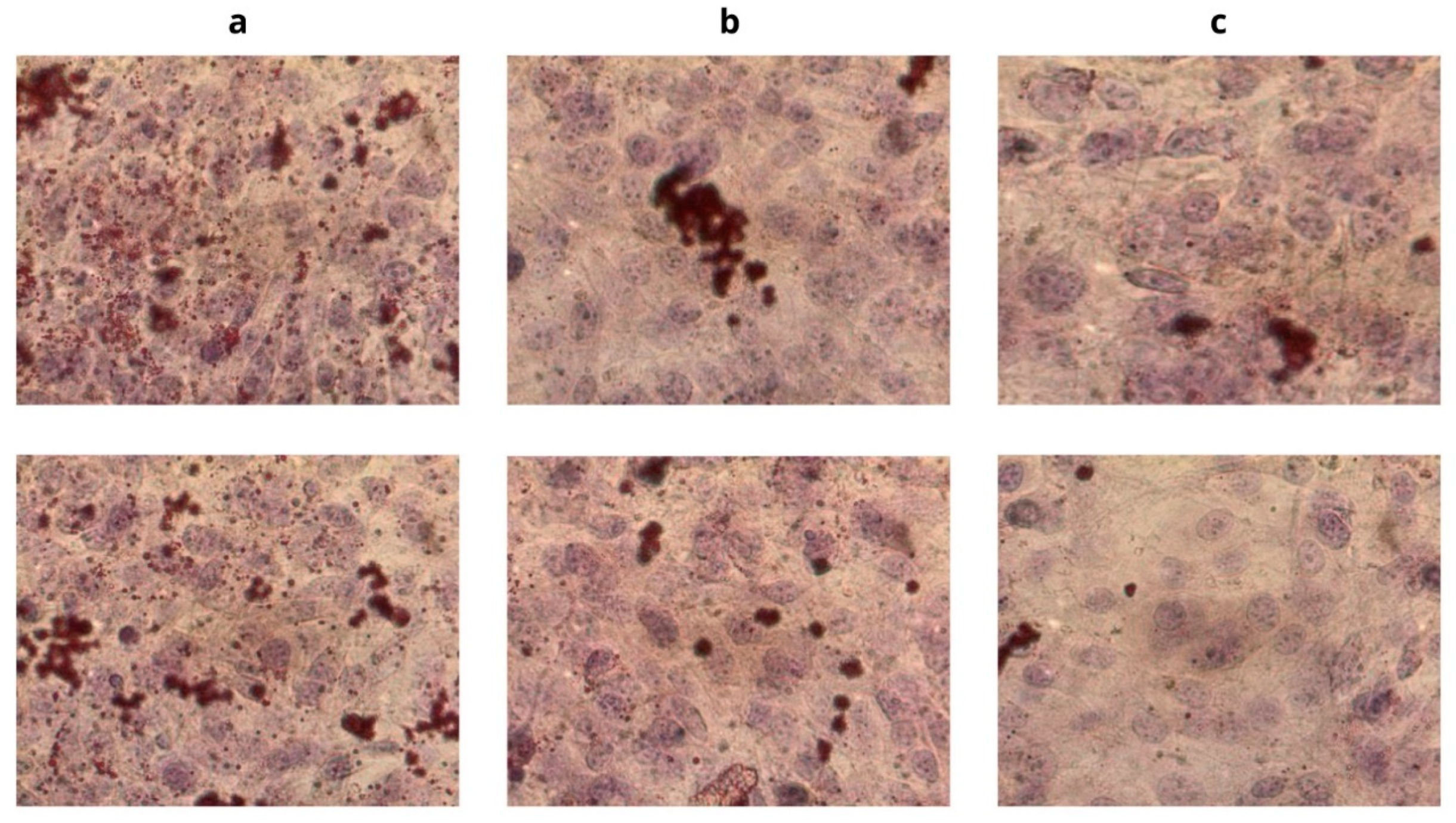
The Oil Red O staining showed that in the presence of the product, the lipid accumulation into the cells was reduced by 32% with respect to the control, with a concentration of 0.1%, and by 51% with a concentration of 0.2% (data not shown). The obtained results highlight the ability of the nutraceutical ingredient to inhibit 3T3-L1 preadipocytes adipogenesis, thus reducing lipid accumulation (Figure 2). Figure 2a shows the images of cells without any treatment with the nutraceutical ingredient; Figure 2b,c show the images of cells treated with the nutraceutical ingredient at different concentrations. The upper-row images show cell cultures before active treatment, while the lower-row ones show the results after the incubation period. Dark red spots are pre-adipocytes differentiated into mature adipocytes. It is then possible to see a reduction in lipid accumulation, but it is impossible to verify if it is for reduced adipogenesis or for increased lipolysis. The percentage of reduction refers to the obtained values of absorbance between the tested product and control.

3.4. Clinical Trial

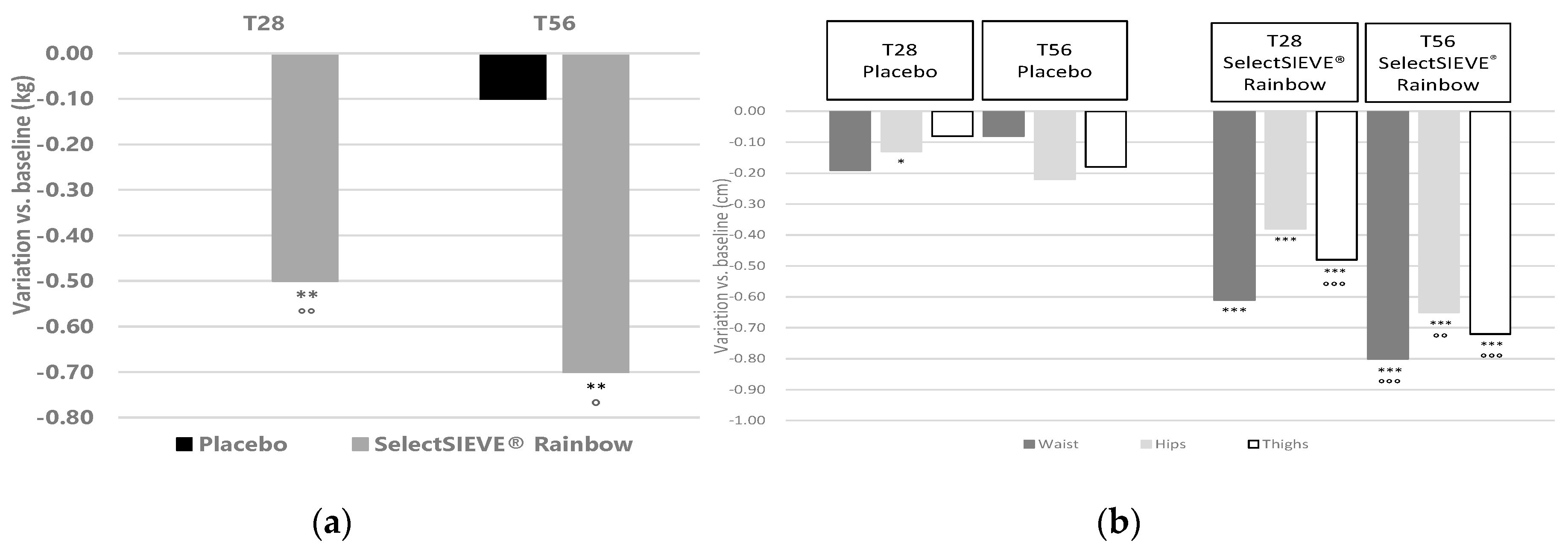
The subjects (Table 3) who took the nutraceutical ingredient lost an average of 0.5 kg after the 28 day-treatment and an average of 0.7 kg at the end of the study (Figure 3a). This weight loss was statistically significant vs. T0 and significantly higher with respect to the one achieved with the placebo.
As for the body circumference, after the 28-day treatment, and more consistently at the end of the study, a statistically significant reduction of all considered circumferences was recorded (Figure 3b). At T56, the mean reduction found for the active-treated group was −0.80 cm at the level of the waist, −0.65 cm at the level of the hips, and −0.72 cm at the level of the thighs. The ultrasound fat tissue thickness measurement confirmed the previous results, showing a significant reduction of the thigh fat tissue thickness already at 28 days of treatment and more markedly at the end of the study, with a mean reduction of 7.2% of the fat thickness. The reduction was −1.24 mm, with a maximum decrease of −4.2 mm (data not shown). These diminutions were statistically significant when compared to the variations obtained in the placebo.
Regarding the evaluation of skin elasticity (Figure 4), already after the first month of treatment, and more consistently at the end of the trial, a statistically significant improvement was recorded in the active-treated group for all three parameters tested in comparison to T0. For the R2 parameter, the values recorded were +3.6% at T28 and +5.7% at T56; for the R6 parameter, 4.1% at T28 and 5.4% at T56; and finally, for the R0 parameter, the values obtained were −3.0% and −5.5% for T28 and T56, respectively. These variations were statistically significant compared to those obtained in the placebo-treated group.
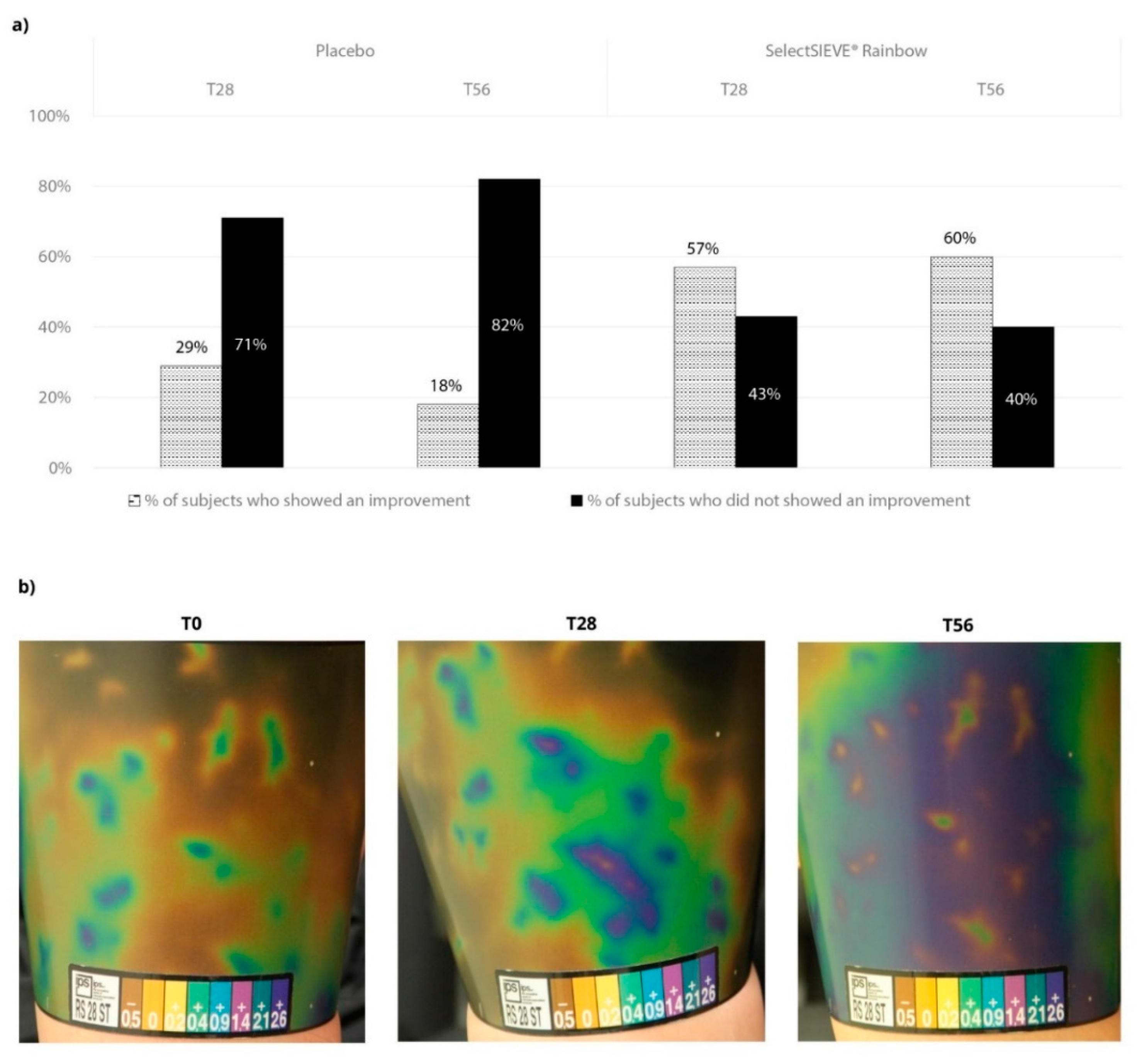
Thermographic evaluations of the skin microcirculation revealed that the active treatment determined an improvement of the cellulite thermographic grade in 57% of the subjects at T28 and in 60% of the subjects at the end of the study, while in the placebo-treated group, the improvement percentages were very low (Figure 5a). Contact thermography pictures showed that from an initial stage of 6 (Grade III—severe of the thermographic scale—leopard spots—cold temperature), a slight improvement was noticed at T28 (bigger warm areas) and a moderate improvement at T56 (smaller spots—higher temperature) (Figure 5b).
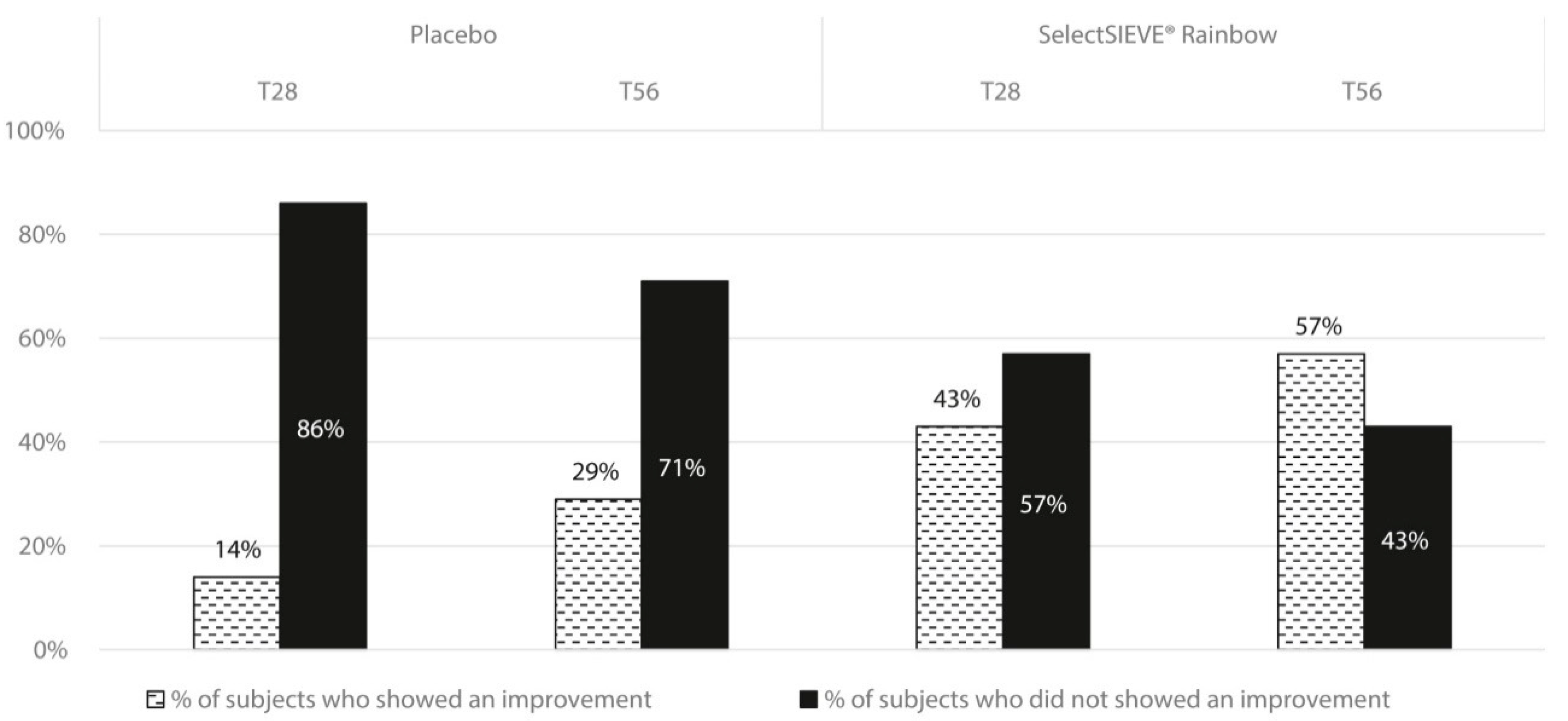
Regarding the cellulite-derived skin imperfections (“orange peel”), the active treatment determined an improvement of the orange peel skin aspect in 43% of the subjects at T28 and in 57% of the subjects at T56. In the placebo-treated group, lower improvement percentages were obtained (Figure 6).
The treatment was well tolerated by all the subjects participating to the study and it was positively judged for most of the investigated aspects; in particular, the volunteers underlined the refining activity on the body silhouette and the improvement of the orange peel skin aspect. After 56 days of treatment, the majority of subjects (70%) who used the active product would suggest the use of the product. On the contrary, for the same experimental time, only 25% of the subjects who used the placebo would suggest the treatment.

4. Discussion

Gynoid lipodystrophy (cellulite) is a disorder with a multifactorial etiology reflected by an incidence of about 85% in adult females [76]. This edematous fibrosclerotic panniculopathy affects adipose tissues, leading to changes in the appearance of the skin surface topography that results in dimpling typical of an orange peel appearance [77].
The pathophysiology of cellulite is essentially an altered connective tissue structure due to microcirculation failure that produces a disturbed dermal extracellular matrix. Several causes can contribute to the triggering and worsening of this skin condition: Hereditary factors, lack of exercise, hormone dysfunction, blood circulation problems, connective tissue weakness, premature skin ageing, poor nutrition, excessive alcohol consumption, etc. [78].
Nowadays, a broad panel of cellulite treatments are available, ranging from topically applied products to laser treatment, and from surgery to the use of pharmacological agents [79].
Nutricosmetics, with their advantages of quick good results and ease of use, could meet customers’ needs. Furthermore, the botanical extracts contained in topical products need to pass through the corneal stratum, which represents the main barrier, and reach the inner layers of the skin to carry out their tasks. Therefore, the clinical efficacy of many active ingredients is limited, owing to their inability to penetrate this barrier. Oral solutions could bypass this limitation and offer a greater solution to cellulite treatment [14]. Most of the classical approaches for cellulite treatment only focus on the stimulation of lipolysis. However, there are many other factors that must be taken into account to improve the overall cellulite appearance.
New therapeutic approaches involve the use of food supplements, such as polyphenol-rich juices, bromelain-based products, and botanical extracts [14,30,56]. Nevertheless, only the use of different combined natural ingredients can ensure simultaneous action on the different biochemical pathways responsible for cellulite-derived blemishes. In this context, the nutritional supplement used in this study was especially designed with this purpose.
One of the biochemical pathways involved in the etiopathology of cellulite is the oxidation of free radicals in adipocytes and connective tissue [80]. A high content of flavonoids, in particular their sub-class anthocyanins, and polyphenols can help in reducing cellular oxidation and thus preventing the consequent inflammatory process. In this study, SelectSIEVE® Rainbow showed a high presence of polyphenols and anthocyanins, which exert considerable antioxidant activity against both hydrophilic and lipophilic radicals.
The aim of this research study was to evaluate the antioxidant power of the tested sample and to evaluate the total polyphenols and total anthocyanins content. The DPPH and ABTS assays allow an evaluation of the ability of a sample to work as a reducing agent and so as a scavenger for these two radicals. These assays are mostly used to evaluate the antioxidant activity of plant extracts because they allow determination of the free radical scavenging activity of the tested sample. Moreover, aiming to evaluate the total polyphenols and total anthocyanins content, the equation obtained from the calibration curves of the antioxidant compounds was used. By comparing the obtained data with the catechin and delphinidin calibration curves, a value of 527 and 55 mg eq./g, respectively, was recorded.
The obtained results provide evidence that the tested product has good antioxidant activity, while the high recorded values for the catechin and delphinidin equivalent make this product very interesting as an antioxidant food supplement.
Bromelain is a proteolytic enzyme used in medicine to counteract edematous swellings caused by inflammatory processes. This enzyme in fact acts on fibrin, facilitating the drainage of the inflamed part and reabsorption of the edema [81]. Additionally, this activity was tested for the nutraceutical ingredient, which showed high proteolytic activity, most likely due to the action of the kiwi and pineapple extracts.
The hypolipidemic effect of SelectSIEVE® Rainbow was confirmed by the high level recorded in the pancreatic lipase assay, where the quantity of product necessary to inhibit the activity was doubled compared to the well-known drug sold globally for obesity treatment. The obtained results evidence that 12 μg/mL of the tested product was able to inhibit 50% of the activity of pancreatic lipase and it is a good result if compared with the IC50 of the positive control of 6 μg/mL. Further investigations are needed to evaluate the real interaction between the product, enzyme, and substrate. Additionally, lipid accumulation was significantly reduced in cells by inhibiting 3T3-L1 preadipocytes’ adipogenesis. These results were not surprising since almost all the ingredients present in the product are reported to stimulate lipolysis and reduce lipid accumulation in cells [82,83]. The same hypolipidemic effect was also evident in the results obtained in the human trial. After 56 days of treatment, subjects showed a reduction in body weight, subcutaneous fat layer, and, most of all, body circumferences. The combination of these results showed that the body shaping was most likely due to the reduction of fat, confirming the lipolysis effect already theorized after the in vitro test. This outcome was strengthened by the positive opinion given by the volunteers, which underlined the refining of body silhouette.
The skin biochemical parameters demonstrated a positive improvement of skin firmness and elasticity, especially after 56 days of supplementation. The effects substantiate the claimed activity on skin and collagen of the product components, already reported in the literature [37,57,84].
Other factors beyond the skin and adipose tissues were involved in the positive effect generated by SelectSIEVE® Rainbow. In fact, microcirculation was consistently increased, as demonstrated by the thermographic evaluation, allowing better oxygenation and nourishment of the tissues and, at the same time, enhancing the swelling and toxin displacement from the extracellular matrixes.

5. Conclusions

In conclusion, the abovementioned results indicate that the use of SelectSIEVE® Rainbow, through the reduction of adipogenesis, lipid accumulation, and swelling, provides visible and measurable results in the improvement of cellulite signs. In particular, a statistically significant decrease of body weight, an important and significant decrease of all considered circumferences and thigh fat tissue thickness, and a significant improvement of biomechanical items were achieved. All the abovementioned results showed a statistical significance compared to the baseline and placebo.
The dermatologist endorsed a considerable improvement of the “orange peel” appearance and microcirculation, corroborated by the volunteers’ evaluation. Indeed, due to all the mentioned positive effects produced by SelectSIEVE® Rainbow, almost 70% of the subjects involved in the study would recommend the use of the tested product.
This first study provides interesting inputs on the effectiveness of the nutraceutical complex standardized in polyphenols, anthocyanins and proteolytic enzymes to counteract cellulite blemishes and improve local microcirculation. The positive response encourages deeper studies and further investigation.

Author Contributions

Conceptualization, V.N. and A.M.; methodology, I.D. and F.P.; software, V.N.; validation, V.N., A.M. and F.P.; formal analysis, V.N.; investigation, F.P., E.C. and M.P.; data curation, M.P. and F.P.; writing—original draft preparation, V.N.; writing—review and editing, F.P.; visualization, V.N.; supervision, V.N.; project administration, A.M.; funding acquisition, A.M. All authors have read and agreed to the published version of the manuscript.

Funding

The study was funded by ROELMI HPC Srl, Origgio (VA), Italy.

Acknowledgments

The authors want to acknowledge all the study staff for their work and professionalism.

Conflicts of Interest

The authors declare no conflict of interest. The funders had no role in the design of the study; in the collection, analyses, or interpretation of data; in the writing of the manuscript, or in the decision to publish the results.

References

  1. Emanuele, E.; Minoretti, P.; Altabas, K.; Gaeta, E.; Altabas, V. Adiponectin expression in subcutaneous adipose tissue is reduced in women with cellulite. Int. J. Dermatol. 2011, 50, 412–416. [Google Scholar] [CrossRef] [PubMed]
  2. Hexsel, D.M.; Siega, C.; Schilling-Souza, J.; Porto, M.D.; Rodrigues, T.C. A bipolar radiofrequency, infrared, vacuum and mechanical massage device for treatment of cellulite: A pilot study. J. Cosmet. Laser Ther. 2011, 13, 297–302. [Google Scholar] [CrossRef] [PubMed]
  3. Khan, M.H.; Victor, F.; Rao, B.; Sadick, N.S. Treatment of cellulite: Part I Pathophysiology. J. Am. Acad. Dermatol. 2010, 62, 361–370. [Google Scholar] [CrossRef] [PubMed]
  4. Smalls, L.K.; Hicks, M.; Passeretti, D.; Gersin, K.; Kitzmiller, W.J.; Bakhsh, A.; Wickett, R.R.; Whitestone, J.; Visscher, M.O. Effect of weight loss on cellulite: Gynoid lipodystrophy. Plast. Reconstr. Surg. 2006, 118, 510–516. [Google Scholar] [CrossRef] [PubMed]
  5. Rossi, A.B.R.; Vergnanini, A.L. Cellulite: A review. J. Eur. Acad. Dermatol. Venereol. 2000, 14, 251–262. [Google Scholar] [CrossRef]
  6. Okay, D.M.; Jackson, P.V.; Marcinkiewicz, M.; Papino, M.N. Exercise and obesity. Prim. Care 2009, 36, 379–393. [Google Scholar] [CrossRef] [PubMed]
  7. Ab Rahman, W.N.A.W.; Lee, K.S.; Yee, J.C.W.; Gupta, M.; Ming, L.C. Evidence of Garcinia cambogia as a fat burning and appetite suppressing agents. Arch. Pharm. Pract. 2016, 7, 22. [Google Scholar] [CrossRef]
  8. Dell’Agli, M.; Bosisio, E. Biflavones of Ginkgo biloba stimulate lipolysis in 3T3-L1 adipocytes. Planta Medica 2002, 68, 76–79. [Google Scholar] [CrossRef]
  9. Zahara, K.; Bibi, Y.; Tabassum, S. Clinical and therapeutic benefits of Centella asiatica. Pure Appl. Biol. 2014, 3, 152. [Google Scholar] [CrossRef]
  10. Kazemipoor, M.; Radzi, C.W.J.W.M.; Cordell, G.A.; Yaze, I. Potential of traditional medicinal plants for treating obesity: A review. arXiv 2012, arXiv:1208.1923. [Google Scholar]
  11. Emanuele, E. Cellulite: Advances in treatment: Facts and controversies. Clin. Dermatol. 2013, 31, 725–730. [Google Scholar] [CrossRef] [PubMed]
  12. Goldman, M.P.; Hexsel, D. Cellulite: Pathophysiology and Treatment; CRC Press: Boca Raton, FL, USA, 2010. [Google Scholar]
  13. Sparavigna, A.; Guglielmini, G.; Togni, S.; Cristoni, A.; Maramaldi, G. Evaluation of anti-cellulite efficacy: A topical cosmetic treatment for cellulite blemishes—A multifunctional formulation. Int. J. Cosmet. Sci. 2011, 623, 305. [Google Scholar]
  14. Hexsel, D.; Orlandi, C.; Zechmeister do Prado, D. Botanical extracts used in the treatment of cellulite. Dermatol. Surg. 2005, 31, 866–873. [Google Scholar] [CrossRef] [PubMed]
  15. Anunciato, T.P.; da Rocha Filho, P.A. Carotenoids and polyphenols in nutricosmetics, nutraceuticals, and cosmeceuticals. J. Cosmet. Dermatol. 2012, 11, 51–54. [Google Scholar] [CrossRef] [PubMed]
  16. Rona, C.; Berardesca, E. Aging skin and food supplements: The myth and the truth. Clin. Dermatol. 2008, 26, 641–647. [Google Scholar] [CrossRef] [PubMed]
  17. Embuscado, M.E. Spices and herbs: Natural sources of antioxidants–a mini review. J. Funct. Foods 2015, 18, 811–819. [Google Scholar] [CrossRef]
  18. Huyut, Z.; Beydemir, Ş.; Gülçin, İ. Antioxidant and antiradical properties of selected flavonoids and phenolic compounds. Biochem. Res. Int. 2017, 2017, 7616791. [Google Scholar] [CrossRef]
  19. Nimse, S.B.; Pal, D. Free radicals, natural antioxidants, and their reaction mechanisms. RSC Adv. 2015, 5, 27986–28006. [Google Scholar] [CrossRef] [Green Version]
  20. Pietta, P.G. Flavonoids as antioxidants. J. Nat. Prod. 2000, 63, 1035–1042. [Google Scholar] [CrossRef]
  21. George, V.C.; Dellaire, G.; Rupasinghe, H.V. Plant flavonoids in cancer chemoprevention: Role in genome stability. J. Nutr. Biochem. 2017, 45, 1–14. [Google Scholar] [CrossRef]
  22. Jannesar, M.; Shoushtari, M.S.; Majd, A.; Pourpak, Z. Bee pollen flavonoids as a therapeutic agent in allergic and immunological disorders. Iran. J. Allergy Asthma Immunol. 2017, 16, 171–182. [Google Scholar] [PubMed]
  23. Marino, A.; Paterniti, I.; Cordaro, M.; Morabito, R.; Campolo, M.; Navarra, M.; Esposito, E.; Cuzzocrea, S. Role of natural antioxidants and potential use of bergamot in treating rheumatoid arthritis. Pharma Nutr. 2015, 3, 53–59. [Google Scholar] [CrossRef]
  24. Solanki, I.; Parihar, P.; Mansuri, M.L.; Parihar, M.S. Flavonoid-based therapies in the early management of neurodegenerative diseases. Adv. Nutr. 2015, 61, 64–72. [Google Scholar] [CrossRef] [PubMed] [Green Version]
  25. Sun, G.W.; Qiu, Z.D.; Wang, W.N.; Sui, X.; Sui, D.J. Flavonoids extraction from propolis attenuates pathological cardiac hypertrophy through PI3K/AKT signaling pathway. J. Evid. Based Complement. Altern. Med. 2016, 2016, 6281376. [Google Scholar] [CrossRef] [PubMed] [Green Version]
  26. Khoo, H.E.; Azlan, A.; Tang, S.T.; Lim, S.M. Anthocyanidins and anthocyanins: Colored pigments as food, pharmaceutical ingredients, and the potential health benefits. Food Nutr. Res. 2017, 61, 1361779. [Google Scholar] [CrossRef] [PubMed] [Green Version]
  27. Pedro, A.C.; Granato, D.; Rosso, N.D. Extraction of anthocyanins and polyphenols from black rice (Oryza sativa L.) by modeling and assessing their reversibility and stability. Food Chem. 2016, 191, 12–20. [Google Scholar] [CrossRef] [PubMed] [Green Version]
  28. Yousuf, B.; Gul, K.; Wani, A.A.; Singh, P. Health benefits of anthocyanins and their encapsulation for potential use in food systems: A review. Crit. Rev. Food Sci. Nutr. 2016, 5613, 2223–2230. [Google Scholar] [CrossRef] [PubMed]
  29. Bratman, S.; Kroll, D.J. Clinical Evaluation of Medicinal Herbs and Other Therapeutic Natural Products; Prima Health: Rocklin, CA, USA, 1999. [Google Scholar]
  30. Šavikin, K.; Menković, N.; Zdunić, G.; Pljevljakušić, D.; Spasić, S.; Kardum, N.; Konić-Ristić, A. Dietary supplementation with polyphenol-rich chokeberry juice improves skin morphology in cellulite. J. Med. Food 2014, 17, 582–587. [Google Scholar] [CrossRef]
  31. Wagner, H. New plant phenolics of pharmaceutical interest. Annal. Proc. Phytochem. Soc. Eur. 1985, 25, 409–425. [Google Scholar]
  32. Tsuda, T.; Ueno, Y.; Aoki, H.; Koda, T.; Horio, F.; Takahashi, N.; Kawada, T.; Osawa, T. Anthocyanin enhances adipocytokine secretion and adipocyte-specific gene expression in isolated rat adipocytes. Biochem. Biophys. Res. Commun. 2004, 3161, 149–157. [Google Scholar] [CrossRef]
  33. Tsuda, T. Recent progress in anti-obesity and anti-diabetes effect of berries. Antioxidants 2016, 5, 13. [Google Scholar] [CrossRef] [PubMed] [Green Version]
  34. Fabroni, S.; Ballistreri, G.; Amenta, M.; Romeo, F.V.; Rapisarda, P. Screening of the anthocyanin profile and in vitro pancreatic lipase inhibition by anthocyanin-containing extracts of fruits, vegetables, legumes and cereals. J. Sci. Food Agric. 2016, 96, 4713–4723. [Google Scholar] [CrossRef] [PubMed]
  35. Kim, S.; Hong, J.; Jeon, R.; Kim, H.S. Adzuki bean ameliorates hepatic lipogenesis and proinflammatory mediator expression in mice fed a high-cholesterol and high-fat diet to induce nonalcoholic fatty liver disease. Nutr. Res. 2016, 36, 90–100. [Google Scholar] [CrossRef] [PubMed]
  36. Park, C.H.; Kim, J.H.; Lee, E.B.; Hur, W.; Kwon, O.J.; Park, H.J.; Yoon, S.K. Aronia melanocarpa extract ameliorates hepatic lipid metabolism through PPARγ2 downregulation. PLoS ONE 2017, 12, e0169685. [Google Scholar] [CrossRef] [PubMed] [Green Version]
  37. Nanashima, N.; Horie, K.; Maeda, H.; Tomisawa, T.; Kitajima, M.; Nakamura, T. Blackcurrant Anthocyanins increase the levels of collagen, elastin, and hyaluronic acid in human skin fibroblasts and ovariectomized rats. Nutrients 2018, 10, 495. [Google Scholar] [CrossRef] [Green Version]
  38. Phetpornpaisan, P.; Tippayawat, P.; Jay, M.; Sutthanut, K. A local Thai cultivar glutinous black rice bran: A source of functional compounds in immunomodulation, cell viability and collagen synthesis, and matrix metalloproteinase-2 and-9 inhibition. J. Funct. Foods 2014, 7, 650–661. [Google Scholar] [CrossRef]
  39. Etebu, E.; Nwauzoma, A.B. A review on sweet orange (Citrus sinensis L Osbeck): Health, diseases and management. Am. J. Res. Commun. 2014, 2, 33–70. [Google Scholar]
  40. Hwang, S.L.; Shih, P.H.; Yen, G.C. Neuroprotective effects of citrus flavonoids. J. Agric. Food Chem. 2012, 60, 877–885. [Google Scholar] [CrossRef]
  41. Ejaz, S.; Ejaz, A.; Matsuda, K.; Chae, W.L. Limonoids as cancer chemopreventive agents. J. Sci. Food Agric. 2006, 86, 339–345. [Google Scholar] [CrossRef]
  42. Stohs, S.J.; Badmaev, V. A review of natural stimulant and non-stimulant thermogenic agents. Phytother. Res. 2016, 30, 732–740. [Google Scholar] [CrossRef] [Green Version]
  43. Graziano, A.C.; Cardile, V.; Crascì, L.; Caggia, S.; Dugo, P.; Bonina, F.; Panico, A. Protective effects of an extract from Citrus bergamia against inflammatory injury in interferon-gamma and histamine exposed human keratinocytes. Life Sci. 2012, 90, 968–974. [Google Scholar] [CrossRef] [PubMed]
  44. Kang, G.J.; Han, S.C.; Ock, J.W.; Kang, H.K.; Yoo, E.S. Anti-inflammatory effect of quercetagetin, an active component of immature Citrus unshiu, in HaCaT human keratinocytes. Biomol. Ther. 2013, 21, 138. [Google Scholar] [CrossRef] [PubMed] [Green Version]
  45. Alam, M.A.; Subhan, N.; Rahman, M.M.; Uddin, S.J.; Reza, H.M.; Sarker, S.D. Effect of citrus flavonoids, naringin and naringenin, on metabolic syndrome and their mechanisms of action. Adv. Nutr. 2014, 5, 404–417. [Google Scholar] [CrossRef] [PubMed]
  46. Akiyama, S.; Katsumata, S.I.; Suzuki, K.; Ishimi, Y.; Wu, J.; Uehara, M. Dietary hesperidin exerts hypoglycemic and hypolipidemic effects in streptozotocin-induced marginal type 1 diabetic rats. J. Clin. Biochem. Nutr. 2009, 46, 87–92. [Google Scholar] [CrossRef] [PubMed] [Green Version]
  47. Ghanim, H.; Sia, C.L.; Upadhyay, M.; Korzeniewski, K.; Viswanathan, P.; Abuaysheh, S.; Mohanty, P.; Dandona, P. Orange juice neutralizes the proinflammatory effect of a high-fat, high-carbohydrate meal and prevents endotoxin increase and Toll-like receptor expression. Am. J. Clin. Nutr. 2010, 914, 940–949. [Google Scholar] [CrossRef] [PubMed] [Green Version]
  48. Mulvihill, E.E.; Burke, A.C.; Huff, M.W. Citrus flavonoids as regulators of lipoprotein metabolism and atherosclerosis. Annu. Rev. Nutr. 2016, 36, 275–299. [Google Scholar] [CrossRef]
  49. Kang, S.I.; Shin, H.S.; Kim, H.M.; Hong, Y.S.; Yoon, S.A.; Kang, S.W.; Kim, J.H.; Kim, M.H.; Ko, H.C.; Kim, S.J. Immature Citrus sunki peel extract exhibits antiobesity effects by β-oxidation and lipolysis in high-fat diet-induced obese mice. Biol. Pharm. Bull. 2012, 35, 223–230. [Google Scholar] [CrossRef] [Green Version]
  50. Kim, G.S.; Park, H.J.; Woo, J.H.; Kim, M.K.; Koh, P.O.; Min, W.; Ko, Y.G.; Kim, C.H.; Won, C.K.; Cho, J.H. Citrus aurantium flavonoids inhibit adipogenesis through the Akt signaling pathway in 3T3-L1 cells. BMC Complement. Altern. Med. 2012, 12, 31. [Google Scholar] [CrossRef] [Green Version]
  51. Anilkumar, M. Ethnomedicinal plants as anti-inflammatory and analgesic agents. In Ethnomedicine: A Source of Complementary Therapeutics; Chattopadhyay, D., Ed.; Research Signpost: Kolkata, India, 2010; pp. 267–293. [Google Scholar]
  52. Hale, L.P.; Greer, P.K.; Trinh, C.T.; Gottfried, M.R. Treatment with oral bromelain decreases colonic inflammation in the IL-10-deficient murine model of inflammatory bowel disease. Clin. Immunol. 2005, 116, 135–142. [Google Scholar] [CrossRef]
  53. Pandey, P.; Tiwari, S. Therapeutic potential of Indian plants for the treatment of rheumatoid arthritis. J. Pharmacogn. Phytochem. 2018, 7, 37–41. [Google Scholar]
  54. Secor, E.R.; Carson, W.F.; Cloutier, M.M.; Guernsey, L.A.; Schramm, C.M.; Wu, C.A.; Thrall, R.S. Bromelain exerts anti-inflammatory effects in an ovalbumin-induced murine model of allergic airway disease. Cell Immunol. 2005, 237, 68–75. [Google Scholar] [CrossRef] [PubMed] [Green Version]
  55. Graf, J. Herbal anti-inflammatory agents for skin disease. Skin Ther. Lett. 2000, 5, 3–5. [Google Scholar]
  56. Rathnavelu, V.; Alitheen, N.B.; Sohila, S.; Kanagesan, S.; Ramesh, R. Potential role of bromelain in clinical and therapeutic applications. Biomed Rep. 2016, 5, 283–288. [Google Scholar] [CrossRef] [PubMed] [Green Version]
  57. Tochi, B.N.; Wang, Z.; Xu, S.Y.; Zhang, W. Therapeutic application of pineapple protease (bromelain): A review. Pak. J. Nutr. 2008, 74, 513–520. [Google Scholar]
  58. Bhattacharyya, B.K. Bromelain: An overview. Nat. Prod. Rad. 2008, 7, 359–363. [Google Scholar]
  59. Manzoor, Z.; Nawaz, A.; Mukhtar, H.; Haq, I. Bromelain: Methods of extraction, purification and therapeutic applications. Braz. Arch. Biol. Technol. 2016, 59. [Google Scholar] [CrossRef] [Green Version]
  60. Maurer, H.R. Bromelain: Biochemistry, pharmacology and medical use. Cell Mol. Life Sci. 2001, 589, 1234–1245. [Google Scholar] [CrossRef]
  61. Pavan, R.; Jain, S.; Kumar, A. Properties and therapeutic application of bromelain: A review. Biotechnol. Res. Int. 2012, 2012, 976203. [Google Scholar] [CrossRef] [Green Version]
  62. Grover, A.K.; Samson, S.E. Benefits of antioxidant supplements for knee osteoarthritis: Rationale and reality. Nutr. J. 2015, 15, 1. [Google Scholar] [CrossRef] [Green Version]
  63. Leontowicz, H.; Leontowicz, M.; Latocha, P.; Jesion, I.; Park, Y.S.; Katrich, E.; Barasch, D.; Nemirovski, A.; Gorinstein, S. Bioactivity and nutritional properties of hardy kiwi fruit Actinidia arguta in comparison with Actinidia deliciosa ‘Hayward’and Actinidia eriantha ‘Bidan’. Food Chem. 2016, 196, 281–291. [Google Scholar] [CrossRef]
  64. Park, Y.S.; Im, M.H.; Ham, K.S.; Kang, S.G.; Park, Y.K.; Namiesnik, J.; Leontowicz, H.; Leontowicz, M.; Trakhtenberg, S.; Gorinstein, S. Quantitative assessment of the main antioxidant compounds, antioxidant activities and FTIR spectra from commonly consumed fruits, compared to standard kiwi fruit. LWT Food Sci. Technol. 2015, 63, 346–352. [Google Scholar] [CrossRef]
  65. Cheng, C.H.; Seal, A.G.; Boldingh, H.L.; Marsh, K.B.; MacRae, E.A.; Murphy, S.J.; Ferguson, A.R. Inheritance of taste characters and fruit size and number in a diploid Actinidia chinensis (kiwifruit) population. Euphytica 2004, 138, 185–195. [Google Scholar] [CrossRef]
  66. D’Evoli, L.; Moscatello, S.; Lucarini, M.; Aguzzi, A.; Gabrielli, P.; Proietti, S.; Battistelli, A.; Famiani, F.; Böhm, V.; Lombardi-Boccia, G. Nutritional traits and antioxidant capacity of kiwifruit (Actinidia deliciosa Planch., cv. Hayward) grown in Italy. J. Food Compost. Anal. 2015, 37, 25–29. [Google Scholar] [CrossRef]
  67. Deters, A.M.; Schröder, K.R.; Hensel, A. Kiwi fruit (Actinidia chinensis L.) polysaccharides exert stimulating effects on cell proliferation via enhanced growth factor receptors, energy production, and collagen synthesis of human keratinocytes, fibroblasts, and skin equivalents. J. Cell Physiol. Suppl. 2005, 202, 717–722. [Google Scholar] [CrossRef] [PubMed]
  68. Stonehouse, W.; Gammon, C.S.; Beck, K.L.; Conlon, C.A.; Kruger, R. Kiwifruit: Our daily prescription for health. Can. J. Physiol. Pharmacol. 2013, 91, 442–447. [Google Scholar] [CrossRef] [PubMed]
  69. Baranowska-Wójcik, E.; Szwajgier, D. Characteristics and pro-health properties of mini kiwi (Actinidia argute). Hortic. Environ. Biotechnol. 2019, 60, 217–225. [Google Scholar] [CrossRef]
  70. Martin, H. Proteinase activities of kiwifruit, pineapple and papaya using ovalbumin, soy protein, casein and bovine serum albumin as substrates. J. Food Nutr. Res. 2017, 5, 214–225. [Google Scholar]
  71. Ivanova, D.; Gerova, D.; Chervenkov, T.; Yankova, T. Polyphenols and antioxidant capacity of Bulgarian medicinal plants. J. Ethnopharmacol. 2005, 96, 145–150. [Google Scholar] [CrossRef]
  72. Curcio, M.; Puoci, F.; Iemma, F.; Parisi, O.I.; Cirillo, G.; Spizzirri, U.G.; Picci, N. Covalent insertion of antioxidant molecules on chitosan by a free radical grafting procedure. J. Agric. Food Chem. 2009, 57, 5933–5938. [Google Scholar] [CrossRef]
  73. Pagliarulo, C.; De Vito, V.; Picariello, G.; Colicchio, R.; Pastore, G.; Salvatore, P.; Volpe, M.G. Inhibitory effect of pomegranate (Punica granatum L.) polyphenol extracts on the bacterial growth and survival of clinical isolates of pathogenic Staphylococcus aureus and Escherichia coli. Food Chem. 2016, 190, 824–831. [Google Scholar] [CrossRef]
  74. Dash, P.; Ghosh, G. Proteolytic and antioxidant activity of protein fractions of seeds of Cucurbita moschata. Food Biosci. 2017, 18, 1–8. [Google Scholar] [CrossRef]
  75. Kim, S.P.; Nam, S.H.; Friedman, M. Mechanism of the antiadipogenic-antiobesity effects of a rice hull smoke extract in 3t3-l1 preadipocyte cells and in mice on a high-fat diet. Food Funct. 2015, 6, 2939–2948. [Google Scholar] [CrossRef] [PubMed]
  76. Schunck, M.; Zague, V.; Oesser, S.; Proksch, E. Dietary supplementation with specific collagen peptides has a body mass index-dependent beneficial effect on cellulite morphology. J. Med. Food. 2015, 18, 1340–1348. [Google Scholar] [CrossRef] [PubMed] [Green Version]
  77. Wilczyński, S.; Koprowski, R.; Deda, A.; Janiczek, M.; Kuleczka, N.; Błońska-Fajfrowska, B. Thermographic mapping of the skin surface in biometric evaluation of cellulite treatment effectiveness. Skin Res. Technol. 2017, 231, 61–69. [Google Scholar] [CrossRef] [PubMed]
  78. David, R.B.; Paula, R.F.D.; Schneider, A.P. Lipodistrofia ginoide: Conceito, etiopatogenia e manejo nutricional. Rev. Bras. Nutr. Clin. 2011, 26, 202–206. [Google Scholar]
  79. Avram, M.M. Cellulite: A review of its physiology and treatment. J. Cosmet. Laser Ther. 2004, 6, 181–185. [Google Scholar] [CrossRef]
  80. Distante, F.; Bacci, P.A.; Carrera, M. Efficacy of a multifunctional plant complex in the treatment of the so-called ‘cellulite’: Clinical and instrumental evaluation. Int. J. Cosmet. Sci. 2006, 28, 191–206. [Google Scholar] [CrossRef]
  81. Errasti, M.E.; Prospitti, A.; Viana, C.A.; Gonzalez, M.M.; Ramos, M.V.; Rotelli, A.E.; Caffini, N.O. Effects on fibrinogen, fibrin, and blood coagulation of proteolytic extracts from fruits of Pseudananas macrodontes, Bromelia balansae, and B. hieronymi (Bromeliaceae) in comparison with bromelain. Blood Coagul. Fibrinolysis 2016, 274, 441–449. [Google Scholar] [CrossRef]
  82. Nakajima, V.M.; Madeira, J.V., Jr.; Macedo, G.A.; Macedo, J.A. Biotransformation effects on anti lipogenic activity of citrus extracts. Food Chem. 2016, 197, 1046–1053. [Google Scholar] [CrossRef]
  83. van der Heijden, R.A.; Morrison, M.C.; Sheedfar, F.; Mulder, P.; Schreurs, M.; Hommelberg, P.P.; Hofker, M.H.; Schalkwijk, C.; Kleemann, R.; Tietge, U.J.F.; et al. Effects of anthocyanin and flavanol compounds on lipid metabolism and adipose tissue associated systemic inflammation in diet-induced obesity. Mediat. Inflamm. 2016, 2016, 2042107. [Google Scholar] [CrossRef]
  84. Packianathan, N.; Kandasamy, R. Skin care with herbal exfoliants. Funct. Plant. Sci. Biotechnol. 2011, 5, 94–97. [Google Scholar]
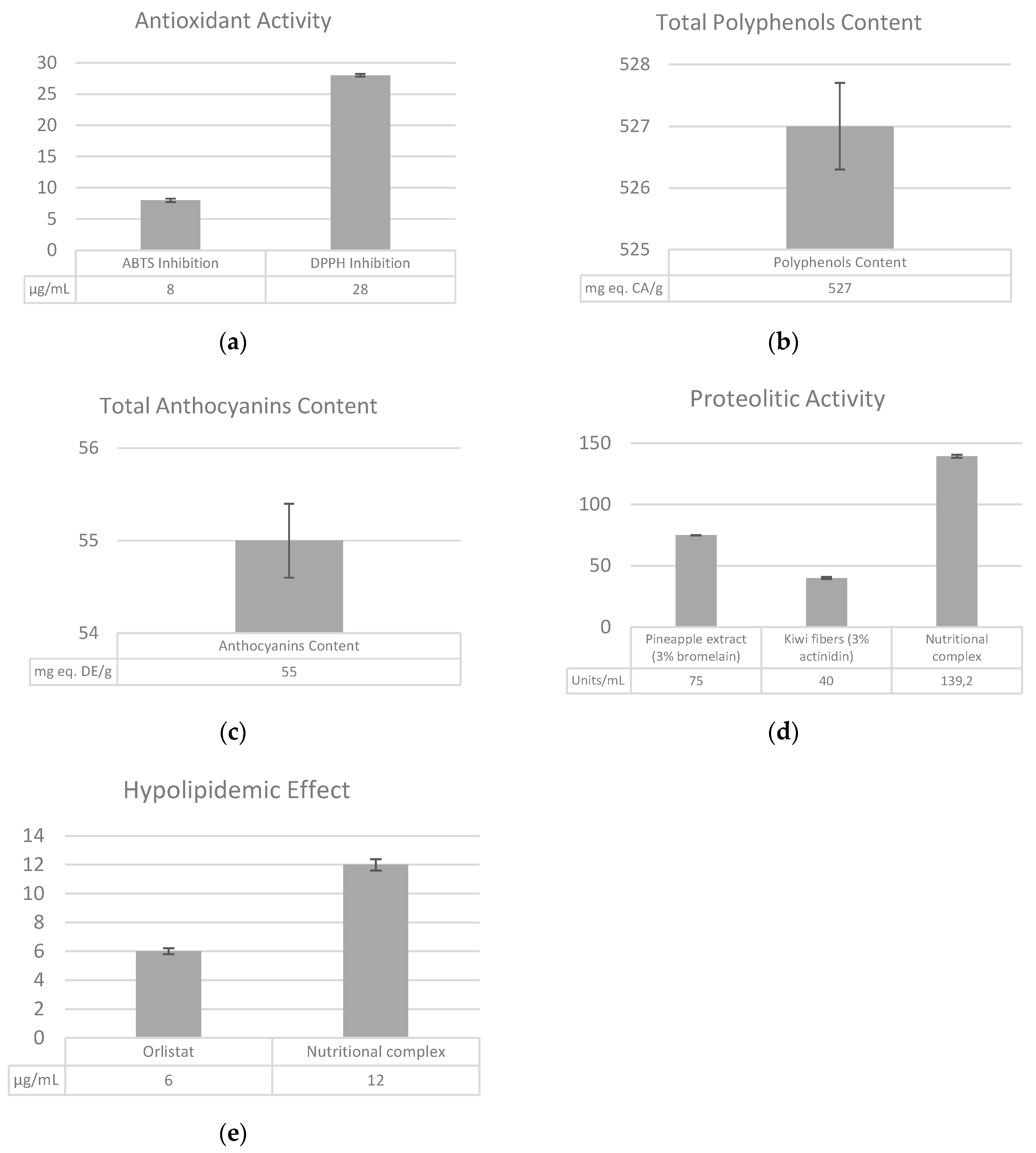
Figure 1. (a) Antioxidant activity: Results are expressed for both the ABTS and DPPH assay as the concentration providing 50% of the radical inhibition. (b) Total polyphenols content: Data are expressed as milligram of catechin equivalent per gram of sample (mg eq. CA/g). (c) Total anthocyanins content: Data are expressed as milligram of delphinidin equivalent per gram of sample (mg eq. DE/g). (d) Proteolytic activity: The results are expressed in units/mL. (e) Hypolipidemic effect-Pancreatic lipase inhibition. Data are expressed as μg/mL. Each measurement was carried out in triplicate. Data are expressed as means ± SEM.
Cosmetics 07 00048 g001
Figure 2. Oil Red O staining assay (40×). Images refers to the stained differentiated adipocytes after incubation with control (a) (no nutraceutical ingredient) and two different concentrations of the tested SelectSIEVE® Rainbow, 0.1% (b) and 0.2% (c).
Cosmetics 07 00048 g002
Figure 3. (a) Body weight: The graph reports the mean variation (as kg) vs. T0. ° Indicates a statistically significant change compared to T0, p < 0.005; °° Indicates a statistically significant change compared to T0, p < 0.001. * Indicates a statistically significant change compared to placebo, p < 0.05; ** Indicates a statistically significant change compared to placebo, p < 0.01. (b) Body circumference: The graph reports the mean variation (as cm) vs. T0. ° Indicates a statistically significant change compared to placebo, p < 0.05; °° Indicates a statistically significant change compared to placebo, p < 0.01. °°° Indicates a statistically significant change compared to placebo, p < 0.001. * Indicates a statistically significant change compared to T0, p < 0.05; ** Indicates a statistically significant change compared to T0, p < 0.01. *** Indicates a statistically significant change compared to T0, p < 0.001. Data are expressed as means ± SEM.
Cosmetics 07 00048 g003
Figure 4. (a) Skin elasticity (R2): R2 values are directly proportional to the degree of skin elasticity: An increase of this parameter is indicative of an improvement of skin elasticity. (b) Skin draining (R6): R6 values are inversely proportional to the degree of skin viscoelasticity: A decrease of this parameter is indicative of an improvement of skin distensibility. (c) Skin tonicity (R0): R0 values are inversely proportional to the degree of skin firmness: A reduction of this parameter is indicative of an improvement of skin firmness. The graphs show the mean variation obtained for each parameter. *** Indicates a statistically significant change compared to T0, p < 0.001. °°° Indicates a statistically significant change compared to placebo, p < 0.001. Data are expressed as means ± SEM.
Cosmetics 07 00048 g004
Figure 5. Evaluation of skin microcirculation: (a) The graph shows the percentage of subjects, belonging to the placebo and the SelectSIEVE® Rainbow group, who showed or did not show an improvement based on clinical evaluation of thermographic analysis. (b) The figure shows the microcirculation improvement by thermographic analysis of subject 22. Thermographic evaluations are performed at the level of the thighs and scored by the dermatologist in relation to the extent of the local variations of the temperature in the thermographic image.
Cosmetics 07 00048 g005
Figure 6. Clinical evaluation of the cellulite skin-derived imperfections (“orange peel”). The graph shows the percentage of subjects, belonging to the placebo and the SelectSIEVE® Rainbow group, who showed or did not show an improvement based on the clinical evaluation.
Cosmetics 07 00048 g006
Table 1. SelectSIEVE® Rainbow components (ROELMI HPC, Origgio (VA), Italy). The composition column refers to quantities expressed as a percentage.
DescriptionBotanical NamePlant PartComposition
Black rice extractOryza sativa L.Semen (Seed)35–45%
Kiwi dried powderActinidia chinensis Planch.Fructus (Fruit)30–40%
Orange extractCitrus sinensis (L.) OsbeckFructus (Fruit)20–30%
Pineapple extract enriched with BromelainAnanas comosus (L.) MerrStipites (Stem)1–5%
Table 2. (a) Termographic evaluation of cellulite-induced alteration of skin microcirculation. (b) Dermatological evaluation of cellulite-derived skin imperfections (“orange peel”). (c) Scoring of both thermographic evaluation and cellulite-derived skin imperfections after 28 and 56 days of product use.
(a) Clinical Classification of the Thermographic Stage at T0Score(b) Dermatological Classification of Orange Peel Skin at T0Score
IVSevere9Slight skin roughness which is not yet clearly visible as “orange peel” skin1
Medium8
Initial7Appearance of “orange peel” skin in a skin region2
IIISevere6
Medium5Appearance of “orange peel” skin in several skin regions3
Initial4
IISevere3Skin appearance like “mattress”4
Medium2
Initial1(c) Scoring of Thermographic Stage and of Improvement of “Orange Peel” SkinScore
INormality0
No variation1
Slight improvement2
Moderate improvement3
Remarkable improvement4
Table 3. Emographic and baseline characteristics.
ActivePlacebo
Sex
Female3030
Age48.3 ± 1.347.0 ± 1.7
Body mass index25.3 ± 0.425.6 ± 0.5
Weight (kg)65.5 ± 1.465.6 ± 2.0
Skin elasticity
R20.7902 ± 0.0140.7934 ± 0.013
R60.4347 ± 0.0190.4270 ± 0.013
R00.3055 ± 0.0110.3202 ± 0.009