Article

Efficacy and Safety of an Oral Nutritional (Dietary) Supplement Containing Pinus pinaster Bark Extract and Grape Seed Extract in Combination with a High SPF Sunscreen in the Treatment of Mild-to-Moderate Melasma: A Prospective Clinical Study

1
Innovation and Development, ISDIN S.A., 08019 Barcelona, Spain
2
Dermik. Clínica Dermatológica multidisciplinar, 08019 Barcelona, Spain
*
Author to whom correspondence should be addressed.
Cosmetics 2019, 6(1), 15; https://doi.org/10.3390/cosmetics6010015
Submission received: 25 January 2019 / Revised: 20 February 2019 / Accepted: 25 February 2019 / Published: 1 March 2019
(This article belongs to the Special Issue Melanogenesis and Melanin-Related Compounds: A Cosmetic Perspective)

Abstract

:
Background: Melasma is a common hyperpigmentation disorder, characterized by light-to-dark brown patches, usually distributed on sun-exposed areas of the body. The objective of this study was to evaluate the efficacy and tolerability of an oral nutritional supplement containing Pinus pinaster and Grape seed extract, vitamins and minerals, used concomitantly with a high SPF sunscreen in 30 women with mild-to-moderate facial melasma. Methods: Efficacy was assessed by measurement of the Melasma Area and Severity Index (MASI), instrumental analysis of the lesions (Mexameter®, VISIA®)) and Patient’s and Physician’s Global Assessment (PGA). Results: The MASI score decreased significantly compared with baseline at days 28, 56, and 84. Mexameter® analysis showed a significant decrease of ∆M (difference in the melanin index between melasma and adjacent area). VISIA® results also showed a reduction in the number and areas of UV pigmented spots and in the areas of melasma overtime. Both the Patient’s and Physician’s Global Assessment showed that the product led to an improvement of the lesions in terms of depigmentation and had positive cosmetic features without adverse events. Conclusion: The oral supplement subject of this study in combination with high SPF sunscreen was effective and well-tolerated for treatment of mild to moderate facial melasma.

1. Introduction

Melasma is a common chronic acquired hyperpigmentation disorder of the skin, characterized by light-to-dark brown patches or macules with symmetrical disposition and irregular borders. Although it is usually present on sun-exposed areas of the body, particularly the regions of the face that are rich in sebaceous glands [1], it can also appear in other body areas such as forearms and neck [2].
This disorder affects both sexes although it is more frequent in females and in darker skin phenotypes (Fitzpatrick skin types III-V). Melasma is a common request for dermatological care and, due to its effect on facial appearance, the disease can be a source of embarrassment for subjects, negatively impacting their quality of life [3].
The occurrence of this disorder has been strongly associated with melanocyte malfunction leading to overproduction of melanin, the naturally occurring pigment in humans responsible for skin, hair and eye colour [4,5]. However, as shown by several studies, the aetiology of the disorder is not limited to melanocyte malfunction [1]. A comparative transcriptional analysis of melasma and healthy surrounding skin revealed the complexity of this disorder in which almost 300 genes related not only to melanocytes, but also to other skin components, seem to be differentially regulated [6].
The aetiology of melasma is not fully understood. However, sun exposure, pregnancy, genetic predisposition, inflammatory processes of the skin, and use of cosmetics, steroids and photosensitizing drugs are considered among the most frequent triggering factors [3,7].
For many years melasma has been strongly linked to female hormone stimulation in women with a genetic predisposition. However, the latest evidence has reduced the impact of female hormones and introduced additional triggering factors pointing out that melasma is a photoaging skin disorder affecting genetically predisposed individuals [1]. Over 40% of melasma subjects report having relatives affected with the disease [3].
New insights also contribute to the understanding of the effect of light exposure on melasma. Recently, beyond ultraviolet (UV) radiation, melasma has been shown to be negatively impacted by other radiation wavelengths such as the shorter wavelengths of visible light [8,9].
Although there are multiple topical therapies, their limited efficacy and recurrence rates have promoted the interest and use of dietary supplements for improvement of melasma symptoms [10]. Thus, there is increased interest in oral medications and nutritional (dietary) supplements for improvement of melasma symptoms as they are easy to administer and provide skin protection at a systemic level. Different oral ingredients have been evaluated, including the bark extract of Pinus pinaster [11] among others [10,12,13] demonstrating that they can strengthen physiological antioxidant skin defences, protect skin from the damaging processes involved in photoaging, and lead to an improvement in skin appearance and pigmentation.
French Maritime Pine Bark Extract (Pinus pinaster) is a natural plant extract containing proanthocyanidins and other antioxidant compounds. A study demonstrated that a daily intake of 75 mg of Pinus pinaster bark extract was therapeutically effective against melasma after 30 days of treatment. No side effects or untoward reactions were observed during the treatment [12].
Grape seed extract (GSE) is also rich in proanthocyanidins. In a one-year study of Japanese women with chloasma, oral administration of 67 mg of GSE three times a day effectively reduced the hyperpigmentation and the extract was shown to be safe and well tolerated [13].
Vitamins are organic compounds which are essential nutrients for humans. They play a significant role in skin health, displaying antioxidant activity and promoting collagen synthesis, keratinization and sebum regulation and photoprotection [14].
The photoprotective effects of vitamins E and C have been intensely studied. A combination of vitamins E and C showed a protective effect in a double-blind, placebo-controlled study. This study showed that combined vitamins C and E reduced the sunburn reaction, UV-induced skin damage and cutaneous blood flow, which increased in the placebo group [15]. In another study, eighteen volunteers received oral supplementation with vitamin E and vitamin C for 3 months to investigate the effect of long-term oral administration of this combination for UVB-induced epidermal damage. After 3 months of treatment, the intake of vitamin C and vitamin E significantly reduced sunburn reaction to UVB irradiation and the thymine dimers induced by irradiation also were significantly reduced [16].
The protective effect of selenium by itself or associated with tocopherol (vitamin E) or other antioxidants is well known in the literature. Supplementation with selenium significantly enhanced the global DNA repair capacities (reviewed in [17]).
Nicotinamide (vitamin B3) is a safe, widely available vitamin that reduces the immune suppressive effects of UV, enhances DNA repair in keratinocytes and has shown promise in the chemoprevention of non-melanoma skin cancer [18].
Finally, the treatment scheme includes the topical use of a high Sun Protection Factor (SPF+50) as a local photoprotection barrier against UV radiation-induced photosensitivity.
The goal of treating melasma is to minimize hyperpigmentation of the affected areas with the lowest impact on surrounding skin in terms of hypopigmentation and irritation. The objective of this study was to evaluate the efficacy and tolerability of an oral nutritional (dietary) supplement containing Pinus pinaster bark extract, GSE, vitamin C and E, nicotinamide and selenium, when used concomitantly with a very high-SPF sunscreen in women with mild to moderate melasma.

2. Materials and Methods

2.1. Subjects

Thirty women from eighteen to fifty years of age and with Fitzpatrick skin type I-V, and facial melasma ranging from mild to moderate were enrolled in this study. All the subjects fulfilled the inclusion criteria.
Subjects with the following conditions were excluded: skin marks in the experimental area that could interfere with results (scars, abundant hair, high quantity of ephelides and nevus, sunburns), history of allergy or reactivity to similar products, history of previous malignant melanoma, use of skin lightening creams during the three weeks previous to the study, use of vitamin A acid or isotretinoin during the three months prior to the study, having received hormonal therapy or hydroquinone within the six months prior to the study, hormonal imbalances, or upcoming changes in lifestyle habits.
The study protocol and the informed consent were approved both by an Independent Ethics Committee (IEC). All the subjects gave written consent before entering the trial.

2.2. Study Design

The study was an open, prospective, monocentric and controlled trial. All subjects took 2 capsules daily with a glass of water during breakfast. The study lasted 84 days and efficacy and tolerability were assessed at Baseline (D0) and Days 28 (D28), 56 (D56) and 84 (D84). Fulfilment of conditions of use was controlled through a questionnaire. Additionally, this questionnaire allowed the collection of each subject’s experience while using this regimen.
The study was performed by EVIC Hispania according to corresponding regulations including the International Conference on Harmonization Guideline for Good Clinical Practice (GCP) and its corresponding ethical principles based on Helsinki Declaration. The study started in December 2012 and was completed in March 2013. Prior to evaluations, subjects were preconditioned in a temperature- and humidity-controlled environment (20°C ± 2°C and Rh of 45% ± 15%) for 15 minutes. All measurements were taken in the same conditions.

2.3. Investigational Products

The investigational product (IP) was an oral nutritional (dietary) supplement containing Pinus pinaster bark extract, Grape seed extract, Vitamin C, Vitamin E, Niacinamide, and selenium. The regimen was 2 capsules daily in the morning.
To avoid bias, subjects were provided with a SPF 50+ sunscreen for use during the entire course of the study. The sunscreen had a broad spectrum with very high protection against UVB and a reinforced high protection against UVA (3 times the minimal UVA protection requested for a SPF 50+). Subjects were requested to apply 2 mg/cm2 of sunscreen to protect at least the face pigmented area before sun exposure and to reapply as many times as needed every 2 hours.

2.4. MASI

Melasma Area and Severity Index was scored by dermatologist assessment according to the method developed by Kimbrough-Green et al (1994) [19].

2.5. Physician’s Global Assessment

The dermatologist completed the Physician’s Global Assessment (PGA) at D28, D56 and D84 evaluating the improvement of melasma by giving a numeric value from 0 to 6 (0 = clear; 1 = almost clear; 2 = Marked improvement; 3 = Moderate improvement; 4 = Slight improvement; 5 = No improvement; and 6 = Worse).

2.6. Patient’s Global Assessment

Subjects completed the Patient’s Global Assessment (PGA) at D28, D56 and D84. PGAs were evaluated by given a numeric value from 0 to 6 (0 = clear; 1 = almost clear; 2 = Marked improvement; 3 = Moderate improvement; 4 = Slight improvement; 5 = No improvement; and 6 = Worse).

2.7. Instrumental Evaluation of Efficacy

2.7.1. Measurements of Skin Depigmenting Properties by Mexameter®

The capability of the investigational product to effectively decrease the melasma pigmentation intensity was evaluated by the comparison of skin color of the hyperpigmented area before (D0) and during treatment (D28, D56 and D84). Melasma spots at the left and right hemispheres and the frontal region of the face of at least 5 mm were selected and evaluated during the whole study.
Measurements were carried out using a Mexameter® (MX 18, Courage & Khazaka Electronic, Cologne, Germany) on three points allocated within the melasma areas and on three different points located within the non-pigmented area adjacent to the melasmas, as control measurements. The Melanin Index (MI) and ΔMI were calculated.

2.7.2. Melasma Assessment by VISIA®-CA and Image J

Standardized images for the left and right hemispheres and the frontal region of the face were obtained before (D0) and during treatment (D28, D56 and D84) using a VISIA® Complexion Analysis system (Canfield Scientific, NJ, USA). Initially, these standardized photos identify visible spots in the evaluated area. Then, mask was applied in the subsequent measurement in order to evaluate the reduction of number and area of these spots. Through the images obtained from VISIA® where a melasma spot is identified, the ImageJ software calculates the area of melasma and to assess the reduction of the pigment area (D28, D56 and D84).
In addition, as exploratory objective, using VISIA®, UV spots were evaluated as a preliminary analysis of anti-ageing parameters.

2.8. Subject Self-Assessment of the Properties of the Product

All participants completed a questionnaire at D28, D56 and D84. The survey included questions regarding the product’s characteristics and intake, efficacy on pigmentation changes and skin qualities, and global evaluation.

2.9. Evaluation of Tolerability

2.9.1. Self-Evaluation

Tolerability was self-evaluated daily by recording all observed reactions and relevant findings using a personal evaluation form starting the first day of the study. Subjects were asked several questions in order to detect potential unpleasant experiences from use of the product (heat, burning, itching, tightness) and to evaluate adverse reactions (erythema, oedema, blisters, scabs, dryness, colouring, spots) and their intensity (mild, moderate, severe).

2.9.2. Visual Examination by Dermatologist

Tolerability was assessed through visual examination of the experimental skin area by an experienced dermatologist. Evaluation was done on the first day of the study and on D28, D56 and D84.

2.10. Statistical Analyses

Values are expressed as mean and standard error of the mean (SEM). A Fisher’s exact test was employed to compare differences in categorical variables. A Student’s t-test for paired samples was performed to evaluate changes between the data obtained at baseline and at each time-point. Differences were considered significant at p < 0.05. Statistical analysis was performed using the SAS software (version 9.3, SAS Institute Inc., Cary, N.C.).

3. Results

3.1. Subjects

30 women were included in the study and completed it. Population characteristics are shown in Table 1.

3.2. Melasma Area and Severity Index (MASI)

After examination of the 30 subjects, a statistically significant reduction in MASI score at D28, D56, and D84 in comparison with D0 (p < 0.0001, all) was observed (Figure 1).
The mean variation percentage of MASI was −18.2% at D28, −33.7 at D56 and −41.4% at D84, showing a progressive reduction of the degree of melasma affectation.

3.3. Melasma Decrease by Mexameter®

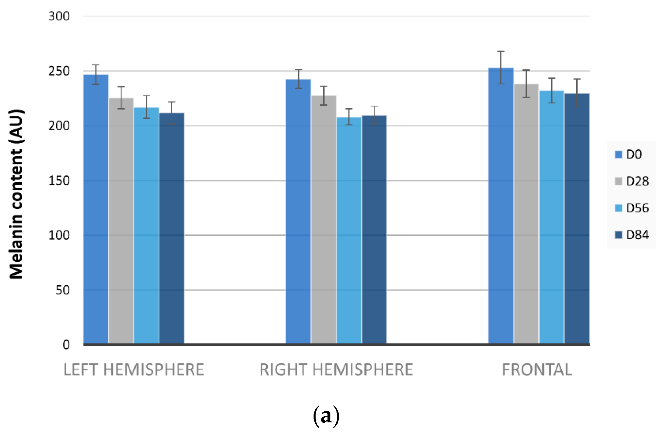
A statistically significant reduction in the pigmentation of melasma for the three assessed facial areas was observed at each timepoints (Figure 2A). In contrast, the melanin index of the majority of adjacent skin areas was maintained at both timepoints.
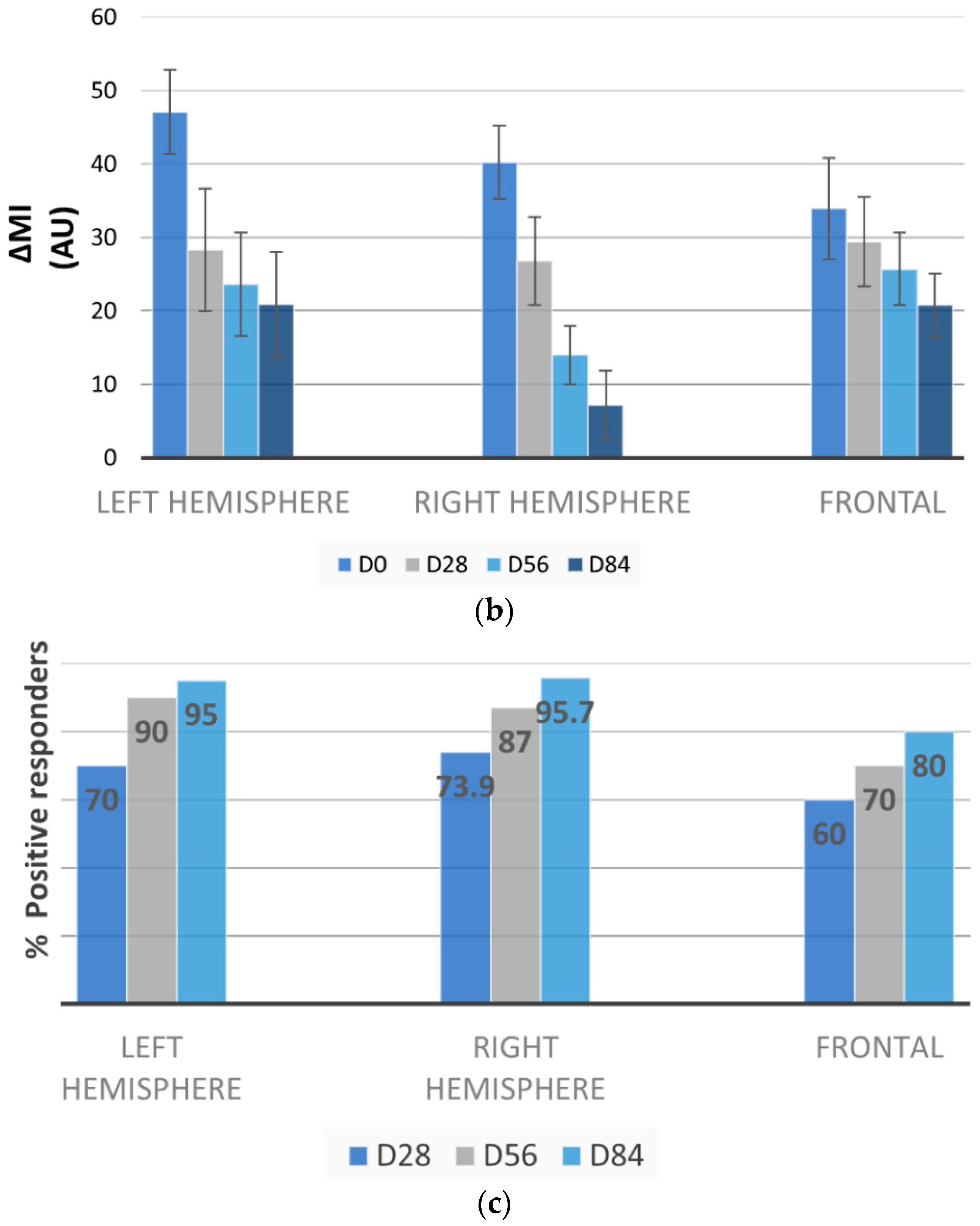
Moreover, values of ΔMI (difference between the values of melanin index obtained in the area of melasma and the ones obtained in adjacent areas) showed a significant reduction in both hemispheres (p < 0.01) at each timepoint and in the front area at D84 (p = 0.009) (Figure 2B). This reduction translates into a decrease of contrast between the colour of melasma spot and adjacent area.
In addition, the percentage of reactive subjects, meaning the percentage of patients presenting a decrease of at least 10% of melanin index between melasma and adjacent areas, versus D0, was analysed.
The percentage of reactive subjects was 70% at D28, 90% at D56 and 95% at D84 for the left facial hemisphere and 73.9% at D28, 87.0% at D56 and 95.7% at D84 for the right facial hemisphere. Reactive responders for the frontal facial areas were 60.0% at D28 70.0% at D56 and 80% at D84 (Figure 2C).
16.7% of the reactive subjects presented melasma at the 3 evaluated areas (right and left hemispheres and frontal area), 43.3% in 2 areas, and 40.0% in 1 area.

3.4. Physician’s and Patient’s Global Assessment

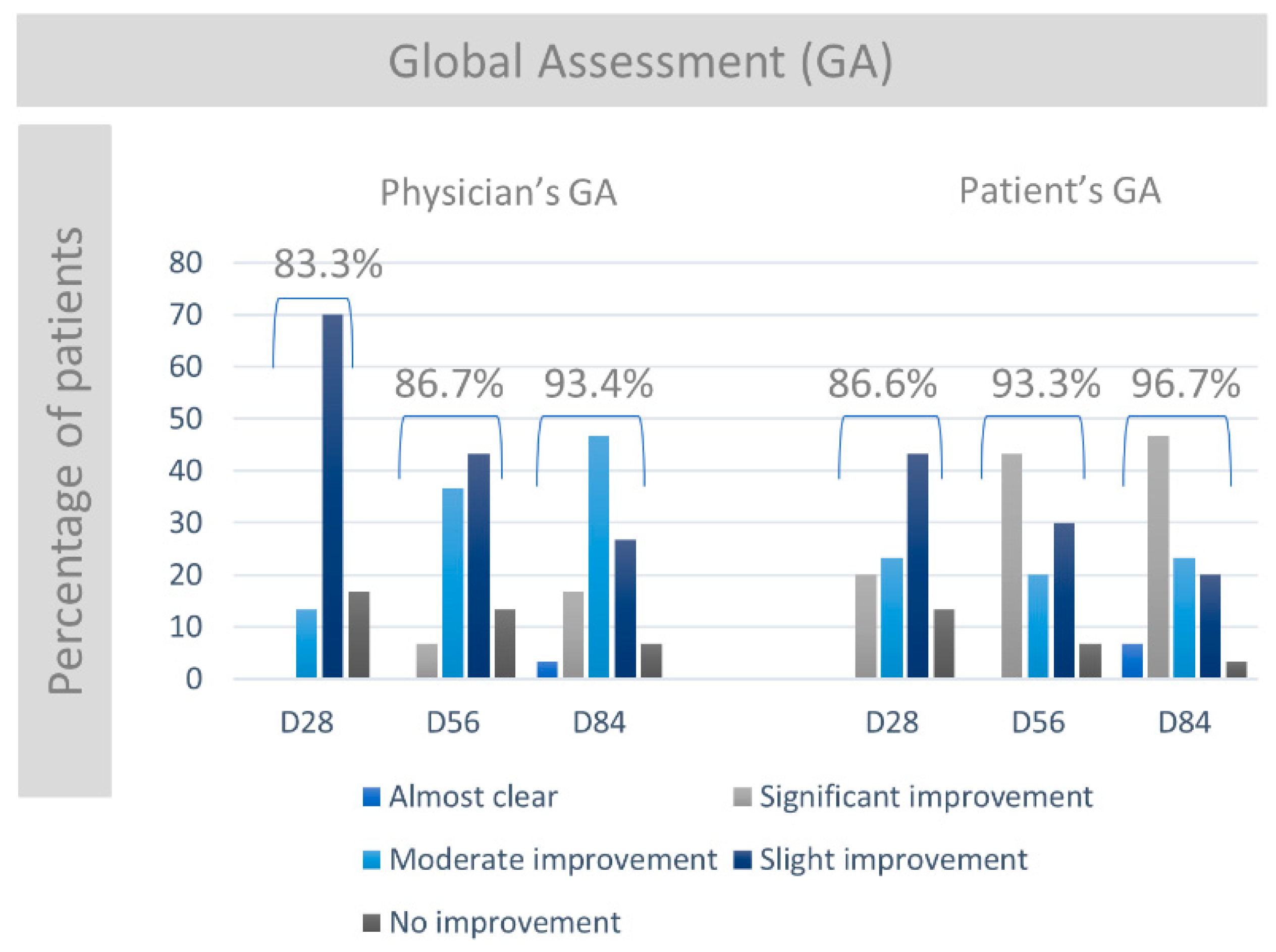
The efficacy of the product was assessed at day D28, D56 and D84 by the Physician and the subjects through the Global Assessment form (PGA). At D28, 83.3% of subjects manifested improvement according to the Physician’s Global Assessment. The percentage of subjects self-reporting improvement according to subjects Global Assessment was 86.6%. These percentages increased up to 86.7% (physicians) and 93.3% (patient) at D56 and 93.3% (physician) and 96.7% (patient) at D84 (Figure 3). Additionally, 53.4% of subjects and 20% of physicians reported either elimination of hyperpigmentation or significant improvement at day 84 of the study.

3.5. Melasma Assessment by VISIA-CA® and Image J

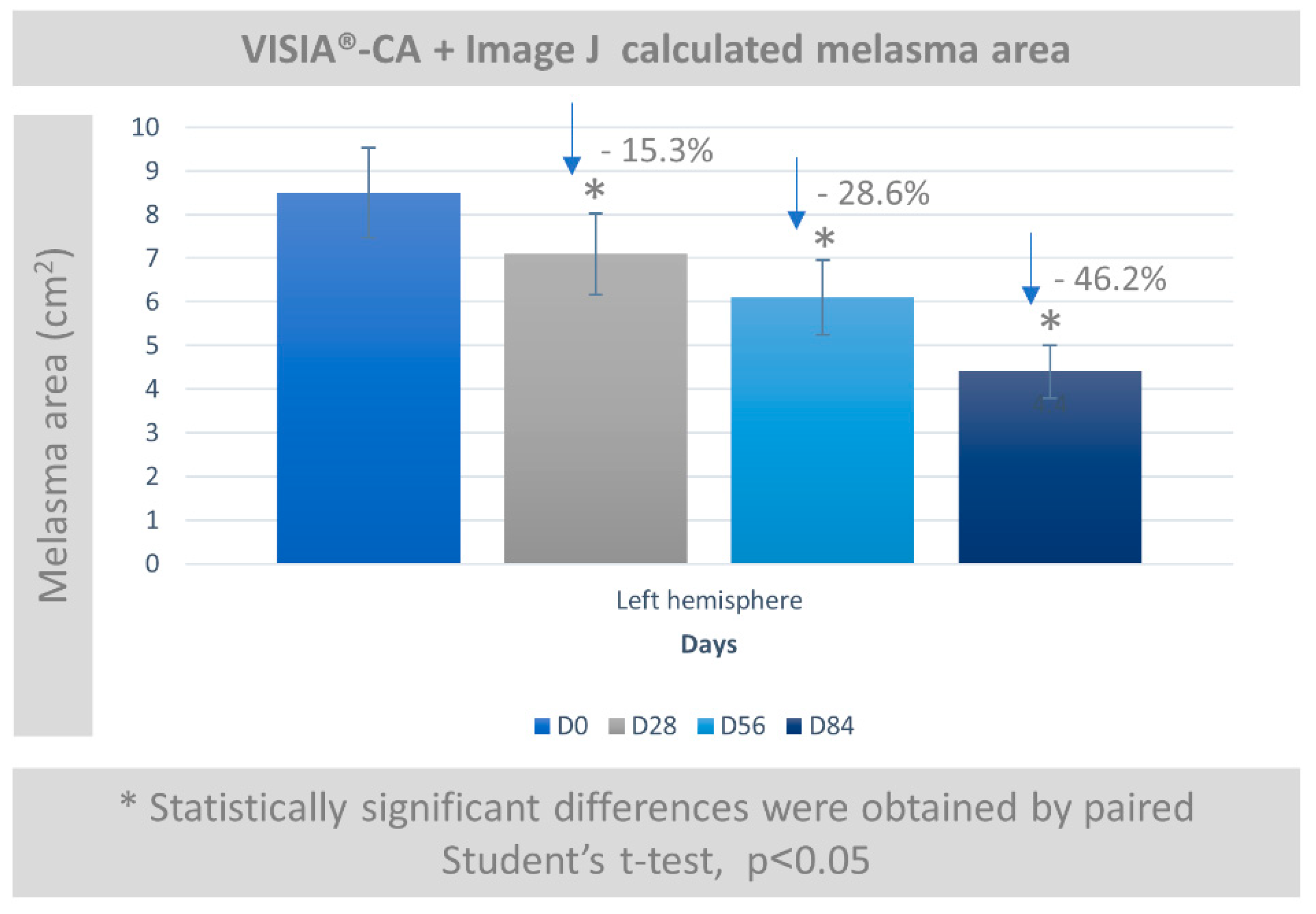
The effect of a measurable decrease in melasma by use of the product on the left hemisphere of the face was evaluated by VISIA-CA® and surface was calculated by using Image J as can be seen in Figure 4. A statistically significant reduction in spots was seen (p < 0.05) with a reduction of 15.3% at D28, 28.6% at D56 and 46.2% at D84 for the mean percentage of variation.

3.6. Subjects Self-Assessment of the Depigmenting Properties of the Product

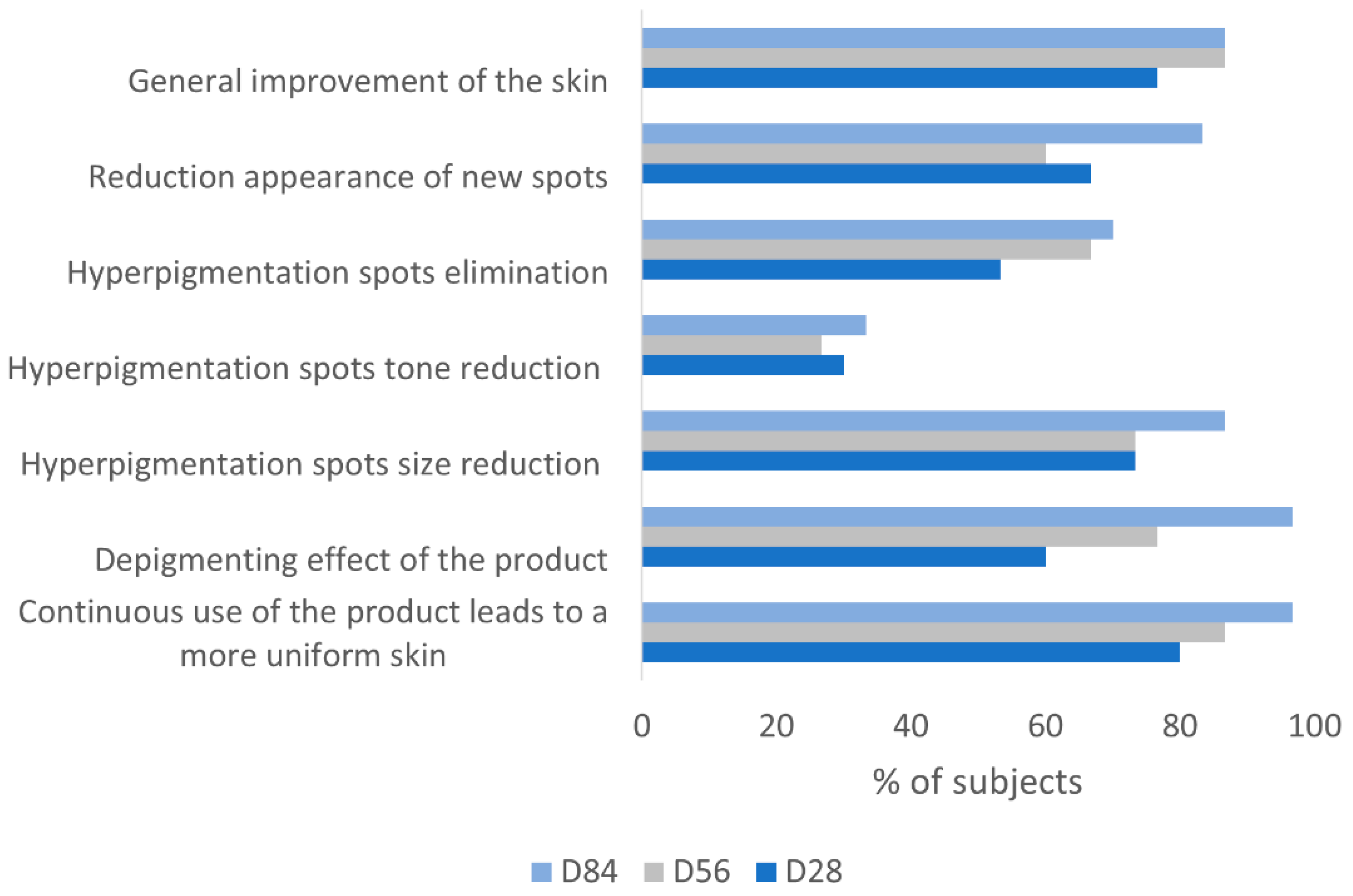
The subjective and qualitative efficacy evaluation shows that according to study subjects continuous use of the supplement (for 84 days) product has “depigmenting and uniformity effect” in 96.7%, “reduces the spot size” in 86.7%, “reduces the appearance of new spots” in 83.3%, and “eliminates the spots” in 70% of cases (Figure 5). Globally, 27 out of the 30 subjects in the study reported a positive overall assessment of the product (quite like it or like it very much).

3.7. Exploratory Analysis: Anti-ageing Parameters

3.7.1. Detection UV Spots by VISIA-CA®

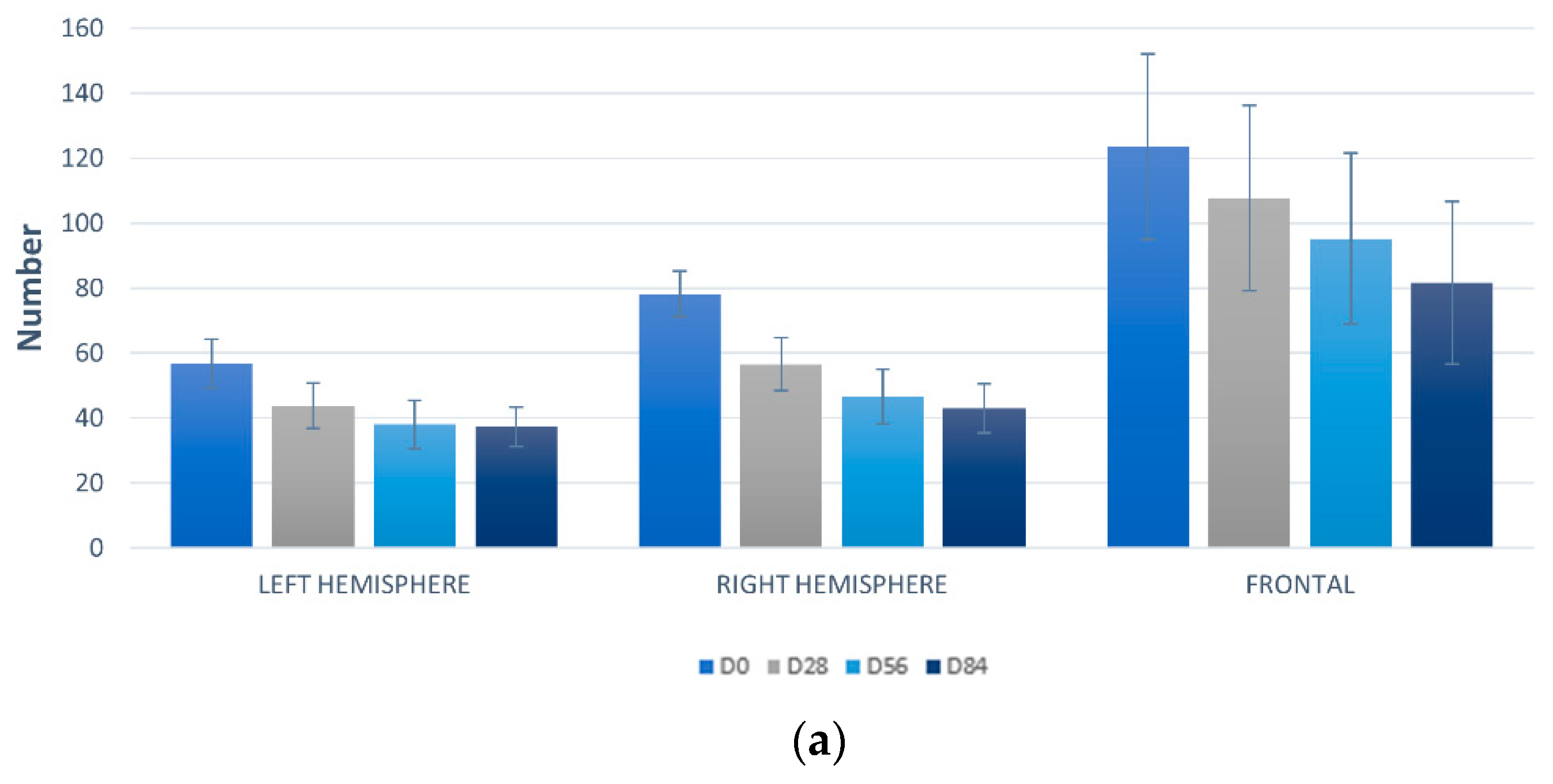
A statistically significant reduction of the number of UV spots at D56 and D84 (Student’s t-test p <0.05) was detected in the left hemisphere and in the frontal facial region in comparison with D0. Additionally, in the right facial hemisphere the number of UV spots also showed a significant decrease at D28 (Student’s t-test p < 0.05) (Figure 6A).
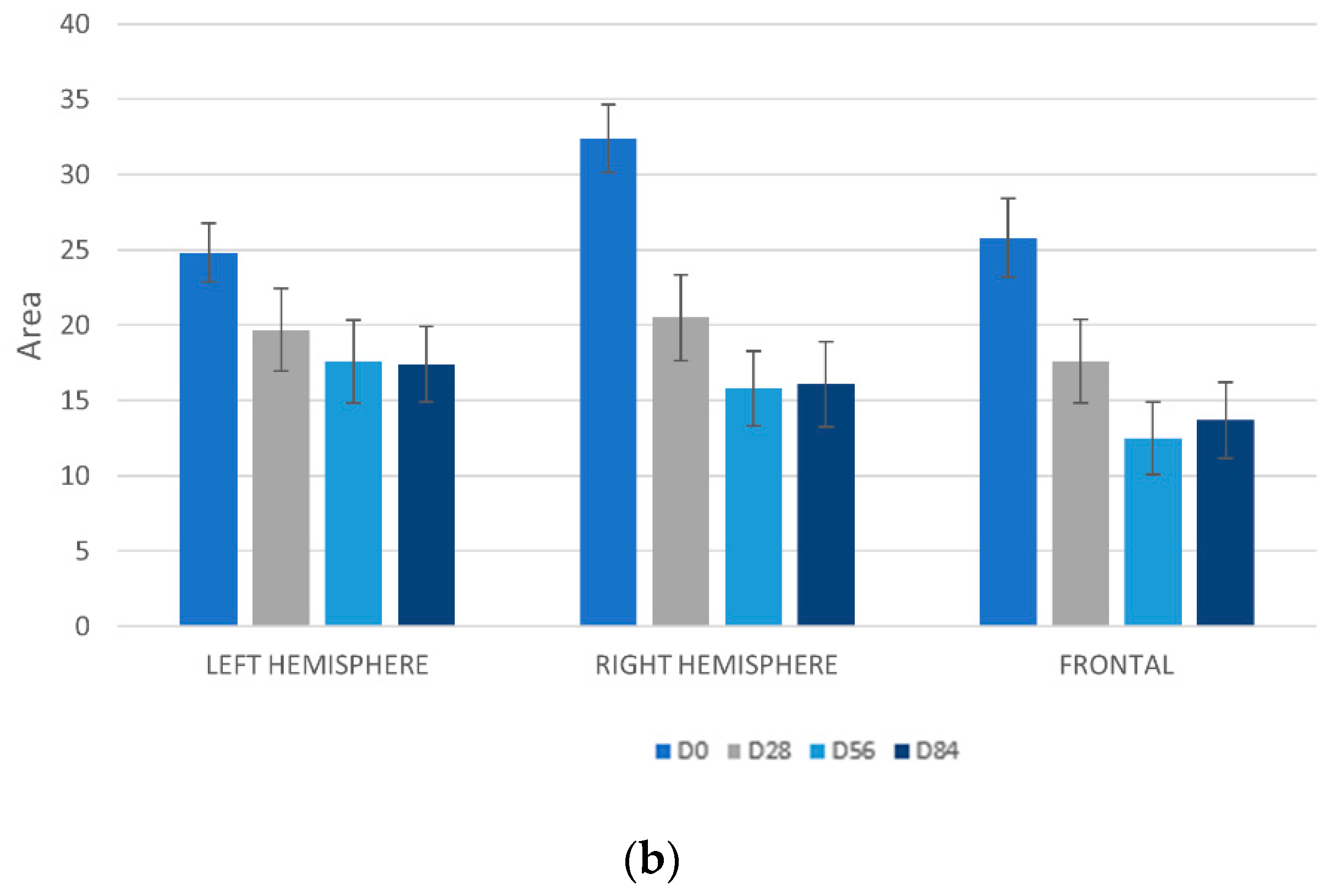
A statistically significant reduction in the area of UV spots was detected in the three facial regions (right and left hemispheres and frontal region) in comparison with D0 at all the assayed times (D28, D56 and D84) (p < 0.05) (Figure 6B).

3.7.2. Subjects Self-Assessment of the Anti-Ageing Properties of the Product

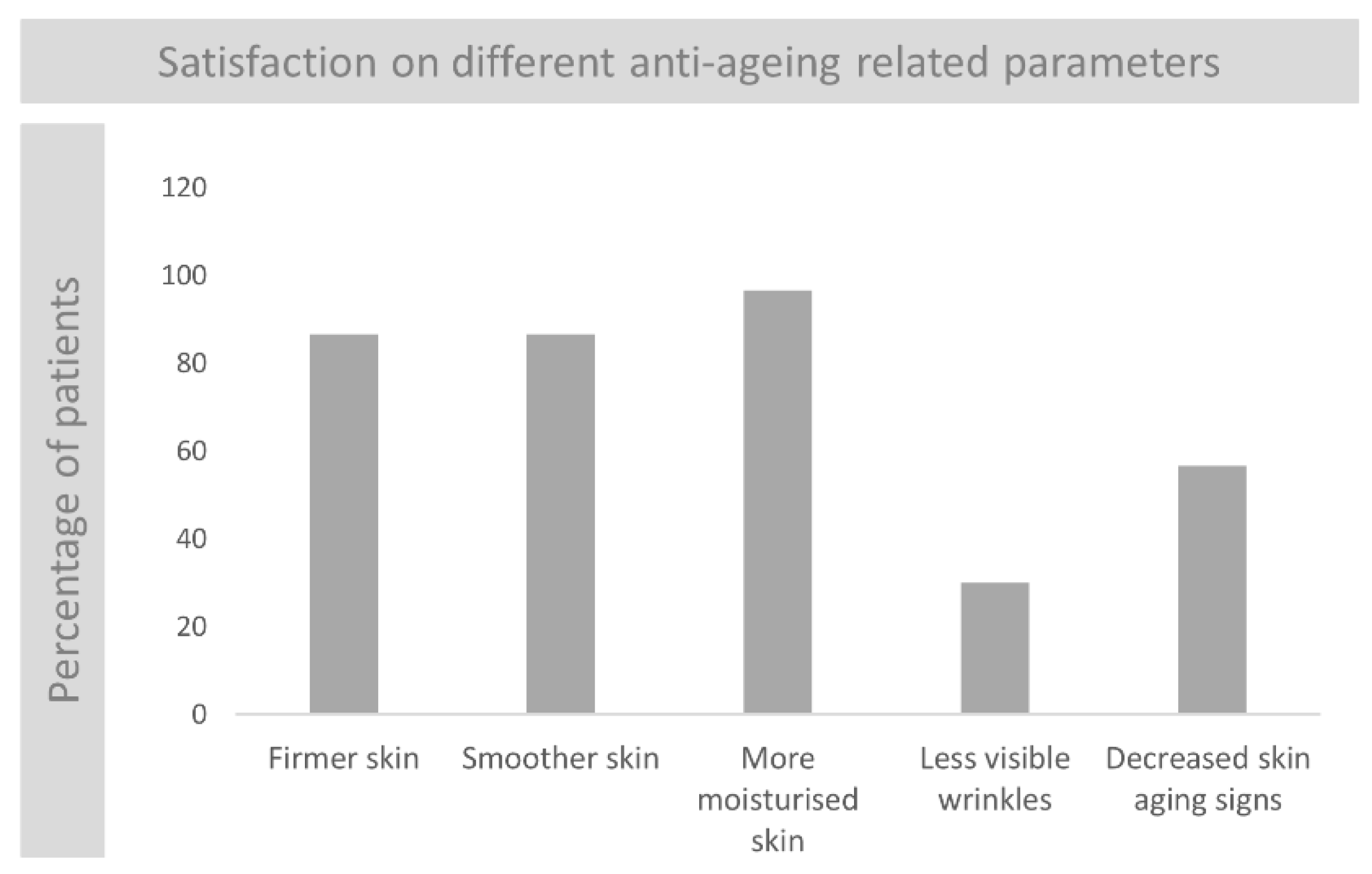
At the 12th-week visit, the patients indicated that the product had “improved skin moisturisation” in 96.7%, “improved the general aspect of skin” in 86.7%, “improved skin firmness and smoothness” in 86.7% and “reduced signs of age” in 56.7% of the cases (Figure 7).

3.7.3. Evaluation of Tolerability

The nutritional (dietary) supplement was very well tolerated after 84 days of treatment, as none of the subjects reported any unpleasant sensations or showed clinical signs that could be attributed to the use of the product under study. Additionally, when patients were asked to report their subjective evaluation of different parameters related to the ease of use of the product (including size, taste and smell of the capsule) their feedback was positive (100% ease of use, 83.3% ease of ingestion, 86,6% correct or small size of capsule, 96.7% correct or weak smell of the capsule and 100% neutral to likeable taste of capsule) (Table S1).

4. Discussion

Melasma is a common hyperpigmentation disorder affecting mostly individuals with darker skin phototypes (higher Fitzpatrick Skin types) and is more prevalent in women. The aetiology and pathogenesis of melasma is not fully understood and its treatment remains a challenge because treatments are frequently disappointing and not free from side effects. Melasma, appearing predominantly on the face, can have a significant psychological impact resulting in a negative effect on quality of life. Individuals with melasma commonly request dermatological care for well tolerated treatment strategies.
Common current therapies for melasma range from topical agents to laser therapies with sometimes suboptimal results or linked to undesirable adverse effects [2]. Due to the complexity of the disorder there is still a need of developing well tolerated treatments that in addition with adequate sun protection, can efficiently lead to reduction of melasma.
Oral supplements with antioxidant, anti-inflammatory and depigmenting properties, are useful and may act synergistically with topical treatments and lasers.
A novel oral nutritional (dietary) supplement based on Pinus pinaster bark extract and proanthocyanidin-rich grape seed extract, has been developed to be incorporated in the armamentarium for the treatment of melasma. Vitamin E and C, selenium and nicotinamide have also been shown to reduce the negative effects of UV radiation on skin by eliminating reactive oxygen species and reducing the erythema associated with UV [18,20,21].
In this study conducted with 30 women with mild to moderate facial melasma and different skin phenotypes (II-IV), efficacy and tolerability of an oral formulation with melasma decreasing properties has been evaluated. Since UV radiation is one of the main triggering factors of the disease (impact worsens during the summer season), limiting sun exposure is crucial [1,22]. Therefore, the intervention was designed as a co-adjuvant treatment to the application of topical sun protection with a high Sun Protection Factor index (SPF 50+). The study was performed during winter months when the intensity of the sun is reduced.
The product demonstrated very good tolerance. The dermatologist did not detect any clinical signs attributable to the use of the product in any of the 30 subjects at any of the study timepoints. The method chosen to evaluate tolerability has been previously used in other studies [23,24,25].
In terms of product efficacy, the nutritional (dietary) supplement subject of this study demonstrated skin melasma reduction properties as shown by instrumental methods, clinical evaluation and subject self-assessment.
Melasma Area and Severity Index (MASI) is a widely used outcome measure in clinical studies on melasma [26,27]. The clinical evaluation of the surface, homogeneity and intensity of pigmentation performed by the dermatologist showed that the Melasma Area and Severity Index (MASI) was significantly reduced at the three assayed times (D28, D56 and D84) in comparison to day 0 (D0). The average percentage of reduction at D84 with respect to D0 was −41.4%.
Instrumental measurement of melasma skin pigmentation by means of melanin index determination demonstrated decrease in the severity of the melasma as a result of the intake of the nutritional (dietary) supplement and an increasing percentage of subjects with a positive response in all facial regions at all timepoints in the course of the study reaching 95% for the left facial hemisphere 95.7% for the right facial hemisphere and 80% for the frontal facial hemisphere at day 84.
Instrumental measurement using Image J images obtained through VISIA-CA® showed that the surface of melasma from the three facial areas was statistically reduced compared to before initiation of treatment and. This was in line with results obtained after processing the images with ImageJ software. The analysis proved that the size of melasma was significantly reduced in the three facial regions at all experimental timepoints. A similar methodology has been used in previous studies [28,29,30].
Evaluation of data on the efficacy of the supplement obtained from the Global Assessment performed by the subjects and the physician concluded that both physician and subjects, reported that the use of the product translated into an improvement of melasma-affected skin. In addition, this improvement increased as the duration of treatment progressed. At D28 improvement of the pigmentation (mild, moderate or significant) was observed in 83.3% of subjects according to physician and in 86.6% of subjects according to subjects themselves. These percentages increased up to 86.7% (physician) and 93.3% (patient) at D56 and 93.3% (physician) and 96.7% (patient) at D84 respectively. Additionally, at D84 of the study either elimination of hyperpigmentation or significant improvement was observed in 53.4% of the subjects according to patients, and in 20% of subjects according to the physician.
VISIA-CA® results showed a statistically significant reduction in the number of UV spots at several timepoints in the three assessed facial areas. A statistically significant reduction was also detected in the area of UV spots in all assessed facial regions in comparison with D0 at all the assayed times.
Subjects showed a high degree of overall satisfaction with the use of the product since 27/30 (90%) users reported liking the product (quite like it, like it much), the remaining 3 (10%) stated to be indifferent (neither like or dislike). Subjects reported that with the continuous use of the nutritional (dietary) supplement the skin acquired a more uniform tone aspect and that the product showed a depigmenting effect reducing/eliminating hyperpigmented spots and reducing the appearance of new spots. Additionally, at the 12th-week visit, a high percentage of patients reported that the product had “improved skin moisturisation”, “improved the general aspect of skin”, “improved skin firmness and smoothness” and “reduced signs of age”.
One limitation of the study is the lack of a very high SPF sunscreen arm without the nutritional (dietary) supplement. Considering the positive outcomes of this trial, a randomized controlled study with increased sample size could be considered in the future to further evaluate the efficacy of this nutritional (dietary) supplement.

5. Conclusions

The studied oral nutritional dietary supplement containing Pinus pinaster bark extract, Grape seed extract, vitamins and minerals is effective and well tolerated as a treatment option for mild to moderate facial melasma in combination with high SPF sunscreen.

Supplementary Materials

The following are available online at https://www.mdpi.com/2079-9284/6/1/15/s1, Table S1. Number of subjects (and percentages) reporting their subjective evaluation of different quality parameters.

Author Contributions

S.A. designed and conducted research, analyzed data and performed statistical analysis; SPRIM Health Group prepared a first draft of the paper; and A.G., P.V.-M., S.A., C.G., J.P.-C. critically reviewed the manuscript. C.G. had the responsibility for the final content. All the authors have read and approved the final manuscript.

Funding

The study has been funded by Laboratorios ISDIN (Spain).

Acknowledgments

The authors wish to thank SPRIM Health Group for their contribution in preparing a first draft of the manuscript.

Conflicts of Interest

Sonia Aladrén; Aurora Garre, MD, Palmira Valderas-Martínez; Corinne Granger, MD are ISDIN employees. Jaime Piquero Casals, Dermatologist is an employee of Dermik, Clínica Dermatológica multidisciplinar, Barcelona, Spain. All authors have participated in the interpretation of the data and in the decision to publish the results. No other conflict of interest were declared.

References

  1. Passeron, T.; Picardo, M. Melasma, a photoaging disorder. Pigment Cell Melanoma Res. 2018, 31, 461–465. [Google Scholar] [CrossRef] [PubMed] [Green Version]
  2. Gupta, A.K.; Gover, M.D.; Nouri, K.; Taylor, S. The treatment of melasma: A review of clinical trials. J. Am. Acad. Dermatol. 2006, 55, 1048–1065. [Google Scholar] [CrossRef] [PubMed]
  3. Handel, A.C.; Miot, L.D.; Miot, H.A. Melasma: A clinical and epidemiological review. An. Bras. Dermatol. 2014, 89, 771–782. [Google Scholar] [CrossRef] [PubMed]
  4. Kang, W.H.; Yoon, K.H.; Lee, E.S.; Kim, J.; Lee, K.B.; Yim, H.; Sohn, S.; Im, S. Melasma: Histopathological characteristics in 56 Korean subjects. Br. J. Dermatol. 2002, 146, 228–237. [Google Scholar] [CrossRef] [PubMed]
  5. Grimes, P.E.; Yamada, N.; Bhawan, J. Light microscopic, immunohistochemical, and ultrastructural alterations in subjects with melasma. Am. J. Dermatopathol. 2005, 27, 96–101. [Google Scholar] [CrossRef] [PubMed]
  6. Kang, H.Y.; Suzuki, I.; Lee, D.J.; Ha, J.; Reiniche, P.; Aubert, J.; Deret, S.; Zugaj, D.; Voegel, J.J.; Ortonne, J.P. Transcriptional profiling shows altered expression of wnt pathway- and lipid metabolism-related genes as well as melanogenesis-related genes in melasma. J. Investig. Dermatol. 2011, 131, 1692–1700. [Google Scholar] [CrossRef] [PubMed]
  7. Lee, A.Y. Recent progress in melasma pathogenesis. Pigment Cell Melanoma Res. 2015, 28, 648–660. [Google Scholar] [CrossRef] [PubMed]
  8. Duteil, L.; Cardot-Leccia, N.; Queille-Roussel, C.; Maubert, Y.; Harmelin, Y.; Boukari, F.; Ambrosetti, D.; Lacour, J.P.; Passeron, T. Differences in visible light-induced pigmentation according to wavelengths: A clinical and histological study in comparison with UVB exposure. Pigment Cell Melanoma Res. 2014, 27, 822–826. [Google Scholar] [CrossRef] [PubMed]
  9. Regazzetti, C.; Sormani, L.; Debayle, D.; Bernerd, F.; Tulic, M.K.; De Donatis, G.M.; Chignon-Sicard, B.; Rocchi, S.; Passeron, T. Melanocytes Sense Blue Light and Regulate Pigmentation through Opsin-3. J. Investig. Dermatol. 2018, 138, 171–178. [Google Scholar] [CrossRef] [PubMed]
  10. Juhasz, M.L.W.; Levin, M.K. The role of systemic treatments for skin lightening. J. Cosmet. Dermatol. 2018, 21. [Google Scholar] [CrossRef] [PubMed]
  11. Zhou, L.L.; Baibergenova, A. Melasma: Systematic review of the systemic treatments. Int. J. Dermatol. 2017, 56, 902–908. [Google Scholar] [CrossRef] [PubMed]
  12. Ni, Z.; Mu, Y.; Gulati, O. Treatment of melasma with Pycnogenol. Phytother. Res. 2002, 16, 567–571. [Google Scholar] [CrossRef] [PubMed]
  13. Yamakoshi, J.; Sano, A.; Tokutake, S.; Saito, M.; Kikuchi, M.; Kubota, Y.; Kawachi, Y.; Otsuka, F. Oral intake of proanthocyanidin-rich extract from grape seeds improves chloasma. Phytother. Res. 2004, 18, 895–899. [Google Scholar] [CrossRef] [PubMed]
  14. Shapiro, S.S.; Saliou, C. Role of vitamins in skin care. Nutrition 2001, 17, 839–844. [Google Scholar] [CrossRef]
  15. Eberlein-Konig, B.; Placzek, M.; Przybilla, B. Protective effect against sunburn of combined systemic ascorbic acid (vitamin C) and D-alpha-tocopherol (vitamin E). J. Am. Acad. Dermatol. 1998, 38, 45–48. [Google Scholar] [CrossRef]
  16. Placzek, M.; Gaube, S.; Kerkmann, U.; Gilbertz, K.P.; Herzinger, T.; Haen, E.; Przybilla, B. Ultraviolet B-induced DNA damage in human epidermis is modified by the antioxidants ascorbic acid and D-alpha-tocopherol. J. Investig. Dermatol. 2005, 124, 304–307. [Google Scholar] [CrossRef] [PubMed]
  17. Bera, S.; De Rosa, V.; Rachidi, W.; Diamond, A.M. Does a role for selenium in DNA damage repair explain apparent controversies in its use in chemoprevention? Mutagenesis 2013, 28, 127–134. [Google Scholar] [CrossRef] [PubMed]
  18. Thompson, B.C.; Surjana, D.; Halliday, G.M.; Damian, D.L. Nicotinamide enhances repair of ultraviolet radiation-induced DNA damage in primary melanocytes. Exp. Dermatol. 2014, 23, 509–511. [Google Scholar] [CrossRef] [PubMed] [Green Version]
  19. Kimbrough-Green, C.K.; Griffiths, C.E.; Finkel, L.J.; Hamilton, T.A.; Bulengo-Ransby, S.M.; Ellis, C.N.; Voorhees, J.J. Topical retinoic acid (tretinoin) for melasma in black patients. A vehicle-controlled clinical trial. Arch. Dermatol. 1994, 130, 727–733. [Google Scholar] [CrossRef] [PubMed]
  20. Fuchs, J.; Kern, H. Modulation of UV-light-induced skin inflammation by D-alpha-tocopherol and L-ascorbic acid: A clinical study using solar simulated radiation. Free Radic. Biol. Med. 1998, 25, 1006–1012. [Google Scholar] [CrossRef]
  21. Favrot, C.; Beal, D.; Blouin, E.; Leccia, M.T.; Roussel, A.M.; Rachidi, W. Age-Dependent Protective Effect of Selenium against UVA Irradiation in Primary Human Keratinocytes and the Associated DNA Repair Signature. Oxid. Med. Cell. Longev. 2018, 5895439. [Google Scholar] [CrossRef] [PubMed]
  22. Demirkan, S.; Gündüz, Ö.; Sayan, C.D. Retrospective Analysis of Endemic Melasma subjects. Dermatol. Rep. 2017, 9, 7027. [Google Scholar] [CrossRef] [PubMed]
  23. Jackson, E.M.; Robillard, N.F. The controlled use test in a cosmetic product safety substantation program. J. Toxicol. Cutan. Ocul. Toxicol. 1982, 20, 117–132. [Google Scholar]
  24. Keswick, B.H.; Ertel, K.D.; Visscher, M.O. Comparison of exaggerated and normal use techniques for assessing the mildness of personal cleansers. J. Soc. Cosm. Chem. 1992, 43, 187–193. [Google Scholar]
  25. Jackwerth, B.; Krächter, H.U.; Matthies, W. Dermatological test methods for optimising mild tenside preparations. Parfüm. Kosmet. 1993, 74, 134–141. [Google Scholar]
  26. Pandya, A.; Berneburg, M.; Ortonne, J.P.; Picardo, M. Guidelines for clinical trials in melasma. Pigmentation Disorders Academy. Br. J. Dermatol. 2006, 156 (Suppl. 1), 21–28. [Google Scholar] [PubMed]
  27. Pandya, A.G. Reliability assessment and validation of the Melasma Area and Severity Index (MASI) and a new modified MASI scoring method. J. Am. Acad. Dermatol. 2011, 64, 78–83. [Google Scholar] [CrossRef] [PubMed]
  28. Clarys, P.; Alewaeters, K.; Barel, A.O. Comparative Study of Skin Color Using Different Bioengineering Methods. Skin Res. Technol. 1999, 5, 129. [Google Scholar]
  29. Costa, A.; Moisés, T.A.; Cordero, T.; Alves, C.R.; Marmirori, J. Association of emblica, licorice and belides as an alternative to hydroquinone in the clinical treatment of melasma. An. Bras. Dermatol. 2010, 85, 613–620. [Google Scholar] [CrossRef] [PubMed]
  30. Dell’Acqua, G.; Wagner, C. Lightening and Illuminating Skin with Acetylated Hydroxystilbenes from Rheum rhaponticum. Cosmet. Toilet. 2011, 126, 634–642. [Google Scholar]
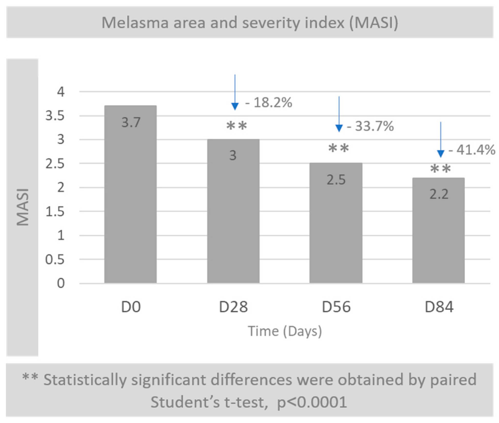
Figure 1. MASI score at D0, D28, D56 and D84.
Cosmetics 06 00015 g001
Figure 2. Efficacy of the nutritional (dietary) supplement on different facial regions assessed by Mexameter®. (a) Melanin index of the melasma at different timepoints in the different facial areas; (b) ΔMI represents the difference between the values of melanin index obtained in the area of melasma and the ones obtained in adjacent areas; (c) Percentage of subjects responding positively to the treatment.
Cosmetics 06 00015 g002aCosmetics 06 00015 g002b
Figure 3. Percentage of subjects reporting different levels of response (almost clear, significant, moderate, slight or no improvement) determined both by Physician’s and by Patient’s Global Assessment at different times (D28, D56 and D84).
Cosmetics 06 00015 g003
Figure 4. Melasma area (cm2) in left hemisphere obtained by analysis of images by Image J software.
Cosmetics 06 00015 g004
Figure 5. Percentage of subjects manifesting satisfaction on several pigmentation parameters evaluated throughout the study (D28, D56 and D84).
Cosmetics 06 00015 g005
Figure 6. Measurements of UV spots in different facial regions (left and right hemisphere and frontal area) at different times (D0, D28, D56 and D84). (a) Number of UV spots in evaluated areas; (b) Surface of UV spots in evaluated areas.
Cosmetics 06 00015 g006aCosmetics 06 00015 g006b
Figure 7. Evaluation of satisfaction at D84: percentage of subjects manifesting satisfaction on different anti-ageing related parameters.
Cosmetics 06 00015 g007
Table 1. Baseline characteristics of subjects.
Subject Baseline Characteristics
Total30 women
Age - Mean (Range)39 (23–63)
Skin Phototype (Fitzpatrick *)
II3 (10%)
III23 (76.7%)
IV4 (13.3%)
* Established after a 30–40 min sun exposure following winter or an equivalent period of no sun exposure. II: burns easily, tans slightly; III: burns moderately, tans progressively; IV: burns minimally, tans easily.