Phytochemical-Loaded Nanotherapeutics in Cosmetic Surgery Wound Healing: A Narrative Review

 ,
,  ,
,  , , and
Abstract
1. Introduction
1.1. Skin and Its Anatomy: A Brief
1.2. Types of Cosmetic Surgery
- Breast Augmentation—Surgical treatment that entails the insertion of breast implants to enhance the volume and contour of the breasts.
- Liposuction—Surgical technique that eliminates surplus adipose tissue from targeted regions of the body, such as the belly, hips, thighs, or buttocks.
- Rhinoplasty—commonly referred to as a nose job—is a surgical operation that alters the shape of the nose to enhance its aesthetic appeal and functionality.
- Facelift—Surgical operation that elevates and firms the skin of the face and neck to diminish the visibility of wrinkles and lax skin.
- Stomach Tuck—Abdominoplasty is a surgical operation that excises surplus skin and adipose tissue from the abdomen to achieve a more streamlined and contoured appearance.
- Blepharoplasty—An eyelid surgery procedure that excises surplus skin and adipose tissue from the upper and lower eyelids to achieve a more youthful and refreshed appearance.
- Breast Reduction—This treatment involves the excision of surplus breast tissue and skin to diminish breast size and enhance shape.
- Botox—Non-invasive therapy that entails the injection of a neurotoxin to temporarily immobilize the muscles responsible for wrinkles and creases on the face.
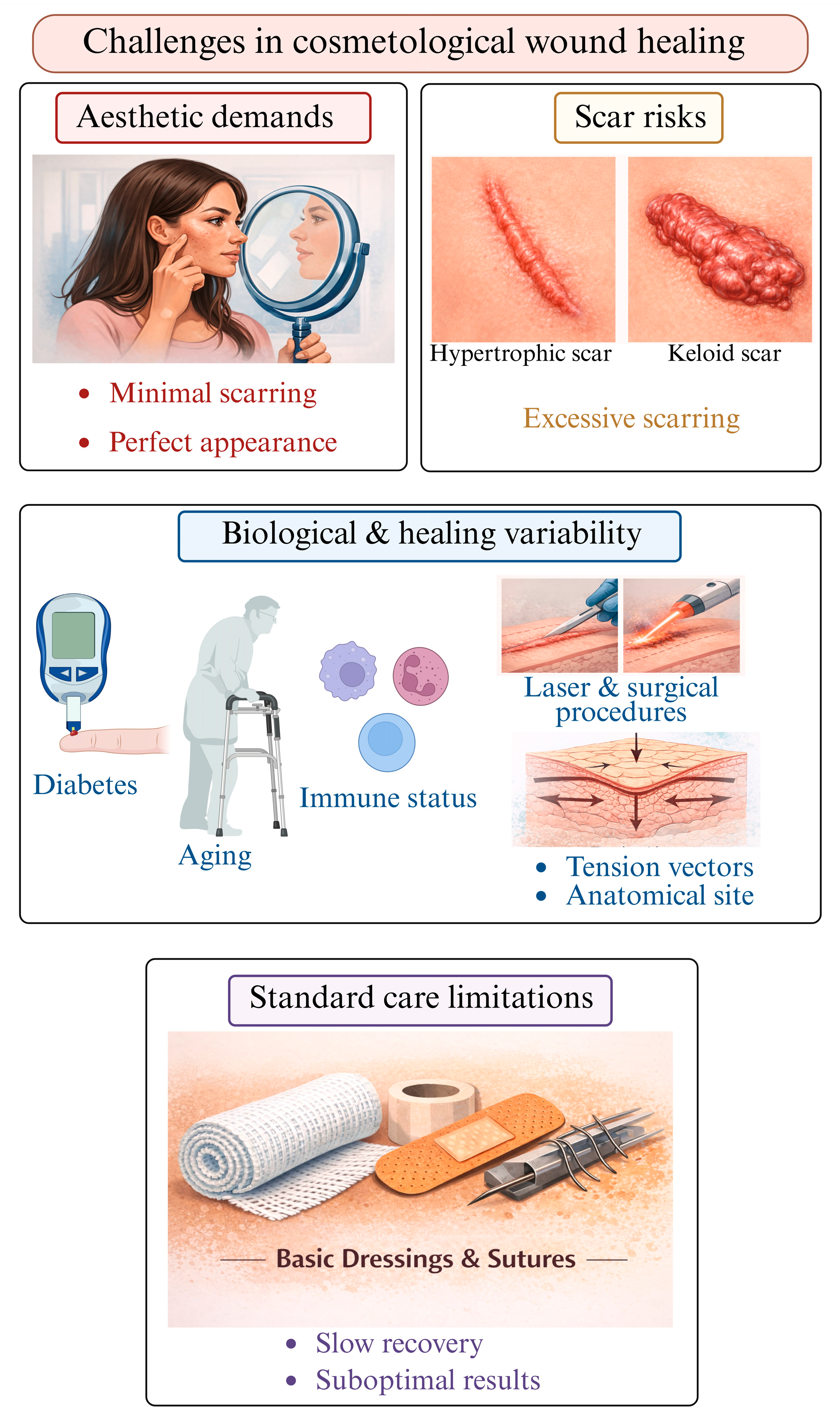
1.3. Challenges in Cosmetological Wound Healing
1.4. Need for Advanced Therapeutics in Cosmetological Wound Healing
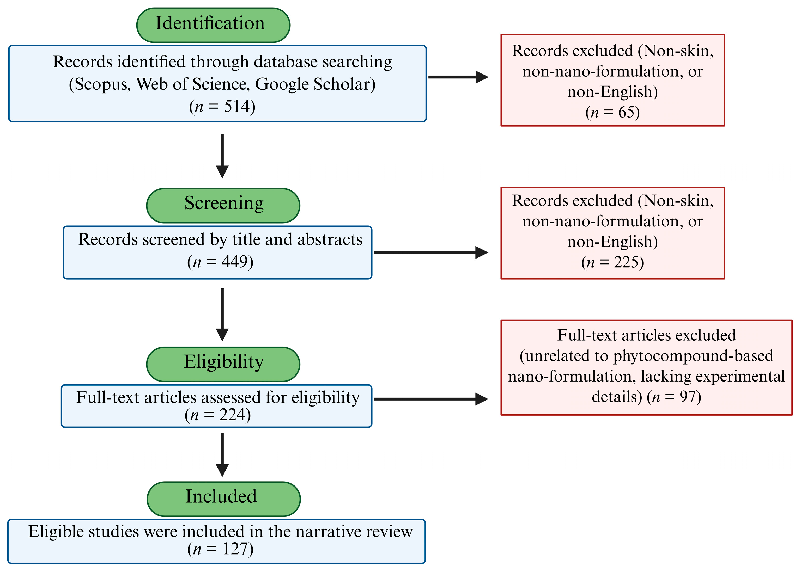
2. Methodology
3. Phytochemicals with Wound Healing Properties
4. Current Clinical Strategies for Surgical Wound Management
4.1. Infection Prevention
4.2. Surgical Closure Materials
4.3. Advanced Wound Devices [Closed-Incision Negative-Pressure Therapy (ciNPT)]
4.4. Scar Modulation Strategies
5. Emerging Adjunct Therapies
6. Nanotechnology in Wound Healing (Nanotechnology-Based Delivery Systems)
6.1. Polymeric Nanocarriers
6.2. Hydrogel Nanoparticles
6.3. Nanofibers
6.4. Nanocapsules/Nanospheres
7. Lipid Nanocarriers
7.1. Solid Lipid Nanoparticles (SLNs)
7.2. Nanostructured Lipid Carriers (NLCs)
8. Vesicular Nanosystems
8.1. Liposomes
8.2. Transferosomes
8.3. Herbosomes
8.4. Ethosomes
8.5. Niosomes
8.6. Cubosomes
9. Inorganic Nanocarriers
9.1. Representative Inorganic Nanoparticles in Wound Healing
9.1.1. Silver Nanoparticles (Ag NPs)
9.1.2. Copper Nanoparticles (CuNPs)
9.1.3. Gold Nanoparticles (AuNPs)
9.1.4. Zinc Oxide Nanoparticles (ZnO NPs)
10. Major Possible Mechanisms

11. Phytochemical-Based Nanocarriers for Cosmetic Surgery
12. Limitations
13. Future Perspectives
14. Conclusions
Author Contributions
Funding
Institutional Review Board Statement
Informed Consent Statement
Data Availability Statement
Acknowledgments
Conflicts of Interest
References
- Abdo, J.M.; Sopko, N.A.; Milner, S.M. The applied anatomy of human skin: A model for regeneration. Wound Med. 2020, 28, 100179. [Google Scholar] [CrossRef]
- Mohamed, S.A.; Hargest, R. Surgical anatomy of the skin. Surgery 2022, 40, 1–7. [Google Scholar] [CrossRef]
- Shastri, M.; Sharma, M.; Sharma, K.; Sharma, A.; Minz, R.W.; Dogra, S.; Chhabra, S. Cutaneous-immuno-neuro-endocrine (CINE) system: A complex enterprise transforming skin into a super organ. Exp. Dermatol. 2024, 33, e15029. [Google Scholar] [CrossRef]
- Jin, R.; Luo, L.; Zheng, J. The Trinity of Skin: Skin Homeostasis as a Neuro–Endocrine–Immune Organ. Life 2022, 12, 725. [Google Scholar] [CrossRef]
- Gupta, R.K.; Wasnik, P.; Mondal, D.; Shukla, D. Critical role of keratinocytes in cutaneous immune responses. Explor. Immunol. 2024, 4, 502–522. [Google Scholar] [CrossRef]
- Skobowiat, C.; Dowdy, J.C.; Sayre, R.M.; Tuckey, R.C.; Slominski, A. Cutaneous hypothalamic-pituitary-adrenal axis homolog: Regulation by ultraviolet radiation. Am. J. Physiol. Endocrinol. Metab. 2011, 301, E484–E493. [Google Scholar] [CrossRef]
- Bikle, D.D. Vitamin D and the skin: Physiology and pathophysiology. Rev. Endocr. Metab. Disord. 2012, 13, 3–19. [Google Scholar] [CrossRef]
- Roosterman, D.; Goerge, T.; Schneider, S.W.; Bunnett, N.W.; Steinhoff, M. Neuronal control of skin function: The skin as a neuroimmunoendocrine organ. Physiol. Rev. 2006, 86, 1309–1379. [Google Scholar] [CrossRef]
- Lotfollahi, Z. The anatomy, physiology and function of all skin layers and the impact of ageing on the skin. Wound Pract. Res. 2024, 32, 6–10. [Google Scholar] [CrossRef]
- McGrath, J.A.; Uitto, J. Structure and Function of the Skin. In Rook’s Textbook of Dermatology, 10th ed.; Griffiths, C.E.M., Barker, J., Bleiker, T.O., Hussain, W., Simpson, R.C., Eds.; Wiley-Blackwell: Hoboken, NJ, USA, 2024; Volume 1. [Google Scholar]
- Brito, S.; Baek, M.; Bin, B.H. Skin Structure, Physiology, and Pathology in Topical and Transdermal Drug Delivery. Pharmaceutics 2024, 16, 1403. [Google Scholar] [CrossRef]
- Dermitzakis, I.; Chatzi, D.; Kyriakoudi, S.A.; Evangelidis, N.; Vakirlis, E.; Meditskou, S.; Theotokis, P.; Manthou, M.E. Skin Development and Disease: A Molecular Perspective. Curr. Issues Mol. Biol. 2024, 46, 8239–8267. [Google Scholar] [CrossRef]
- Maksimovic, S.; Nakatani, M.; Baba, Y.; Nelson, A.M.; Marshall, K.L.; Wellnitz, S.A.; Firozi, P.; Woo, S.-H.; Ranade, S.; Patapoutian, A.; et al. Epidermal Merkel cells are mechanosensory cells that tune mammalian touch receptors. Nature 2014, 509, 617–621. [Google Scholar] [CrossRef]
- Hemat Jouy, S.; Mohan, S.; Scichilone, G.; Mostafa, A.; Mahmoud, A.M. Adipokines in the Crosstalk between Adipose Tissues and Other Organs: Implications in Cardiometabolic Diseases. Biomedicines 2024, 12, 2129. [Google Scholar] [CrossRef]
- Richard, A.J.; White, U.; Elks, C.M.; Stephens, J.M. Adipose Tissue: Physiology to Metabolic Dysfunction. In Endotext [Internet]; Feingold, K.R., Adler, R.A., Ahmed, S.F., Anawalt, B., Blackman, M.R., Chrousos, G., Corpas, E., de Herder, W.W., Dhatariya, K., Dungan, K., et al., Eds.; MDText.com, Inc.: South Dartmouth, MA, USA, 2000. Available online: https://www.ncbi.nlm.nih.gov/books/NBK555602/?utm_ (accessed on 16 March 2026).
- Blüher, M. Clinical Relevance of Adipokines. Diabetes Metab. J. 2012, 36, 317–327. [Google Scholar] [CrossRef] [PubMed]
- Ioannidou, A.; Fisher, R.M.; Hagberg, C.E. The multifaceted roles of the adipose tissue vasculature. Obes. Rev. 2022, 23, e13403. [Google Scholar] [CrossRef]
- Wollina, U. An overview of plastic surgery: Types, benefits and considerations. J. Cosmo. Tricho. 2023, 9, 203. [Google Scholar]
- Sorg, H.; Sorg, C.G.G. Skin Wound Healing: Of Players, Patterns, and Processes. Eur. Surg. Res. 2023, 64, 141–157. [Google Scholar] [CrossRef]
- Mony, M.P.; Harmon, K.A.; Hess, R.; Dorafshar, A.H.; Shafikhani, S.H. An Updated Review of Hypertrophic Scarring. Cells 2023, 12, 678. [Google Scholar] [CrossRef]
- Landers, K.; Hwang, M.; Cottrill, E. A narrative review of scars after surgery: What to expect. Ann. Thyroid 2023, 8, 9. [Google Scholar] [CrossRef]
- Lee, E.H. Patient expectations and performance measures in dermatologic surgery. Clin. Dermatol. 2016, 34, 111–113. [Google Scholar] [CrossRef] [PubMed]
- Monstrey, S.; Middelkoop, E.; Vranckx, J.J.; Bassetto, F.; Ziegler, U.E.; Meaume, S.; Téot, L. Updated scar management practical guidelines: Non-invasive and invasive measures. J. Plast. Reconstr. Aesthetic Surg. 2014, 67, 1017–1025. [Google Scholar] [CrossRef]
- Alster, T.S.; Lupton, J.R. Treatment of complications of laser skin resurfacing. Arch. Facial Plast. Surg. 2000, 2, 279–284. [Google Scholar] [CrossRef]
- Ogawa, R. Mechanobiology of scarring. Wound Rep. Reg. 2011, 19, S2–S9. [Google Scholar] [CrossRef]
- Mamun, A.A.; Shao, C.; Geng, P.; Wang, S.; Xiao, J. Recent advances in molecular mechanisms of skin wound healing and its treatments. Front. Immunol. 2024, 15, 1395479. [Google Scholar] [CrossRef]
- Almadani, Y.H.; Vorstenbosch, J.; Davison, P.G.; Murphy, A.M. Wound Healing: A Comprehensive Review. Semin. Plast. Surg. 2021, 35, 141–144. [Google Scholar] [CrossRef]
- Wang, P.; Huang, B.; Horng, H.; Yeh, C.; Chen, Y. Wound healing. J. Chin. Med. Assoc. 2018, 81, 94–101. [Google Scholar] [CrossRef]
- Ozgok Kangal, M.K.; Kopitnik, N.L. Physiology, Wound Healing. In StatPearls [Internet]; StatPearls Publishing: Treasure Island, FL, USA, 2025. Available online: https://www.ncbi.nlm.nih.gov/books/NBK535406/?utm_ (accessed on 16 March 2026).
- Fernández-Guarino, M.; Hernández-Bule, M.L.; Bacci, S. Cellular and Molecular Processes in Wound Healing. Biomedicines 2023, 11, 2526. [Google Scholar] [CrossRef]
- Wang, Z.C.; Zhao, W.Y.; Cao, Y.; Liu, Y.Q.; Sun, Q.; Shi, P.; Cai, J.-Q.; Shen, X.Z.; Tan, W.-Q. The Roles of Inflammation in Keloid and Hypertrophic Scars. Front. Immunol. 2020, 11, 603187. [Google Scholar] [CrossRef]
- Kohlhauser, M.; Mayrhofer, M.; Kamolz, L.P.; Smolle, C. An Update on Molecular Mechanisms of Scarring-A Narrative Review. Int. J. Mol. Sci. 2024, 25, 11579. [Google Scholar] [CrossRef]
- Darby, I.A.; Desmoulière, A. Scar Formation: Cellular Mechanisms. In Textbook on Scar Management: State of the Art Management and Emerging Technologies; Téot, L., Mustoe, T.A., Middelkoop, E., Gauglitz, G.G., Eds.; Springer: Cham, Switzerland, 2020. Available online: https://www.ncbi.nlm.nih.gov/books/NBK586083/ (accessed on 16 March 2026).
- Wallace, H.A.; Basehore, B.M.; Zito, P.M. Wound Healing Phases. In StatPearls [Internet]; StatPearls Publishing: Treasure Island, FL, USA, 2025. Available online: https://www.ncbi.nlm.nih.gov/books/NBK470443/?utm_source.com (accessed on 16 March 2026).
- Fang, X.; Wang, Y.; Chen, H.; Yan, Z.; Jin, S.; Wu, Y.; Shu, F.; Xiao, S. Hypertrophic Scarring and Keloids: Epidemiology, Molecular Pathogenesis, and Therapeutic Interventions. MedComm 2025, 6, e70381. [Google Scholar] [CrossRef]
- Kazemeini, S.; Nadeem-Tariq, A.; Hajian, P.; Anil, B.; Easterly, J.; Sraa, K.; Pokharel, S.; Metellus, R.; Kazemeini, M. Hypertrophic and Keloid Scar Management: Advances in Diagnosis, Perioperative Care, and Anesthetic Modulation. Cureus 2025, 17, e88810. [Google Scholar] [CrossRef]
- Latoni, D.I.; McDaniel, D.C.; Tsao, H.; Tsao, S.S. Update on the Pathogenesis of Keloid Formation. JID Innov. 2024, 4, 100299. [Google Scholar] [CrossRef]
- Berman, B.; Maderal, A.; Raphael, B. Keloids and Hypertrophic Scars: Pathophysiology, Classification, and Treatment. Dermatol. Surg. 2017, 43, S3–S18. [Google Scholar] [CrossRef]
- Cedillo-Cortezano, M.; Martinez-Cuevas, L.R.; López, J.A.M.; Barrera López, I.L.; Escutia-Perez, S.; Petricevich, V.L. Use of Medicinal Plants in the Process of Wound Healing: A Literature Review. Pharmaceuticals 2024, 17, 303. [Google Scholar] [CrossRef]
- Vitale, S.; Colanero, S.; Placidi, M.; Di Emidio, G.; Tatone, C.; Amicarelli, F.; D’Alessandro, A.M. Phytochemistry and Biological Activity of Medicinal Plants in Wound Healing: An Overview of Current Research. Molecules 2022, 27, 3566. [Google Scholar] [CrossRef]
- Fuloria, S.; Mehta, J.; Chandel, A.; Sekar, M.; Rani, N.N.I.M.; Begum, M.Y.; Subramaniyan, V.; Chidambaram, K.; Thangavelu, L.; Nordin, R.; et al. A Comprehensive Review on the Therapeutic Potential of Curcuma longa Linn. in Relation to its Major Active Constituent Curcumin. Front. Pharmacol. 2022, 13, 820806. [Google Scholar] [CrossRef]
- Khameneh, B.; Eskin, N.A.M.; Iranshahy, M.; Fazly Bazzaz, B.S. Phytochemicals: A Promising Weapon in the Arsenal against Antibiotic-Resistant Bacteria. Antibiotics 2021, 10, 1044. [Google Scholar] [CrossRef]
- Mitra, A.; Shahid, A.; Kumari, S.; Mukherjee, T.; Pramanick, S.; Mohanty, S.; Ansari, M.A.; Adhikary, K.; Prabhakar, P.K.; Kesari, K.K. Optimizing wound healing: Insights from phytochemicals and advanced therapies. Inflammopharmacology 2025, 33, 4009–4035. [Google Scholar] [CrossRef]
- Witkowska, K.; Paczkowska-Walendowska, M.; Garbiec, E.; Cielecka-Piontek, J. Topical Application of Centella asiatica in Wound Healing: Recent Insights into Mechanisms and Clinical Efficacy. Pharmaceutics 2024, 16, 1252. [Google Scholar] [CrossRef]
- Vijayaraghavan, K.; Rajkumar, J.; Bukhari, S.N.; Al Sayed, B.; Seyed, M.A. Chromolaena odorata: A neglected weed with a wide spectrum of pharmacological activities (Review). Mol. Med. Rep. 2017, 15, 1007–1016. [Google Scholar] [CrossRef]
- Ceriani, R.; Cherif-Pino, D.A.; Pérez-Basáez, P.; Escobar, M.; Leyton, P.; Weinstein-Oppenheimer, C.R.; Moraga-Espinoza, D.F.; Bahamondez-Canas, T.F. Tailoring Polymeric Scaffolds with Buddleja globosa Extract for Dual Antimicrobial and Biocompatible Wound Healing Applications. Molecules 2025, 30, 2428. [Google Scholar] [CrossRef] [PubMed]
- Berry, C.E.; Brenac, C.; Gonzalez, C.E.; Kendig, C.B.; Le, T.; An, N.; Griffin, M.F. Natural Compounds and Biomimetic Engineering to Influence Fibroblast Behavior in Wound Healing. Int. J. Mol. Sci. 2024, 25, 3274. [Google Scholar] [CrossRef] [PubMed]
- Berríos-Torres, S.I.; Umscheid, C.A.; Bratzler, D.W.; Leas, B.; Stone, E.C.; Kelz, R.R.; Reinke, C.E.; Morgan, S.; Solomkin, J.S.; Mazuski, J.E.; et al. Centers for Disease Control and Prevention Guideline for the Prevention of Surgical Site Infection, 2017. JAMA Surg. 2017, 152, 784–791, Erratum in: JAMA Surg. 2017, 152, 803. [Google Scholar] [CrossRef]
- Wolfhagen, N.; Boldingh, Q.J.J.; Boermeester, M.A.; de Jonge, S.W. Perioperative care bundles for the prevention of surgical-site infections: Meta-analysis. Br. J. Surg. 2022, 109, 933–942. [Google Scholar] [CrossRef]
- Darouiche, R.O.; Wall, M.J., Jr.; Itani, K.M.; Otterson, M.F.; Webb, A.L.; Carrick, M.M.; Miller, H.J.; Awad, S.S.; Crosby, C.T.; Mosier, M.C.; et al. Chlorhexidine-Alcohol versus Povidone-Iodine for Surgical-Site Antisepsis. N. Engl. J. Med. 2010, 362, 18–26. [Google Scholar] [CrossRef] [PubMed]
- Wade, R.G.; Burr, N.E.; McCauley, G.; Bourke, G.; Efthimiou, O. The Comparative Efficacy of Chlorhexidine Gluconate and Povidone-iodine Antiseptics for the Prevention of Infection in Clean Surgery: A Systematic Review and Network Meta-analysis. Ann. Surg. 2021, 274, e481–e488. [Google Scholar] [CrossRef]
- Children’s Mercy Kansas City. SSI Postoperative Surgical Bundle: Summary. Clin. Crit. Apprais. Top. 2020, 12. Available online: https://scholarlyexchange.childrensmercy.org/clinical-critically-appraised-topics/12 (accessed on 19 March 2026).
- Jalalzadeh, H.; Timmer, A.S.; Buis, D.R.; Dreissen, Y.E.M.; Goosen, J.H.M.; Graveland, H.; Griekspoor, M.; IJpma, F.F.A.; van der Laan, M.J.; Schaad, R.R.; et al. Triclosan-Containing Sutures for the Prevention of Surgical Site Infection: A Systematic Review and Meta-Analysis. JAMA Netw. Open 2025, 8, e250306. [Google Scholar] [CrossRef]
- Depuydt, M.; Van Egmond, S.; Petersen, S.M.; Muysoms, F.; Henriksen, N.; Deerenberg, E. Systematic review and meta-analysis comparing surgical site infection in abdominal surgery between triclosan-coated and uncoated sutures. Hernia 2024, 28, 1017–1027. [Google Scholar] [CrossRef]
- Kachapila, M.; Nepogodiev, D.; Kadir, B.; Picciochi, M.; Kamarajah, S.K.; Bhangu, A.; Oppong, R. Economic analysis of triclosan-coated versus uncoated sutures at preventing surgical site infection in patients undergoing abdominal surgery. BMJ Surg. Interv. Health Technol. 2025, 7, e000383. [Google Scholar] [CrossRef]
- Charoenlux, P.; Utoomprurkporn, N.; Seresirikachorn, K. Cyanoacrylate Tissue Adhesives Compared with Sutures on Facial and Neck Wounds: A Meta-analysis. OTO Open 2023, 7, e73. [Google Scholar] [CrossRef]
- Tsai, Y.C.; Huang, D.W.; Chou, Y.Y.; An, Y.C.; Cheng, Y.S.; Chen, P.H.; Tzeng, Y.S. Comparative Evaluation of Tissue Adhesives and Sutures in the Management of Facial Laceration Wounds in Children. J. Pers. Med. 2023, 13, 1350. [Google Scholar] [CrossRef]
- Singer, A.J.; Quinn, J.V.; Clark, R.E.; Hollander, J.E.; TraumaSeal Study Group. Closure of lacerations and incisions with octylcyanoacrylate: A multicenter randomized controlled trial. Surgery 2002, 131, 270–276. [Google Scholar] [CrossRef]
- Wang, J.; Chapman, Z.; Cole, E.; Koide, S.; Mah, E.; Overstall, S.; Trotter, D. Use of Closed Incision Negative Pressure Therapy (ciNPT) in Breast Reconstruction Abdominal Free Flap Donor Sites. J. Clin. Med. 2021, 10, 5176. [Google Scholar] [CrossRef]
- Abul, A.; Abel, A.; Al-Saffar, M.; Iskeirjeh, S.; Badran, S. Efficacy of closed-incision negative pressure wound therapy in abdominal-based autologous breast reconstruction: A systematic review and meta-analysis. J. Plast. Reconstr. Aesthet. Surg. 2025, 107, 151–161. [Google Scholar] [CrossRef] [PubMed]
- Cooper, H.J.; Singh, D.P.; Gabriel, A.; Mantyh, C.; Silverman, R.; Griffin, L. Closed Incision Negative Pressure Therapy versus Standard of Care in Reduction of Surgical Site Complications: A Systematic Review and Meta-analysis. Plast. Reconstr. Surg. Glob. Open 2023, 11, e4722. [Google Scholar] [CrossRef] [PubMed]
- Le, P.B.; Martinez, O.P.; Mujadzic, T.; Yturralde, K.; Gilstrap, J.N. Closed Incision Negative-Pressure Therapy on Free Flap Donor Sites: A Meta-analysis of Postoperative Outcomes. Plast. Reconstr. Surg. Glob. Open 2024, 12, e5995. [Google Scholar] [CrossRef]
- Badia, J.M.; Rubio-Pérez, I.; Morales-Pérez, C.; Bueno-Lledó, J.; Rodríguez-Lorenzo, L.M.; Sancho, J.; Marín-Peña, O.; López, S.; Masià, J.; Balibrea, J.M. Closed-Incision Negative Pressure Therapy: Scoping Review and Multidisciplinary Consensus Recommendations of the Spanish Observatory of Infection in Surgery. Int. Wound J. 2025, 22, e70750. [Google Scholar] [CrossRef] [PubMed]
- Gallo, L.; Kim, P.; Dunn, E.; Churchill, I.; Yuan, M.; Avram, R.; McRae, M.; Thoma, A.; Coroneos, C.J.; Voineskos, S.H. Closed-Incision Negative Pressure Therapy Compared to Conventional Dressing Following Autologous Abdominal Tissue Breast Reconstruction: The MACVAC Pilot Randomized Control Trial. Plast. Surg 2025. Online ahead of print. [Google Scholar] [CrossRef]
- Levit, T.; Olaiya, O.; Lavoie, D.C.T.; Avram, R.; Coroneos, C.J. The Use of Negative Pressure Wound Therapy for Breast Surgeries: A Systematic Review and Meta-Analysis. Plast. Surg. 2025. Online ahead of print. [Google Scholar] [CrossRef]
- Longaker, M.T.; Rohrich, R.J.; Greenberg, L.; Furnas, H.; Wald, R.; Bansal, V.; Seify, H.; Tran, A.; Weston, J.; Korman, J.M.; et al. A randomized controlled trial of the embrace advanced scar therapy device to reduce incisional scar formation. Plast. Reconstr. Surg. 2014, 134, 536–546. [Google Scholar] [CrossRef]
- Lim, A.F.; Weintraub, J.; Kaplan, E.N.; Januszyk, M.; Cowley, C.; McLaughlin, P.; Beasley, B.; Gurtner, G.C.; Longaker, M.T. The embrace device significantly decreases scarring following scar revision surgery in a randomized controlled trial. Plast. Reconstr. Surg. 2014, 133, 398–405. [Google Scholar] [CrossRef]
- Zhang, D.Z.; Liu, X.Y.; Xiao, W.L.; Xu, Y.X. Botulinum Toxin Type A and the Prevention of Hypertrophic Scars on the Maxillofacial Area and Neck: A Meta-Analysis of Randomized Controlled Trials. PLoS ONE 2016, 11, e0151627. [Google Scholar] [CrossRef] [PubMed]
- Jiang, A.; Jiang, R.; Liu, T. A Meta-Analysis of the Efficacy and Safety of Botulinum Toxin Type A for the Management of Scars After Facial Surgery. J. Cosmet. Dermatol. 2025, 24, e70111. [Google Scholar] [CrossRef]
- Winayanuwattikun, W.; Vachiramon, V.; Rattananukrom, T.; Palakornkitti, P.; Sitpahul, N. Efficacy of Botulinum Toxin Type A for Prevention of Post-Mastectomy Scar in Transmen: A Prospective, Randomized Study. Toxins 2023, 15, 636. [Google Scholar] [CrossRef]
- Wang, F.; Li, X.; Wang, X.; Jiang, X. Efficacy of topical silicone gel in scar management: A systematic review and meta-analysis of randomised controlled trials. Int. Wound J. 2020, 17, 765–773. [Google Scholar] [CrossRef] [PubMed]
- Jiang, Q.; Chen, J.; Tian, F.; Liu, Z. Silicone gel sheeting for treating hypertrophic scars. Cochrane Database Syst. Rev. 2021, 9, CD013357. [Google Scholar] [CrossRef]
- Srivastava, S.; Patil, A.; Prakash, C.; Kumari, H. Comparison of Intralesional Triamcinolone Acetonide, 5-Fluorouracil, and Their Combination in Treatment of Keloids. World J. Plast. Surg. 2018, 7, 212–219. [Google Scholar] [CrossRef] [PubMed]
- Acharya, R.; Agrawal, S.; Khadka, D.K.; Pant, A.R. Efficacy and safety of intralesional triamcinolone acetonide alone and its combination with 5-fluorouracil in keloids and hypertrophic scars: Randomized, parallel group, and double blinded trial. Ski. Health Dis. 2024, 4, e450. [Google Scholar] [CrossRef]
- King, A.; Guirguis, M.; Satkunanathan, S.; Saad, M.; Bose, R. Intralesional 5-Fluorouracil for Keloids: A Systematic Review. J. Cutan. Med. Surg. 2024, 28, 381–386. [Google Scholar] [CrossRef]
- Yenyuwadee, S.; Achavanuntakul, P.; Phisalprapa, P.; Levin, M.; Saokaew, S.; Kanchanasurakit, S.; Manuskiatti, W. Effect of Laser and Energy-based Device Therapies to Minimize Surgical Scar Formation: A Systematic Review and Network Meta-analysis. Acta Derm. Venereol. 2024, 104, adv18477. [Google Scholar] [CrossRef] [PubMed]
- Ji, Q.; Luo, L.; Ni, J.; Pu, X.; Qiu, H.; Wu, D. Fractional CO2 Laser to Treat Surgical Scars: A System Review and Meta-Analysis on Optimal Timing. J. Cosmet. Dermatol. 2025, 24, e16708. [Google Scholar] [CrossRef]
- Hasiba-Pappas, S.K.; Tuca, A.C.; Luze, H.; Nischwitz, S.P.; Zrim, R.; Geißler, J.C.J.; Lumenta, D.B.; Kamolz, L.P.; Winter, R. Platelet-Rich Plasma in Plastic Surgery: A Systematic Review. Transfus. Med. Hemother. 2022, 49, 129–142. [Google Scholar] [CrossRef] [PubMed]
- Li, Y.; Wang, X.; Li, Y.; Li, D.; Li, S.; Shen, C. Efficacy and safety of allogeneic platelet-rich plasma in chronic wound treatment: A meta-analysis of randomized controlled trials. Sci. Rep. 2024, 14, 25209. [Google Scholar] [CrossRef]
- Zawadzki, T.; Sitek, A.; Antoszewski, B.; Kasielska-Trojan, A. Do Intraoperative Platelet-Rich Plasma Injections Influence the Final Appearance of Vertical Scars after Breast Reduction? Spectrophotometric Analysis. J. Clin. Med. 2024, 13, 691. [Google Scholar] [CrossRef]
- Cavallo, I.; Sivori, F.; Mastrofrancesco, A.; Abril, E.; Pontone, M.; Di Domenico, E.G.; Pimpinelli, F. Bacterial Biofilm in Chronic Wounds and Possible Therapeutic Approaches. Biology 2024, 13, 109. [Google Scholar] [CrossRef]
- Hrynyshyn, A.; Simões, M.; Borges, A. Biofilms in Surgical Site Infections: Recent Advances and Novel Prevention and Eradication Strategies. Antibiotics 2022, 11, 69. [Google Scholar] [CrossRef]
- Yang, Q.; Larose, C.; Della Porta, A.C.; Schultz, G.S.; Gibson, D.J. A surfactant-based wound dressing can reduce bacterial biofilms in a porcine skin explant model. Int. Wound J. 2017, 14, 408–413. [Google Scholar] [CrossRef]
- Phillips, P.L.; Yang, Q.; Davis, S.; Sampson, E.M.; Azeke, J.I.; Hamad, A.; Schultz, G.S. Antimicrobial dressing efficacy against mature Pseudomonas aeruginosa biofilm on porcine skin explants. Int. Wound J. 2015, 12, 469–483. [Google Scholar] [CrossRef]
- Malone, M.; Johani, K.; Jensen, S.O.; Gosbell, I.B.; Dickson, H.G.; McLennan, S.; Hu, H.; Vickery, K. Effect of cadexomer iodine on the microbial load and diversity of chronic non-healing diabetic foot ulcers complicated by biofilm in vivo. J. Antimicrob. Chemother. 2017, 72, 2093–2101. [Google Scholar] [CrossRef]
- Malone, M.; Schwarzer, S.; Radzieta, M.; Jeffries, T.; Walsh, A.; Dickson, H.G.; Micali, G.; Jensen, S.O. Effect on total microbial load and community composition with two vs. six-week topical Cadexomer Iodine for treating chronic biofilm infections in diabetic foot ulcers. Int. Wound J. 2019, 16, 1477–1486. [Google Scholar] [CrossRef]
- Hsieh, P.Y.; Chen, K.Y.; Chen, H.Y.; Sheng, W.H.; Chang, C.H.; Wang, C.L.; Chiag, P.-Y.; Chen, H.-P.; Shiao, C.-W.; Lee, P.-C.; et al. Postoperative Showering for Clean and Clean-contaminated Wounds: A Prospective, Randomized Controlled Trial. Ann. Surg. 2016, 263, 931–936. [Google Scholar] [CrossRef]
- Samaan, C.; Kim, Y.; Zhou, S.; Kirby, J.S.; Cartee, T.V. Early postoperative water exposure does not increase complications in cutaneous surgeries: A randomized, investigator-blinded, controlled trial. J. Am. Acad. Dermatol. 2024, 91, 896–903. [Google Scholar] [CrossRef]
- Xu, Z.; Dong, M.; Yin, S.; Dong, J.; Zhang, M.; Tian, R.; Min, W.; Zeng, L.; Qiao, H.; Chen, J. Why traditional herbal medicine promotes wound healing: Research from immune response, wound microbiome to controlled delivery. Adv. Drug Deliv. Rev. 2023, 195, 114764. [Google Scholar] [CrossRef]
- Zhu, J.; Xia, F.; Wang, S.; Guan, Y.; Hu, F.; Yu, F. Recent advances in nanomaterials and their mechanisms for infected wounds management. Mater. Today Bio 2025, 31, 101553. [Google Scholar] [CrossRef]
- Patil, S.B.; Patil, P.P.; Gore, S.D.; Patil, S.C.; Koli, R. Nanoparticle-enabled herbal therapeutics for wound healing: Bridging traditional medicine and modern nanotechnology. Nano Trends 2026, 13, 100170. [Google Scholar] [CrossRef]
- Nandhini, J.; Karthikeyan, E.; Rajeshkumar, S. Nanomaterials for wound healing: Current status and futuristic frontier. Biomed. Technol. 2024, 6, 26–45. [Google Scholar] [CrossRef]
- Mendes, C.; Thirupathi, A.; Corrêa, M.E.A.B.; Gu, Y.; Silveira, P.C.L. The Use of Metallic Nanoparticles in Wound Healing: New Perspectives. Int. J. Mol. Sci. 2022, 23, 15376. [Google Scholar] [CrossRef]
- Madaninasab, P.; Mohammadzadeh, M.; Labbaf, S. Carbon nanomaterials: Exploring new frontiers in wound healing therapy. Bioeng. Transl. Med. 2025, 10, e70071. [Google Scholar] [CrossRef]
- Sultana, A.; Zare, M.; Thomas, V.; Kumar, S.T.S.; Seeram, R. Nano-based drug delivery systems: Conventional drug delivery routes, recent developments and future prospects. Med. Drug Discov. 2022, 15, 100134. [Google Scholar] [CrossRef]
- Kumar, M.; Mahmood, S.; Chopra, S.; Bhatia, A. Biopolymer based nanoparticles and their therapeutic potential in wound. Int. J. Biol. Macromol. 2024, 267, 131335. [Google Scholar] [CrossRef]
- Sahana, T.G.; Rekha, P.D. Biopolymers: Applications in wound healing and skin tissue engineering. Mol. Biol. Rep. 2018, 45, 2857–2867. [Google Scholar] [CrossRef]
- Manconi, M.; Manca, M.L.; Caddeo, C.; Cencetti, C.; Di Meo, C.; Zoratto, N.; Nacher, A.; Fadda, A.M.; Matricardi, P. Preparation of gellan-cholesterol nanohydrogels embedding baicalin and evaluation of their wound healing activity. Eur. J. Pharm. Biopharm. 2018, 127, 244–249. [Google Scholar] [CrossRef]
- Nurkesh, A.; Jaguparov, S.; Jimi, A. Saparov Recent advances in the controlled release of growth factors and cytokines for improving cutaneous wound healing. Front. Cell Dev. Biol. 2020, 8, 638. [Google Scholar] [CrossRef]
- Kumari, A.; Raina, N.; Wahi, A.; Goh, K.W.; Sharma, P.; Nagpal, R.; Jain, A.; Ming, L.C.; Gupta, M. Wound-Healing Effects of Curcumin and Its Nanoformulations: A Comprehensive Review. Pharmaceutic 2022, 14, 2288. [Google Scholar] [CrossRef]
- Nagvenkar, A.P.; Deokar, A.; Perelshtein, I.; Gedanken, A. A one-step sonochemical synthesis of stable ZnO–PVA nanocolloid as a potential biocidal agent. J. Mater. Chem. B 2016, 4, 2124–2132. [Google Scholar] [CrossRef] [PubMed]
- Li, P.; Chen, X.; Shen, Y.; Li, H.; Zou, Y.; Yuan, G.; Hu, P.; Hu, H. Mucus penetration enhanced lipid polymer nanoparticles improve the eradication rate of Helicobacter pylori biofilm. J. Control. Release 2019, 300, 52–63. [Google Scholar] [CrossRef] [PubMed]
- Liu, Y.; Shi, L.; Su, L.; van der Mei, H.C.; Jutte, P.C.; Ren, Y.; Busscher, H.J. Nanotechnology-based antimicrobials and delivery systems for biofilm-infection control. Chem. Soc. Rev. 2019, 48, 428–446. [Google Scholar] [CrossRef]
- Ke, C.L.; Deng, F.S.; Chuang, C.Y.; Lin, C.H. Antimicrobial Actions and Applications of Chitosan. Polymers 2021, 13, 904. [Google Scholar] [CrossRef] [PubMed]
- Wu, J.; Zheng, Y.; Song, W.; Luan, J.; Wen, X.; Wu, Z.; Chen, X.; Wang, Q.; Guo, S. In situ synthesis of silver-nanoparticles/bacterial cellulose composites for slow-released antimicrobial wound dressing. Carbohydr. Polym. 2014, 102, 762–771. [Google Scholar] [CrossRef]
- Oliveira, C.; Sousa, D.; Teixeira, J.A.; Ferreira-Santos, P.; Botelho, C.M. Polymeric biomaterials for wound healing. Front. Bioeng. Biotechnol. 2023, 11, 1136077. [Google Scholar] [CrossRef]
- Han, G.; Nguyen, L.N.; Macherla, C.; Chi, Y.; Friedman, J.M.; Nosanchuk, J.D.; Martinez, L.R. Nitric oxide-releasing nanoparticles accelerate wound healing by promoting fibroblast migration and collagen deposition. Am. J. Pathol. 2012, 180, 1465–1473. [Google Scholar] [CrossRef]
- Lan, J.; Teng, Z.; Huang, Q.; Qin, F.; Zheng, Y.; Liu, Y.; Chang, Y.; Zhou, X.; Li, X.; Wan, W.; et al. Fibroblast-Targeted Nanodelivery Systems: Mechanisms of Collagen Remodeling Regulation and Novel Strategies for Scar Repair. Pharmaceutics 2026, 18, 172. [Google Scholar] [CrossRef]
- Liu, S.; Zhao, Y.; Li, M.; Nie, L.; Wei, Q.; Okoro, O.V.; Jafari, H.; Wang, S.; Deng, J.; Chen, J.; et al. Bioactive wound dressing based on decellularized tendon and GelMA with incorporation of PDA-loaded asiaticoside nanoparticles for scarless wound healing. Chem. Eng. J. 2023, 466, 143016. [Google Scholar] [CrossRef]
- Narisepalli, S.; Salunkhe, S.A.; Chitkara, D.; Mittal, A. Asiaticoside polymeric nanoparticles for effective diabetic wound healing through increased collagen biosynthesis: In-vitro and in-vivo evaluation. Int. J. Pharm. 2023, 631, 122508. [Google Scholar] [CrossRef]
- Liang, T.; Lu, C.; Zhao, M.; Cao, X.; Hao, J.; Zhang, X.; Fu, H.; Cao, Q.; Li, L.; Jiang, J. Multifunctional Quercetin-Hordein-Chitosan Nanoparticles: A Non-Antibiotic Strategy for Accelerated Wound Healing. Int. J. Biol. Macromol. 2025, 305, 140943. [Google Scholar] [CrossRef]
- Mamgain, A.; Kenwat, R.; Paliwal, R. Biopolymer Zein Nanoparticles Loaded with Moringa oleifera Extract for Improved Wound Healing Activity: Development, Qbd Based Optimization and in Vivo Study. Int. J. Biol. Macromol. 2024, 263, 130314. [Google Scholar] [CrossRef]
- Dong, Z.; Yin, J.; Zhou, X.; Li, S.; Fu, Z.; Liu, P.; Shen, L.; Shi, W. Natural and biocompatible dressing unit based on tea carbon dots modified core-shell electrospun fiber for diabetic wound disinfection and healing. Colloids Surf. B Biointerfaces 2023, 226, 113325. [Google Scholar] [CrossRef]
- Shefa, A.A.; Sultana, T.; Park, M.K.; Lee, S.Y.; Gwon, J.G.; Lee, B.T. Curcumin incorporation into an oxidized cellulose nanofiber-polyvinyl alcohol hydrogel system promotes wound healing. Mater. Des. 2020, 186, 108313. [Google Scholar] [CrossRef]
- Fakhraei, F.; Marouzi, S.; Yazdian-Robati, R.; Kalalinia, F.; Hashemi, M. Formulation and In Vivo Evaluation of Perovskia Abrotanoides Kar. Essential Oil Nanoemulsion Incorporated Chitosan Gel as a Novel Paradigm in Promoting Wound Healing. Environ. Technol. Innov. 2025, 37, 104001. [Google Scholar] [CrossRef]
- Chu, W.; Wang, P.; Ma, Z.; Peng, L.; Guo, C.; Fu, Y.; Ding, L. Lupeol-Loaded Chitosan-Ag+ Nanoparticle/Sericin Hydrogel Accelerates Wound Healing and Effectively Inhibits Bacterial Infection. Int. J. Biol. Macromol. 2023, 243, 125310. [Google Scholar] [CrossRef]
- Palani, N.; Vijayakumar, P.; Monisha, P.; Ayyadurai, S.; Rajadesingu, S. Electrospun nanofibers synthesized from polymers incorporated with bioactive compounds for wound healing. J. Nanobiotechnol. 2024, 22, 211. [Google Scholar] [CrossRef]
- Panichpakdee, J.; Pavasant, P.; Supaphol, P. Electrospun cellulose acetate fiber mats containing emodin with potential for use as wound dressing. Chiang Mai J. Sci. 2016, 43, 1249–1259. [Google Scholar]
- Suwantong, O.; Ruktanonchai, U.; Supaphol, P. In vitro biological evaluation of electrospun cellulose acetate fiber mats containing asiaticoside or curcumin. J. Biomed. Mater. Res. Part A 2010, 94, 1216–1225. [Google Scholar] [CrossRef]
- Ahn, S.; Ardoña, H.A.M.; Campbell, P.H.; Gonzalez, G.M.; Parker, K.K. Alfalfa Nanofibers for Dermal Wound Healing. ACS Appl. Mater. Interfaces. 2019, 11, 33535–33547. [Google Scholar] [CrossRef]
- Stojko, M.; Włodarczyk, J.; Sobota, M.; Karpeta-Jarząbek, P.; Pastusiak, M.; Janeczek, H.; Dobrzyński, P.; Starczynowska, G.; Orchel, A.; Stojko, J.; et al. Biodegradable Electrospun Nonwovens Releasing Propolis as a Promising Dressing Material for Burn Wound Treatment. Pharmaceutics 2020, 12, 883. [Google Scholar] [CrossRef]
- Liakos, I.; Rizzello, L.; Hajiali, H.; Brunetti, V.; Carzino, R.; Pompa, P.P.; Athanassiou, A.; Mele, E. Fibrous wound dressings encapsulating essential oils as natural antimicrobial agents. J. Mater. Chem. B 2015, 3, 1583–1589. [Google Scholar] [CrossRef]
- Karamat-Iradmousa, M.; Karimi, H.; Mahboubi, A.; Rabbani, S.; Kamalinejad, M.; Haeri, A. Bi-Layered Nanofibers Loaded with Pomegranate Flowers Extract as a Novel Wound Dressing: Fabrication, Characterization, and in Vivo Healing Promotion. Ind. Crop. Prod. 2023, 202, 117042. [Google Scholar] [CrossRef]
- Mouro, C.; Gomes, A.P.; Gouveia, I.C. Emulsion Electrospinning of PLLA/PVA/Chitosan with Hypericum perforatum L. as an Antibacterial Nanofibrous Wound Dressing. Gels 2023, 9, 353. [Google Scholar] [CrossRef]
- Hajati Ziabari, A.; Ebrahimi, S.; Jafari, K.; Doodmani, S.M.; Natouri, O.; Nobakht, A.; Mouseli, S. Bilayer Nanofibers Loaded with Malva Sylvestris Extract for Enhanced Wound Healing Applications. J. Drug Deliv. Sci. Technol. 2024, 93, 105373. [Google Scholar] [CrossRef]
- Kandaswamy, K.; Prasad Panda, S.; Subramanian, R.; Khan, H.; Rafi Shaik, M.; Althaf Hussain, S.; Guru, A.; Arockiaraj, J. Synergistic Berberine Chloride and Curcumin-Loaded Nanofiber Therapies against Methicillin-Resistant Staphylococcus Aureus Infection: Augmented Immune and Inflammatory Responses in Zebrafish Wound Healing. Int. Immunopharmacol. 2024, 140, 112856. [Google Scholar] [CrossRef]
- Maleki, H.; Doostan, M.; Khoshnevisan, K.; Baharifar, H.; Maleki, S.A.; Fatahi, M.A. Zingiber Officinale and Thymus vulgaris Extracts Co-Loaded Polyvinyl Alcohol and Chitosan Electrospun Nanofibers for Tackling Infection and Wound Healing Promotion. Heliyon 2024, 10, e23719. [Google Scholar] [CrossRef]
- Mirhaj, M.; Varshosaz, J.; Nasab, P.M.; Al-Musawi, M.H.; Almajidi, Y.Q.; Shahriari-Khalaji, M.; Tavakoli, M.; Alizadeh, M.; Sharifianjazi, F.; Mehrjoo, M.; et al. A Double-Layer Cellulose/Pectin-Soy Protein Isolate-Pomegranate Peel Extract Micro/Nanofiber Dressing for Acceleration of Wound Healing. Int. J. Biol. Macromol. 2024, 255, 128198. [Google Scholar] [CrossRef]
- Akbarpour, A.; Rahimnejad, M.; Sadeghi-Aghbash, M.; Feizi, F. Bioactive Nanofibrous Mats Constructs: Separate Efficacy of Lawsonia inermis and Scrophularia Striata Extracts in PVA/Alginate Matrices for Enhanced Wound Healing. Int. J. Biol. Macromol. 2024, 277, 134545. [Google Scholar] [CrossRef]
- Adamu, B.F.; Gao, J.; Xiangnan, Y.; Tan, S.; Zhao, H.; Jhatial, A.K. Rhamnus prinoides Leaf Extract Loaded Polycaprolactone-Cellulose Acetate Nanofibrous Scaffold as Potential Wound Dressing: An in Vitro Study. Int. J. Biol. Macromol. 2024, 279, 134934. [Google Scholar] [CrossRef]
- Guo, H.; Ran, W.; Jin, X.; Huang, Y.; Long, F.; Xiao, Y.; Gan, R.-Y.; Wu, Y.; Gao, H. Development of Pectin/Chitosan-Based Electrospun Biomimetic Nanofiber Membranes Loaded with Dihydromyricetin Inclusion Complexes for Wound Healing Application. Int. J. Biol. Macromol. 2024, 278, 134526. [Google Scholar] [CrossRef]
- Zhang, M.; Wang, Y.; Yin, X.; Xue, M.; Zhao, X.; Zheng, R.; Qiu, J.; Zhu, Z. Chlorogenic Acid-Assisted Dopamine-sodium Alginate Composite Nanofiber Membranes for Promoting Wound Healing. Carbohydr. Polym. 2025, 354, 123298. [Google Scholar] [CrossRef]
- Cheriyan, S.; Shin, H.; Razack, S.A.; Kang, M.; Boopathi, T.S.; Kang, H.W.; Mani, K. In-Vitro and in-Vivo Studies of Tridax Procumbens Leaf Extract Incorporated Bilayer Polycaprolactone/Polyvinyl Alcohol-Chitosan Electrospun Nanofiber for Wound Dressing Application. Int. J. Biol. Macromol. 2025, 299, 139920. [Google Scholar] [CrossRef]
- Nazemoroaia, M.; Bagheri, F.; Mirahmadi-Zare, S.Z.; Eslami-kaliji, F.; Derakhshan, A. Asymmetric Natural Wound Dressing Based on Porous Chitosan-Alginate Hydrogel/Electrospun PCL-Silk Sericin Loaded by 10-HDA for Skin Wound Healing: In Vitro and in Vivo Studies. Int. J. Pharm. 2025, 668, 124976. [Google Scholar] [CrossRef]
- Li, W.; He, J.; Chen, Q.; Bao, F.; Huo, Y.; Deng, J.; Lin, Q.; Luo, F. Enhancement of Oryzanol Application by Constructing Modified β-CD Inclusion Complex and Polycaprolactone-Chitosan Electrospun Fiber Membranes: Perspectives on Wound Dressings and Grape Preservation. Food Chem. 2025, 473, 143025. [Google Scholar] [CrossRef]
- Abdelazim, E.B.; Abed, T.; Goher, S.S.; Alya, S.H.; El-Nashar, H.A.S.; El-Moslamy, S.H.; El-Fakharany, E.M.; Abdul-Baki, E.A.; Shakweer, M.M.; Eissa, N.G.; et al. In vitro and in vivo studies of Syzygium cumini-loaded electrospun PLGA/PMMA/collagen nanofibers for accelerating topical wound healing. RSC Adv. 2024, 14, 101–117. [Google Scholar] [CrossRef]
- Yu, H.; Chen, D.; Lu, W.; Zhang, C.; Wang, H.; Peng, Z.; Jiang, H.; Xiao, C. Characterization of Polyvinyl Alcohol/Chitosan Nanofibers Loaded with Royal Jelly by Blending Electrospinning for Potential Wound Dressings. Int. J. Biol. Macromol. 2025, 307, 141977. [Google Scholar] [CrossRef]
- Khan, F.; Mondal, B.; Bairagi, B.; Mandal, S.; Mandal, D.; Nath, D. Fabrication of Chitosan/PEO/Rosmarinic Acid Based Nanofibrous Mat for Diabetic Burn Wound Healing and Its Anti-Bacterial Efficacy in Mice. Int. J. Biol. Macromol. 2025, 301, 140416. [Google Scholar] [CrossRef]
- Abdelaziz, A.G.; Nageh, H.; Abdalla, M.S.; Abdo, S.M.; Amer, A.A.; Loutfy, S.A.; Alsalme, A.; Cornu, D.; Bechelany, M.; Barhoum, A. Enhanced Wound Healing with Flaxseed Extract-Loaded Polyvinyl Alcohol Nanofibrous Scaffolds: Phytochemical Composition, Antioxidant Activity, and Antimicrobial Properties. J. Sci. Adv. Mater. Devices 2025, 10, 100862. [Google Scholar] [CrossRef]
- de Souza, M.L.; Dos Santos, W.M.; de Sousa, A.L.M.D.; de Albuquerque Wanderley Sales, V.; Nóbrega, F.P.; de Oliveira, M.V.G.; Rolim-Neto, P.J. Lipid Nanoparticles as a Skin Wound Healing Drug Delivery System: Discoveries and Advances. Curr. Pharm. Des. 2020, 26, 4536–4550. [Google Scholar] [CrossRef]
- Motsoene, F.; Abrahamse, H.; Dhilip Kumar, S.S. Multifunctional lipid-based nanoparticles for wound healing and antibacterial applications: A review. Adv. Colloid Interface Sci. 2023, 321, 103002. [Google Scholar] [CrossRef]
- Duan, Y.; Dhar, A.; Patel, C.; Khimani, M.; Neogi, S.; Sharma, P.; Siva Kumar, N.; Vekariya, R.L. A brief review on solid lipid nanoparticles: Part and parcel of contemporary drug delivery systems. RSC Adv. 2020, 10, 26777–26791. [Google Scholar] [CrossRef]
- Nasiri, F.; Faghfouri, L.; Hamidi, M. Preparation, optimization, and in-vitro characterization of α-tocopherol-loaded solid lipid nanoparticles (SLNs). Drug Dev. Ind. Pharm. 2020, 46, 159–171. [Google Scholar] [CrossRef]
- Gad, H.A.; Abd El-Rahman, F.A.A.; Hamdy, G.M. Chamomile oil loaded solid lipid nanoparticles: A naturally formulated remedy to enhance the wound healing. J. Drug Deliv. Sci. Technol. 2019, 50, 329–338. [Google Scholar] [CrossRef]
- Bogadi, S.; Rahamathulla, M.; Karri, V.V.S.R.; Kuppusamy, G.; Alamri, A.; Alhamhoom, Y.; Farhana, S.A.; Ahmed, M.M.; Pasha, I. Silk fibroin-coated resveratrol solid lipid nanoparticles for diabetic wound healing. Sci. Rep. 2026, 16, 8518. [Google Scholar] [CrossRef]
- Sandhu, S.K.; Kumar, S.; Raut, J.; Singh, M.; Kaur, S.; Sharma, G.; Roldan, T.L.; Trehan, S.; Holloway, J.; Wahler, G.; et al. Systematic Development and Characterization of Novel, High Drug-Loaded, Photostable, Curcumin Solid Lipid Nanoparticle Hydrogel for Wound Healing. Antioxidants 2021, 10, 725. [Google Scholar] [CrossRef]
- Chauhan, I.; Yasir, M.; Verma, M.; Singh, A.P. Nanostructured lipid carriers: A groundbreaking approach for transdermal drug delivery. Adv. Pharm. Bull. 2020, 10, 150–165. [Google Scholar] [CrossRef]
- Lúcio, M.; Giannino, N.; Barreira, S.; Catita, J.; Gonçalves, H.; Ribeiro, A.; Fernandes, E.; Carvalho, I.; Pinho, H.; Cerqueira, F.; et al. Nanostructured Lipid Carriers Enriched Hydrogels for Skin Topical Administration of Quercetin and Omega-3 Fatty Acid. Pharmaceutics 2023, 15, 2078. [Google Scholar] [CrossRef]
- Alquraisy, A.; Ramadhani, K.; Mohammed, A.F.A.; Wilar, G.; Osman, W.; Elamin, K.M.; Wathoni, N. Nanostructured Lipid Carrier-Gels for Wound Healing: A Narrative Review of Formulation Strategies, Mechanisms, and Translational Potential. Nanotechnol. Sci. Appl. 2026, 19, 585159. [Google Scholar] [CrossRef]
- Sotirova, Y.; Gugleva, V.; Stoeva, S.; Kolev, I.; Nikolova, R.; Marudova, M.; Nikolova, K.; Kiselova-Kaneva, Y.; Hristova, M.; Andonova, V. Bigel Formulations of Nanoencapsulated St. John’s Wort Extract—An Approach for Enhanced Wound Healing. Gels 2023, 9, 360. [Google Scholar] [CrossRef]
- Emad, N.A.; Pandit, J.; Ali, A.; Rathee, A.; Solanki, P.; Imtiyaz, K.; Rizvi, M.M.A.; Aqil, M.; Khan, M.A.; Sultana, Y. Beeswax-based nanoconstructs enriched dual responsive hydrogel for diabetic foot ulcers in streptozotocin-induced diabetic rats. Int. J. Biol. Macromol. 2025, 288, 138500, Erratum in: Int. J. Biol. Macromol. 2025, 298, 139997. [Google Scholar] [CrossRef]
- Husain, M.; Agrawal, Y. Development and optimization of doxycycline hyclate and Aloe-Emodin NLC’s loaded gel for diabetic wound healing. J. Clust. Sci. 2025, 36, 128. [Google Scholar] [CrossRef]
- Barari, F.; Maghsoudian, S.; Alinezhad, V.; Ahmadi, S.M.; Amiri, F.T.; Fatahi, Y.; Barari, M.; Ebrahimnejad, P.; Akbari, J.; Atyabi, F.; et al. Effective topical delivery of astaxanthin via optimized chitosan-coated nanostructured lipid carriers: A promising strategy for enhanced wound healing and tissue regeneration. J. Drug Deliv. Sci. Technol. 2025, 110, 107052. [Google Scholar] [CrossRef]
- Elkhateeb, O.M.; Badawy, M.E.I.; Noreldin, A.E.; Abou-Ahmed, H.M.; El-Kammar, M.H.; Elkhenany, H.A. Comparative evaluation of propolis nanostructured lipid carriers and its crude extract for antioxidants, antimicrobial activity, and skin regeneration potential. BMC Complement. Med. Ther. 2022, 22, 256. [Google Scholar] [CrossRef]
- Albaayit, S.F.A.; Abdullah, R.; Noor, M.H.M. Zerumbone-loaded nanostructured lipid carrier gel enhances wound healing in diabetic rats. BioMed Res. Int. 2022, 2022, 1129297. [Google Scholar] [CrossRef]
- Imran, M.; Iqubal, M.K.; Imtiyaz, K.; Saleem, S.; Mittal, S.; Rizvi, M.M.A.; Ali, J.; Baboota, S. Topical nanostructured lipid carrier gel of quercetin and resveratrol: Formulation, optimization, in vitro and ex vivo study for the treatment of skin cancer. Int. J. Pharm. 2020, 587, 119705. [Google Scholar] [CrossRef]
- Khezri, K.; Farahpour, M.R.; Rad, S.M. Efficacy of Mentha pulegium essential oil encapsulated into nanostructured lipid carriers as an in vitro antibacterial and infected wound healing agent. Colloids Surf. A Physicochem. Eng. Asp. 2020, 589, 124414. [Google Scholar] [CrossRef]
- Khezri, K.; Farahpour, M.R.; Rad, S.M. Accelerated infected wound healing by topical application of encapsulated Rosemary essential oil into nanostructured lipid carriers. Artif. Cells Nanomed. Biotechnol. 2019, 47, 980–988. [Google Scholar] [CrossRef]
- Pivetta, T.P.; Simões, S.; Araújo, M.M.; Carvalho, T.; Arruda, C.; Marcato, P.D. Development of nanoparticles from natural lipids for topical delivery of thymol: Investigation of its anti-inflammatory properties. Colloids Surf. B Biointerfaces 2018, 164, 281–290. [Google Scholar] [CrossRef] [PubMed]
- Sun, D.; Guo, S.Y.; Yang, L.; Wang, Y.R.; Wei, X.H.; Song, S.; Yang, Y.W.; Gan, Y.; Wang, Z.T. Silicone elastomer gel impregnated with 20(S)-protopanaxadiol-loaded nanostructured lipid carriers for ordered diabetic ulcer recovery. Acta Pharmacol. Sin. 2020, 41, 119–128. [Google Scholar] [CrossRef] [PubMed]
- Alexander, H.R.; Syed Alwi, S.S.; Yazan, L.S.; Zakarial Ansar, F.H.; Ong, Y.S. Migration and proliferation effects of thymoquinone-loaded nanostructured lipid carrier (TQ-NLC) and thymoquinone (TQ) on in vitro wound healing models. Evid.-Based Complement. Altern. Med. 2019, 2019, 9725738. [Google Scholar] [CrossRef]
- Tazehjani, D.A.J.; Farahpour, M.R.; Hamishehkar, H. Effectiveness of topical caraway essential oil loaded into nanostructured lipid carrier as a promising platform for the treatment of infected wounds. Colloids Surf. A Physicochem. Eng. Asp. 2021, 610, 125748. [Google Scholar] [CrossRef]
- Allaw, M.; Manconi, M.; Caboni, P.; Bacchetta, G.; Escribano-Ferrer, E.; Peris, J.E.; Nacher, A.; Diez-Sales, O.; Manca, M.L. Formulation of Liposomes Loading Lentisk Oil to Ameliorate Topical Delivery, Attenuate Oxidative Stress Damage and Improve Cell Migration in Scratch Assay. Biomed. Pharmacother. 2021, 144, 112351. [Google Scholar] [CrossRef]
- Takahashi, M.; Kitamoto, D.; Asikin, Y.; Takara, K.; Wada, K. Liposomes Encapsulating Aloe Vera Leaf Gel Extract Significantly Enhance Proliferation and Collagen Synthesis in Human Skin Cell Lines. J. Oleo Sci. 2009, 58, 643–650. [Google Scholar] [CrossRef]
- Nunes, P.S.; Albuquerque, R.L., Jr.; Cavalcante, D.R.; Dantas, M.D.; Cardoso, J.C.; Bezerra, M.S.; Souza, J.C.; Serafini, M.R.; Quitans, L.J., Jr.; Bonjardim, L.R.; et al. Collagen-based films containing liposome-loaded usnic acid as dressing for dermal burn healing. J. Biomed. Biotechnol. 2011, 2011, 761593. [Google Scholar] [CrossRef]
- Wang, H.; Liu, M.; Du, S. Optimization of Madecassoside Liposomes Using Response Surface Methodology and Evaluation of Its Stability. Int. J. Pharm. 2014, 473, 280–285. [Google Scholar] [CrossRef]
- Jangde, R.; Singh, D. Preparation and optimization of quercetin-loaded liposomes for wound healing, using response surface methodology. Artif. Cells Nanomed. Biotechnol. 2016, 44, 635–641. [Google Scholar] [CrossRef]
- Kianvash, N.; Bahador, A.; Pourhajibagher, M.; Ghafari, H.; Nikoui, V.; Rezayat, S.M.; Dehpour, A.R.; Partoazar, A. Evaluation of Propylene Glycol Nanoliposomes Containing Curcumin on Burn Wound Model in Rat: Biocompatibility, Wound Healing, and Anti-Bacterial Effects. Drug Deliv. Transl. Res. 2017, 7, 654–663. [Google Scholar] [CrossRef] [PubMed]
- Karatoprak, G.Ş.; Yücel, Ç.; Göger, F.; Sobarzo-Sánchez, E.; Küpeli Akkol, E. Potential Antioxidant and Enzyme Inhibitory Effects of Nanoliposomal Formulation Prepared from Salvia aramiensis Rech. f. Extract. Antioxidants 2020, 9, 293. [Google Scholar] [CrossRef]
- Gunal, M.; Ayla, S.; Caglayan, B.; Beker, M.; Bedri, N.; Aslan, I.; Ozdemir, E.; Kilic, E.Y. Can Carpobrotus edulis, Heal Incisional and Excisional Wounds on the Skin? Bratisl. Med. J. 2021, 112, 912–917. [Google Scholar] [CrossRef]
- Castangia, I.; Caddeo, C.; Manca, M.L.; Casu, L.; Latorre, A.C.; Díez-Sales, O.; Ruiz-Saurí, A.; Bacchetta, G.; Fadda, A.M.; Manconi, M. Delivery of Liquorice Extract by Liposomes and Hyalurosomes to Protect the Skin against Oxidative Stress Injuries. Carbohydr. Polym. 2015, 134, 657–663. [Google Scholar] [CrossRef]
- Manca, M.L.; Manconi, M.; Meloni, M.C.; Marongiu, F.; Allaw, M.; Usach, I.; Peris, J.E.; Escribano-Ferrer, E.; Tuberoso, C.I.G.; Gutierrez, G.; et al. Nanotechnology for Natural Medicine: Formulation of Neem Oil Loaded Phospholipid Vesicles Modified with Argan Oil as a Strategy to Protect the Skin from Oxidative Stress and Promote Wound Healing. Antioxidants 2021, 10, 670. [Google Scholar] [CrossRef]
- Manca, M.L.; Castangia, I.; Zaru, M.; Nácher, A.; Valenti, D.; Fernàndez-Busquets, X.; Fadda, A.M.; Manconi, M. Development of Curcumin Loaded Sodium Hyaluronate Immobilized Vesicles (Hyalurosomes) and Their Potential on Skin Inflammation and Wound Restoring. Biomaterials 2015, 71, 100–109. [Google Scholar] [CrossRef]
- Un, R.N.; Barlas, F.B.; Yavuz, M.; Ag Seleci, D.; Seleci, M.; Gumus, Z.P.; Guler, E.; Demir, B.; Can, M.; Coskunol, H.; et al. Phyto-Niosomes: In Vitro Assessment of the Novel Nanovesicles Containing Marigold Extract. Int. J. Polym. Mater. Polym. Biomater. 2015, 64, 927–937. [Google Scholar] [CrossRef]
- Ali, M.; Motaal, A.A.; Ahmed, M.A.; Alsayari, A.; El-Gazayerly, O.N. An in Vivo Study of Hypericum perforatum in a Niosomal Topical Drug Delivery System. Drug Deliv. 2018, 25, 417–425. [Google Scholar] [CrossRef]
- Castangia, I.; Manca, M.L.; Allaw, M.; Hellström, J.; Granato, D.; Manconi, M. Jabuticaba (Myrciaria jaboticaba) Peel as a Sustainable Source of Anthocyanins and Ellagitannins Delivered by Phospholipid Vesicles for Alleviating Oxidative Stress in Human Keratinocytes. Molecules 2021, 26, 6697. [Google Scholar] [CrossRef]
- Lim, A.W.; Ng, P.Y.; Chieng, N.; Ng, S.F. Moringa oleifera Leaf Extract–Loaded Phytophospholipid Complex for Potential Application as Wound Dressing. J. Drug Deliv. Sci. Technol. 2019, 54, 101329. [Google Scholar] [CrossRef]
- Darvishi, B.; Dinarvand, R.; Mohammadpour, H.; Kamarul, T.; Sharifi, A.M. Dual l-Carnosine/Aloe Vera Nanophytosomes with Synergistically Enhanced Protective Effects against Methylglyoxal-Induced Angiogenesis Impairment. Mol. Pharm. 2021, 18, 3302–3325. [Google Scholar] [CrossRef]
- Demir, B.; Barlas, F.B.; Guler, E.; Gumus, P.Z.; Can, M.; Yavuz, M.; Coskunol, H.; Timur, S. Gold Nanoparticle Loaded Phytosomal Systems: Synthesis, Characterization and in Vitro Investigations. RSC Adv. 2014, 4, 34687–34695. [Google Scholar] [CrossRef]
- Partoazar, A.; Kianvash, N.; Darvishi, M.H.; Nasoohi, S.; Rezayat, S.M.; Bahador, A. Ethosomal curcumin promoted wound healing and reduced bacterial flora in second degree burn in rat. Drug Res. 2016, 66, 660–665. [Google Scholar] [CrossRef]
- Moulaoui, K.; Caddeo, C.; Manca, M.L.; Castangia, I.; Valenti, D.; Escribano, E.; Atmani, D.; Fadda, A.M.; Manconi, M. Identification and Nanoentrapment of Polyphenolic Phytocomplex from Fraxinus Angustifolia: In Vitro and in Vivo Wound Healing Potential. Eur. J. Med. Chem. 2015, 89, 179–188. [Google Scholar] [CrossRef]
- Jahanfar, S.; Gahavami, M.; Khosravi-Darani, K.; Jahadi, M.; Mozafari, M.R. Entrapment of Rosemary Extract by Liposomes Formulated by Mozafari Method: Physicochemical Characterization and Optimization. Heliyon 2021, 7, e08632. [Google Scholar] [CrossRef]
- Manconi, M.; Petretto, G.; D’hallewin, G.; Escribano, E.; Milia, E.; Pinna, R.; Palmieri, A.; Firoznezhad, M.; Peris, J.E.; Usach, I.; et al. Thymus Essential Oil Extraction, Characterization and Incorporation in Phospholipid Vesicles for the Antioxidant/Antibacterial Treatment of Oral Cavity Diseases. Colloids Surf. B Biointerfaces 2018, 171, 115–122. [Google Scholar] [CrossRef]
- Manconi, M.; Manca, M.L.; Marongiu, F.; Caddeo, C.; Castangia, I.; Petretto, G.L.; Pintore, G.; Sarais, G.; D’hallewin, G.; Zaru, M.; et al. Chemical Characterization of Citrus limon Var. Pompia and Incorporation in Phospholipid Vesicles for Skin Delivery. Int. J. Pharm. 2016, 506, 449–457. [Google Scholar] [CrossRef]
- Nasab, M.E.; Takzaree, N.; Saffaria, P.M. In Vitro Antioxidant Activity and in Vivo Wound-Healing Effect of Lecithin Liposomes: A Comparative Study. J. Comp. Eff. Res. 2019, 8, 633–643, Corrigendum. J. Comp Eff. Res. 2019, 8, 1343. [Google Scholar] [CrossRef]
- Cui, M.D.; Pan, Z.H.; Pan, L.Q. Danggui Buxue extract-loaded liposomes in thermosensitive gel enhance in vivo dermal wound healing via activation of the VEGF/PI3K/Akt and TGF-β/Smads signaling pathway. Evid.-Based Complement. Altern. Med. 2017, 2017, 8407249. [Google Scholar] [CrossRef]
- Castangia, I.; Nácher, A.; Caddeo, C.; Valenti, D.; Fadda, A.M.; Díez-Sales, O.; Ruiz-Saurí, A.; Manconi, M. Fabrication of quercetin and curcumin bionanovesicles for the prevention and rapid regeneration of full-thickness skin defects on mice. Acta Biomater. 2014, 10, 1292–1300. [Google Scholar] [CrossRef]
- Nguyen, M.H.; Vu, N.B.D.; Nguyen, T.H.N.; Le, H.S.; Le, H.T.; Tran, T.T.; Le, X.-C.; Le, V.-T.; Nguyen, T.-T.; Bui, C.-B.; et al. In Vivo Comparison of Wound Healing and Scar Treatment Effect between Curcumin-Oligochitosan Nanoparticle Complex and Oligochitosan-Coated Curcumin-Loaded-Liposome. J. Microencapsul. 2019, 36, 158–168. [Google Scholar] [CrossRef]
- Sebaaly, C.; Charcosset, C.; Stainmesse, S.; Fessi, H.; Greige-Gerges, H. Clove Essential Oil-in-Cyclodextrin-in-Liposomes in the Aqueous and Lyophilized States: From Laboratory to Large Scale Using a Membrane Contactor. Carbohydr. Polym. 2016, 138, 75–85. [Google Scholar] [CrossRef]
- Safta, D.A.; Bogdan, C.; Moldovan, M.L. Vesicular Nanocarriers for Phytocompounds in Wound Care: Preparation and Characterization. Pharmaceutics 2022, 14, 991. [Google Scholar] [CrossRef]
- Zylberberg, C.; Matosevic, S. Pharmaceutical liposomal drug delivery: A review of new delivery systems and a look at the regulatory landscape. Drug Deliv. 2016, 23, 3319–3329. [Google Scholar] [CrossRef]
- Manconi, M.; Manca, M.L.; Caddeo, C.; Valenti, D.; Cencetti, C.; Diez-Sales, O.; Nacher, A.; Mir-Palomo, S.; Terencio, M.C.; Demurtas, D.; et al. Nanodesign of new self-assembling core-shell gellan-transfersomes loading baicalin and in vivo evaluation of repair response in skin. Nanomedicine 2018, 14, 569–579. [Google Scholar] [CrossRef]
- Lu, M.; Qiu, Q.; Luo, X.; Liu, X.; Sun, J.; Wang, C.; Lin, X.; Deng, Y.; Song, Y. Phyto-phospholipid complexes (phytosomes): A novel strategy to improve the bioavailability of active constituents. Asian J. Pharm. Sci. 2019, 14, 265–274. [Google Scholar] [CrossRef]
- Mazumder, A.; Dwivedi, A.; Du Preez, J.L.; Du Plessis, J. In vitro wound healing and cytotoxic effects of sinigrin–phytosome complex. Int. J. Pharm. 2016, 498, 283–293. [Google Scholar] [CrossRef]
- Allaw, M.; Manconi, M.; Aroffu, M.; Marongiu, F.; Porceddu, M.; Bacchetta, G.; Usach, I.; Rached, R.A.; Rajha, H.N.; Maroun, R.G.; et al. Extraction, Characterization and Incorporation of Hypericum scruglii Extract in Ad Hoc Formulated Phospholipid Vesicles Designed for the Treatment of Skin Diseases Connected with Oxidative Stress. Pharmaceutics 2020, 12, 1010. [Google Scholar] [CrossRef]
- Pleguezuelos-Villa, M.; Diez-Sales, O.; Manca, M.L.; Manconi, M.; Sauri, A.R.; Escribano-Ferrer, E.; Nácher, A. Mangiferin Glycethosomes as a New Potential Adjuvant for the Treatment of Psoriasis. Int. J. Pharm. 2020, 573, 118844. [Google Scholar] [CrossRef]
- Ibrahim, S.; Tagami, T.; Kishi, T.; Ozeki, T. Curcumin Marinosomes as Promising Nano-Drug Delivery System for Lung Cancer. Int. J. Pharm. 2018, 540, 40–49. [Google Scholar] [CrossRef]
- Lopez, C.; Mériadec, C.; David-Briand, E.; Dupont, A.; Bizien, T.; Artzner, F.; Riaublanc, A.; Anton, M. Loading of Lutein in Egg-Sphingomyelin Vesicles as Lipid Carriers: Thermotropic Phase Behaviour, Structure of Sphingosome Membranes and Lutein Crystals. Food Res. Int. 2020, 138, 109770. [Google Scholar] [CrossRef]
- Sasindran, S.; Easwaran, M.; Shyamala, G.; Karuppaiah, A.; Siram, K.; Veintramuthu, S. Phytochemical Screening and Cytotoxicity Evaluation of Crude Extracts: Toxicity Comparison of Crude Extracts and Its Ethosomal Formulations. J. Cosmet. Dermatol. 2020, 19, 1794–1803. [Google Scholar] [CrossRef]
- Fadaei, M.S.; Fadaei, M.R.; Kheirieh, A.E.; Rahmanian-Devin, P.; Dabbaghi, M.M.; Nazari Tavallaei, K.; Shafaghi, A.; Hatami, H.; Rahimi, V.B.; Nokhodchi, A.; et al. Niosome as a promising tool for increasing the effectiveness of anti-inflammatory compounds. EXCLI J. 2024, 23, 212–263. [Google Scholar]
- Haritha, K.; Arun Kumar, K.V.; Mohan, A.; Roshni, M.; Sandhra, S.; Sindhu, V.; Megha, V.S. Herbal Niosomes- Nature’s Secret for Accelerated Wound Healing: A Review. Int. J. Pharm. Sci. 2024, 2, 2256–2265. [Google Scholar]
- Sivadasan, D.; Sultan, M.H.; Alqahtani, S.S.; Javed, S. Cubosomes in Drug Delivery-A Comprehensive Review on Its Structural Components, Preparation Techniques and Therapeutic Applications. Biomedicines 2023, 11, 1114. [Google Scholar] [CrossRef]
- Thakkar, V.; Korat, V.; Baldaniya, L.; Gohel, M.; Gandhi, T.; Patel, N. Development and Characterization of Novel Hydrogel Containing Antimicrobial Drug for Treatment of Burns. Int. J. Pharm. Investig. 2016, 6, 158. [Google Scholar] [CrossRef]
- Rehman, U.; Sheikh, A.; Alsayari, A.; Wahab, S.; Kesharwani, P. Hesperidin-loaded cubogel as a novel therapeutic armamentarium for full-thickness wound healing. Colloids Surf. B Biointerfaces 2024, 234, 113728. [Google Scholar] [CrossRef]
- Puri, A.; Mohite, P.; Maitra, S.; Subramaniyan, V.; Kumarasamy, V.; Uti, D.E.; Sayed, A.A.; El-Demerdash, F.M.; Algahtani, M.; El-Kott, A.F.; et al. From nature to nanotechnology: The interplay of traditional medicine, green chemistry, and biogenic metallic phytonanoparticles in modern healthcare innovation and sustainability. Biomed. Pharmacother. 2024, 170, 116083. [Google Scholar] [CrossRef]
- Radulescu, D.M.; Surdu, V.A.; Ficai, A.; Ficai, D.; Grumezescu, A.M.; Andronescu, E. Green synthesis of metal and metal oxide nanoparticles: A review of the principles and biomedical applications. Int. J. Mol. Sci. 2023, 24, 15397. [Google Scholar] [CrossRef]
- Rybka, M.; Mazurek, Ł.; Konop, M. Beneficial effect of wound dressings containing silver and silver nanoparticles in wound healing-from experimental studies to clinical practice. Life 2022, 13, 69. [Google Scholar] [CrossRef]
- Singh, M.; Thakur, V.; Kumar, V.; Raj, M.; Gupta, S.; Devi, N.; Upadhyay, S.K.; Macho, M.; Banerjee, A.; Ewe, D.; et al. Silver nanoparticles and its mechanistic insight for chronic wound healing: Review on recent progress. Molecules 2022, 27, 5587. [Google Scholar] [CrossRef]
- Gunasekaran, T.; Nigusse, T.; Dhanaraju, M.D. Silver nanoparticles as real topical bullets for wound healing. J. Am. Coll. Clin. Wound Spec. 2011, 3, 82–96. [Google Scholar] [CrossRef] [PubMed]
- Lakkim, V.; Reddy, M.C.; Pallavali, R.R.; Reddy, K.R.; Reddy, C.V.; Inamuddin; Bilgrami, A.L.; Lomada, D. Green Synthesis of Silver Nanoparticles and Evaluation of Their Antibacterial Activity against Multidrug-Resistant Bacteria and Wound Healing Efficacy Using a Murine Model. Antibiotics 2020, 9, 902. [Google Scholar] [CrossRef]
- Iurilli, M.; Porrelli, D.; Turco, G.; Lagatolla, C.; Camurri Piloni, A.; Medagli, B.; Nicolin, V.; Papa, G. Electrospun Collagen-Coated Nanofiber Membranes Functionalized with Silver Nanoparticles for Advanced Wound Healing Applications. Membranes 2025, 15, 39. [Google Scholar] [CrossRef]
- Aldakheel, F.M.; Wickramasinghe, R.; Thamaraiselvan, C.; El Sayed, M.M.; Fagir, M.H.; El Dein, D.K.; Mohsen, D. Green silver nanoparticle-embedded chitosan-alginate hydrogel: A novel antibacterial approach for potential wound healing. Polym. Polym. Compos. 2025, 33, 09673911251320463. [Google Scholar] [CrossRef]
- Diao, W.; Li, P.; Jiang, X.; Zhou, J.; Yang, S. Progress in copper-based materials for wound healing. Wound Repair Regen. 2024, 32, 314–322. [Google Scholar] [CrossRef]
- Mahmoodi, S.; Elmi, A.; Hallaj Nezhadi, S. Copper nanoparticles as antibacterial agents. J. Mol. Pharm. Org. Process Res. 2018, 6, 140. [Google Scholar] [CrossRef]
- Geng, X.; Liu, K.; Wang, J.; Su, X.; Shi, Y.; Zhao, L. Preparation of Ultra-Small Copper Nanoparticles-Loaded Self-Healing Hydrogels with Antibacterial, Inflammation-Suppressing and Angiogenesis-Enhancing Properties for Promoting Diabetic Wound Healing. Int. J. Nanomed. 2023, 18, 3339–3358. [Google Scholar] [CrossRef]
- Kim, J.; Kang, S.H.; Choi, Y.; Lee, W.; Kim, N.; Tanaka, M.; Kang, S.H.; Choi, J. Antibacterial and biofilm-inhibiting cotton fabrics decorated with copper nanoparticles grown on graphene nanosheets. Sci. Rep. 2023, 13, 11947. [Google Scholar] [CrossRef]
- Butsyk, A.; Varava, Y.; Moskalenko, R.; Husak, Y.; Piddubnyi, A.; Denysenko, A.; Korniienko, V.; Ramanaviciute, A.; Banasiuk, R.; Pogorielov, M.; et al. Copper Nanoparticle Loaded Electrospun Patches for Infected Wound Treatment: From Development to In-Vivo Application. Polymers 2024, 16, 2733. [Google Scholar] [CrossRef] [PubMed]
- Toczek, J.; Sadłocha, M.; Major, K.; Stojko, R. Benefit of silver and gold nanoparticles in wound healing process after endometrial cancer protocol. Biomedicines 2022, 10, 679. [Google Scholar] [CrossRef] [PubMed]
- Poomrattanangoon, S.; Pissuwan, D. Gold nanoparticles coated with collagen-I and their wound healing activity in human skin fibroblast cells. Heliyon 2024, 10, e33302. [Google Scholar] [CrossRef]
- Meng, H.; Zhao, Y.; Cai, H.; You, D.; Wang, Y.; Wu, S.; Wang, Y.; Guo, W.; Qu, W. Hydrogels containing chitosan-modified gold nanoparticles show significant efficacy in healing diabetic wounds infected with antibiotic-resistant bacteria. Int. J. Nanomed. 2024, 19, 1539–1556. [Google Scholar] [CrossRef]
- Ma, P.; Da, J.; Zhao, G.; Suo, F.; Li, Y.; Zhou, X.; Li, Y.; Han, Y.; Zou, M.; Dou, X. Injectable light-responsive hydrogel dressing promotes diabetic wound healing by enhancing wound angiogenesis and inhibiting inflammation. Polymers 2025, 17, 607. [Google Scholar] [CrossRef] [PubMed]
- Asif, N.; Amir, M.; Fatma, T. Recent advances in the synthesis, characterization and biomedical applications of zinc oxide nanoparticles. Bioprocess Biosyst. Eng. 2023, 46, 1377–1398. [Google Scholar] [CrossRef] [PubMed]
- Mandal, A.K.; Katuwal, S.; Tettey, F.; Gupta, A.; Bhattarai, S.; Jaisi, S.; Bhandari, D.P.; Shah, A.K.; Bhattarai, N.; Parajuli, N. Current research on zinc oxide nanoparticles: Synthesis, Characterization, and biomedical applications. Nanomaterials 2022, 12, 3066. [Google Scholar] [CrossRef] [PubMed]
- Asif, M.; Chaudhry, A.S.; Ashar, A.; Hamad Bin, R.; Muhammad Hassan, S.; Hassaan Bin, A.; Abdul, A. Zinc oxide nanoparticles accelerate the healing of methicillin-resistant Staphylococcus aureus (MRSA)-infected wounds in rabbits. Asian Pac. J. Trop. Biomed. 2023, 13, 488–496. [Google Scholar] [CrossRef]
- Liu, H.; Yang, Y.; Deng, L.; Shen, Z.; Huang, Q.; Shah, N.G.; Chen, W.; Zhang, Y.; Wang, X.; Yu, L.; et al. Antibacterial and antioxidative hydrogel dressings based on tannic acid-gelatin/oxidized sodium alginate loaded with zinc oxide nanoparticles for promoting wound healing. Int. J. Biol. Macromol. 2024, 279, 135177. [Google Scholar] [CrossRef]
- Xu, L.; Wang, H.; Zhao, L. Biosynthesis of AgNPs and their effective wound healing activity in nursing care in children after surgery. J. Drug Deliv. Sci. Technol. 2020, 55, 101425. [Google Scholar] [CrossRef]
- Perumalsamy, H.; Balusamy, S.R.; Sukweenadhi, J.; Nag, S.; MubarakAli, D.; Farh, M.E.-A.; Vijay, H.; Rahimi, S. A comprehensive review on Moringa oleifera nanoparticles: Importance of polyphenols in nanoparticle synthesis, nanoparticle efficacy and their applications. J. Nanobiotechnol. 2024, 22, 71. [Google Scholar] [CrossRef]
- Malik, S.; Niazi, M.; Khan, M.; Rauff, B.; Anwar, S.; Amin, F.; Hanif, R. Cytotoxicity Study of Gold Nanoparticle Synthesis Using Aloe vera, Honey, and Gymnema sylvestre Leaf Extract. ACS Omega 2023, 8, 6325–6336. [Google Scholar] [CrossRef]
- Anwar, S.; Altaf, S.; Babar, M.S.U.R.; Aslam, B.; Muzaffar, H.; Iftikhar, A. Camellia sinensis- and Cocos nucifera-Derived Gold Nanoparticles for Treatment of Infections Caused by Antibiotic-Resistant Staphylococcus aureus. Mater. Proc. 2023, 14, 67. [Google Scholar]
- Taay, Y.M.; Mohammed, M.T.; Alwan, A.H.; Ismail, A.H. Broccoli-mediated gold nanoparticles: Eco-friendly synthesis and nano-bio interactions promoting wound healing and targeted cytotoxicity. J. Genet. Eng. Biotechnol. 2026, 24, 100635. [Google Scholar] [CrossRef] [PubMed]
- Wafaey, A.A.; El-Hawary, S.S.; Abdelhameed, M.F.; El Raey, M.A.; Abdelrahman, S.S.; Ali, A.M.; Kirollos, F.N. Green synthesis of zinc oxide nanoparticles using ethanolic extract of Gliricidia sepium (Jacq.) Kunth. ex. Walp. stem: Characterizations and their gastroprotective effect on ethanol-induced gastritis in rats. Bioorg. Chem. 2024, 145, 107225. [Google Scholar] [CrossRef]
- Ansari, N.; Kavish, M.; Wagay, J.A.; Khan, Z.; Khan, A.R.; Ahmed, S.; Rahman, Q.I. Sustainable synthesis of ZnO nanostructures using Ficus religiosa leaf extract with enhanced photocatalytic and antibacterial activity. Mater. Sci. Eng. B 2024, 310, 117752. [Google Scholar] [CrossRef]
- Kota, S.; Dumpala, P.; Sajja, R.; Anantha, R. Investigation of functional characteristics of copper/copper oxide nanoparticles synthesized with Moringa oleifera and Musa sps. extracts: In-vitro and porcine study. Sci. Rep. 2024, 14, 30857. [Google Scholar] [CrossRef]
- Korde, S.A.; Thombre, P.B.; Dipake, S.S.; Sangshetti, J.N.; Rajbhoj, A.S.; Gaikwad, S.T. Neem gum (Azadirachta indicia) facilitated green synthesis of TiO2 and ZrO2 nanoparticles as antimicrobial agents. Inorg. Chem. Commun. 2023, 153, 110777. [Google Scholar] [CrossRef]
- Barroso, A.; Mestre, H.; Ascenso, A.; Simões, S.; Reis, C. Nanomaterials in wound healing: From material sciences to wound healing applications. Nano Sel. 2020, 1, 443–460. [Google Scholar] [CrossRef]
- Tanwar, S.N.; Parauha, Y.R.; There, Y.; Swart, H.C.; Dhoble, S.J. Plant-Based Biosynthesis of Metal and Metal Oxide Nanoparticles: An Update on Antimicrobial and Anticancer Activity. ChemBioEng Rev. 2024, 11, e202400012. [Google Scholar] [CrossRef]
- Shenashen, M.A.; Sherif, E.; Emad, E. Synthesis, Morphological Control, and Properties of Silver Nanoparticles in Potential Applications. Part. Syst. Charact. 2014, 31, 293–316. [Google Scholar] [CrossRef]
- Nayal, R.; Mejjo, D.; Abajy, M.Y. Anti-inflammatory properties and safety of green synthesized metal and metal oxide nanoparticles: A review article. Eur. J. Med. Chem. Rep. 2024, 11, 100169. [Google Scholar] [CrossRef]
- Yoshida, Y.G.; Yan, S.; Xu, H.; Yang, J. Novel metal nanomaterials to promote angiogenesis in tissue regeneration. Eng. Regen. 2023, 4, 265–276. [Google Scholar] [CrossRef]
- Kusnadi, K.; Herdiana, Y.; Rochima, E.; Putra, O.N.; Mohd Gazzali, A.; Muchtaridi, M. Collagen-Based Nanoparticles as Drug Delivery System in Wound Healing Applications. Int. J. Nanomed. 2024, 19, 11321–11341. [Google Scholar] [CrossRef]
- Zheng, Q.; Chen, C.; Liu, Y.; Gao, J.; Li, L.; Yin, C.; Yuan, X. Metal Nanoparticles: Advanced and Promising Technology in Diabetic Wound Therapy. Int. J. Nanomed. 2024, 19, 965–992. [Google Scholar] [CrossRef]
- Wang, M.; Luo, Y.; Yang, Q.; Chen, J.; Feng, M.; Tang, Y.; Xiao, W.; Tang, Z.; Zheng, Y.; Li, L. Optimization of Metal-Based Nanoparticle Composite Formulations and Their Application in Wound Dressings. Int. J. Nanomed. 2025, 20, 2813–2846. [Google Scholar] [CrossRef]
- Ganesan, P.; Choi, D.K. Current application of phytocompound-based nanocosmeceuticals for beauty and skin therapy. Int. J. Nanomed. 2016, 11, 1987–2007. [Google Scholar] [CrossRef] [PubMed]
- Bose, D.; Thakur, G.; Chidananda, C. From phytochemistry to biomaterials: Curcumin-based scaffolds in wound management. Phytochem. Rev. 2026. [Google Scholar] [CrossRef]
- Cruz, A.T.; Di Filippo, L.D.; Duarte, J.L.; Guillot, A.J.; Pérez-García, A.; Melero, A.; Chorilli, M. Solid lipid nanoparticles for skin delivery of trans-resveratrol: Impact of preparation methods on formulation stability. Cosmetics 2025, 12, 7. [Google Scholar] [CrossRef]
- Chen-yu, G.; Chun-fen, Y.; Qi-lu, L.; Qi, T.; Yan-wei, X.; Wei-na, L.; Guang-xi, Z. Development of a quercetin-loaded nanostructured lipid carrier formulation for topical delivery. Int. J. Pharm. 2012, 430, 292–298. [Google Scholar] [CrossRef]
- Stoeva-Grigorova, S.; Ivanova, N.; Sotirova, Y.; Radeva-Ilieva, M.; Hvarchanova, N.; Georgiev, K. Lipid-based nanotechnologies for delivery of green tea catechins: Advances, challenges, and therapeutic potential. Pharmaceutics 2025, 17, 985. [Google Scholar] [CrossRef]
- Harwansh, R.K.; Rahman, M.A.; Deshmukh, R. Development of genistein-loaded nanogel for skin aging: An in vitro and in vivo study. Indian J. Pharm. Educ. Res. 2025, 59, 901–912. [Google Scholar] [CrossRef]
- Ascenso, A.; Pinho, S.; Eleutério, C.; Praça, F.G.; Bentley, M.V.L.B.; Oliveira, H.; Santos, C.; Silva, O.; Simões, S. Lycopene from Tomatoes: Vesicular Nanocarrier Formulations for Dermal Delivery. J. Agric. Food Chem. 2013, 61, 7284–7293. [Google Scholar] [CrossRef]
- Chaves, M.A.; Ferreira, L.S.; Baldino, L.; Pinho, S.C.; Reverchon, E. Current Applications of Liposomes for the Delivery of Vitamins: A Systematic Review. Nanomaterials 2023, 13, 1557. [Google Scholar] [CrossRef]
- Dior. Available online: https://www.dior.com/en_th/beauty/products/dior-capture-le-s%C3%A9rum-Y0998028.html (accessed on 26 April 2026).
- Lohani, A.; Verma, A.; Joshi, H.; Yadav, N.; Karki, N. Nanotechnology-based cosmeceuticals. ISRN Dermatol. 2014, 2014, 843687. [Google Scholar] [CrossRef]
- Cardoza, C.; Nagtode, V.; Pratap, A.; Mali, S.N. Emerging applications of nanotechnology in cosmeceutical health science: Latest updates. Health Sci. Rev. 2022, 4, 100051. [Google Scholar] [CrossRef]
- Pardeike, J.; Schwabe, K.; Müller, R.H. Influence of nanostructured lipid carriers (NLC) on the physical properties of the Cutanova Nanorepair Q10 cream and the in vivo skin hydration effect. Int. J. Pharm. 2010, 396, 166–173. [Google Scholar] [CrossRef] [PubMed]
- Zhou, H.; Luo, D.; Chen, D.; Tan, X.; Bai, X.; Liu, Z.; Yang, X.; Liu, W. Current Advances of Nanocarrier Technology-Based Active Cosmetic Ingredients for Beauty Applications. Clin. Cosmet. Investig. Dermatol. 2021, 14, 867–887. [Google Scholar] [CrossRef]
- Gupta, V.; Mohapatra, S.; Mishra, H.; Farooq, U.; Kumar, K.; Ansari, M.J.; Aldawsari, M.F.; Alalaiwe, A.S.; Mirza, M.A.; Iqbal, Z. Nanotechnology in Cosmetics and Cosmeceuticals-A Review of Latest Advancements. Gels 2022, 8, 173. [Google Scholar] [CrossRef]
- Oselusi, S.O.; Sibuyi, N.R.S.; Meyer, M.; Madiehe, A.M. Anti-inflammatory, cytotoxic, and potential wound healing effects of phytofabricated Ehretia rigida leaf aqueous extract-synthesized silver nanoparticles. Sci. Rep. 2025, 15, 40301. [Google Scholar] [CrossRef] [PubMed]
- Varsha, A.; Bharathraj, A.; Shivanee, K.; Sahana, R.K.; Babu, S.; Selvamurugan, N. Unveiling the potential of inorganic nanoparticle-based scaffolds in wound healing: Advances in antimicrobial and regenerative strategies. Nanoscale Adv. 2025, 8, 395–421. [Google Scholar] [CrossRef]



| Nanofiber Matrix | Loaded Phytochemical/Extract | Properties | Biological Activities Related to Wound Healing | Reference |
|---|---|---|---|---|
| Cellulose acetate nanofibers | Asiaticoside (C. asiatica) | Porous nanofibrous structure, ECM-like morphology, good drug loading and release | Increased antioxidant activity, stimulated collagen synthesis (types I & III procollagen), and promoting tissue regeneration | [119] |
| Plant-derived nanofibers | Alfalfa extract (Medicago sativa) | Bioactive fibrous matrix with good porosity and cell compatibility | Antibacterial activity and phytoestrogenic effects supporting skin regeneration | [120] |
| PLGA nanofibers | Propolis | Controlled release, biodegradable | Promoted burn wound healing | [121] |
| Cellulose nanofibers | Essential oils (cinnamon, lemongrass, peppermint) | High surface area, porous structure, good vapor permeability, non-cytotoxic | Antimicrobial activity against E. coli | [122] |
| CS/PVA active layer + PCL backing (bilayer) | Pomegranate flower extract | Liquid absorption capacity, diameter ~400 nm | Antimicrobial activity against S. aureus and P. aeruginosa; ~88% wound healing within 2 weeks | [123] |
| PLA/PVA/Chitosan nanofibers | Hypericum perforatum extract | Extracellular matrix-like characteristics—porous morphology, good swelling, wettability, and WVTR | Cytocompatibility and antibacterial activity against S. aureus | [124] |
| PCL/collagen multilayer nanofibers | Malva sylvestris extract | Fiber diameter 120–140 nm, good WVTR similar to commercial dressings | Improved water uptake, blood clotting, and tissue regeneration | [125] |
| PCL nanofibers | Curcumin + berberine | Improved mechanical strength, swelling ability, thermal stability | Strong antibacterial activity against MRSA; rapid wound closure in the zebrafish model | [126] |
| PVA/CS nanofibers | Thymus vulgaris and Zingiber officinale extracts | Porous structure, enhanced wettability, high liquid absorption, diameter ~380 nm | Antioxidant and antibacterial activity; improved healing in infected rat wounds | [127] |
| Cellulose microfiber layer + pectin/soy protein/pomegranate peel layer | Pomegranate peel extract | Bilayer structure mimicking skin and improved mechanical strength | Promoted angiogenesis, cell adhesion, and rapid wound healing in vivo | [128] |
| PVA/alginate nanofibers | Lawsonia inermis and Scrophularia striata extracts | Biocompatible matrices with improved physico-mechanical properties | Antimicrobial activity and enhanced tissue regeneration in vivo | [129] |
| PCL/cellulose acetate nanofibers | Rhamnus prinoides leaf extract | Smooth morphology, fiber diameter 330–380 nm | Antibacterial activity; enhanced cell migration and hydrophilicity | [130] |
| Pectin–CS–PVA nanofibers | Dihydromyricetin–cyclodextrin complex | Hydrophilic, interconnected ECM-like structure, fiber diameter 200–400 nm | Antioxidant and antibacterial activity; improved wound healing in vitro and in vivo | [131] |
| PVA nanofibers with dopamine-functionalized alginate | Chlorogenic acid | Improved water absorption, hydrophilicity, desirable WVTR, porous structure | Antioxidant and antibacterial activity; protection against oxidative stress and improved wound healing | [132] |
| PCL/PVA-CS nanofibers | Tridax procumbens leaf extract | Good mechanical properties and biocompatibility | Greater inhibition of E. coli than S. aureus; enhanced tissue regeneration in wounds | [133] |
| PCL–silk sericin nanofiber layer + CS–alginate hydrogel layer | 10-hydroxydecanoic acid (10-HDA) | Controlled swelling, multilayered structure | Improved cell proliferation, antimicrobial activity, and accelerated wound healing in rats | [134] |
| PCL-CS nanofibers | Oryzanol–β-cyclodextrin complex | Improved wettability and antibacterial activity | Reduced inflammation and enhanced re-epithelialization | [135] |
| PLGA/PMMA/collagen/glycine nanofibers | Syzygium cumini leaf extract | Biocompatible electrospun scaffold | Antimicrobial activity against multiple bacteria and fungi; improved healing In vivo | [136] |
| PVA/CS nanofibers | Royal jelly | Hydrophilic, good vapor permeability, fiber diameter ~200–300 nm | Infection control—Antibacterial activity against S. aureus and E. coli | [137] |
| Chitosan/Poly(ethylene oxide) nanofibers | Rosmarinic acid | Uniform electrospun fibers with good wettability and controlled release capability | Increased α-smooth muscle actin and elastin expression, accelerating wound contraction and tissue regeneration | [138] |
| PVA nanofibers | Flaxseed extract (Linum usitatissimum) | Hydrophilic nanofibers with high surface area and good mechanical stability | Rapid wound closure with antimicrobial activity against Gram-positive and Gram-negative bacteria | [139] |
| Phytocompound | Source | NLC Composition (Solid/Liquid Lipid/Surfactant) | Mode of Action in Wound Healing | Experimental Model System | Reference |
|---|---|---|---|---|---|
| Hyperforin | H. perforatum | Compritol 888 ATO/Almond/Borage oil; Polysorbate 80 | Anti-inflammatory activity; stimulates keratinocyte proliferation and tissue regeneration | HaCaT keratinocyte scratch assay; Streptozotocin-induced diabetic mouse excisional wound model | [150] |
| Rutin | Ruta graveolens | Beeswax/sesame oil; Tween 80 | Antioxidant activity enhances collagen deposition and angiogenesis. | Full-thickness excisional wound model in Wistar rats | [151] |
| Aloe-emodin | Aloe vera | Stearic acid/oleic acid; Tween 80 | Antibacterial and anti-inflammatory; promotes epithelialization | Fibroblast proliferation assay (NIH-3T3 cells); rat excisional wound model | [152] |
| Astaxanthin | Haematococcus pluvialis | Glyceryl monostearate/oleic acid; Span 60/Tween 60 | Potent antioxidant; enhances fibroblast migration and collagen synthesis | Human dermal fibroblast assay; mouse excisional wound model | [153] |
| Propolis | Bee product | Glyceryl monostearate/capric acid; lecithin/Tween 80 | Antimicrobial and anti-inflammatory; promotes granulation tissue formation | Rat-burn wound model | [154] |
| Zerumbone | Zingiber zerumbet | Hydrogenated palm oil/olive oil; Tween 80 | Suppresses TNF-α and IL-6 and increases IL-10; enhances wound contraction | Sprague-Dawley rat excisional wound model | [155] |
| Quercetin-Resveratrol | Fruits and vegetables Vitis vinifera | Labrafil lipids; Cremophor RH40S. | Antioxidant activity promotes fibroblast proliferation and angiogenesis, anti-inflammatory; improves tissue remodeling. | L929 fibroblast cell proliferation assay, human dermal fibroblast migration assay | [156] |
| Mentha pulegium essential oil | M. pulegium | Precirol ATO5/Miglyol 812; Poloxamer 407 | Antibacterial; increases IL-10 and TGF-β and suppresses NF-κB. | Rat-infected excisional wound model | [157] |
| Rosemary essential oil | Rosmarinus officinalis | Precirol ATO5/Miglyol 812; Poloxamer 407 | Antimicrobial and antioxidant; enhances angiogenesis | Rat excisional wound healing model | [158] |
| Thymol | T. vulgaris | Illipe butter/Calendula oil; Pluronic F68 | Anti-inflammatory and antimicrobial; accelerates wound closure | Rat full-thickness wound model | [159] |
| 20(S)-Protopanaxadiol | Panax ginseng | Glyceryl monostearate/MCT; Tween 80/Pluronic F68 | Stimulates fibroblast proliferation and collagen deposition | NIH-3T3 fibroblast assay; mouse excisional wound model | [160] |
| Thymoquinone | Nigella sativa | Compritol 888 ATO, Miglyol 812, and Poloxamer 188 | Promotes fibroblast proliferation and migration, attenuates ROS levels, enhances fibroblast proliferation and migration, and decreases apoptosis. | 3T3-L1 cells, streptozotocin-induced diabetic mouse wound | [161] |
| Caraway essential oil | Carum carvi | Precirol ATO5 + Miglyol 812, Poloxamer 407 | Antibacterial activity. Suppresses TNF-α and IL-1β, enhances granulation tissue formation | Rat excisional wound model | [162] |
| Vesicular Nano System | Principal Constituents of Vesicles | Encapsulated Herbal Extract/ Phytocompound | Bioactive Compounds | Wound Healing Affect | Quantitative Wound-Healing Improvement | References |
|---|---|---|---|---|---|---|
| Liposomes | Soy lecithin | A. vera gel extract | Glycoproteins, Aloesin | Tissue regeneration | Increased the human epidermal keratinocytes’ proliferation by 77% at the concentration of 4 µg/mL of A. vera gel extract. | [164] |
| Phosphatidylcholine | Usnic acid | Lichen metabolite | Antibacterial, wound healing | Exact % epithelialization not reported. | [165] | |
| Egg lecithin | Madecassoside | Triterpenoid | Wound healing, collagen synthesis | Not reported | [166] | |
| Phosphatidylcholine | Quercetin | Flavonoid | Antioxidant | Not reported | [167] | |
| Lecithin, cholesterol | Curcumin | Polyphenol | Anti-inflammatory, antioxidant | After 8 days, significant recovery of wound-repair effects was observed; after 18 days, wound contraction was significantly greater than in other groups, but the exact % wound-closure values were not reported. | [168] | |
| Dipalmitoylphosphatidylcholine, cholesterol | Salvia aramiensis extract | Phenolics | Antioxidant | Not reported | [169] | |
| Hydrogenated phosphatidylcholine, cholesterol | Carpobrotus edulis extract | Flavonoids | Antioxidant and wound healing | Wound healing and contraction, cell migration and invasion, and angiogenesis were significantly improved compared to the control. | [170] | |
| Liposomes/Hyalurosomes | Phosphatidyl choline, hyaluronate | Glycyrrhiza glabra extract | Glycyrrhizin, Polyphenols | Anti-inflammatory | Not reported | [171] |
| Soy lecithin, sodium hyaluronate | Neem oil (Azadirachta indica) | Flavonoids, Fatty acids | Antimicrobial, healing | The wound closure effect of neem oil-loaded liposomes, argan liposomes, and argan-hyalurosomes was 85% within 24 h and reached ~100% by 48 h. | [172] | |
| Hyalurosomes | Phosphatidylcholine | Curcumin | Polyphenol | Antioxidant, healing | 0.5 hyalurosomes showed ~100% wound closure at 48 h, demonstrating superior efficacy compared to other formulations (liposomes and 0.1 hyalurosomes). | [173] |
| Niosomes | Tween 60, cholesterol | Calendula officinalis extract | Flavonoids, Terpenoids | Anti-inflammatory, healing | Exact % wound-closure values were not reported. | [174] |
| Span surfactants, cholesterol | H.perforatum extract | Hyperforin, Hypericin | Antibacterial, wound repair | The niosomal gel significantly reduced inflammatory cell count (18.4 ± 5.3) by day 7 and accelerated the transition to the proliferative phase of healing. By day 21, it achieved complete re-epithelialization and a marked wound size reduction compared to the control. | [175] | |
| Transferosomes | Lipoid S75, Tween 80 | Myrciaria jaboticaba extract | Flavonoids, Anthocyanins | Antioxidant, skin repair. | The nanoformulations achieved ~90–100% wound closure at 48 h, compared to 40% in untreated cells and 50% with free extract, indicating an approximate 4–60% improvement in wound healing. | [176] |
| Phytosomes | Lecithin, cholesterol | M. oleifera extract | Quercetin, chlorogenic acid | Anti-inflammatory, healing | Not reported | [177] |
| Soy lecithin | A. vera extract | Acemannan, β-sitosterol | Anti-inflammatory, ↑ VEGF expression | At 500 µg/mL, nanophytosomes improved: wound scratch healing rate (4.92 ± 0.3 mm/h vs. 3.07 ± 0.3 mm/h), tube formation (15 ± 3 vs. 2 ± 0.3), transwell migration (586 ± 32 vs. 394 ± 18), invasion (172 ± 9 vs. 115 ± 5), and nitric oxide synthesis (26.11 ± 0.19 vs. 5.1 ± 0.33). | [178] | |
| AuNP-Phytosomes/Liposomes | Egg phosphatidylcholine | C. officinalis extract | Chlorogenic acid, quercetin | Antioxidant, wound healing | Antioxidant protection: 81% cell viability for AuNP-phytosomes vs. 74% for free AuNP and 48.8% for free Calendula extract under H2O2 stress. In the scratch assay, plain liposomes gave 23.5% gap closure at 8 h, while free Calendula gave 27.42% at 8 h; results showed stronger wound healing, but the exact percentage for AuNP-phytosomes was not reported. | [179] |
| Ethosomes | Egg lecithin | Curcumin | Polyphenol | Anti-inflammatory | Burn bacterial flora reduction was ~11% better than free curcumin; complete wound contraction occurred by day 16, with a significant p < 0.001. | [180] |
| Ethosomes/PEVs | Phospholipon®, fatty acids | Fraxinus angustifolia extract | Polyphenols | Antioxidant and antimicrobial | Not reported | [181] |
| Glycerosomes | Phosphatidylcholine, glycerol | R. officinalis extract | Polyphenols | Antioxidant, antimicrobial | Not reported | [182] |
| Liposomes/Glycerosomes/PEVs | Soy lecithin | Thymus capitatus oil | Carvacrol | Antimicrobial | ~100% wound closure was observed after 48 h of the Thymus essential oil extract-containing nanovesicle. | [183] |
| Glycerosomes/Hyalurosomes | Lipoid S75, hyaluronate | Citrus limon extract | Flavonoids | Anti-inflammatory, antioxidant | Not reported | [184] |
| Metal Nanoparticle | Plant Source | Phytochemical | Biological Functions | Wound-Healing Property | Reference |
|---|---|---|---|---|---|
| Silver nanoparticles (AgNPs) | C. roseus | Alkaloids, flavonoids, phenolics | Antibacterial, antioxidant | Increased collagen deposition and accelerated wound closure | [210] |
| A. indica | Terpenoids, flavonoids, phenolics | Antimicrobial, anti-inflammatory | Promotes re-epithelialization and tissue regeneration | [210] | |
| Ocimum sanctum | Flavonoids, polyphenols | Antibacterial, antioxidant | Enhances fibroblast proliferation and wound contraction | [226] | |
| M. oleifera | Polyphenols, proteins | Antioxidant, antimicrobial | Improves tissue repair and cellular regeneration | [227] | |
| Gold nanoparticles (AuNPs) | A. vera | Polysaccharides, phenolics | Anti-inflammatory, antioxidant | Promotes angiogenesis and fibroblast migration | [228] |
| Camellia sinensis | Catechins, flavonoids | Antioxidant, antimicrobial | Supports wound contraction and collagen formation | [229] | |
| Brassica oleracea | Flavonoids and isothiocyanides | Antioxidant, Antimicrobial | Enhanced collagen deposition and complete epithelialization. | [230] | |
| Zinc oxide nanoparticles (ZnONPs) | Gliricidia sepium | Apigenin-7-O-glucoside, kaempferol, protocatechuic acid | Anti-inflammatory, antibacterial | Accelerates diabetic wound healing and reduces inflammation | [231] |
| Ficus religiosa | Phenolics, tannins | Antimicrobial, antioxidant | Enhances collagen synthesis and tissue remodeling | [232] | |
| Copper oxide nanoparticles (CuONPs) | M. oleifera | Polyphenols, flavonoids | Antibacterial, pro-angiogenic | Stimulates growth factor release and wound closure | [233] |
| Titanium dioxide nanoparticles (TiO2 NPs) | A. indica | Phenolic compounds | Antimicrobial | Prevents infection and supports wound healing | [234] |
| Cosmetic Procedure | Phytochemical | Nanocarrier System | Role in Cosmetic Surgery/Dermatology | Reference |
|---|---|---|---|---|
| Breast augmentation (post-surgical healing and scar reduction) | Curcumin | Chitosan nanoparticles/SLNs | Anti-inflammatory, antimicrobial, promotes wound healing, and reduces scar formation. | [244] |
| Breast augmentation (skin regeneration) | Resveratrol | SLNs, nanocapsules | Antioxidant protection; stimulates collagen synthesis and tissue repair | [245] |
| Breast augmentation recovery | Quercetin | NLCs | Anti-oxidative and anti-inflammatory effects that reduce post-surgical inflammation | [246] |
| Liposuction/body contouring | A. vera extract | Liposomes/nanoemulsions | Enhances wound healing, improves skin hydration and regeneration | [164] |
| Facial cosmetic surgery (facelift, rhinoplasty healing) | Green tea catechins | Liposomes/nanoemulsions | Antioxidant activity and protection against oxidative stress in damaged skin | [247] |
| Scar reduction after cosmetic procedures | Genistein | Nanoemulsions | Stimulates collagen production and improves skin elasticity | [248] |
| Skin resurfacing/laser cosmetic procedures | Lycopene | Transfersomes/ethosomes | Photoprotection and reduction in oxidative damage | [249] |
| Post-surgical skin rejuvenation | Vitamin C | Liposomes/SLNs | Promotes collagen synthesis and improves wound healing | [250] |
| Product Name | Company/Brand | Nanotechnology System | Key Active Ingredient | Cosmetic/Post-Procedure Use | Reference |
|---|---|---|---|---|---|
| Capture Le Sérum | Dior | Nanoliposomes | Anti-aging peptides and antioxidants | Skin rejuvenation and wrinkle reduction after facial cosmetic treatments | [251] |
| Plénitude Revitalift | L’Oréal Paris | Polymeric nanocapsules | Retinol | Anti-aging therapy and collagen stimulation after aesthetic procedures | [252,253] |
| Cutanova Cream Nanorepair Q10 | Dr. Rimpler | Nanostructured lipid carriers (NLCs) | Coenzyme Q10 | Skin repair and anti-wrinkle care after dermatological procedures | [254] |
| LR Nano Gold Day Cream | LR Zeitgard | Gold nanoparticles | Nano-gold | Antioxidant protection and skin regeneration after cosmetic treatments | [253] |
| Nano Gold BB Cream SPF 50 | Tony Moly | Gold nanoparticles | Nano-gold | Skin protection and cosmetic coverage after aesthetic skin procedures | [253] |
| Soleil Soft-Touch Anti-Wrinkle Sun Cream SPF 15 | Lancôme | Nanocapsules | Vitamin-based antioxidants | UV protection and prevention of photodamage after laser or peeling treatments | [255] |
| PhytoRx UV Defense Sunblock SPF 100 | Lotus Professionals | ZnO/TiO2 nanoparticles | Mineral UV filters | Sun protectio n for sensitive skin following cosmetic dermatology procedures | [256] |
Disclaimer/Publisher’s Note: The statements, opinions and data contained in all publications are solely those of the individual author(s) and contributor(s) and not of MDPI and/or the editor(s). MDPI and/or the editor(s) disclaim responsibility for any injury to people or property resulting from any ideas, methods, instructions or products referred to in the content. |
© 2026 by the authors. Licensee MDPI, Basel, Switzerland. This article is an open access article distributed under the terms and conditions of the Creative Commons Attribution (CC BY) license.
Share and Cite
Sivamaruthi, B.S.; Suganthy, N.; Kesika, P.; Chaiyasut, K.; Waditee-Sirisattha, R.; Rungseevijitprapa, W.; Chaiyasut, C. Phytochemical-Loaded Nanotherapeutics in Cosmetic Surgery Wound Healing: A Narrative Review. Cosmetics 2026, 13, 111. https://doi.org/10.3390/cosmetics13030111
Sivamaruthi BS, Suganthy N, Kesika P, Chaiyasut K, Waditee-Sirisattha R, Rungseevijitprapa W, Chaiyasut C. Phytochemical-Loaded Nanotherapeutics in Cosmetic Surgery Wound Healing: A Narrative Review. Cosmetics. 2026; 13(3):111. https://doi.org/10.3390/cosmetics13030111
Chicago/Turabian StyleSivamaruthi, Bhagavathi Sundaram, Natarajan Suganthy, Periyanaina Kesika, Khontaros Chaiyasut, Rungaroon Waditee-Sirisattha, Wandee Rungseevijitprapa, and Chaiyavat Chaiyasut. 2026. "Phytochemical-Loaded Nanotherapeutics in Cosmetic Surgery Wound Healing: A Narrative Review" Cosmetics 13, no. 3: 111. https://doi.org/10.3390/cosmetics13030111
APA StyleSivamaruthi, B. S., Suganthy, N., Kesika, P., Chaiyasut, K., Waditee-Sirisattha, R., Rungseevijitprapa, W., & Chaiyasut, C. (2026). Phytochemical-Loaded Nanotherapeutics in Cosmetic Surgery Wound Healing: A Narrative Review. Cosmetics, 13(3), 111. https://doi.org/10.3390/cosmetics13030111