


Abstract
Background/Objectives: Amniotic-derived biologics have emerged as powerful modulators of tissue regeneration. This study evaluates the composition and characteristics of a human stem cell-conditioned media (hSCCM) that is a sterile, cell-free, amniotic-derived solution, and the presumed efficacy of hSCCM as an active ingredient in an enriched cosmetic lotion. Methods: Data from preclinical benchtop studies and an exploratory observational assessment were reviewed. First, an investigation of the active ingredient, hSCCM, was completed. Flow cytometry assays were completed for mesenchymal stem cell (MSC) characterization. Cellular proliferation assays were conducted to evaluate concentration response, shelf life, and temperature stability. ELISA and LC-MS/MS were used to specify and detail the proteomics of the hSCCM. Second, the hSCCM-enriched lotion’s cosmetic safety and efficacy were evaluated. Preliminary microbial, stability, and early-stage nonclinical retrospective user evaluation of the hSCCM-enriched lotion was conducted to help characterize the cosmetic and evaluate topical safety and efficacy. Results: Flow cytometry demonstrated alignment with ISCT (International Society for Cell and Gene Therapy) characterization for MSCs. Initial in vitro data demonstrated enhanced proliferative effects at hSCCM concentrations as low as 5% (p-value < 0.0001); no statistically significant trend in proliferative capability in aged samples (p-value = 0.79), and no significant effect on proliferative capability when exposed to acute temperature changes (p-values all above 0.05) were observed. Proteomic characterization showed an enriched amniotic-derived solution. Microbial testing of the enriched lotion demonstrated success with multiple unique preservative formulations. hSCCM-enriched lotion demonstrated stability across acute cold- and heat-stress representative scenarios. An exploratory retrospective observational analysis revealed promising trends. Conclusions: The hSCCM demonstrates topical efficacy across in vitro dermal and follicular assays via proliferative and regenerative mechanisms and protein enrichment. The enriched lotion showed success in early-stage microbial and stability testing and demonstrates positive trends in topical skin outcomes. These findings support their potential translational application in dermatologic and aesthetic usage, and broader integumentary contexts.
1. Introduction
Stem cells have become central to modern regenerative medicine due to their defining abilities to self-renew [1] and differentiate into diverse specialized cell types, including neurons, myocytes, and erythrocytes [2]. Essentially, stem cells are the raw materials used for building various structures in the human body. These properties have made stem cells important tools in therapeutic development, tissue repair, and areas of interest in dermatology and cosmetic science [3].
Advances in stem cell biology have augmented treatments for neurodegenerative diseases, cardiovascular disease, diabetes, muscle degeneration, and various types of cancer and lymphoma [4]. Stem cells are classified by both their tissue of origin, such as embryonic stem cells (ESCs), tissue-specific progenitor stem cells (TSPSCs), mesenchymal stem cells (MSCs), umbilical cord stem cells (UCSCs), bone marrow stem cells (BMSCs), and induced pluripotent stem cells (iPSCs), and by their developmental potency, ranging from unipotent to totipotent [4,5].
Among these cell types, human amniotic mesenchymal stem cells (hAMSCs) represent a promising source of restorative endogenous biological agents. Derived from the mesenchymal layer of the amniotic membrane, hAMSCs are multipotent, highly proliferative, and exhibit low immunogenicity, while avoiding the ethical concerns associated with embryonic stem cells [6,7]. As hAMSCs grow, they secrete a rich mixture of cytokines, growth factors, chemokines, and extracellular vesicles that support tissue repair, angiogenesis, immunomodulation, and anti-inflammatory processes [8,9,10]. These biological components can be collected as a human-derived stem cell-conditioned medium, enabling therapeutic benefits without direct cell transplantation.
The process of creating conditioned media from human stem cells begins with culturing the cells until they reach a confluence of 80–90%. At this stage, the cells are metabolically active, producing a nutrient-rich supernatant that is essential for cellular growth and function. This supernatant is harvested and filtered to capture the valuable components released by the cells. To further enhance safety and extend shelf life, the medium undergoes irradiation, a process that eliminates contaminants and results in a sterile, high-quality conditioned medium, rich in growth factors and natural biochemicals [11,12,13].
Prior studies have reported that stem cell-conditioned media may support tissue repair through paracrine mechanisms involving enhanced keratinocyte activity, angiogenesis, and ECM (extracellular matrix) deposition across both in vitro and in vivo models [13,14,15]. A 2023 study demonstrated how hAMSC-conditioned medium lowered inflammation, restored function, and reduced oxidative stress in activated monocytes in vitro [16]. Previous research investigating the efficacy of a hSCCM containing cosmetics found that these products benefited skin moisture content, transepidermal water loss, eye wrinkles, and skin tone compared to control groups (n = 64, n = 60) [17,18]. Such findings highlight the broad therapeutic relevance of amniotic-derived fluids and motivate further characterization of their composition, stability, and functional activity [19,20].
Axolotl Biologix has developed a patented human amniotic stem cell-derived conditioned medium and conducted extensive analyses to characterize its proteomics, biological activity, and stability. Additional in-house studies, including a fibroblast proliferation assay and follicular assessment assay, have been conducted to evaluate the potential of a hSCCM as a key active ingredient in a cosmetic formulation that incorporates hyaluronic acid and a ceramide complex, two well-established components in skin care cosmetic chemistry. Hyaluronic acid (HA) is a naturally occurring, highly hydrophilic polysaccharide essential for skin hydration and repair [21,22,23,24,25]. Ceramides constitute a major portion of the skin barrier and help maintain moisture retention and barrier integrity [26,27].
The presented work investigates the scientific foundations of a human stem cell-derived conditioned medium and the subsequent cosmetic application. By integrating in-house benchtop assays and third-party analyses to characterize the bioactive components, evaluation of the stability and proliferative activity to assess the potential as a novel bioactive ingredient in topical formulations occurred. Analysis of the hSCCM will help position the biofluid as a unique and supportive active ingredient in topical cosmetics. An early-stage, retrospective, exploratory observational analysis of the cosmetic was completed to explore its safety and preliminary efficacy in improving dermal appearance and user-reported feedback on the cosmetic.
2. Materials and Methods
2.1. Ingredient Synthesis and Testing
2.1.1. MSC Acquisition, Flow Cytometry and hSCCM Synthesis
MSCs were isolated from the amniotic membrane layer of placental tissue collected following full-term live births from consenting mothers under protocols consistent with ethical guidelines and applicable regulatory standards. The tissue procurement agency is registered as a tissue bank with the Food and Drug Administration (FDA) and is also accredited with the Association for Advancing Tissue and Biologics (AATB). Immediately following delivery, placentas were recovered under sterile conditions and transported to the processing facility in temperature-controlled transporters. Maternal donors provided informed consent prior to tissue collection and were screened according to established medical and social history criteria by the tissue bank. In the laboratory, the placenta was aseptically dissected to separate the amniotic membrane from the underlying chorionic layer, after which the amniotic layer was washed to remove residual blood and mechanically sectioned. The tissue was then subjected to controlled enzymatic digestion to dissociate the stromal cell populations contained within the amniotic membrane following internal proprietary protocols. The resulting cell suspension was filtered and plated into tissue culture flasks containing defined MSC expansion media. Cells were cultured under standard incubator conditions (37 °C, 5% CO2, humidified atmosphere), allowing for the adherence-based selection and expansion of MSC populations through four passages. During the cell culture process, both adherent cellular populations and hSCCM were collected. The hSCCM, representing the MSC-derived secretome, was harvested at each passage, combined, and terminally irradiated, while MSCs were expanded through four passages and collected for downstream characterization and analysis.
Flow cytometry analysis was performed by JangoCell (Fitchburg, WI, USA), an independent contract research organization, using the antibody panel described below. The sponsor prepared and shipped the cell samples, while JangoCell carried out staining, data acquisition, and analysis according to the agreed statement of work.
MSC samples were prepared and analyzed by flow cytometry to evaluate the expression of characteristic surface markers. Approximately two million cells per sample were cultured and shipped on wet ice overnight in culture medium. Upon receipt, JangoCell immediately processed the samples using a predefined antibody panel consisting of human-specific antibodies targeting CD73, CD105, CD90, CD31, CD45, and CD34. The panel was designed to include both positive MSC markers (CD73, CD105, CD90) and negative lineage markers (CD31, CD45, CD34).
The antibody panel was chosen based on the ISCT criteria for MSC characterization [28]. Positive markers included CD73, CD90, and CD105, which are consistently expressed on human MSCs and are defining features of this cell type. Negative markers comprised CD31, CD34, and CD45, which are typically found on endothelial and hematopoietic lineages but are absent in MSCs. Including both positive and negative markers confirmed MSC identity and excluded contaminating non-MSC populations, ensuring accurate assessment of sample purity and compliance with established MSC validation standards.
Each sample underwent a single flow cytometry analysis, with parallel testing performed on a control sample (JangoCell BM-MSC, passage 3, 9 runs total). Cells were stained with single-color labeling for each antibody in the panel. Cell viability was evaluated using the LIVE/DEAD™ Fixable Aqua Dead Cell Stain Kit (Thermo Fisher, Waltham, MA, USA, L34966) according to the manufacturer’s instructions. Flow cytometry was conducted following the general methodology provided by ThermoFisher’s Flow Cytometry Division, with a total of 9 runs per sample. Data acquisition was performed using the Attune NxT Flow Cytometer (Thermo Fisher, Waltham, WA, USA). A Certificate of Analysis (CoA) was created to document the results of each assay.
After data compilation, statistical analysis was completed to determine differences in proliferation of the control and experimental groups. Welch’s t-test was conducted to determine if there was a significant difference between the two groups. A two one-sided test was also used to test equivalence within a ±2% equivalence bound.
2.1.2. Proliferative Assays
Cell proliferation assays are widely used to assess the activity of cosmetic and bioactive ingredients on dermal fibroblasts, the primary cells responsible for extracellular matrix production and skin maintenance. In these in-house studies, the proliferative activity of the hSCCM was quantified using the FluoReporter™ Hoechst (Molecular Probes, Inc., Eugene, OR, USA) dye assay, a sensitive and well-established method for measuring total double-stranded DNA as a proxy for cell number and proliferation. Hoechst-based DNA quantification assays are widely used in cell biology because DNA content correlates directly with cell number, providing a robust and reliable measure of proliferation [29]. All staining and quantification steps followed the manufacturer’s established Hoechst 33342 protocol (Thermo Fisher Scientific).
For the following proliferation assays, human neonatal dermal fibroblasts (HDFns) were cultured under standard conditions and seeded at 12,000 cells per mL in 96-well plates. After treatment, cells were lysed and stained with the Hoechst dye, which binds to AT-rich regions of DNA and produces fluorescence proportional to DNA content. Plates were incubated briefly at room temperature, protected from light, and fluorescence was measured using a BioTek Cytation 1 plate reader (BioTek Instruments, Winooski, VT, USA) (excitation ~350 nm, emission ~460 nm). DNA concentrations were interpolated from a standard curve to determine relative proliferative activity across all experiments [30,31].
Three complementary proliferation assays were conducted to evaluate the following: (1) the minimum effective concentration of hSCCM necessary to sustain and enhance the proliferation of dermal fibroblasts, (2) the functional shelf life of the finished hSCCM product, and (3) the stability of the hSCCM after exposure to various storage and shipping temperatures.
Concentration–Response Assay
An internal concentration–response assay was performed to determine the lowest concentration of hSCCM capable of maintaining proliferative activity in HDFn. Five concentrations of hSCCM (20%, 10%, 5%, 2%, and 1%) were prepared in Dulbecco’s Modified Eagle Medium (DMEM) supplemented with 1% fetal bovine serum (FBS), with DMEM + 1% FBS serving as the assay control. Primary HDFn at passage five were seeded into 96-well plates with six replicates per condition, followed by the addition of the designated treatment dilutions. Plates were incubated for 48 h at 37 °C with 5% CO2 in a humidified incubator (Eppendorf SE, Hamburg, Germany) before quantifying proliferative activity.
A linear mixed-effects model was created to determine if concentration had a significant effect on the proliferative activity, accounting for variability between assays. In addition, post hoc comparisons using Dunnett’s adjustment were used to measure significant differences between the control group and various concentration levels of hSCCM.
Shelf Life
Internal experimental studies assessing the proliferative effect of hSCCM on HDFn cells were conducted from March 2019 through August 2020. All hSCCM samples were derived from the same donor tissue and tested using an established 48 h cellular proliferation assay that quantifies total DNA using a fluorescent DNA dye. hSCCM samples aged 12 to 29 months from the date of media collection (time zero) were evaluated to determine functional shelf life. Ten proliferation assays were examined to assess whether the hSCCM sustained its proliferative activity over the full 29-month period. Each assay involved fibroblasts that were seeded at 12,000 cells/mL in 96-well plates and treated with either DMEM + 1% FBS (control) or hSCCM for 48 h at 37 °C and 5% CO2. Following lysis, total DNA was quantified using the FluoReporter™ Hoechst assay with a calf thymus DNA standard curve to determine relative cell number.
To determine if the age of hSCCM influenced proliferative activity, Spearman’s rank correlation test was used. This test gives insight into the strength and direction of the relationship between two variables, does not assume linearity, is robust to outliers, and is useful for small sample sizes and non-normal data.
Temperature Stability Assay
A temperature stability study was conducted to assess the range of storage and shipping temperatures at which the hSCCM retains biological activity. Combined sub-lots from a single donor were aliquoted and incubated for 24 h at 4 °C, 15 °C, 22 °C, 30 °C, 40 °C, or 50 °C. After temperature exposure, samples were held at room temperature until testing, compared to a control sample of DMEM plus 1% FBS. Three independent proliferation assays were performed, generating 12 assay plates. All assays used the same fibroblast seeding density and culture conditions described above. Variability across plates and technicians was low (<25% for all conditions except the room-temperature sample).
Again, a linear mixed-effects model was created to account for variability between individual plates and assays to determine if storage temperature affected proliferative activity. A post hoc comparison was used to assess the difference between experimental groups.
2.1.3. HRM Mass Spectrometry Proteomic Analysis
A comprehensive proteomic analysis was performed to characterize the extracellular proteins, growth factors, and cytokines present in the hSCCM compared with a Prime XV control biofluid. Proteomics provides a high-resolution assessment of the molecular composition of biofluids and is increasingly used in research to evaluate the regenerative potential of cosmetic ingredients.
Nine biofluid samples were provided to an external third-party testing facility for analysis, consisting of six hSCCM samples and three Prime XV samples. All samples were processed by Biognosys AG (Next Generation Proteomics, Switzerland) using their standardized preparation for global proteome profiling. Samples were shipped and stored at ambient temperature before analysis, then subjected to albumin depletion (Pierce™ Albumin Depletion Kit, Thermo Scientific), reduction, alkylation, peptide purification, and peptide quantification. Proteomic profiling was performed using Biognosys’ Hyper Reaction Monitoring (HRM™) LC-MS/MS platform, which enables broad, peptide-level quantification in complex biofluids.
HRM maps were generated for each sample, and a combined spectral library was created with quality control applied throughout. Data was processed in Spectronaut Pulsar (version 16) to produce proteome-wide quantitative profiles. Across the hSCCM samples, an average of 1115 proteins, 6138 peptides, and 7348 ion variants were identified.
Downstream analysis included statistical testing to compare hSCCM and Prime XV cell culture media samples and hierarchical clustering utilizing the Manhattan distance again for comparisons. Additionally, pathway enrichment analysis was completed to better understand the interactions and influence that enriched proteins may have on known pathways. Data transformations were performed to better visualize and understand increases in protein intensities. Finally, observationally abundant and highly enriched proteins were explored for their roles and functions.
2.1.4. Hair Follicle Assessment
An exploratory histological assessment was conducted to examine hair follicle presence in treated and control mouse tissue (Jackson Laboratory murine model C57BKS.Cg-m+/+Leprdb/J mice (db/db)) as part of a broader evaluation of skin-associated biological responses (Northern Arizona University (NAU) IACUC approval, Protocol #12-006R1, 3 August 2015). This study was a preliminary pilot study, investigating the follicular regeneration of wounds treated with hSCCM in comparison with those that were treated with Prime growth medium (control group). Prime medium served as the control group, as this is the growth medium from which the hSCCM is enriched. It was hypothesized that follicular regeneration would increase in full-thickness wounds treated with the hSCCM compared to the control group. The modified methods follow standard practices of previous studies demonstrating the use of murine models for histological analysis [32,33,34].
A total of 7 wounds were analyzed using six separate mice (one mouse had 2 wounds). Power analysis on the sample size was not conducted as this was a preliminary pilot study. There were no exclusion criteria for mice or wounds, and no observations were excluded from the analysis. The full-thickness wounds were created on the dorsum of each animal using a sterile 6 mm dermal punch and tissue excision following standardized methods.
The mice arrived in healthy condition at NAU’s Research Annex and were allowed a 3-day acclimation period. The surgical study followed a 6-day research protocol: day 0 = full-thickness wound creation; day 6 = euthanization. The husbandry and pre/postoperative treatment followed institutional animal research annex protocols. Following the application of the treatments, the wounds were wrapped with a non-adhesive sterile gauze pad and CobanTM wrap (3M Company, St. Paul, MN, USA) to prevent animal interference and contamination of the wound sites. The mice recovered in warmed cages; all mice survived the surgery and had no complications. On the sixth day of wound healing, the mice were euthanized using CO2 asphyxiation, and death was confirmed with cervical dislocation. At this point, final gross images were captured. All pictures were measured using blinding methods in which the analyst did not know the treatment of the wound, only animal numbers and wound site numbers. The skin from the dorsum was removed and displayed flat on a cutting board. An eight millimeter (8 mm) dermal punch was used to dissect the healed wound site for histological evaluation. No experimental interventions beyond the study design were administered that would confound wound healing outcomes, and all procedures adhered to established animal welfare guidelines. No adverse side effects were observed in the control or experimental groups.
A total of three wound sites were randomly assigned to the control group, and four wound sites were randomly assigned to the hSCCM treatment group. This allowed for comparison of hSCCM versus control (Prime media) samples at the tissue level for a broader evaluation of skin-associated biological responses. Histological evaluation was performed on seven wound sites following hematoxylin- and eosin-staining (H&E); the tissue sections were collected from three control sites and four hSCCM-treated sites. All images were compiled into a blinded, deidentified dataset and evaluated by an independent researcher, who examined each wound margin for identifiable hair follicles and recorded both the total count and the count normalized to tissue area. Despite known interspecies differences in dermal structure and healing dynamics, murine models serve as an important preclinical platform for assessing regenerative pathways.
A two-tailed t-test was used to compare the follicular counts between the control and hSCCM treated sites. Statistical analysis was conducted in RStudio Desktop version 2024.12.0 (Posit Software, PBC, Boston, MA, USA). Although this pilot dataset did not reach statistical significance, the analysis provided preliminary information on follicular patterns in treated skin.
2.2. Cosmetic Testing
Early-Stage Safety and Efficacy Exploratory Observational Analysis:
To complement the ingredient-level safety and quality assessments, a series of in-house evaluations were completed on the finished topical-lotion formulation. These additional tests were designed to verify that the final lotion product with 10% hSCCM met microbiological, physical, and safety expectations under practical use and storage conditions for a cosmetic topical product. Following the completion of a cosmetic safety substantiation for each ingredient, the finished lotion formula was crafted and subjected to the following stability assessments.
The final finished lotion was manufactured using proprietary protocols which contained the following ingredients: distilled water, human stem cell-conditioned media, glycerin, Simmondsia chinensis (jojoba) seed oil, Rosa moschata (rosehip) seed oil, ceteareth-20, cetearyl alcohol, ceramide complex, glyceryl stearate, hyaluronic acid, xanthan gum, disodium EDTA, diazolidinyl urea, and 3-Iodo-2-propynyl butyl carbamate (preservative).
2.2.1. Microbiological Quality Assessment
Microbiological quality was assessed using the LotionCrafter Microbial Test (NUT/RB BioPaddles), which incorporated Nutrient TTC Agar for the detection of Gram-negative bacteria and Rose Bengal Agar for selective enumeration of yeasts and molds (USP<61> and ISO 16212 guidelines) [35]. This dip-slide system was employed solely for preliminary, in-house quality screening and is not intended to replace full regulatory microbiological validation. All procedures were performed in a certified biological safety cabinet (The Baker Company, Sanford, ME, USA) (BSC, level II). A total of five separate batches of lotion were tested individually, using sterile serological pipette tips to apply a thin, even smear of product across each agar surface of each paddle to avoid cross-contamination. Following inoculation, the paddles were incubated at 30 °C for 24–96 h. Plates were examined visually at multiple time points (every 24 h) for evidence of microbial growth. Incubation temperatures remained within the recommended 30–32 °C range throughout the study.
2.2.2. Stability Assessment
The physical stability of the lotion formulation containing 10% hSCCM was evaluated using three independently manufactured batches (C3, C4, C5). To address emulsion robustness, three standard cosmetic stability tests were performed in-house: cold-stress testing, heat-stress testing, and centrifugal-stress testing, following principles adapted from ISO/TR 18811 for cosmetic stability evaluation [36].
Cold-stress testing was conducted to assess the formulation’s resistance to phase separation and freeze–thaw instability. Two aliquots of each batch (~2 mL) were stored at 4 °C or −20 °C for 72 h. Samples were then thawed to room temperature and visually inspected for changes in texture, color or separation, before undergoing centrifugation at 2000 rpm for 5 min, inspected, then followed by 3000 rpm for 5 min. A second freeze–thaw cycle was performed by storing the same batches at −40° for 48 h. The thawed samples were centrifuged and inspected sequentially after 10,500, 12,000, 13,000, 14,000, and 15,000 rpm for 5 min each with visual inspection after every cycle.
Heat-stress testing was performed to evaluate thermal stability and potential heat-induced destabilization. The in-house heat-stress test was performed by storing aliquots of all three batches in a 40 °C water bath for 48 h, followed by 60 °C for 24 h. After heating, samples were centrifuged at 10,500 and 12,500 rpm for 5 min and inspected for evidence of creaming or phase separation.
2.2.3. Single-User Retrospective Observational Analysis
In addition to the microbial testing, an exploratory evaluation was completed to begin a preliminary investigation into the efficacy of the final lotion product with 10% hSCCM. A single exploratory qualitative case exploration was completed. The topical user was a 65-year-old male and a former professional athlete who presented with a completely closed burn injury sustained approximately 20 years prior in his career. The study was conducted for 3 months, with approximately 1 mL of lotion being self-applied to the site two times daily (morning and evening). This study was conducted for cosmetic evaluation purposes only. Observations were limited to cosmetic appearance and qualitative user feedback.
3. Results
3.1. Ingredient Testing
3.1.1. Flow Cytometry
Flow cytometry analysis was completed by a third-party research organization, JangoBio. The analysis was completed using an Attune NxT flow cytometer. Cell viability was assessed for BM-MSG cells (control) and hAMSCs (line 614.1.1C). The mean viability for the control BM-MSC group was 95.3% (SD = 0.46, %CV = 0.49%), while the hAMSC line demonstrated a mean viability of 96.3% (SD = 0.53, %CV = 0.55%). These results show consistently high viability across both the control and experimental cell groups with exceptionally low run-to-run variability (Figure 1).
Figure 1.
Flow Cytometry results for cell viability of BM-MSC (control, n = 9) and hAMSC (experimental, n = 9) groups. Average of 9 runs with SD, expressed as % viability. All figures were generated using GraphPad Prism version 11.0.0 (GraphPad Software, Boston, MA, USA). Abbreviations: BM-MSC—Bone Marrow Mesenchymal Stem Cell; hAMSC—human amniotic mesenchymal stem cell.
A Welch’s T-Test conducted on the control BM-MSC group and experimental hAMSC group showed that the experimental group had a statistically significant higher average viability (t(15.75) = 4.37, p-value = 0.0004877). This analysis demonstrated greater viability in the hAMSC group; however, the difference is small. Additional analysis using the two one-sided test (TOST) procedure shows the groups are equivalent within a ±2% equivalence bound (90% CI = [0.615, 1.434], p-value = 0.0004). Overall, these results demonstrate high viability for both groups, with the hAMSC showing slightly elevated viability. The groups fall within a ±2% equivalence margin, demonstrating high similarity (see Table 1).
Table 1.
Flow cytometry viability results for each of 9 runs for BM-MSC and hAMSC groups.
The second half of the flow cytometry analysis analyzed three positive (CD73+, CD105+, and CD90+) and three negative (CD31+, CD45+, and CD34+) MSC biomarkers. For positive biomarkers CD73+, CD105+, and CD90+, the control group has 96.2%, 98.4%, and 99.2% of cell positives for these MSC biomarkers respectively. Similarly, the hAMSC group had 95.9%, 96.6%, and 99.9% of cells positive for these MSC biomarkers respectively. The results show extremely high expression percentages for both the control BM-MSC group and the experimental hAMSC group. Negative biomarkers CD31+, CD45+, and CD34+ were found in 0.5%, 0.1%, and 43.7% of cells for the control group and 0.6%, 0.1%, and 23.1% of the experimental group, respectively (see Table 2). While CD31+ and CD45+ expression was low and consistent across both cell populations, CD34+ expression was elevated relative to MSC expectations and warrants further investigation, as expression of CD34+ was predicted to be low, similar to those of CD31+ and CD45+ (Figure 2).
Table 2.
Summary of MSC biomarker results by flow cytometry for BM-MSC and hAMSC groups, expressed as % viability.
Figure 2.
Flow Cytometry results for MSC biomarkers, BM-MSC and hAMSC groups, expressed as % viability. Positive biomarkers CD73, CD105, and CD90 (squares) and negative biomarkers CD31, CD45, and CD34 (circles). Abbreviations: BM-MSC—Bone Marrow Mesenchymal Stem Cell; hAMSC—human amniotic mesenchymal stem cell.
3.1.2. Proliferative Assays
Concentration–Response Assay
A FluoReporter™ Hoechst dye assay using HDFn was used to assess the proliferation of dermal fibroblast cells treated with varying concentrations of hSCCM. Two separate but identical assays were completed. The average increase in DNA concentration across both assays is normalized and presented as a percentage increase for varying hSCCM concentrations compared to the control group (1% FBS) in Figure 3. Concentrations of 1%, 2%, 5%, 10%, and 20% hSCCM were investigated. Increasing hSCCM concentration was associated with higher DNA abundance relative to control, as expected (see Table 3).
Figure 3.
Concentration effect on DNA abundance represented as a % increase above control. SEM and pairwise comparison between experimental groups (n = 3) and control (n = 3) provided significance (<0.05). Statistically significant differences (p-value < 0.05) indicated by ****, ns = not significant. Abbreviations: hSCCM—Human stem cell-conditioned media.
Table 3.
Summary statistics of average DNA abundance sorted by concentration of hSCCM across assay replicates as a proxy for proliferation.
A linear mixed-effects model analysis was performed on the raw interpolated DNA concentrations. This model looked to evaluate the effect of the hSCCM concentration on the resulting DNA concentration, whilst accounting for the variability across assays. A significant effect was observed (p-value = approximately 0, F5,64 = 86.97). Post hoc comparisons using Dunnett’s adjustment revealed no significant differences from control at 1% or 2% hSCCM. In contrast, significant increases in DNA content were observed at 5%, 10%, and 20% hSCCM, with the largest effect at 20% (see Table 4). These results indicate a concentration-dependent increase in HDFn DNA, with a response threshold between 2% and 5%.
Table 4.
Post hoc comparison of hSCCM concentrations compared to the control group.
Shelf Life
To test the shelf life stability of the hSCCM, HDFn proliferation proxy assays were carried out on hSCCM samples ranging from 12 to 29 months (see Figure 4). A total of six assays were completed on the hSCCM aged 12, 14, 16, 17, 18, and 29 months old. A summary of the percentage increase relative to the control is provided in Table 5.
Figure 4.
Results of the shelf life HDFn assay. DNA concentration as a % increase relative to the control group for ages 12–29 (n = 3 for each group) months hSCCM with SEM.
Table 5.
Results of shelf life HDFn assay represented as an average % increase relative to the control group, % SEM provided.
Visual inspection of the proliferative responses across sample ages suggested that the magnitude of DNA increase remained relatively consistent over the tested age range with no discernible trend. Visually, it appears that at age 29 months, proliferative effects may begin to decline. To formally assess whether age had an association with DNA content, Spearman’s rank correlation coefficient was calculated to determine if there was a trend in DNA concentration associated with the age of the hSCCM. The rho for this test was −0.14 with a p-value of 0.79, meaning there was not a statistically significant trend in age versus DNA content response of this data.
Temperature Stability Assay
FluoReporter™ Hoechst dye assays were used as a proxy to assess the effect of hSCCM storage temperature on HDFn proliferation. Data from three independent assay runs, with multiple plates per assay, were analyzed using a linear mixed-effects model with storage temperature as a fixed effect and treating plates nested within assays as random effects. This approach accounted for inter-assay and inter-plate variability while evaluating temperature-dependent differences in proliferative response.
Overall, storage temperature had a significant effect when compared to the control condition (F6,210 = 14.53, p < 1 × 10⁻¹³), with all hSCCM storage temperatures demonstrating significantly higher DNA abundance than the control (p < 0.0001 for all comparisons). This result is as expected. However, post hoc pairwise comparisons among storage temperatures using Tukey adjustment revealed no statistically significant differences between any of the tested temperatures (4 °C, 15 °C, 22 °C, 30 °C, 40 °C, and 50 °C; all adjusted p-values > 0.17). These findings indicate that the hSCCM retains comparable proliferative activity across a broad range of storage temperatures (Figure 5 and Table 6).
Figure 5.
hSCCM storage temperature effect on DNA abundance as a proxy for proliferation. DNA abundance is represented as a % relative to the control for each group with SEM (n = 3 for each group). p-value > 0.05 across all experimental groups.
Table 6.
All pairwise comparisons between temperature groups to test for significant differences in DNA abundance as a proxy for proliferation.
3.1.3. Third-Party Proteomics
After conducting in-house ELISA assays for the presence of influential proteins, further investigation into the characterization of the proteomics for the hSCCM was appropriate.
A comprehensive panel for the proteomics of the hSCCM and control Prime was conducted by BiogynoSys, utilizing their proprietary Hyper Reaction Monitoring (HRM) technology. A total of 1547 unique proteins, 10,920 unique peptides, and 12,896 unique peptide ion variants were identified in the hSCCM and control growth media (Prime) samples (see Table 7).
Table 7.
Protein, peptide, and peptide ion variants count for hSCCM and Prime (control) samples.
By gross analysis of protein content for the hSCCM and the control group, the hSCCM shows an increase in unique proteins and subsequent peptides and peptide ions in the hSCCM. An average of 1115 unique proteins were found in over six samples of the hSCCM, while an average of 711 unique proteins were found in Prime. This represents a 56.8% increase in hSCCM unique protein count over the control Prime growth media.
Further analysis shows that peptide concentration showed extreme collinearity with the most abundant proteins. The top 50 most abundant peptides were all related peptides to the top 20 most abundant proteins. Due to this, only protein concentration will be examined further. The 20 most abundant proteins that provide the bulk functional properties of the hSCCM in order are Albumin, Zinc finger protein 587B, Serotransferrin, Haptoglobin, Protein–glutamine gamma-glutamyltransferase Z, Hemopexin, Transthyretin, Afamin, Protein PML, Zinc-alpha-2-glycoprotein, Alpha-1B-glycoprotein, Dehydrogenase/reductase SDR family member 13, Serine protease 1; Trypsin-2; Putative trypsin-6, Alpha-2-HS-glycoprotein, Plasma protease C1 inhibitor, Alpha-1-acid glycoprotein 2, Insulin, isoform 2; Insulin, Nck-associated protein 1, Serine/threonine-protein kinase/endoribonuclease IRE1, and Erbin respectively. These intensities ranged from 6,062,152 to 1,471,724,341.
The top three most abundant proteins, Albumin, Zinc finger protein 587B, and Serotransferrin, make up a large proportion of the top 20 protein concentrations (83.76%), and of the total protein concentration as a whole (79.85%). This is expected as Albumin, Zinc finger protein 587B, and Serotransferrin are commonly found in high concentrations of various cellular growth media. Figure 6 visualizes the signal intensities of the top 20 most abundant proteins. Notably, many of the most abundant proteins are commonly found in typical cell growth media.
Figure 6.
Average of the top 20 protein intensities in the hSCCM fluid across 6 samples, represented on a Log10 scale with SD.
This assay also revealed which proteins were being actively enriched the most via hAMSCs by examining the proteins with the highest mean Log2 ratio (hSCCM/Control). The 10 proteins that showed the highest overall increase were Sorting nexin-18, Thioredoxin domain-containing protein 5 (TXNDC5), CD63 antigen, Cell adhesion molecule 3, CCN family member 1, Alpha-actinin-4, Angiopoietin-related protein 4 (ANGPTL4), Slit homolog 2 protein, and Endoplasmic Reticulum Aminopeptidase 2. These top 10 proteins range in Log2 ratio values from 10.47 to 17.74, which represent a strong enrichment in protein concentrations in the hSCCM samples (see Figure 7).
Figure 7.
Highest log2 ratio proteins for hSCCM/Prime. Ratio represents an average of 6 hSCCM samples and 3 Prime samples.
When looking specifically for proteins with known positive effects on dermal and integumentary systems, there were notable enrichments within the hSCCM. Proteins of interest included matricellular signaling protein (CCN1), transforming growth factor beta 1 (TGF-β1), thrombosipondin-1 (THBS1), matrix metalloproteinase-2 (MMP2), fibronectin (FN1), tissue inhibitors of metalloproteinases (TIMP1 and TIMP2), type 1 collagen chains (COL1A1 and COL1A2), plasminogen activator inhibitor-1 (SERPINE1), and basement membrane laminin subunits (LAMB1 and LAMC1). These have known impacts on the systems of interest, with the literature supporting their positive impact (see Table 8).
Table 8.
List of proteins enriched in the hSCCM and their impacts on dermal and integumentary systems. Log2 ratio of hSCCM/Prime, fold increase, and p-value for Log2 ratio of each protein listed.
Unsupervised clustering of the 1547 identified proteins in both the hSCCM and Prime showed that samples naturally grouped according to sample groups (hSCCM or Prime), rather than sample ID. When organized without assigning groups, samples cluster by group primarily, rather than separately based on ID. Hierarchical clustering using Manhattan distance across all protein intensities demonstrated clear separation between sample groups, with additional separation based on individual sample ID. These results are visualized in the accompanying heat map and sample dendrogram (Figure 8). Protein intensity values represent the mean of 1–3 measured peptides per protein. This indicates a clear difference in protein composition of hSCCM and Prime.
Figure 8.
Unsupervised proteomic clustering distinguishes hSCCM and Prime samples. A total of 1547 protein intensities represented on a color scale (blue = low intensity; yellow = high intensity) for each of the 3 control (Prime) samples and 6 hSCCM (Amb) samples. Sample dendrogram (top) shows primary separation based on sample group (Prime or Amb), and some separation according to sample ID.
Pathway and functional enrichment analysis was conducted on the upregulated proteins within the hSCCM compared to the control. The protein selection criteria were any protein with an intensity average ratio between hSCCM:control >1, with a q-value < 0.05. This q-value controls the false-discovery rate for the proteins included in the analysis at about 5%. This criterion identified 89 proteins for further analysis.
STRING-db network analysis was completed to visualize the functional relationships between upregulated proteins, provided in Figure 9 [56]. In the STRING network, each node (bubble) represents a unique protein, each line represents a predicted functional association, and the thickness of the line represents the confidence in that interaction based on current evidence. Functional interactions were identified utilizing a STRING score of >0.4 (medium confidence). STRING scores range from 0.150 to 0.999, with 1 indicating high confidence that the interaction is strongly supported [57].
Figure 9.
STRING-db functional analysis map. Functional interactions depict highly interactive and connected protein enrichment. Nodes represent proteins and lines represent functional association. Line thickness reflects STRING confidence score. The network was generated using a minimum interaction score of 0.4 (medium confidence). Figure created utilizing https://String-db.org.
STRING-db network analysis identified 89 proteins with 334 edges (functional association), which exceeds the predicted 79 edges expected by chance. The PPI (protein–protein interaction) enrichment p-value is <1.0 × 10⁻¹⁶. Together with an average node degree (number of connections) of 7.51, these results indicate that the upregulated proteins within the hSCCM are a highly interconnected non-random network.
Gene Ontology (GO) term enrichment analysis was completed on the same set of eligible proteins. GO term analysis identifies overexpressed molecular functions and processes among significantly enriched proteins. Figures have been organized into GO categories: Biological Process, Molecular Function, and Cellular Component. The top 5 GO terms ranked by signal intensities for enriched proteins are displayed in Figure 10. In String-db, the signal intensity is described as a weighted harmonic mean between observed and expected ratio and −log(FDR). The signal measure is a means to balance both FDR and the observed and expected ratios for the most intuitive ordering of enriched terms.
Figure 10.
Top 5 signal intensities for Biological Process, Molecular Function, and Cellular Component GO terms. The top enriched GO terms indicate that hSCCM-associated proteins were strongly related to relevant biological pathways. Figure created utilizing https://String-db.org. Abbreviations: FDR—False-discovery rate.
3.1.4. Histological Follicular Assessment
The follicle count investigation was conducted on a total of seven murine wound sites. Four sites were treated with hSCCM, and three sites of similar wound margin size treated with Prime media served as the control (Figure 11). After a deidentified image deck was analyzed by an unbiased researcher, the total follicular count for each site was reported (see Figure 12 representation). This exploratory analysis showed an average of 0.33 follicles in the control group, while an average of 22 follicles was reported in the hSCCM-treated experimental group. Additional analysis incorporating the area measured revealed an average follicle count per µm2 of 9.72 × 10⁻⁸ for the control group and 9.56 × 10⁻⁶ for the experimental group.
Figure 11.
Follicular count and follicle count/ µm2 for control- and hSCCM-treated groups with SD. The control group is represented as an average of 3 trials, and the hSCCM-treated group is represented as an average of 4 trials.
Figure 12.
Follicular count of control- and hSCCM (Axolotl Fluid)-treated wounds. Green lines mark where the wound was located, and images have been expanded. Left (control) shows no hair follicles or epidermis present on the wound site. Right (hSCCM) has hair follicles and epidermis present where the wound site was located. Scale bars for the low-magnification image are 2 mm and 300 µm for the high-magnification im-age. Images created in Canva (Canva Pty Ltd., Sydney, Australia).
To assess if the mean follicular count and count per unit area were statistically different, further analysis was conducted. Using an unpaired t-test assuming unequal variance (Welch’s t-test), the one-tailed p-value for follicular count and follicular count/µm2 was 0.0613 and 0.0578, respectively (see Table 9). A one-tailed analysis was appropriate for this context as the investigation was specifically interested in detecting increases in follicular count. Effect size analysis demonstrated a large standardized difference between groups (Hedges’ g = 1.159); however, the 95% confidence interval (−0.38 to 3.04) was wide and crossed zero, reflecting the limited statistical power associated with the small sample size. These findings should therefore be interpreted as preliminary but biologically suggestive. Although statistical significance was not reached, the magnitude of the observed difference was substantial. The small sample size and substantial within-group variability limit statistical power. Practically, this preliminary data seems to show a trend where the hSCCM-treated wound margins had better follicular regeneration compared to the control group.
Table 9.
Summary of statistical analysis for follicular data for the control- and hSCCM-treated groups.
3.2. Cosmetic Testing—Early-Stage Safety and Efficacy Exploratory Observational Analysis
3.2.1. Microbiological Quality Assessment
Microbial testing of the hSCCM-enriched topical lotion was conducted to begin an early-stage safety assessment. In-house microbial testing was completed with the use of the LotionCrafter microbial test kit. This kit included a Nutrient-TTC Agar (NUT) agar for testing the presence of general anaerobic and aerobic bacteria, and on the opposite side is a Rose Bengal Agar (RB) agar for detecting the presence of yeast and mold. A total of five different formulations of the enriched lotion were tested. A summary of the formulations is presented in Table 10.
Table 10.
Formulations of lotion for microbial testing.
No microbial growth was present on either NUT or RB media. Samples were left to incubate for an additional 72 h (total of 96 h) and reassessed. Similarly, no observable growth was present (See Figure 13 on either medium. Note the white smear on each test paddle; this is the lotion spread on the agar surface. Each formulation had no observable microbial growth and performed adequately on the initial safety screening.
Figure 13.
NUT (yellow) and BR (pink) microbial test paddle for samples COS-004–COS-008 after 96 h of incubation. No signs of microbial growth on any paddles with any of the tested formulations. Image created in Canva.
The absence of detectable microbial growth across all samples indicates that the lotions met basic microbiological expectations under the conditions tested, consistent with effective preservation and appropriate manufacturing hygiene. As a qualitative screening tool, the NUT/RB system may not detect organisms present at very low levels or those requiring specialized growth conditions; however, the uniform lack of colony formation provides evidence of product cleanliness at the time of testing. Accordingly, these findings should be interpreted as initial data supporting the safety of the material prior to subsequent, more rigorous analytical evaluation.
3.2.2. Stability Assessment
Across all three manufactured batches, the hSCCM-enriched lotion formulation demonstrated strong physical stability under cold-stress, heat-stress, and centrifugal-stress conditions. Samples stored at 4 °C and −20 °C for approximately 72 h showed no visible phase separation, and subsequent centrifugation at 2000 and 3000 rpm did not induce instability. Additional testing following 48 h of −40 °C storage confirmed similar robustness, with no separation observed until the highest centrifugation speed of 15,000 rpm, at which sample C3 and C4 exhibited early localized separation
Heat exposure reduced viscosity in all samples when stirred but remained visually uniform after 48 h at 40 °C, with only minor condensation noted. After centrifugation at 10,500 rpm, sample C3 remained stable, while C4 and C5 showed minimal surface condensation or small clear regions at the vial base. Exposure to 60 °C for 24 h reduced viscosity further in all samples and produced early signs of separation in C5. High-speed centrifugation at 12,500 rpm further accentuated these effects, resulting in small clear pockets in C3, oil accumulation in C4, and an expanded white-to-clear transition zone in C5 (see Figure 14). Overall, the formulation remained stable under moderate-stress conditions, with separation occurring only under extreme thermal and mechanical challenges.
Figure 14.
Sample C3 (left), C4 (middle), and C5 (right), after 24 h storage at 60 °C and 12,500 rpm centrifugation. Separation begins to occur in C3, is very prominent in C4 and prominent in C5.
3.2.3. Single-User Retrospective Observational Analysis
In addition to in-house testing, a preliminary, single-user observational evaluation was conducted to explore the potential efficacy of the final lotion formulation containing 10% hSCCM. The individual was an adult male with a 20-year-old, healed burn-related skin abnormality on the ankle. The application routine was approximately 1 mL of lotion self-applied topically to the skin site two times per day for three months. The skin coloration showed visual improvements (see Figure 15. timeline of skin coloration resolving), and the user noted that the site was “not bothering [him] for the first time in years” by the end of the three-month trial.
Figure 15.
hSCCM-enriched lotion retrospective, observational analysis progression. Noticeable resolution in the visual redness of the skin spot is observable from start (29 January 2024) to finish (20 March 2024). Image created in Canva.
This study was purely exploratory and provided observational qualitative data based solely on visualization and user-reported results. This exploratory assessment was designed to document real-world use and tolerability rather than to establish clinical efficacy. Cosmetic products are not designed to diagnose, treat, or cure; as such, this was purely observations for aesthetic assessment. The topical application period was completed without reported cosmetic adverse effects, and the user noted progressive improvements in the appearance of the affected area over the course of the topical assessment timeframe. While these observations are limited to a single exploratory case, they provide early qualitative insight supporting further controlled evaluation of the hSCCM formulation’s potential.
4. Discussion
This study provides early-stage evidence that hSCCM is a biologically active and potentially useful cosmetic ingredient. Across in vitro proliferation testing, stability assessment, proteomic profiling, and exploratory follicular assessment, hSCCM showed reproducible biological activity. The hSCCM retained biological activity across a broad range of storage conditions and sample ages. Additionally, the hSCCM demonstrated a protein composition distinct from the base media. Together, these findings support the feasibility of hSCCM-based topical formulations while highlighting the need for more controlled follow-up studies.
The topical-lotion formulation demonstrated success in early-stage in-house microbial and stability assessments. The single-user retrospective exploratory observational analysis demonstrated improvements in skin discoloration and in user-reported skin assessment feedback. While this analysis was limited by the absence of objective outcome measures and other features typical of stronger clinical evaluations, the evidence provides hypothesis-generating preliminary context to support future studies.
4.1. Ingredient Testing
4.1.1. Flow Cytometry
Flow cytometry confirmed the identity of the hAMSCs used to generate the hSCCM, which displayed a phenotype consistent with MSC identity. The analysis conducted by JangoBio utilized BM-MSC as the reference control population.
The results supported that the hAMSC population used to generate the hSCCM was generally consistent with the expected MSC identity and viability, when compared to the control MB-MSC group. Although a small significant difference was observed in the viability between the hAMSC and BM-MSC groups, the groups were comparable within a ±2% equivalence bound, which is unlikely to be biologically relevant. The positive- and negative-biomarker results suggest that the starting cell population was suitable for conditioned-media generation.
Both groups exceeded ISCT criteria for positive MSC markers (CD105, CD73 and CD90) and low or absent expression of negative markers, such as CD45, CD34, CD14 or CD11b; CD79 alpha; or CD19 and HLA-DR surface molecules [28]. CD34 was the only notable deviation, with elevated expression in both hAMSCs and BM-MSCs. While the ISCT includes CD34 in the criteria for MSC characterization, the proposed negative marker has increasingly been shown to be expressed in native MSCs, such as in adipose tissue [58]. The presence of CD34 expression in the control MSC population reinforces the growing consensus that elevated CD34 expression alone does not exclude MSC identity in certain tissue-derived MSCs.
Overall, the viability data and biomarker profiles strongly support the classification of the hAMSCs as a valid MSC source for conditioned media generation. This analysis was limited in not assessing functional MSC properties.
4.1.2. Proliferative Assays
Concentration–Response Assay
The concentration–response assay demonstrated a clear dose-dependent effect of hSCCM on HDFn cell number. Mixed-effects modeling of the raw interpolated DNA concentrations demonstrated a strong concentration-dependent effect of hSCCM on HDFn DNA content while accounting for inter-assay variability.
Mixed-effects modeling showed a highly significant overall concentration effect, with increases emerging at 5% hSCCM and reaching their maximum at 20%. In contrast, 1% and 2% hSCCM produced no detectable change relative to the low-serum control, indicating a threshold between 2% and 5% at which hSCCM-derived factors begin to elicit measurable biological activity.
Importantly, the assay quantifies DNA content and therefore reflects net changes in cell number rather than direct measurements of cell cycle progression or proliferation rate. Thus, the observed increases may reflect enhanced cell survival, reduced cell loss, increased proliferation, or a combination of these factors—collectively interpreted as proliferative effects. The assay does not distinguish between survival and proliferative effects, nor does it identify which specific hSCCM components drive the response. Additional mechanistic assays would be required to clarify the underlying pathways.
Shelf Life
Shelf life testing showed that hSCCM maintained consistent biological activity across samples aged 12 to 29 months. Across six independent experiments, hSCCM samples produced comparable increases in DNA content relative to an internal control, with no indication of a systematic change associated with sample age within the sample range.
A Spearman rank correlation analysis found no statistically significant monotonic relationship between hSCCM age and DNA content (p value = 0.79), indicating that the cellular response remained stable across the evaluated age range. These findings suggest that, under the conditions tested, hSCCM retains its ability to support increases in HDFn DNA content over extended storage periods.
Several limitations of this analysis should be noted. Each experiment was conducted independently with its own internal control, and results were therefore summarized descriptively rather than pooled for inferential statistical testing. In addition, the DNA-based assay reflects net changes in cellular DNA and does not distinguish between increased cell division, enhanced cell survival, or reduced cell loss. The relatively small number of independent experiments further limits the statistical power to detect subtle age-dependent trends.
Despite these constraints, the consistency of responses observed across samples spanning over 2 years of storage provides preliminary evidence supporting the shelf life robustness of the hSCCM in this in vitro model. Future studies incorporating larger sample sizes and complementary measures of cell cycle activity or viability would strengthen these conclusions.
Temperature Stability Assay
The temperature stability assays showed that hSCCM maintained consistent activity following 24 h exposure to temperatures ranging from 4 °C to 50 °C. All treated samples increased HDFn DNA content relative to control, and no temperature-dependent differences were detected.
These results indicate that hSCCM remains functionally stable across a broad thermal range and is unlikely to be adversely affected by temperature fluctuations that may occur during routine storage, handling, or shipping. The consistency of these findings across multiple independent assay runs and technicians, with inter-plate and inter-assay variability accounted for in the statistical model, supports the robustness of this conclusion.
Because the Hoechst assay quantifies total DNA, the results reflect relative net cell abundance rather than direct measurements of cell division. The absence of temperature-dependent differences suggests that prior thermal exposure does not diminish hSCCM’s ability to support dermal fibroblast viability and proliferative effects. This data shows that hSCCM retains consistent biological activity after acute exposure to various storage temperatures, confirming its stability as a cosmetic bioactive ingredient in real-world conditions.
4.1.3. Third-Party Proteomics
Third-party proteomic analysis revealed that hSCCM contains a substantially broader and more complex protein profile than the Prime XV control, with overall protein abundance dominated by a small number of highly expressed proteins typical of conditioned media. Despite this dominance, hSCCM also contained a diverse array of lower-abundance proteins that contribute to its biological complexity. Peptide-level patterns closely mirrored protein-level abundance, supporting the use of protein intensities for interpretation.
Several proteins were enriched in hSCCM, such as Sorting nexin-18, TXNDC5, CD63, CCN1, and ANGPTL4. These elevated ratios reflect strong enrichment in hSCCM, driven primarily by minimal or absent detection in Prime controls. Rather than representing precise quantitative fold changes, these findings indicate the selective presence or accumulation of specific proteins associated with cellular processing, vesicle trafficking, redox regulation, extracellular signaling, and many other cellular processes.
Functional grouping of the most abundant proteins showed representation across several categories: transport and carrier proteins (Albumin, Serotransferrin, Transthyretin, and Afamin), skin-associated glycoproteins (Zinc-alpha-2-glycoprotein, Alpha-1-acid glycoprotein 2, Alpha-1B-glycoprotein, and Fetuin-A), and antioxidant-related proteins (Haptoglobin, Hemopexin) [59,60,61,62,63,64,65,66]. Additionally, highly abundant intracellular proteins—PML, Nck-associated protein 1, Erbin, and Zinc finger protein 587B—reflect the complex biological origin of the conditioned media.
Unsupervised hierarchical clustering further distinguished hSCCM from Prime controls, with samples grouping cleanly by treatment rather than by individual sample ID. Indicating systematic and reproducible proteomic differences across replicates. Secondary clustering by individual sample ID suggests expected biological variability but does not obscure the dominant group-level differences.
STRING-db network mapping and GO term enrichment analysis suggest that the proteins increased in the hSCCM are not randomly distributed but concentrated in biologically related categories that may be relevant to cosmetic applications. The STRING-db network mapping demonstrated functional associations between many proteins and clustering around specific protein hubs. Throughout the GO term categories, top terms like cell adhesion, angiogenesis, collagen binding, ECM structural constituent and basement membrane seem to suggest the pathway enrichment profile of the ECM is not random and settles around a theme of biological pathways that may be beneficial in cosmetic applications.
Together, the increased protein diversity, selective enrichment of functional proteins, and consistent clustering patterns support the conclusion that the hSCCM is a biologically conditioned fluid with a proteomic profile aligned with pathways of potential cosmetic relevance. STRING-db network analysis and GO term enrichment support the idea that the hSCCM-enriched protein profile has high functional association and appears relevant to cosmetic applications. This proteomic foundation provides a strong rationale for future work to define how specific proteins contribute to the observed biological activity and cosmetic utility of hSCCM.
4.1.4. Histological Follicular Assessment
Follicular regeneration post-injury is a meaningful indicator of healthy tissue repair, as mammalian wounds often heal with scar tissue lacking structures such as hair follicles and sweat glands [67]. Furthermore, because hair follicle-associated stem cells contribute to epithelialization and reduced scarring, increasing follicular regeneration can signal a shift toward more functional healing outcomes [68].
The histological assessment demonstrated a positive association between hSCCM treatment and follicular regeneration at wound margins. Although p-values did not reach statistical significance at the 5% level, largely due to the small sample size (n = 7) and variability within the hSCCM group, the pattern consistently favored hSCCM-treated groups, with controls demonstrating minimal or absent regeneration and treated samples demonstrating more frequent follicle formation.
Future research should prioritize increasing the sample size and refining the experimental design to strengthen statistical confidence. A more robust dataset would help transform this analysis from an exploratory preliminary study into one that solidifies hSCCM’s potential role in supporting follicular regeneration.
4.2. Cosmetic Testing
4.2.1. Scientific Rationale for hSCCM-Enriched Topical Cosmetic Lotion
hSCCM is composed of naturally secreted cytokines, growth factors, and extracellular matrix-associated proteins generated during standard cell culture [17,69]. While not a pharmaceutical product, its composition provides a biologically plausible foundation for use as a topical cosmetic ingredient.
The proteomic analysis identified numerous cytokines, growth factors, matricellular proteins, and ECM-associated factors with known roles in skin structure and homeostasis, including CCN1, TGF-β1, THBS1, MMP2, FN1, TIMP1/2, COL1A1 and COL1A2, SERPINE1, LAMB1 and LAMC1. Collectively, these factors participate in ECM organization, cellular adhesion, migration, proliferation, and signaling pathways central to maintaining healthy skin. Among these, TGF-β1 contributes to dermal–epidermal communication and collagen organizations, which cosmetically influences the firmness and integrity of the skin [70,71,72]. Additionally, fibronectin and laminin subunits support matrix assembly and cellular adhesion, while MMP2 and TIMP1/TIMP2 play a delicate balance in matrix turnover and homeostasis.
The presence of these naturally produced signaling molecules provides a coherent biological rationale for incorporating hSCCM into a topical cosmetic formulation. By supporting epidermal communication and homeostatic pathways, hSCCM may help maintain a healthy and stable skin environment associated with smoother, softer, and healthier-looking skin. When combined with established cosmetic ingredients such as hyaluronic acid and ceramides, the formulation is grounded in an established scientific foundation for cosmetic use.
4.2.2. Early-Stage Safety and Efficacy Retrospective Observational Analysis
This study provides an initial screening of an hSCCM-enriched cosmetic lotion formulation through in-house qualitative microbial testing, temperature stability, and observational assessment. The findings suggest acceptable microbial safety and potential cosmetic performance but highlight the need for further controlled evaluations.
Preliminary lotion formulation testing suggested that the hSCCM-enriched lotion had acceptable early-stage microbial resistance and physical stability for further development. The observations showed stronger short-term robustness, with breakdowns occurring under aggressive heat stress, which should be taken into consideration for future formulation optimization. These findings should be interpreted as early-stage data rather than a formal preservative efficacy or long-term storage validation.
Observations from a single user noted improvements in skin discoloration following topical use of hSCCM-enriched lotion. However, the results were uncontrolled and based on subjective reports, thus requiring cautious interpretation. Additionally, this study was conducted for cosmetic evaluation purposes only. Observations were limited to cosmetic appearance and user-reported information.
Limitations of this work include qualitative microbial testing and the lack of standardized outcome measures or control groups in the exploratory case, which means efficacy cannot be established; only a positive outcome can be observed.
Despite these limitations, this study contributes preliminary data supporting the feasibility of hSCCM-based cosmetic formulations and provides a foundation for future investigations. Subsequent studies incorporating standardized preservative efficacy testing, controlled cosmetic assessments, and larger subject cohorts will be necessary to more rigorously evaluate product performance and cosmetic benefit.
5. Conclusions
The hSCCM is a sterile, cell-free, amniotic-derived solution that demonstrated measurable biological activity across multiple benchtop assays and may have utility as a bioactive ingredient in topical cosmetic formulations. In this study, hAMSCs used for hSCCM generation met ISCT criteria for MSC identity, and hSCCM produced a concentration-dependent increase in HDFn DNA content, with observable effects between 2% and 5%. Additional analyses, including proteomic profiling, showed that hSCCM contains a distinct and comparatively diverse protein composition enriched with signaling molecules relevant to skin biology and topical care applications. Stability studies further indicated that proliferative activity was maintained across 12 to 29 months of storage and following exposure to temperatures ranging from 4 °C to 50 °C, supporting the formulation resilience of the conditioned media under the conditions evaluated.
Exploratory follicular assessments suggested a possible regenerative trend; however, interpretation remains limited by the small sample size and preliminary nature of the analysis. Collectively, these findings support the potential of hSCCM as a biologically active cosmetic ingredient and provide early evidence of feasibility for incorporation into topical lotion formulations, including acceptable microbial safety, temperature stability, and preliminary user-perceived improvement in skin appearance. Nevertheless, these findings should be interpreted cautiously, as the study remains early-stage and subject to important limitations. Future investigations should incorporate larger sample sizes, more-standardized assay conditions to reduce variability, direct measures of cell cycle activity, and controlled cosmetic user evaluations to further define the biological performance, cosmetic relevance, and translational potential of hSCCM-based formulations.
Author Contributions
Conceptualization, D.C., A.I. and A.J.T.; methodology, D.C., A.I. and A.J.T.; formal analysis, D.C. and A.I.; investigation, A.J.T.; resources, A.J.T.; data curation, D.C., A.I. and A.J.T.; writing—original draft preparation, D.C. and A.I.; writing—review and editing, D.C. and A.J.T.; visualization, D.C. and A.J.T.; supervision, A.J.T.; project administration, A.J.T. All authors have read and agreed to the published version of the manuscript.
Funding
No external funding was supplied for this research.
Institutional Review Board Statement
The animal study protocol was approved by Northern Arizona University (NAU) IACUC approval Protocol #12-006R1, 3 August 2015. This manuscript describes a retrospective single-client case report based solely on information generated during routine cosmetic care. No prospective intervention, protocol-driven treatment, or systematic investigation was performed. Under U.S. federal regulations governing human subjects research, Institutional Review Board (IRB) review is required only for activities meeting the regulatory definition of research involving human subjects, as defined in 45 CFR §46.102.
Informed Consent Statement
Maternal donors provided informed consent prior to tissue collection and were screened according to established medical and social history criteria by the tissue bank. In the single-user retrospective observational analysis, informed consent was obtained from all subjects involved in the study. Written informed consent has been obtained from the patients to publish this paper.
Data Availability Statement
The datasets presented in this article are not readily available because of corporate policy and an ongoing study with hSCCM. Requests to access the datasets should be directed to Aaron J. Tabor at aaron@mtincubator.com.
Conflicts of Interest
David Cajthaml, Alison Ingraldi, and Aaron Tabor, the authors, are employees of Axolotl Biologix Inc. Aaron Tabor is an employee of MedTech Incubator Inc. The authors declare that the research was conducted in the absence of any commercial or financial relationships that could be construed as a potential conflict of interest. The funders had no role in the design of the study; in the collection, analyses or interpretation of data; in the writing of the manuscript; or in the decision to publish the results.
Abbreviations
The following abbreviations are used in this manuscript:
| AATB | Association for Advancing Tissue and Biologics |
| BMSC/BM-MSC | Bone marrow-derived mesenchymal stem cell |
| BSC | Biological safety cabinet |
| RB | Rose Bengal Agar |
| CCN | CYR61/CTGF/NOV protein family |
| CD | Cluster of differentiation |
| CI | Confidence interval |
| CoA | Certificate of Analysis |
| CV | Coefficient of variation |
| DMEM | Dulbecco’s Modified Eagle Medium |
| DNA | Deoxyribonucleic acid |
| ELISA | Enzyme-linked immunosorbent assay |
| ESC | Embryonic stem cell |
| FBS | Fetal bovine serum |
| FDA | Food and Drug Administration |
| FN1 | Fibronectin |
| hAMSC | Human amniotic mesenchymal stem cell |
| HDFn | Human dermal neonatal fibroblast |
| H&E | Hematoxylin and eosin |
| HA | Hyaluronic acid |
| HLA | Human leukocyte antigen |
| HRM | Hyper Reaction Monitoring |
| hSCCM | Human stem cell-conditioned media |
| iPSC | Induced pluripotent stem cell |
| ISCT | International Society for Cell and Gene Therapy |
| LC–MS/MS | Liquid chromatography–tandem mass spectrometry |
| MMP2 | Matrix metalloproteinase-2 |
| MSC | Mesenchymal stem cell |
| NUT | Nutrient TTC agar |
| PG | Passage group |
| rpm | Revolutions per minute |
| SERPINE1 | PAI-1 Plasminogen activator inhibitor-1 |
| SD | Standard deviation |
| SEM | Standard error of the mean |
| TOST | Two one-sided test |
| TSPSC | Tissue-specific progenitor stem cell |
| TGF-β1 | Transforming growth factor beta 1 |
| THBS1 | Thrombospondin-1 |
| UCSC | Umbilical cord stem cell |
References
- Tian, Z.; Yu, T.; Liu, J.; Wang, T.; Higuchi, A. Introduction to stem cells. Prog. Mol. Biol. Transl. Sci. 2023, 199, 3–32. [Google Scholar] [CrossRef] [Scilit] [PubMed]
- Poliwoda, S.; Noor, N.; Downs, E.; Schaaf, A.; Cantwell, A.; Ganti, L.; Kaye, A.D.; Mosel, L.I.; Carroll, C.B.; Viswanath, O.; et al. Stem cells: A comprehensive review of origins and emerging clinical roles in medical practice. Orthop. Rev. 2022, 14, 667–687. [Google Scholar] [CrossRef] [Scilit] [PubMed]
- Prodinger, C.M.; Reichelt, J.; Bauer, J.W.; Laimer, M. Current and future perspectives of stem cell therapy in dermatology. Ann. Dermatol. 2017, 29, 667–687. [Google Scholar] [CrossRef] [Scilit]
- Mahla, R.S. Stem cells applications in regenerative medicine and disease therapeutics. Int. J. Cell Biol. 2016, 2016, 6940283. [Google Scholar] [CrossRef] [Scilit]
- Zakrzewski, W.; Dobrzyński, M.; Szymonowicz, M.; Rybak, Z. Stem cells: Past, present, and future. Stem Cell Res. Ther. 2019, 10, 68. [Google Scholar] [CrossRef] [Scilit] [PubMed]
- de Laorden, E.H.; Rodilla, B.L.; Arroyo-Hernández, M.; Iglesias, M. Advances in human amniotic placenta membrane-derived mesenchymal stromal cells (hAMSCs) for regenerative medicine: Enhancing therapeutic potential with biomaterials and scaffolds. Front. Bioeng. Biotechnol. 2025, 13, 1590393. [Google Scholar] [CrossRef] [Scilit] [PubMed]
- Li, J.; Zhou, Z.; Wen, J.; Jiang, F.; Xia, Y. Human amniotic mesenchymal stem cells promote endogenous bone regeneration. Front. Endocrinol. 2020, 11, 543623. [Google Scholar] [CrossRef] [Scilit]
- Kang, J.W.; Koo, H.C.; Hwang, S.Y.; Kang, S.K.; Ra, J.C.; Lee, M.H.; Park, Y.H. Immunomodulatory effects of human amniotic membrane-derived mesenchymal stem cells. J. Vet. Sci. 2012, 13, 23–31. [Google Scholar] [CrossRef] [Scilit]
- Muntiu, A.; Papait, A.; Vincenzoni, F.; Vitali, A.; Lattanzi, W.; Romele, P.; Cargnoni, A.; Silini, A.; Parolini, O.; Desiderio, C. Disclosing the molecular profile of the human amniotic mesenchymal stromal cell secretome by filter-aided sample preparation proteomic characterization. Stem Cell Res. Ther. 2023, 14, 339. [Google Scholar] [CrossRef] [Scilit]
- He, D.; Cheng, Y.; Lv, H.; Geng, A.; Zheng, J.; Dang, L.; Li, P. Proteomic profiling of the human amniotic stem cell-highly abundant secreted proteins. PeerJ 2025, 13, e19449. [Google Scholar] [CrossRef] [Scilit] [PubMed]
- Chouaib, B.; Haack-Sørensen, M.; Chaubron, F.; Cuisinier, F.; Collart-Dutilleul, P.Y. Towards the standardization of mesenchymal stem cell secretome-derived product manufacturing for tissue regeneration. Int. J. Mol. Sci. 2023, 24, 12594. [Google Scholar] [CrossRef] [Scilit]
- Acuto, S.; Lo Iacono, M.; Baiamonte, E.; Lo Re, R.; Maggio, A.; Cavalieri, V. An optimized procedure for preparation of conditioned medium from Wharton’s jelly mesenchymal stromal cells isolated from umbilical cord. Front. Mol. Biosci. 2023, 10, 1273814. [Google Scholar] [CrossRef] [Scilit] [PubMed]
- Asgharnezhad, G.; Mohamadi, S.; Mohseni, M.M.; Mousvi-Niri, N.; Naseroleslami, M. Conditioned Medium from Human Amniotic Membrane-Derived Mesenchymal Stem Cells Modulates Inflammatory and Myofibrotic Factors in Vivo. J. Tehran Heart Cent. 2024, 19, 198–205. [Google Scholar] [CrossRef] [Scilit] [PubMed]
- Danieli, P.; Malpasso, G.; Ciuffreda, M.C.; Cervio, E.; Calvillo, L.; Copes, F.; Pisano, F.; Mura, M.; Kleijn, L.; de Boer, R.A.; et al. Conditioned medium from human amniotic mesenchymal stromal cells limits infarct size and enhances angiogenesis. Stem Cells Transl. Med. 2015, 4, 448–458. [Google Scholar] [CrossRef] [Scilit] [PubMed]
- Noh, C.H.; Park, S.; Seong, H.R.; Lee, A.Y.; Tsolmon, K.E.; Geum, D.; Hong, S.C.; Kim, T.M.; Choi, E.K.; Kim, Y.B. An exosome-rich conditioned medium from human amniotic membrane stem cells facilitates wound healing via increased reepithelization, collagen synthesis, and angiogenesis. Cells 2023, 12, 2698. [Google Scholar] [CrossRef] [Scilit]
- Li, M.; Li, T.; Jin, J.; Xie, C.; Zhu, J. Human amniotic mesenchymal stem cell-conditioned medium inhibited LPS-induced cell injury and inflammation by modulating CD14/TLR4-mediated signaling pathway in monocytes. Mol. Immunol. 2023, 158, 10–21. [Google Scholar] [CrossRef] [Scilit]
- Putri, W.E.; Widyaswari, M.S.; Prakoeswa, C.R.S.; Muhammad, D.S.; Romadhani, D.F.; Nisaussholihah, N. The effect of human adipose stem cell-conditioned medium (hASC-CM) in photoaged skin. J. Stem Cells Regen. Med. 2024, 20, 47–52. [Google Scholar] [CrossRef] [Scilit] [PubMed]
- Kim, H.J.; Jung, M.S.; Hur, Y.K.; Jung, A.H. A study on clinical effectiveness of cosmetics containing human stem cell conditioned media. Biomed Dermatol. 2020, 4, 9. [Google Scholar] [CrossRef] [Scilit]
- Nazarinia, D.; Aboutaleb, N.; Gholamzadeh, R.; Nasseri Maleki, S.; Mokhtari, B.; Nikougoftar, M. Conditioned medium obtained from human amniotic mesenchymal stem cells attenuates focal cerebral ischemia/reperfusion injury in rats by targeting mTOR pathway. J. Chem. Neuroanat. 2019, 102, 101707. [Google Scholar] [CrossRef] [Scilit]
- Miceli, V.; Bertani, A.; Chinnici, C.M.; Bulati, M.; Pampalone, M.; Amico, G.; Carcione, C.; Schmelzer, E.; Gerlach, J.C.; Conaldi, P.G. Conditioned medium from human amnion-derived mesenchymal stromal/stem cells attenuating the effects of cold ischemia-reperfusion injury in an in vitro model using human alveolar epithelial cells. Int. J. Mol. Sci. 2021, 22, 510. [Google Scholar] [CrossRef] [Scilit]
- Essendoubi, M.; Gobinet, C.; Reynaud, R.; Angiboust, J.F.; Manfait, M.; Piot, O. Human skin penetration of hyaluronic acid of different molecular weights as probed by Raman spectroscopy. Ski. Res. Technol. 2016, 22, 55–62. [Google Scholar] [CrossRef] [Scilit]
- Draelos, Z.D.; Diaz, I.; Namkoong, J.; Wu, J.; Boyd, T. Efficacy evaluation of a topical hyaluronic acid serum in facial photoaging. Dermatol. Ther. 2021, 11, 1385–1394. [Google Scholar] [CrossRef] [Scilit]
- Jegasothy, S.M.; Zabolotniaia, V.; Bielfeldt, S. Efficacy of a new topical nano-hyaluronic acid in humans. J. Clin. Aesthetic Dermatol. 2014, 7, 27–29. [Google Scholar]
- Bravo, B.; Correia, P.; Gonçalves Junior, J.E.; Sant’Anna, B.; Kerob, D. Benefits of topical hyaluronic acid for skin quality and signs of skin aging: From literature review to clinical evidence. Dermatol. Ther. 2022, 35, e15903. [Google Scholar] [CrossRef] [Scilit]
- Bukhari, S.N.A.; Roswandi, N.L.; Waqas, M.; Habib, H.; Hussain, F.; Khan, S.; Sohail, M.; Ramli, N.A.; Thu, H.E.; Hussain, Z. Hyaluronic acid, a promising skin rejuvenating biomedicine: A review of recent updates and pre-clinical and clinical investigations on cosmetic and nutricosmetic effects. Int. J. Biol. Macromol. 2018, 120, 1682–1695. [Google Scholar] [CrossRef] [Scilit]
- Schild, J.; Kalvodová, A.; Zbytovská, J.; Farwick, M.; Pyko, C. The role of ceramides in skin barrier function and the importance of their correct formulation for skincare applications. Int. J. Cosmet. Sci. 2024, 46, 526–543. [Google Scholar] [CrossRef] [Scilit]
- Mijaljica, D.; Townley, J.P.; Hondros, A.; Hewson, C.; Harrison, I.P.; Spada, F. Considering phytosphingosine-based ceramide formulations for atopic skin care. Dermato 2024, 4, 5–22. [Google Scholar] [CrossRef] [Scilit]
- Dominici, M.; le Blanc, K.; Mueller, I.; Slaper-Cortenbach, I.; Marini, F.C.; Krause, D.S.; Deans, R.J.; Keating, A.; Prockop, D.J.; Horwitz, E.M. Minimal criteria for defining multipotent mesenchymal stromal cells: The International Society for Cellular Therapy position statement. Cytotherapy 2006, 8, 315–317. [Google Scholar] [CrossRef] [Scilit] [PubMed]
- Madorran, E.; Ambrož, M.; Knez, J.; Sobočan, M. An overview of the current state of cell viability assessment methods using OECD classification. Int. J. Mol. Sci. 2025, 26, 220. [Google Scholar] [CrossRef] [Scilit] [PubMed]
- Labarca, C.; Paigen, K. A simple, rapid, and sensitive DNA assay procedure. Anal. Biochem. 1980, 102, 344–352. [Google Scholar] [CrossRef] [Scilit] [PubMed]
- Bohari, S.P.; Grover, L.M.; Hukins, D.W.L. Pulsed low-intensity ultrasound increases proliferation and extracellular matrix production by human dermal fibroblasts in three-dimensional culture. J. Tissue Eng. 2015, 6, 2041731415615777. [Google Scholar] [CrossRef] [Scilit] [PubMed]
- Masson-Meyers, D.S.; Andrade, T.A.M.; Caetano, G.F.; Guimarães, F.R.; Leite, M.N.; Leite, S.N.; Frade, M.A.C. Experimental models and methods for cutaneous wound healing assessment. Int. J. Exp. Pathol. 2020, 101, 21–37. [Google Scholar] [CrossRef] [Scilit]
- Michaels, J.; Churgin, S.S.; Blechman, K.M.; Greives, M.R.; Aarabi, S.; Galiano, R.D.; Gurtner, G.C. db/db mice exhibit severe wound-healing impairments compared with other murine diabetic strains in a silicone-splinted excisional wound model. Wound Repair Regen. 2007, 15, 665–670. [Google Scholar] [CrossRef] [Scilit]
- Galiano, R.D.; Michaels, J.; Dobryansky, M.; Levine, J.P.; Gurtner, G.C. Quantitative and reproducible murine model of excisional wound healing. Wound Repair Regen. 2004, 12, 485–492. [Google Scholar] [CrossRef] [Scilit]
- ISO 16212; Cosmetics—Microbiology—Enumeration of yeast and mold. International Organization for Standardization: Geneva, Switzerland, 2017.
- ISO 18811; Cosmetics—Guidance on the Stability Testing of Cosmetic Products. International Organization for Standardization: Geneva, Switzerland, 2018.
- Zhou, Y.; Li, H.; Liang, X.; Du, H.; Suo, Y.; Chen, H.; Liu, W.; Duan, R.; Huang, X.; Li, Q. The CCN1 (CYR61) protein promotes skin growth by enhancing epithelial-mesenchymal transition during skin expansion. J. Cell. Mol. Med. 2020, 24, 1460–1473. [Google Scholar] [CrossRef] [Scilit]
- Cárdenas-León, C.G.; Mäemets-Allas, K.; Klaas, M.; Lagus, H.; Kankuri, E.; Jaks, V. Matricellular proteins in cutaneous wound healing. Front. Cell Dev. Biol. 2022, 10, 1073320. [Google Scholar] [CrossRef] [Scilit]
- Deng, Z.; Fan, T.; Xiao, C.; Tian, H.; Zheng, Y.; Li, C.; He, J. TGF-β signaling in health, disease, and therapeutics. Signal Transduct. Target. Ther. 2024, 9, 61. [Google Scholar] [CrossRef] [Scilit] [PubMed]
- Pakyari, M.; Farrokhi, A.; Maharlooei, M.K.; Ghahary, A. Critical role of transforming growth factor beta in different phases of wound healing. Adv. Wound Care 2013, 2, 215–224. [Google Scholar] [CrossRef] [Scilit]
- Ruiz-Cañada, C.; Bernabé-García, Á.; Liarte, S.; Rodríguez-Valiente, M.; Nicolás, F.J. Chronic wound healing by amniotic membrane: TGF-β and EGF signaling modulation in re-epithelialization. Front. Bioeng. Biotechnol. 2021, 9, 689328. [Google Scholar] [CrossRef] [Scilit]
- Sweetwyne, M.T.; Murphy-Ullrich, J.E. Thrombospondin1 in tissue repair and fibrosis: TGF-β-dependent and independent mechanisms. Matrix Biol. 2012, 31, 178–186. [Google Scholar] [CrossRef] [Scilit] [PubMed]
- Murphy-Ullrich, J.E.; Suto, M.J. Thrombospondin-1 regulation of latent TGF-β activation: A therapeutic target for fibrotic disease. Matrix Biol. 2018, 68–69, 28–43. [Google Scholar] [CrossRef] [Scilit]
- Kandhwal, M.; Behl, T.; Singh, S.; Sharma, N.; Arora, S.; Bhatia, S.; Al-Harrasi, A.; Sachdeva, M.; Bungau, S. Role of matrix metalloproteinase in wound healing. Am. J. Transl. Res. 2022, 14, 4391–4405. [Google Scholar]
- Bajbouj, K.; Ramakrishnan, R.K.; Hamid, Q. Role of matrix metalloproteinases in angiogenesis and its implications in asthma. J. Immunol. Res. 2021, 2021, 6645072. [Google Scholar] [CrossRef] [Scilit] [PubMed]
- Patten, J.; Wang, K. Fibronectin in development and wound healing. Adv. Drug Deliv. Rev. 2021, 170, 353–368. [Google Scholar] [CrossRef] [Scilit] [PubMed]
- Gimeno-Lluch, I.; Benito-Jardón, M.; Guerrero-Barberà, G.; Burday, N.; Costell, M. The role of the fibronectin synergy site for skin wound healing. Cells 2022, 11, 2100. [Google Scholar] [CrossRef] [Scilit]
- Zhou, P.; Yang, C.; Zhang, S.; Ke, Z.X.; Chen, D.X.; Li, Y.Q.; Li, Q. The imbalance of MMP-2/TIMP-2 and MMP-9/TIMP-1 contributes to collagen deposition disorder in diabetic non-injured skin. Front. Endocrinol. 2021, 12, 734485. [Google Scholar] [CrossRef] [Scilit] [PubMed]
- Cui, N.; Hu, M.; Khalil, R.A. Biochemical and biological attributes of matrix metalloproteinases. Prog. Mol. Biol. Transl. Sci. 2017, 147, 1–73. [Google Scholar] [CrossRef] [Scilit]
- Naomi, R.; Ridzuan, P.M.; Bahari, H. Current insights into collagen type I. Polymers 2021, 13, 2642. [Google Scholar] [CrossRef] [Scilit]
- Sen, C.K.; Friday, A.; Khanna, S.; Roy, S. Collagen-Based Products in Wound, Skin, and Health Care. Adv. Wound Care 2025. [Google Scholar] [CrossRef] [Scilit]
- Deng, G.; Curriden, S.A.; Hu, G.; Czekay, R.P.; Loskutoff, D.J. Plasminogen activator inhibitor-1 regulates cell adhesion by binding to the somatomedin B domain of vitronectin. J. Cell. Physiol. 2001, 189, 23–33. [Google Scholar] [CrossRef] [Scilit]
- Providence, K.M.; Higgins, S.P.; Mullen, A.; Battista, A.; Samarakoon, R.; Higgins, C.E.; Wilkins-Port, C.E.; Higgins, P.J. SERPINE1 (PAI-1) is deposited into keratinocyte migration trails and required for optimal monolayer wound repair. Arch. Dermatol. Res. 2008, 300, 303–310. [Google Scholar] [CrossRef] [Scilit]
- Iorio, V.; Troughton, L.D.; Hamill, K.J. Laminins: Roles and utility in wound repair. Adv. Wound Care 2015, 4, 250–263. [Google Scholar] [CrossRef] [Scilit] [PubMed]
- Tsuruta, D.; Hashimoto, T.; Hamill, K.J.; Jones, J.C.R. Hemidesmosomes and focal contact proteins: Functions and cross-talk in keratinocytes, bullous diseases and wound healing. J. Dermatol. Sci. 2011, 62, 1–7. [Google Scholar] [CrossRef] [Scilit]
- von Mering, C.; Jensen, L.J.; Snel, B.; Hooper, S.D.; Krupp, M.; Foglierini, M.; Jouffre, N.; Huynen, M.A.; Bork, P. STRING: Known and predicted protein–protein associations, integrated and transferred across organisms. Nucleic Acids Res. 2005, 33, D433–D437. [Google Scholar] [CrossRef] [Scilit]
- Szklarczyk, D.; Kirsch, R.; Koutrouli, M.; Nastou, K.; Mehryary, F.; Hachilif, R.; Gable, A.L.; Fang, T.; Doncheva, N.T.; Pyysalo, S.; et al. The STRING database in 2023: Protein-protein association networks and functional enrichment analyses for any sequenced genome of interest. Nucleic Acids Res. 2023, 51, D638–D646. [Google Scholar] [CrossRef] [Scilit] [PubMed]
- Lin, C.S.; Xin, Z.C.; Dai, J.; Lue, T.F. Commonly used mesenchymal stem cell markers and tracking labels: Limitations and challenges. Histol. Histopathol. 2013, 28, 1109–1116. [Google Scholar] [CrossRef] [Scilit]
- Belinskaia, D.A.; Voronina, P.A.; Goncharov, N.V. Integrative role of albumin: Evolutionary, biochemical and pathophysiological aspects. J. Evol. Biochem. Physiol. 2021, 57, 1419–1448. [Google Scholar] [CrossRef] [Scilit] [PubMed]
- Weichhart, T. Transferrin: The iron transporter takes control. Blood 2024, 144, 9–10. [Google Scholar] [CrossRef] [Scilit] [PubMed]
- Voegele, A.F.; Jerković, L.; Wellenzohn, B.; Eller, P.; Kronenberg, F.; Liedl, K.R.; Dieplinger, H. Characterization of the vitamin E-binding properties of human plasma afamin. Biochemistry 2002, 41, 14532–14538. [Google Scholar] [CrossRef] [Scilit] [PubMed]
- Noh, J.Y.; Shin, J.U.; Kim, J.H.; Kim, S.H.; Kim, B.M.; Kim, Y.H.; Park, S.; Kim, T.G.; Shin, K.O.; Park, K.; et al. ZAG regulates the skin barrier and immunity in atopic dermatitis. J. Investig. Dermatol. 2019, 139, 1648–1657.e7. [Google Scholar] [CrossRef] [Scilit] [PubMed]
- Wang, X.Q.; Hayes, M.T.; Kempf, M.; Fraser, J.F.; Liu, P.Y.; Cuttle, L.; Friend, L.R.R.; Rothnagel, J.A.; Saunders, N.A.; Kimble, R.M. Fetuin-A: A major fetal serum protein that promotes “wound closure” and scarless healing. J. Investig. Dermatol. 2008, 128, 753–757. [Google Scholar] [CrossRef] [Scilit]
- Fournier, T.; Medjoubi-N, N.; Porquet, D. Alpha-1-acid glycoprotein. Biochim. Biophys. Acta 2000, 1482, 157–171. [Google Scholar] [CrossRef] [Scilit]
- Smith, A.; McCulloh, R.J. Hemopexin and haptoglobin: Allies against heme toxicity from hemoglobin not contenders. Front. Physiol. 2015, 6, 187. [Google Scholar] [CrossRef] [Scilit]
- Mehta, N.U.; Reddy, S.T. Role of hemoglobin/heme scavenger protein hemopexin in atherosclerosis and inflammatory diseases. Curr. Opin. Lipidol. 2015, 26, 384–387. [Google Scholar] [CrossRef] [Scilit] [PubMed]
- Takeo, M.; Lee, W.; Ito, M. Wound healing and skin regeneration. Cold Spring Harb. Perspect. Med. 2015, 5, a023267. [Google Scholar] [CrossRef] [Scilit]
- Zhang, Y.; Cui, J.; Cang, Z.; Pei, J.; Zhang, X.; Song, B.; Fan, X.; Ma, X.; Li, Y. Hair follicle stem cells promote epidermal regeneration under expanded condition. Front. Physiol. 2024, 15, 1306011. [Google Scholar] [CrossRef] [Scilit]
- Kim, K.H.; Lee, S.; Bae, S. Whitening and moisturizing enhancing effects of three-dimensional human adipose-derived mesenchymal stem cell-conditioned medium-containing cream. J. Cosmet. Dermatol. 2023, 22, 3352–3361. [Google Scholar] [CrossRef] [Scilit] [PubMed]
- Verrecchia, F.; Mauviel, A. Transforming growth factor-β signaling through the Smad pathway: Role in extracellular matrix gene expression and regulation. J. Investig. Dermatol. 2002, 118, 211–215. [Google Scholar] [CrossRef] [Scilit] [PubMed]
- Liarte, S.; Bernabé-García, Á.; Nicolás, F.J. Role of TGF-β in skin chronic wounds: A keratinocyte perspective. Cells 2020, 9, 306. [Google Scholar] [CrossRef] [Scilit] [PubMed]
- Raghow, R.; Postlethwaite, A.E.; Keski-Oja, J.; Moses, H.L.; Kang, A.H. Transforming growth factor β increases steady state levels of type I procollagen and fibronectin messenger RNAs posttranscriptionally in cultured human dermal fibroblasts. J. Clin. Investig. 1987, 79, 1285–1288. [Google Scholar] [CrossRef] [Scilit]
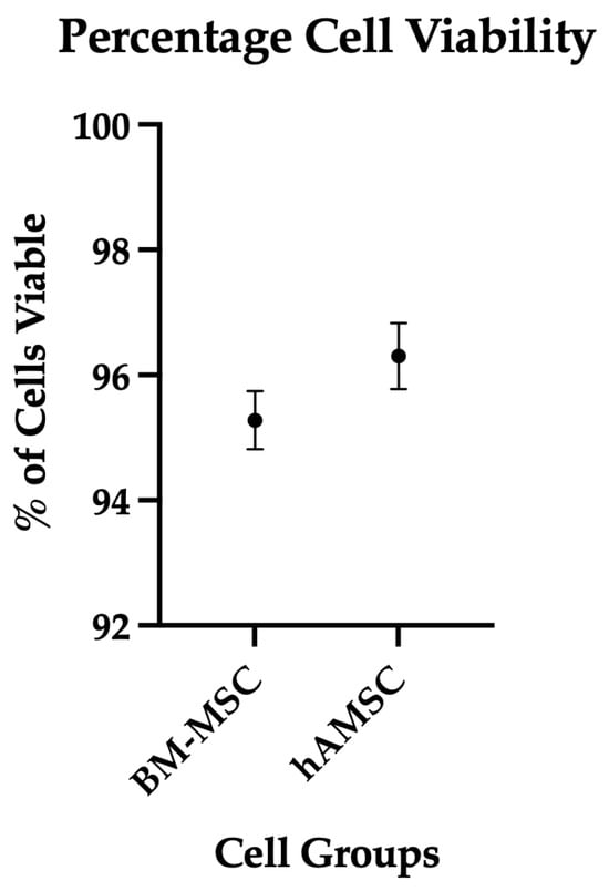
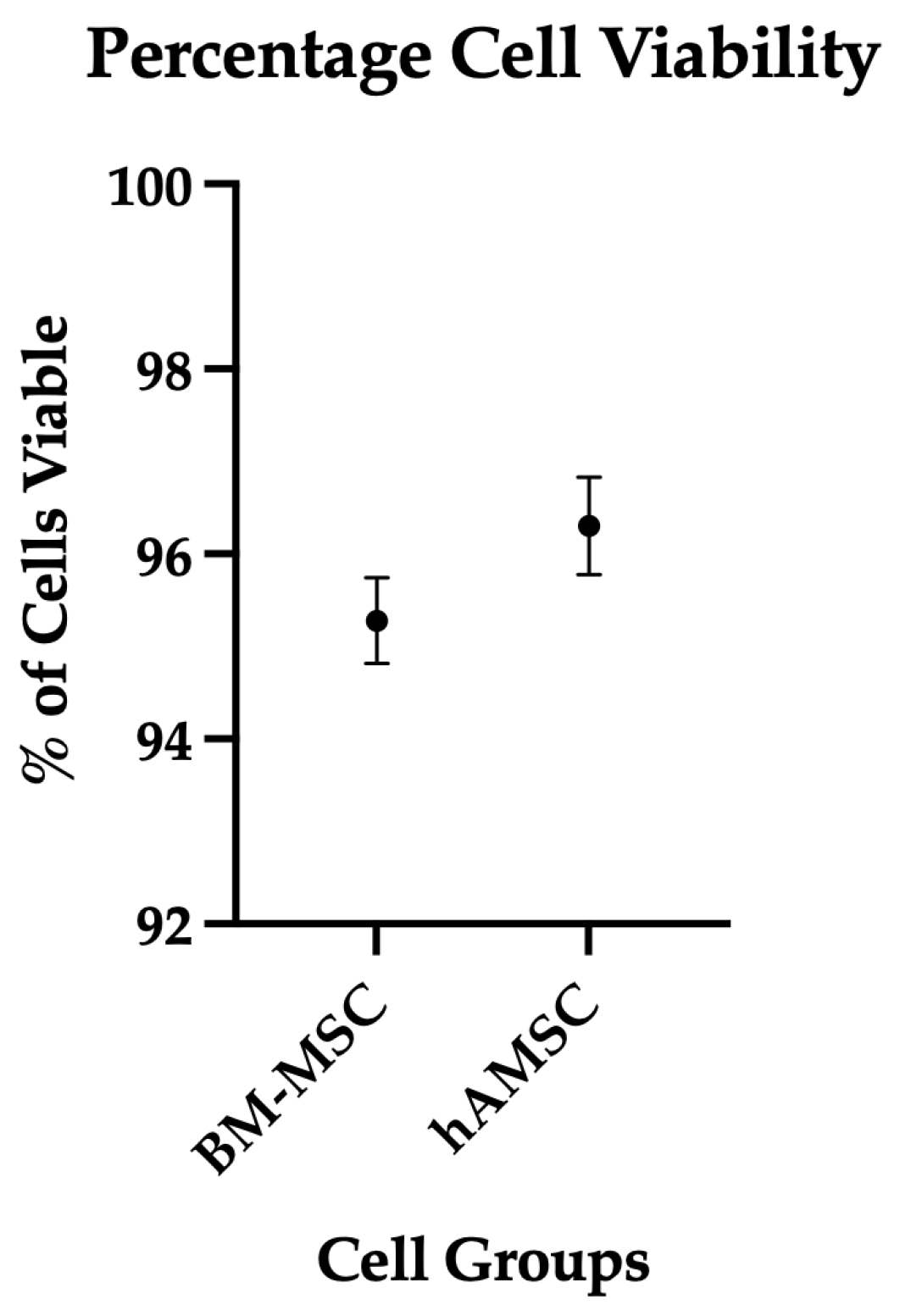
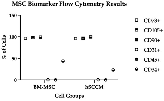
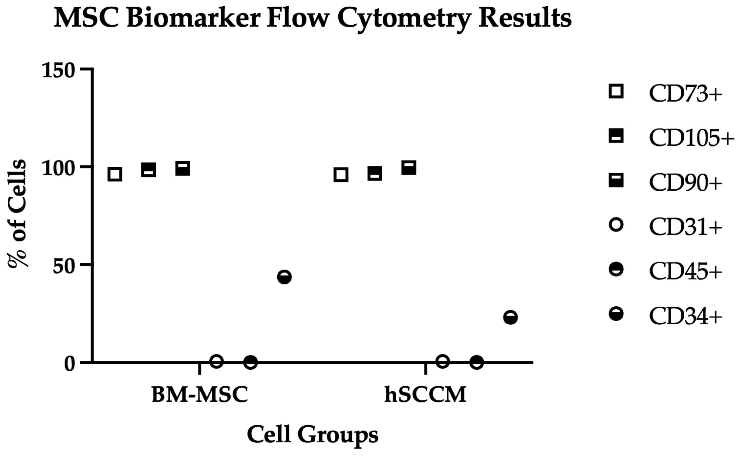
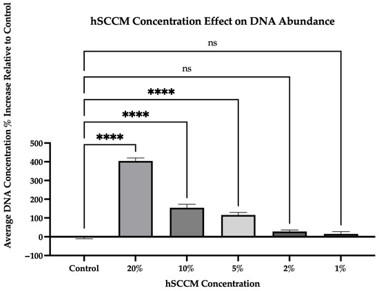
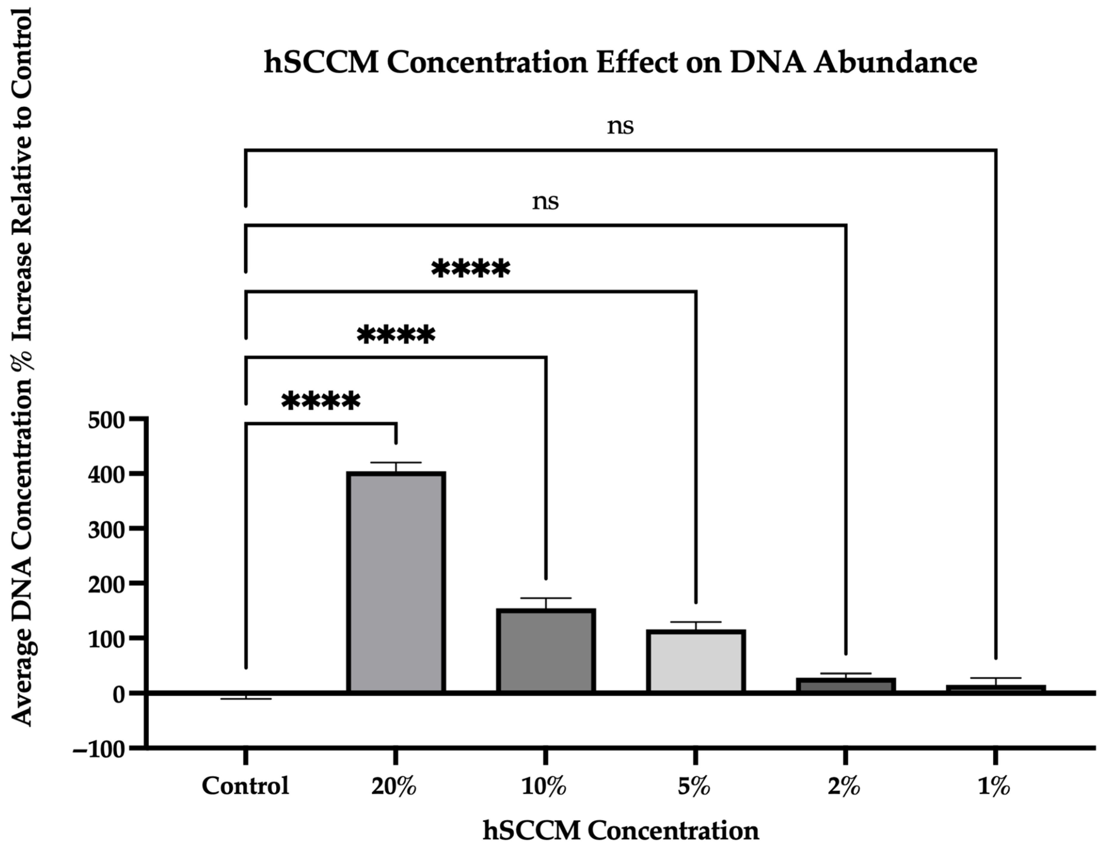
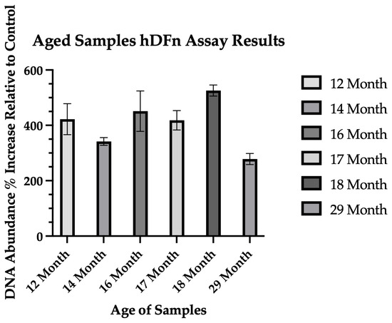
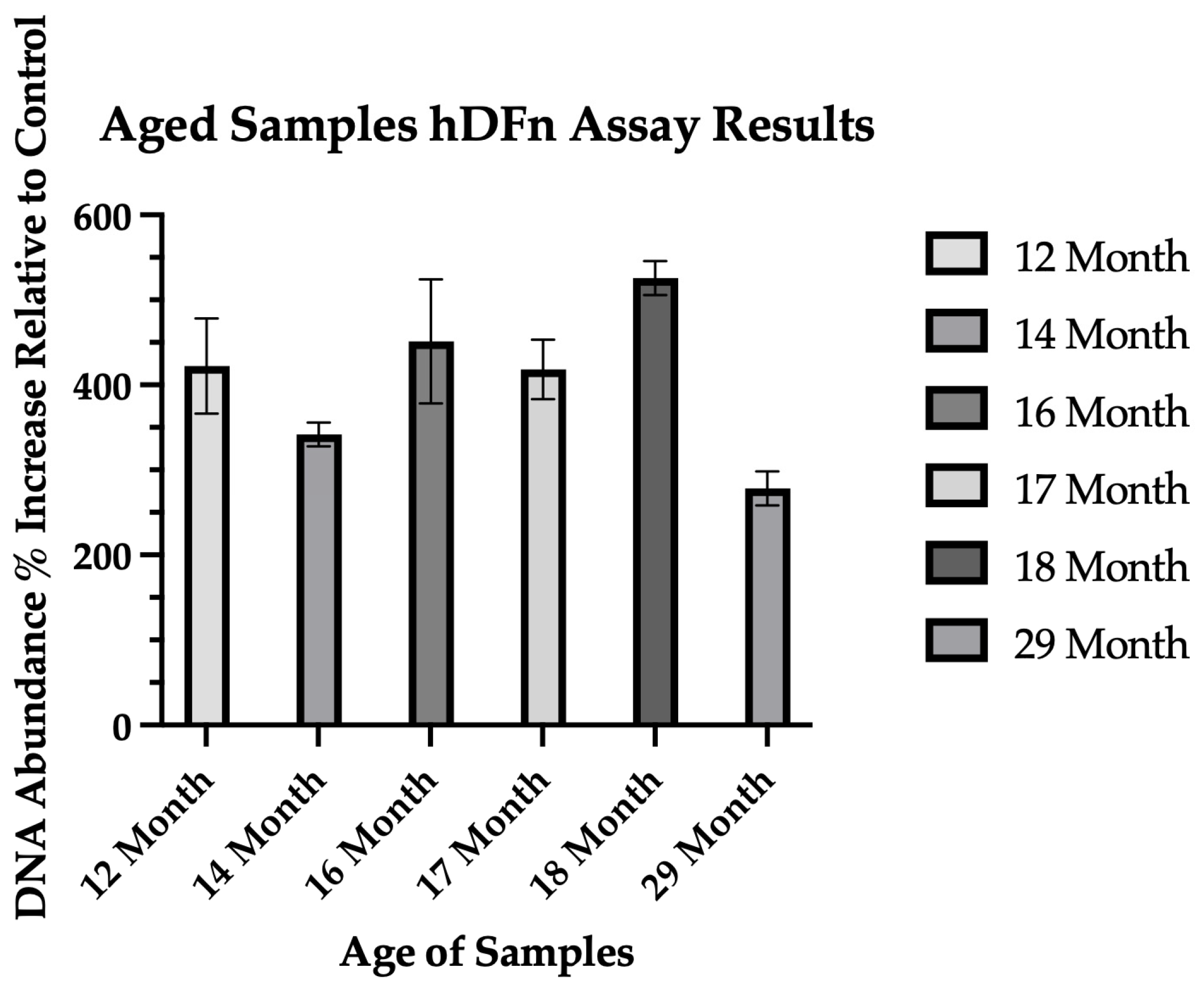
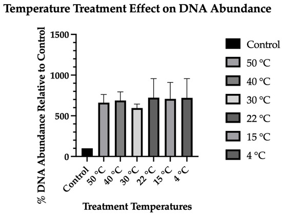
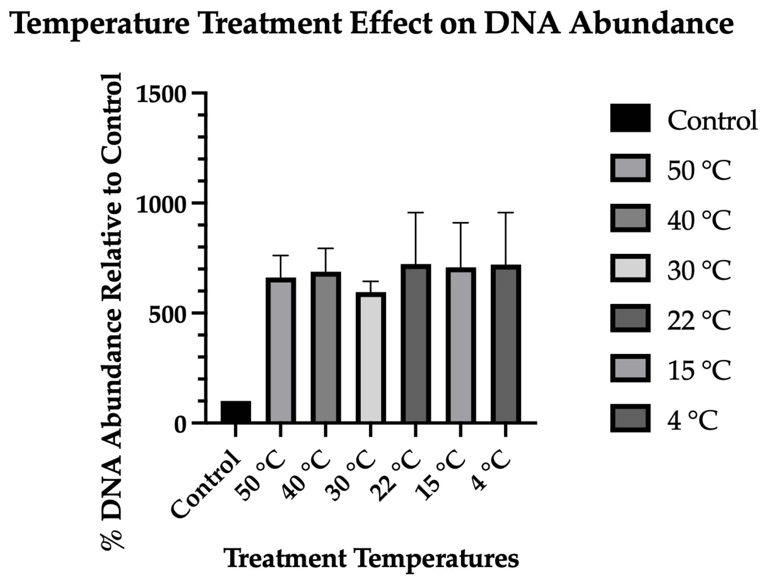
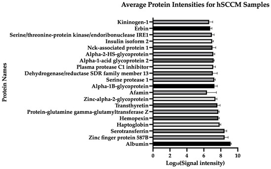
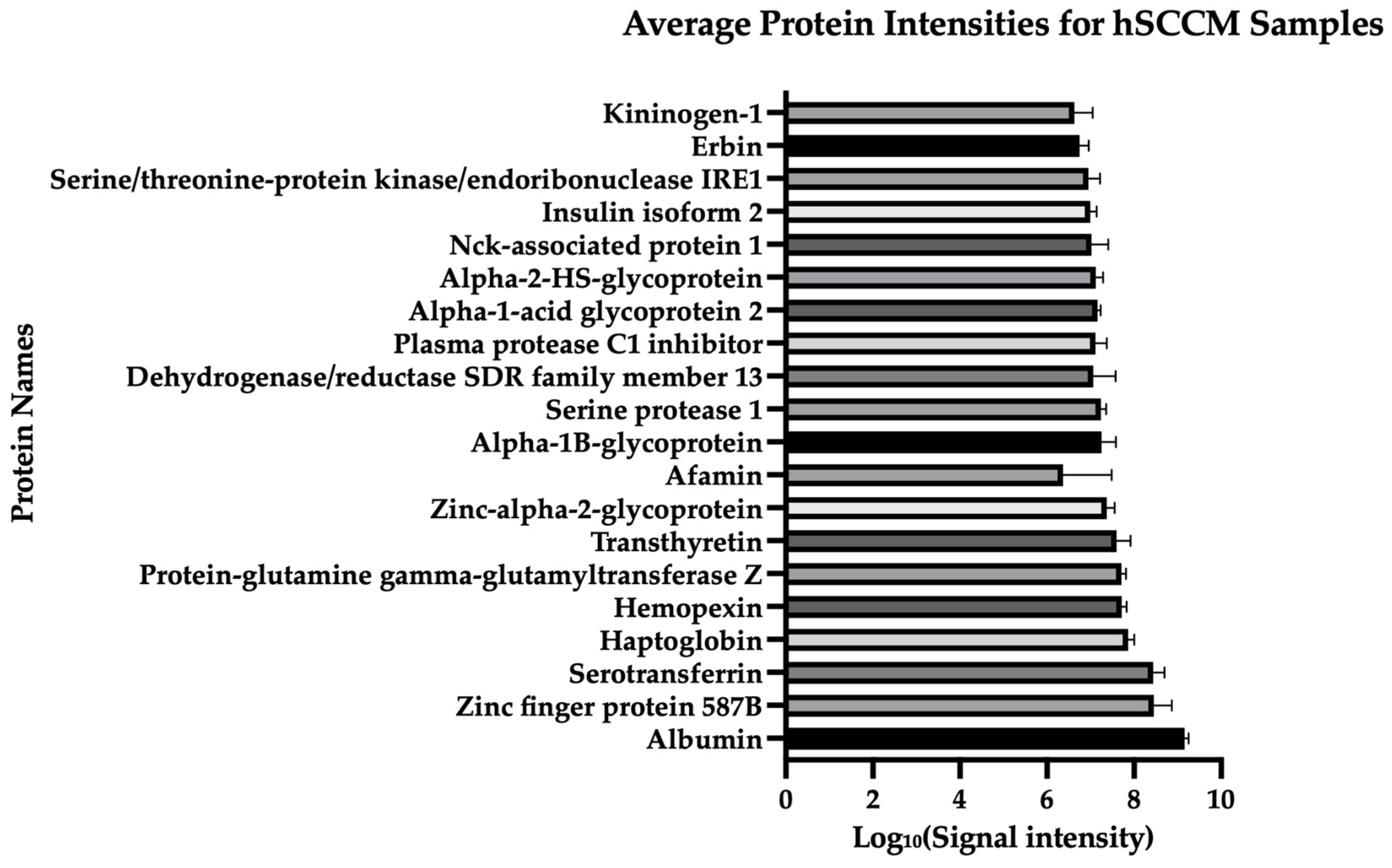
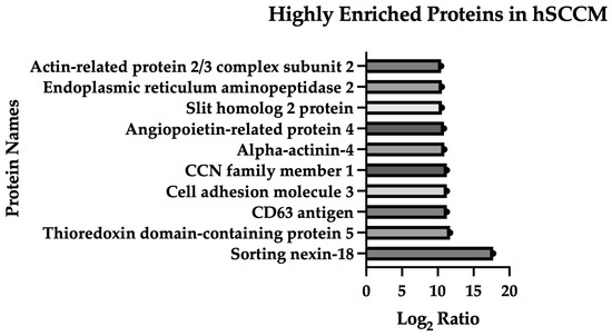
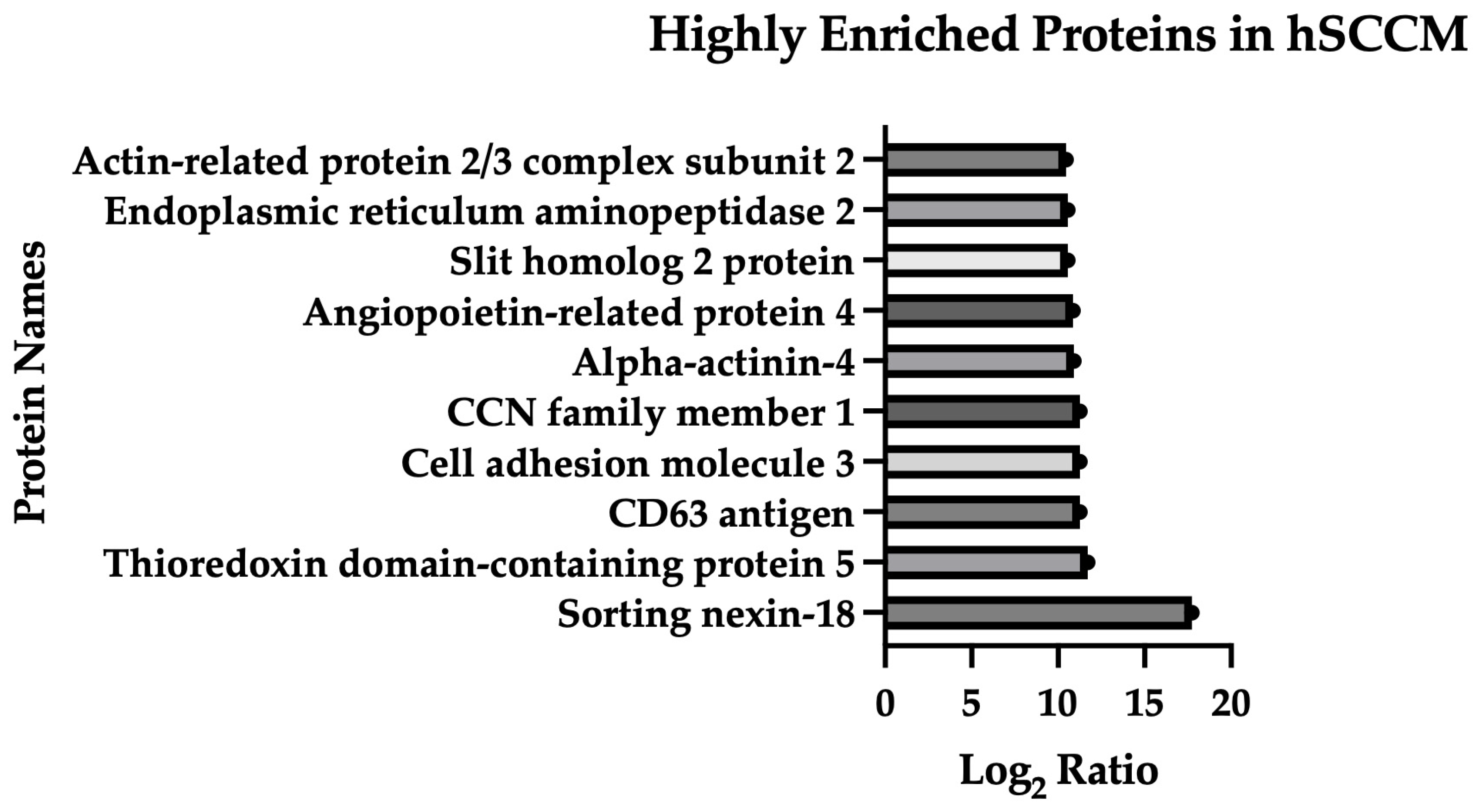
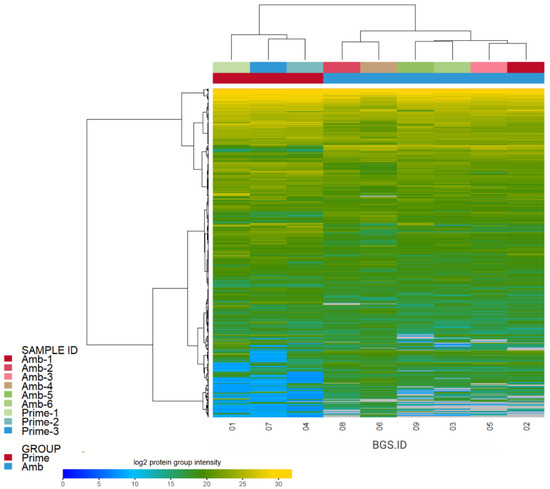
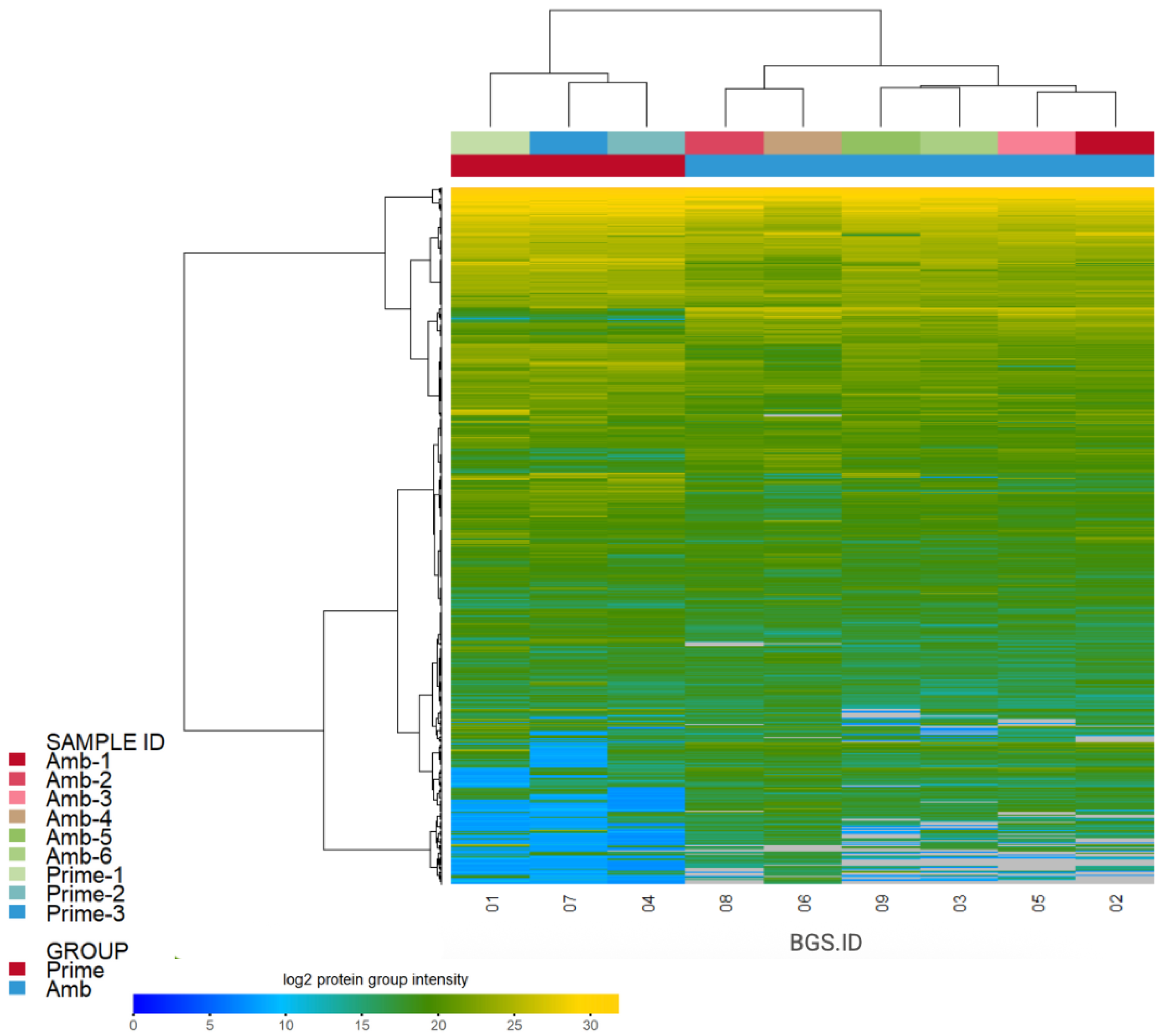
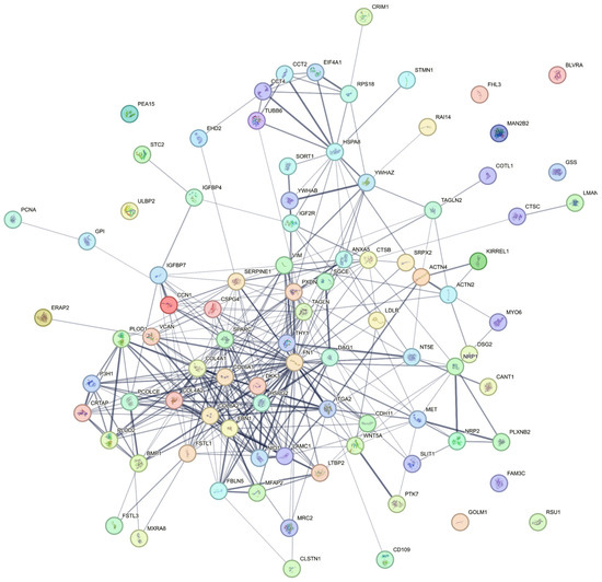
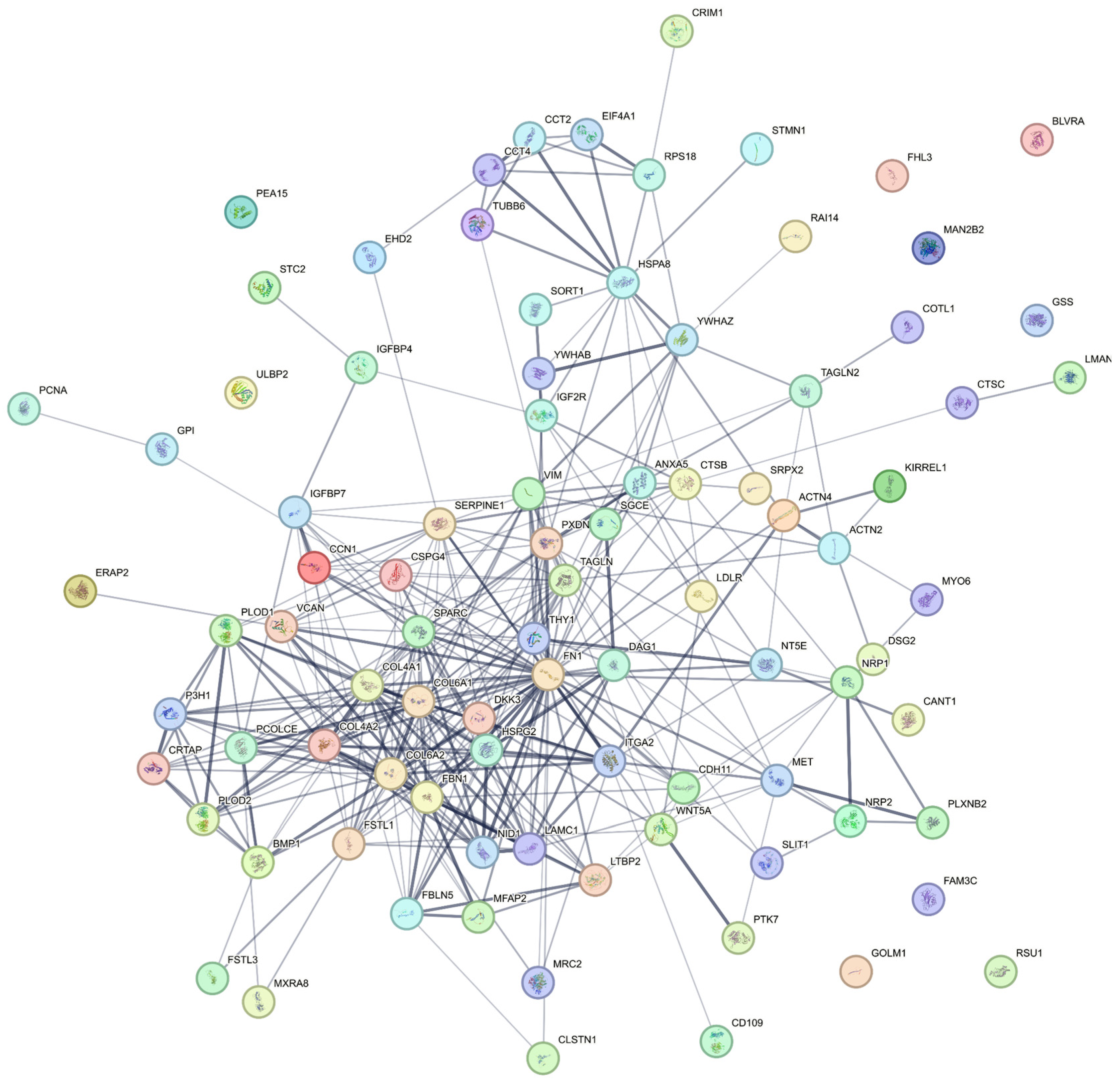
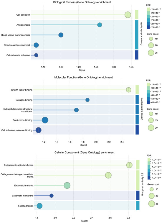
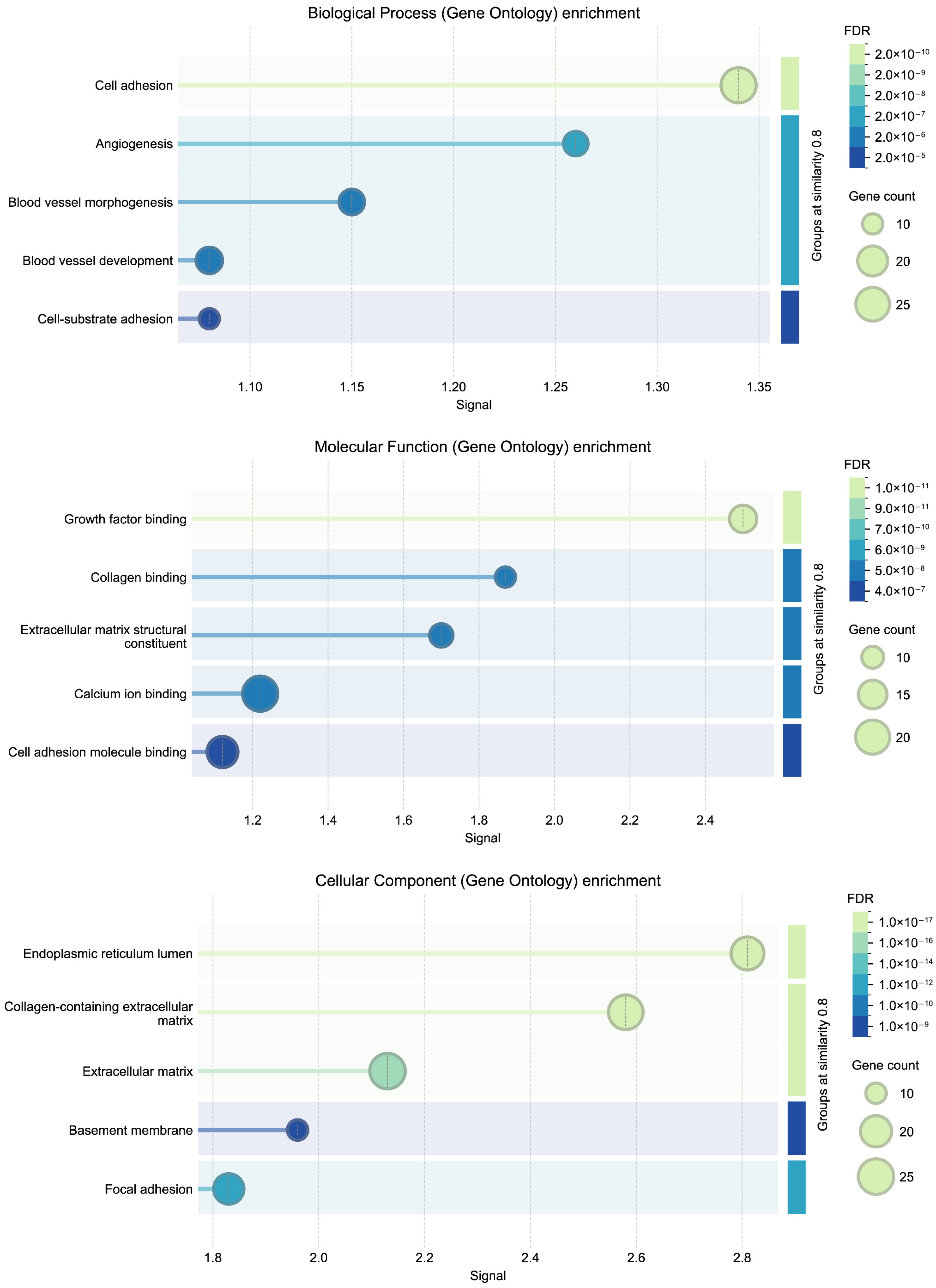
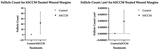
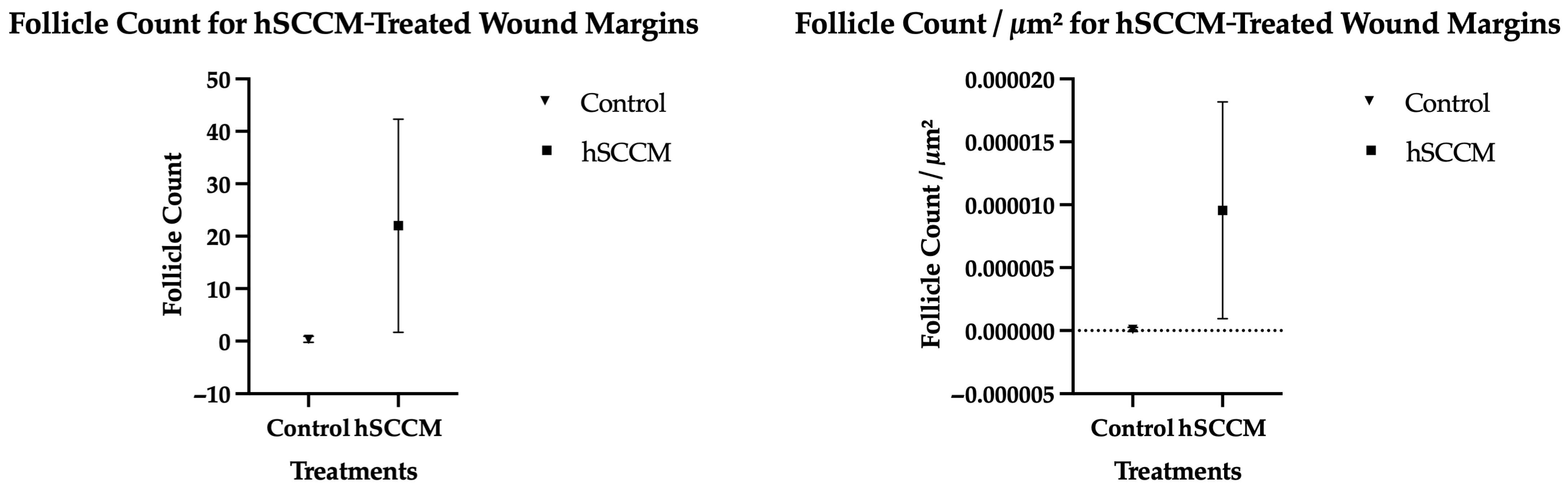
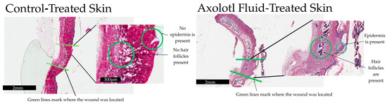
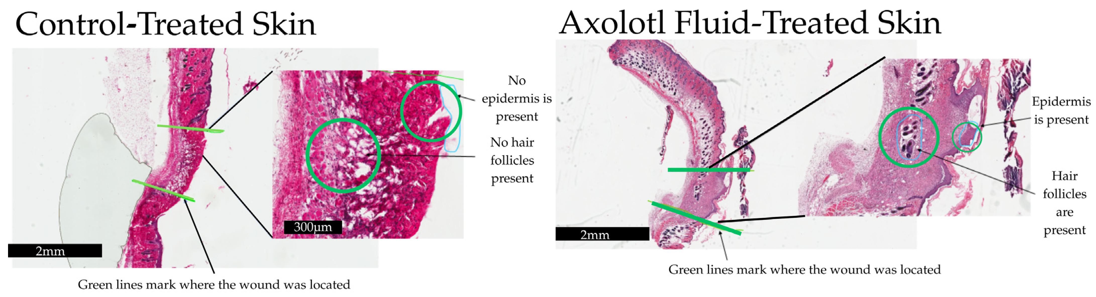
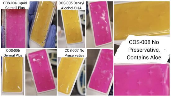
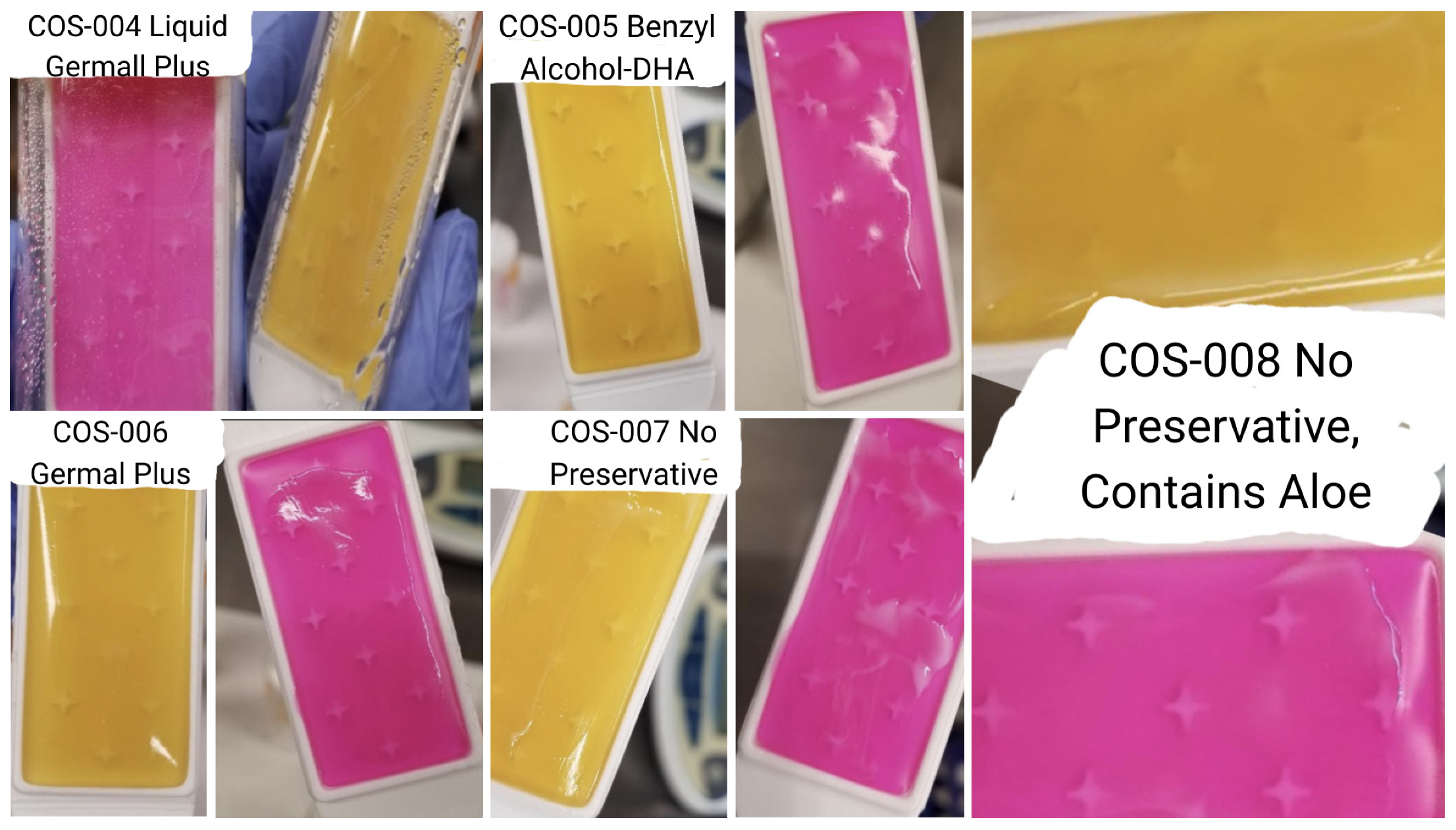
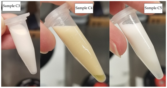
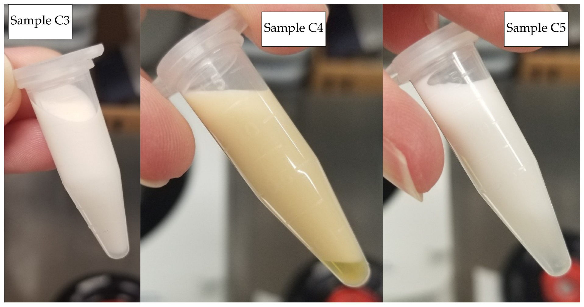
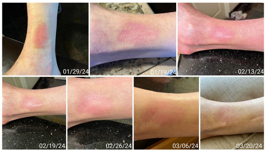
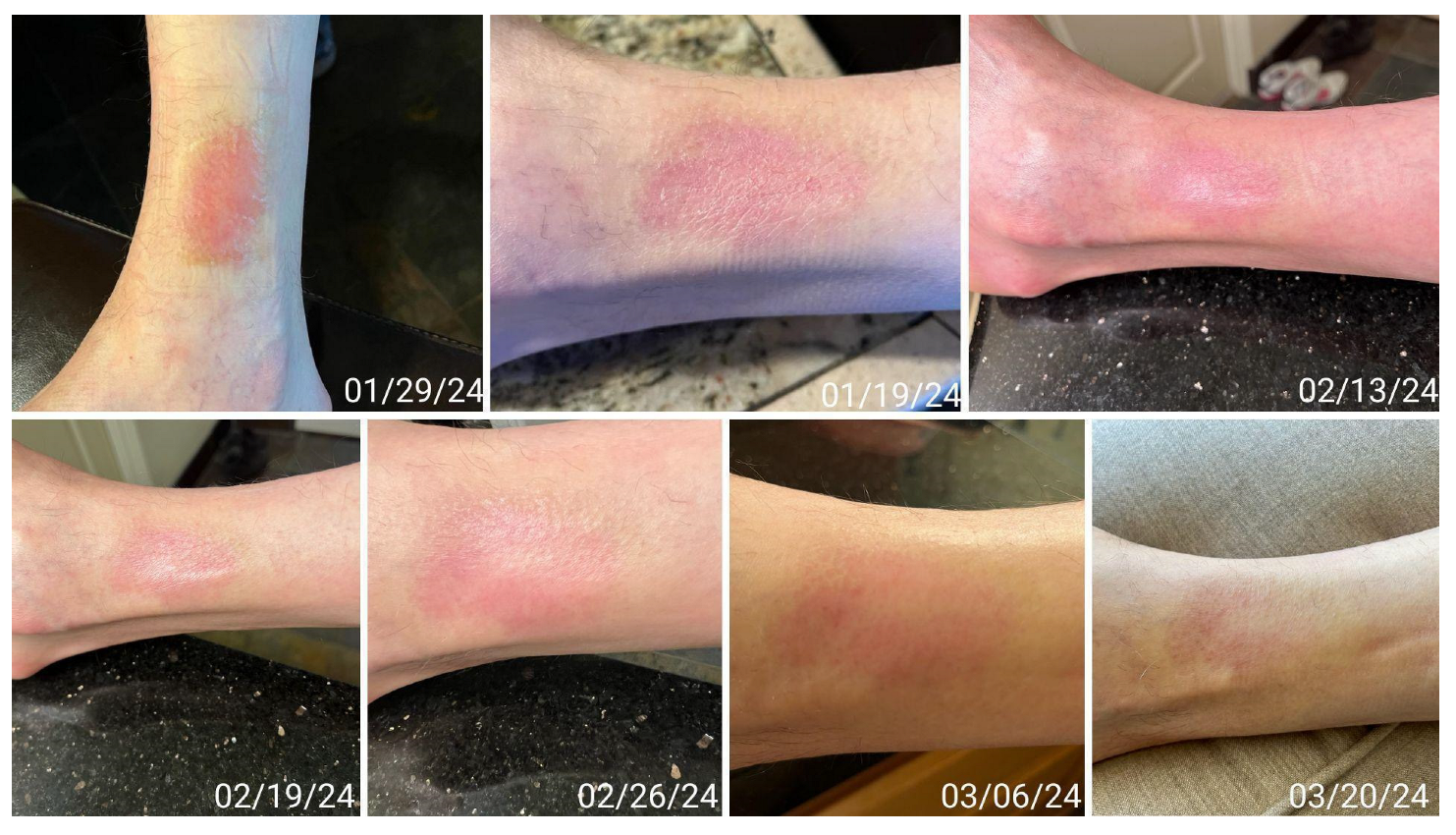

Disclaimer/Publisher’s Note: The statements, opinions and data contained in all publications are solely those of the individual author(s) and contributor(s) and not of MDPI and/or the editor(s). MDPI and/or the editor(s) disclaim responsibility for any injury to people or property resulting from any ideas, methods, instructions or products referred to in the content. |
© 2026 by the authors. Licensee MDPI, Basel, Switzerland. This article is an open access article distributed under the terms and conditions of the Creative Commons Attribution (CC BY) license.