Article

Microencapsulation of Epidermal Growth Factor (EGF) in Arabic Gum/Gelatine A Coacervates and Its Incorporation into Cosmetics: Evaluation of Skin Barrier Function and Ageing Indicators


by
Júlia Cristiê Kessler
1,2,*,
Isabel M. Martins
1,
Yaidelin A. Manrique
1,
Sigrún Dögg Gudjónsdóttir
3,
Alírio E. Rodrigues
1,
Maria Filomena Barreiro
2,* and
Madalena Maria Dias
1
1
LSRE-LCM, ALiCE, Faculty of Engineering, University of Porto, 4200-465 Porto, Portugal
2
CIMO, LA SusTEC, Instituto Politécnico de Bragança, Campus de Santa Apolónia, 5300-253 Bragança, Portugal
3
BIOEFFECT ehf., Víkurhvarf 7, 203 Kópavogur, Iceland
*
Authors to whom correspondence should be addressed.
Cosmetics 2026, 13(2), 89; https://doi.org/10.3390/cosmetics13020089
Submission received: 31 October 2025 / Revised: 19 March 2026 / Accepted: 2 April 2026 / Published: 10 April 2026
(This article belongs to the Special Issue Functional Molecules as Novel Cosmetic Ingredients)

Abstract

Epidermal Growth Factor (EGF) plays an important role in skin regeneration and repair by promoting cell proliferation and collagen synthesis. However, its topical application is limited by low stability, susceptibility to degradation, and poor penetration through the stratum corneum due to its hydrophilic nature and relatively large molecular size. Microencapsulation offers a strategy to protect sensitive bioactives and improve their delivery in cosmetic formulations. In this study, EGF was encapsulated in Arabic gum/gelatine A (AG/GE) coacervate microcapsules and incorporated into a hydrating cream. The work extends previous studies using the same microcapsule composition for lipophilic compounds, demonstrating its applicability for a hydrophilic bioactive and highlighting the versatility of the encapsulation platform. The resulting microcapsules exhibited spherical, multinucleated morphology with an encapsulation efficiency of 78.8 + 1.0%. Although diffusion of microencapsulated EGF in the cream could not be directly determined, the formulation showed trends towards improvement in several skin parameters during the volunteer evaluation, including reduction in surface spots (31%), brown spots (21%) and pore visibility (10%), and improved texture (22%). A 25% decrease in transepidermal water loss and a 33% increase in elasticity suggested improved skin barrier function. Volunteers reported high acceptance regarding non-irritancy, texture, and sensory experience.

1. Introduction

Epidermal Growth Factor (EGF) is a protein essential for skin health, promoting wound healing and delaying skin ageing by stimulating differentiation, proliferation, and growth in the epidermal and epithelial layers [1]. EGF also stimulates collagen and elastin production as a naturally occurring human protein, enhancing skin elasticity and resilience [2]. Structurally, EGF is a small polypeptide with a molecular mass of approximately 6–9 kDa that regulates key processes involved in skin homeostasis, including keratinocyte proliferation, cell migration, and dermal matrix synthesis. These effects are mediated through its interaction with the epidermal growth factor receptor (EGFR), EGF activates intracellular signaling pathways such as MAPK/ERK and PI3K/Akt, promoting epidermal regeneration and tissue repair. Due to these biological mechanisms, EGF has attracted significant interest in dermatology and cosmetic science, particularly in applications related to wound healing, skin barrier recovery, and anti-aging treatments [3,4].
Despite its biological activity, the topical application of EGF remains challenging. The molecule is relatively large and hydrophilic, which significantly limits its ability to penetrate the stratum corneum, the main barrier of the skin. As a result, only small fractions of applied EGF can reach viable epidermal layers where its biological targets are located. Additionally, EGF is susceptible to proteolytic degradation and instability in conventional cosmetic formulations, which further reduces its efficacy when applied topically [5,6,7].
To overcome these limitations, several delivery strategies have been explored to enhance the stability and skin penetration of EGF. These include nanovesicles, liposomes, transferosomes, peptide conjugates, microneedle systems, and other transdermal delivery approaches designed to improve macromolecule transport across the stratum corneum. Although these systems have demonstrated improved delivery in experimental settings, many systems remain complex, expensive, or difficult to integrate into conventional cosmetic formulations, highlighting the need for scalable encapsulation approaches compatible with standard cream formulations [3,8].
Biotechnological advances in recent decades have enabled the large-scale production of EGF for cosmetic applications [1]. One example is ORF Genetics (https://www.orfgenetics.com, Reykjavík, Iceland), which produces plant-based EGF with high stability (over 95%), a molecular mass of 9.5 kDa, and robust biological activity (>10 million U·mg−1), marketed under the brand ISOkine and incorporated into BIOEFFECT skincare products (https://www.bioeffect.com, Reykjavík, Iceland).
Among the available delivery approaches, microencapsulation by complex coacervation (MCC) has emerged as a promising strategy for protecting sensitive bioactives and enabling controlled release [1,9]. In this process, two oppositely charged polyelectrolytes interact through hydrogen bonding, electrostatic interactions, and polarisation effects [9,10]. Studies using gelatin-alginate coacervate have shown that this approach can effectively encapsulate EGF, protecting the protein and enabling controlled release in wound-healing applications [11]. Complex coacervation using Arabic gum and gelatine (AG/GE) offers several advantages for cosmetic applications compared with other delivery systems. Both polymers are biocompatible, biodegradable, and widely used in cosmetic and pharmaceutical formulations, while the coacervation process occurs under mild conditions that are suitable for sensitive biomolecules such as growth factors. AG/GE systems are among the most established coacervation pairs and have been widely applied for the encapsulation of oils, antioxidants, and other bioactive compounds due to their high encapsulation efficiency and controlled release properties [12,13,14]. In contrast to lipid nanoparticles or chitosan-based carriers, complex coacervation systems can be produced under mild aqueous conditions without the need for organic solvents or high-energy processing, which helps preserve the activity of sensitive proteins such as EGF [1,6,8].
Successful encapsulation of lipophilic cosmetic actives such as α-tocopherol and Moringa oleifera extracts, using AG/GE coacervates, has been previously demonstrated [10,12]. However, the applicability of this system to hydrophilic macromolecules such as growth factors has not yet been explored. Demonstrating that the same microcapsule architecture can accommodate both hydrophobic and hydrophilic actives would significantly expand the versatility of this encapsulation platform and potentially enable the simultaneous incorporation of different classes of cosmetic bioactives within a single delivery system.
This study addresses the growing demand for scientifically advanced and eco-friendly skincare by enhancing the performance of EGF in a hydrating cream with an innovative AG/GE coacervate system. Building on previous work with lipophilic compounds, this study investigates the encapsulation of the hydrophilic protein EGF and evaluates its performance in a cosmetic formulation through physicochemical characterisation and volunteer skin assessments.

2. Materials and Methods

2.1. Materials

2.1.1. Microcapsules Production

Gelatine A (GE, derived from porcine skin, 300 g bloom, CAS 9000-70-8), Arabic gum (AG, sourced from the Acacia tree, CAS 90000-01-5), N-hydroxy succinimide (NHS, purity ≥ 97%, CAS 6066-82-6), Polysorbate 80 (P80, HLB 4.3, CAS 9005-65-6), and hydrogen chloride (HCl, purity ≥ 37%, CAS 7647-01-0) were all procured from Sigma Aldrich (Madrid, Spain). Additionally, 1-ethyl-3-(3-dimethylaminopropyl) carbodiimide (EDC, purity ≥ 98%, CAS 25952-53-8) was sourced from Alfa Aesar (Budapest, Hungary). Caprylic triglyceride (CAP, derived from Cocos nucifera oil, batch 14832) was obtained from Plena Natura (Lisbon, Portugal), and the epidermal growth factor (EGF) was provided by ORF Genetics (Reykjavík, Iceland).

2.1.2. Hydrating Cream Production

Cetanol was sourced from Vevy Europe (Genova, Italy), while C12 Acid PEG-8 ester and sodium hyaluronate RM-CHEM-10 were acquired from Shijiazhuang (Hebei, China). Citric acid was provided by Caleo (Graz, Austria), and Carbopol Ultrez 10 carbomer was obtained from Lubrizol (Wickliffe, OH, USA). Additionally, 2-phenoxyethanol, ethylhexylglycerin, and potassium sorbate were supplied by IMD Sweden AB (Malmo, Sweden). Butylene glycol was purchased from Gulf Chemical (Mandai, Singapore), potassium hydroxide from Sigma Aldrich (St. Louis, MO, USA), and α-tocopherol (CAS 10191-41-0, 99%) was obtained from BASF (Ludwigshafen, Germany).

2.2. Methods

2.2.1. Microcapsules Production by Complex Coacervation

The parameters used to produce AG/GE microcapsules were selected based on a previously optimised formulation developed through a fractional factorial Design of Experiments approach, in which key variables such as emulsifier ratio (CAP:P80), stirring rate and time, and crosslinker concentration (EDC/NHS) were systematically evaluated. These studies demonstrated that a CAP:P80 ratio of 3.5:1, emulsification at 9500 rpm for 2 min, and crosslinking with 10% EDC/NHS provided the most stable microcapsules, with spherical morphology, average particle sizes of approximately 60 µm, and high encapsulation efficiency. These parameters represent a balance between efficient droplet formation during emulsification, optimal polymer complexation, and sufficient crosslinking to stabilise the microcapsule shell without promoting aggregation. The optimised formulation was therefore adopted in the present work to investigate the encapsulation of the hydrophilic bioactive EGF. Further details regarding the optimisation procedure can be found in earlier studies [10,12].
Microencapsulation by complex coacervation (MCC) in batch mode was used to encapsulate the growth factor EGF. The production method, initially designed and optimised to protect hydrophobic compounds such as Mo leaf extracts and α-tocopherol [12], is applied here to evaluate its suitability for hydrophilic bioactives. This approach assesses MCC’s versatility in stabilising different compounds, thereby expanding its potential applications in skincare formulations.
The microencapsulation process begins by dissolving approximately 14 mg of lyophilised EGF powder in 2 mL of UV-filtered water and then adding to a pre-prepared GE solution 7.14 g·L−1 maintained at 30 °C. This mixture is combined with the oil phase composed of CAP and P80 at a ratio of 3.5:1 (v/v), initiating the emulsification step at 9500 rpm for 2 min using an Ultra Turrax (IKA Yellowline DI 25, Gravimetra, Lisbon, Portugal). Next, AG 14.3 g·L−1 is added to the emulsion while stirring at 400 rpm with a magnetic stirrer (Agimatic-N, J.P. Selecta, Barcelona, Spain) at 30 °C. Coacervation follows by adjusting the pH from 6.3 to 3.9 using 1 M HCl. Subsequently, the microcapsules are gradually cooled to 7 °C in an ice bath under stirring at 400 rpm. At this stage, 5 mL of 50 mM EDC plus 25 mM NHS (10% of the aqueous phase) is added to initiate the microcapsules crosslinking using continuous stirring for another 60 min. The dispersion of the crosslinked microcapsules is then transferred to a decantation funnel to allow phase separation overnight under refrigerated conditions. The concentrated microcapsule phase is collected from the bottom of the decantation funnel, with residual reagents washed out once using 60 mL of deionised water at 30 °C. The final microcapsule-rich dispersion is stored in a Falcon tube without preservatives and kept refrigerated until further analysis. The microcapsules were maintained as an aqueous dispersion to ensure stability and facilitate direct integration into the cosmetic formulation. Further details on the microencapsulation process and production setup can be found in previous works [10,12].

2.2.2. Characterising the Microcapsule Dispersions

  • Particle Morphology
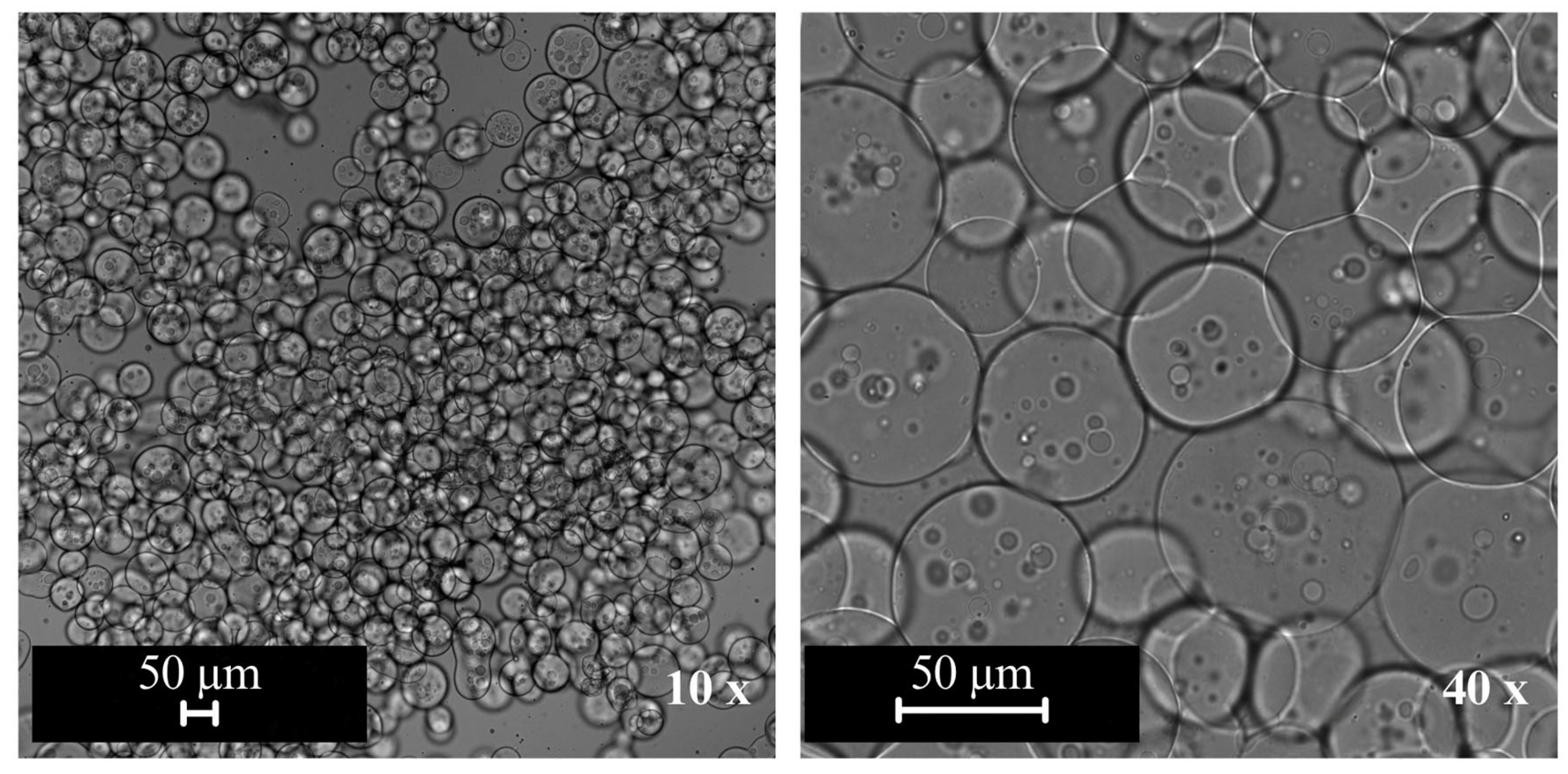
The morphology of the MC EGF particles was analysed using optical microscopy (Eclipse Ci H55OS, equipped with a DS-Qi2 camera, Nikon, Tokyo, Japan). The images were processed with NIS-Elements L software (version 4.50) and captured at 10× and 40× magnifications.
  • Encapsulation Efficiency
The encapsulation efficiency (EE) of EGF was determined using the JESS apparatus (ProteinSimple, Bio-Techne, Minneapolis, MN, USA), which performs automated capillary-based immunoassays analogous to traditional Western blotting. The protocol followed the manufacturer’s instructions using the standard JESS reagent kit.
The kit includes several proprietary buffers designed to support protein denaturation, electrophoretic separation, antibody binding, and chemiluminescent detection. The 10× sample buffer and 5× Master Mix contain detergents and buffering agents that stabilise proteins and maintain the appropriate pH and ionic strength for capillary electrophoresis. Dithiothreitol (DTT) was prepared as a 400 mM solution to reduce disulphide bonds, ensuring protein denaturation and improving electrophoretic separation. Samples were diluted using a 0.1× separation buffer supplied with the kit to maintain optimal protein concentration and compatibility with the capillary system. A wash buffer provided by the manufacturer was used to remove unbound antibodies during the immunodetection steps, reducing background signal. Chemiluminescent detection was performed using the Luminol-S reagent included in the kit, which reacts with the horseradish peroxidase (HRP) enzyme conjugated to the secondary antibody to produce the measurable signal.
First, DTT is diluted in 40 µL of ultrapure water to create Reactant A, a 400 mM solution. Next, 20 µL of 10× sample buffer is combined with a concentrated 5× Master Mix and 20 µL of the 400 mM DTT solution, forming Reactant B. The third reactant, Reactant C, consists of a biotinylated ladder diluted in 20 µL of ultrapure water, serving as a molecular weight marker for proteins. It is employed as a reference to ascertain the molecular weight of proteins separated in the capillary-based system, enabling detection by a streptavidin-based reagent.
A modified version of a previously described protocol [10] is employed for sample preparation. Following the decantation step in the microcapsule production process, dispersions are collected in aliquots from the EGF microcapsules (MC EGF) supernatant. This supernatant contains predominantly the nonencapsulated (free) EGF fraction, which was quantified to determine the amount of protein not retained within the microcapsules. Each sample is diluted in a 0.1× buffer solution to determine the EE.
Then, 40 µL of each diluted sample is combined with 10 µL of Reactant B in a microcentrifuge tube and gently mixed using a pipette. The mixture is homogenised using a vortex mixer, denatured in a water bath at 95 °C for 5 min, and once again homogenised before being cooled further in an ice bath. Luminol-S Mix (Reactant D) is also cooled down in the ice bath.
Finally, 5 µL of Reactant C, 3 µL of the sample, 10 µL of antibody diluent, 10 µL of primary and secondary Anti-Rabbit antibodies, 10 µL of streptavidin-HRP, 15 µL of Reactant D, and 500 µL of wash buffer are added to specific wells on a plate. The plate is homogenised using a benchtop centrifuge (Avanti J-15R, Beckman Coulter, Indianapolis, IN, USA) for 5 min at 2500 rpm at room temperature. After confirming that the liquid has settled in the wells with no air bubbles, the plate is placed in the JESS apparatus, and the Compass for SW software (version 6.0.x) is configured. Once initiated, the JESS system automates the entire process, including protein separation, antibody binding, washing, and detection within capillaries. The software also aids in data analysis, allowing for quantification and visualisation of protein levels through electropherograms or virtual Western blot images. This automated approach reduces hands-on time, minimises variability, and enhances the efficiency and precision of microencapsulated protein quantification.
The samples are analysed and quantified using a standard calibration curve of ISOkine product (EGF, y = 8.26 × 10⁴x − 4.13 × 10⁴; R2 = 0.998) in a range of 1.25 to 40 ng·mL−1. The obtained concentration is used for the indirect calculation of the EGF EE,
EE   ( % ) = total   concentration   -   nonencapsulated   concentration total   concentration   ×   100
This method was used to quantify the nonencapsulated EGF fraction in the supernatant for indirect calculation of encapsulation efficiency. No direct structural or bioactivity assessment of the encapsulated protein was performed in this study.
  • Solid Content
The microcapsule dispersions were submitted to an air-drying process at 105 ± 1 °C until a constant weight was achieved. This process was used to measure the solid content (SC) gravimetrically, determined by [14]:
SC   ( % )   =   dry   mass liquid   mass   ×   100

2.2.3. Cosmetic Production Containing Microencapsulated EGF

A novel hydrating cream formulation was developed and compared to BIOEFFECT’s commercial hydrating cream (control formulation, CF). The new formulation, inspired by the CF’s minimalist composition, was enriched with microencapsulated EGF (MC EGF) instead of its free form. It was designed as a fast-absorbing, oil-free emulsion free of fragrance, alcohol, gluten, and parabens, containing only sixteen carefully selected ingredients, including soft Icelandic water, hyaluronic acid, and α-tocopherol. This approach aimed to evaluate the efficacy of delivery systems incorporating a hydrophilic bioactive and to facilitate a deeper analysis of its effects on human skin. BIOEFFECT adheres to ISO 22716:2007 Cosmetics GMP guidelines [15] and EU Regulation (EC) No 1223/2009 [16] to ensure consistently high-quality cosmetic products.
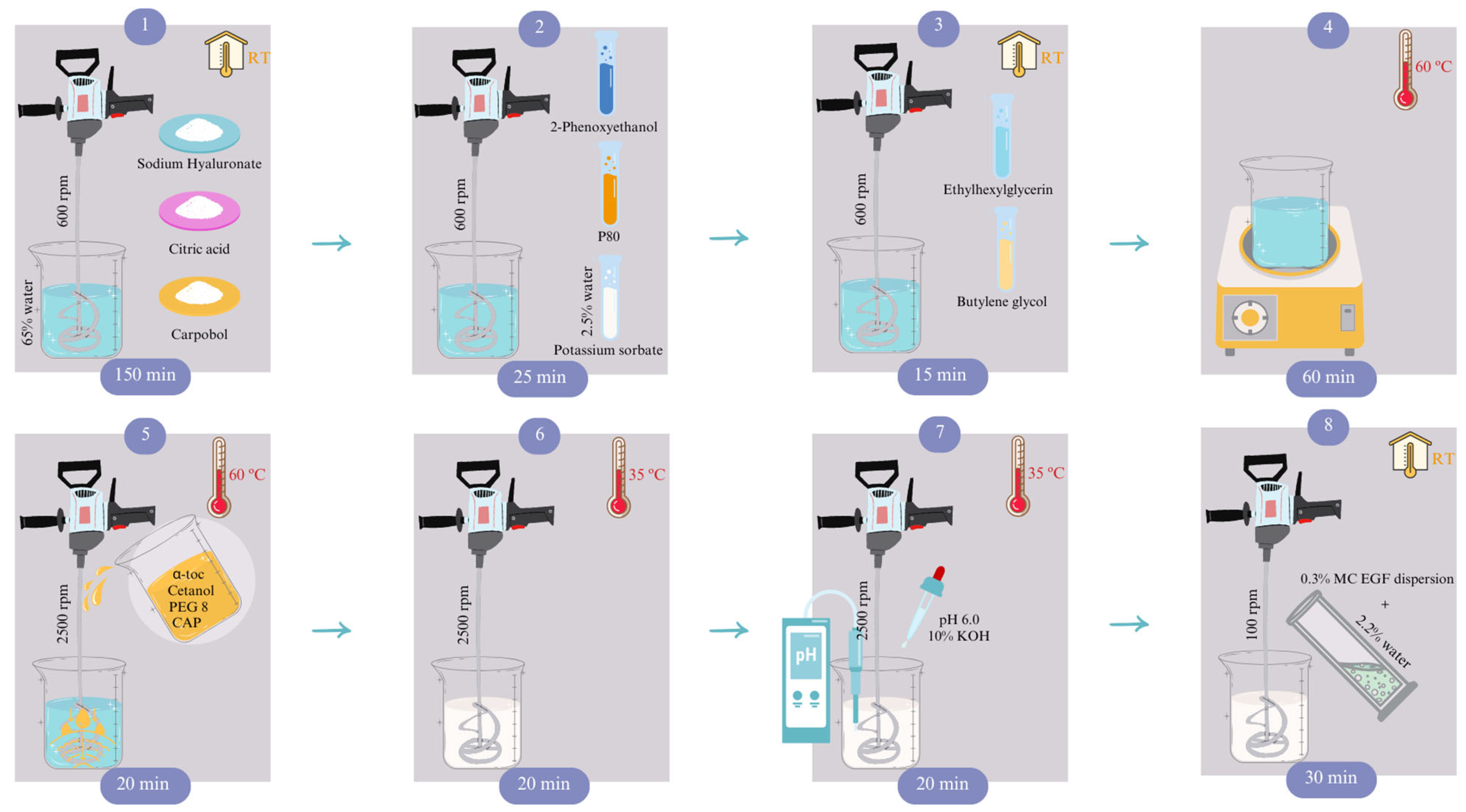
In this study, a novel formulation of the hydrating cream was produced, where the free form of EGF was replaced by microencapsulated EGF (MC EGF), which was incorporated in the final blending step, as illustrated in Figure 1.
First, the water phase is prepared by adding water (65% of the total volume) with humectant (sodium hyaluronate), thickener (Carbopol), preservatives (2-phenoxyethanol, potassium sorbate, ethylhexylglycerin), emulsifier (P80), and texture enhancer (butylene glycol), with its pH adjusted to a range of 3.3 to 3.7 using a chelating agent (citric acid), while maintaining a temperature of 20 °C under constant stirring (Hei-torque Core, Heidolph, Schwabach, Germany). The solution is then heated to 60 °C and combined with the oil phase, also at 60 °C, which contains emollients (Cetanol, PEG 8 and CAP) and α-tocopherol. The mixture is blended for approximately 60 min at 2500 rpm using a laboratory shear mixer (AE500S-H 70 G, Huanyu, Wenzhou, China), which gradually reduces the temperature to 35 °C, at which point the pH is adjusted to 6.0 using a second chelating agent (KOH).
In the final step, 70% of the water content is achieved by incorporating the MC EGF dispersion. This mixture is stirred for 30 min at 100 rpm using a Hei-torque Core stirrer (Heidolph, Schwabach, Germany) fitted with a PR 30 pitched-blade impeller, ensuring thorough homogenisation of the bioactive component. The concentration of the growth factor in the formulation is adjusted to match the amount of EGF present in its free form in the control formulation (CF). This corresponds to the incorporation of 0.3% of the microcapsule dispersion, ensuring comparable active ingredient levels between the encapsulated and non-encapsulated formulations. The exact EGF loading in the commercial formulation is proprietary to the industrial partner and is therefore not disclosed.

2.2.4. Skin Compatibility Test

A skin compatibility assessment of the hydrating cream containing 0.3% MC EGF was carried out by SGS Idea (https://www.ideatestsgroup.com, Marillac, France), a certified testing company. The procedure involves a simple application under occlusive conditions using a Finn Chamber® (Epitest Ltd Oy, Tuusula, Finland) or equivalent device. To evaluate skin compatibility, a 20 μL (0.02 g) sample is applied to a patch placed on the outer arm at 20 °C for 48 h under dermatological supervision.
Twelve healthy individuals between the ages of 18 and 65, without dry or sensitive skin at the test site, were enrolled in the study. All underwent clinical screening to rule out dermatological lesions, active atopy, or a history of severe allergies or reactions to household products. Pregnant or breastfeeding individuals, or those on anti-inflammatory, corticosteroid, or antihistamine medication, were excluded. After agreeing to the study conditions, participants were exposed to a 48-h patch test. Assessments were conducted 30 to 40 min after patch removal, rating skin responses—such as erythema, edema, and signs like dryness or papules—on a 0 to 3 scale. The Mean Irritation Index (M.I.I.) was calculated as the total score divided by the number of volunteers. M.I.I. values ≤ 0.2 indicated non-irritancy.

2.2.5. Stability Tests

Hydrating cream formulations containing MC EGF were subjected to in-house stability tests following ISO/TR 18811:2018 guidelines for cosmetic product stability [17]. The following tests were conducted:
  • Spin Test
A 40 g sample is placed into a 50 mL Falcon tube and incubated in a mini oven (MK II, MWG-Biotech, Bavaria, Germany) at 37 °C for 1 h. After incubation, the sample is centrifuged at 300 rpm for 15 min using a benchtop centrifuge (Avanti J-15R, Beckman Coulter, Indianapolis, IN, USA). This process is repeated for six rounds, with visual inspections conducted after each cycle. The sample is considered stable if no phase separation is observed after all rounds.
  • Temperature Cycle
A 25 g sample is placed into the final container and subjected to a four-day temperature cycle test. Each 24-h cycle involves incubating the sample at −18 °C in a refrigerator (ERB 3030, Electrolux, Stockholm, Sweden), followed by a transfer to room temperature (20 °C), heating to 37 °C in a mini oven (MK II, MWG-Biotech, Bavaria, Germany), and returning to 20 °C for the final 24-h period before visual inspection and image documentation. The analysis is carried out in duplicate, and the sample is considered stable if no phase separation or changes in appearance are observed.
  • Long-term and Heat Stability
Three 25 g samples are placed into different containers and incubated at 4 °C in a refrigerator (ERB 3030, Electrolux, Stockholm, Sweden), 20 °C or room temperature, and 40 °C in an incubator (Hood TH 30, Edmund Buhler, Bodelshausen, Germany). Over 60 days, monthly inspections are performed, coinciding with the skin studies described in Section 3.4. These inspections include homogeneity, visual appearance, and odour, all of which are documented with images.
  • pH Stability
A 40 g sample is placed in a 50 mL Falcon tube and incubated at 20 °C for 60 days. The pH is measured and recorded biweekly using a portable pH meter (Testo 206-pH2, Campinas, Brazil). The test is performed in triplicate for each formulation. The products are considered stable if the pH remains within 6.0 ± 0.3.
  • Rheology Analysis
Viscosity and thixotropy effects are monitored using a coaxial rheometer (RST-CC, Brookfield, Toronto, Canada). A 0.8 mL sample of the MC formulation and CF is subjected to shear rates ranging from 30 to 360 s−1, and shear stresses from 240 to 720 Pa. The equipment operates at 20 °C with parallel plate geometry and a gap of 0.5 mm, employing Rheo3000 software standard edition for PC control and data acquisition. ANOVA and Tukey tests are used to assess significant differences with α = 0.05 [12].

2.2.6. Cosmetic Cream Performance Test

  • Volunteers Selection
Twenty healthy Icelandic volunteers, both female and male, aged 20 and above, participated in a skin performance study conducted between March and May 2024. The participants were evenly divided into two groups: one using MC EGF and the other using CF. The study lasted 60 days and included three visits to the Skin Lab at BIOEFFECT HQ. Volunteers were selected and screened for eligibility through a standard in-house procedure.
Participants were excluded if they had known sensitivities to any ingredients in the formulations or if they had skin conditions affecting the facial area. To maintain consistency, volunteers planning to travel to warmer climates during the study were not included. Additionally, those who had undergone facial cosmetic procedures such as surgery, laser treatments, dermal filler or toxin injections, microneedling, or similar treatments in the 12 months before enrolment or who planned to do so during the study were excluded. Volunteers who had used BIOEFFECT skincare products containing EGF within the 12 months prior to the study were also excluded.
The Icelandic Ethical Committee confirmed that studies of this nature at BIOEFFECT do not require formal approval, as no biological material is collected, the products are non-medical, and no treatment is involved. All participants provided informed consent for the use of their images in research.
  • Analytical Skin Assessment
After selection, participants visited the Skin Lab to provide informed consent and undergo baseline measurements. They were instructed to apply the assigned product twice daily, morning and evening, while abstaining from other skincare products, including hydrating creams, serums, and eye creams, to avoid any confounding effects. Sunscreen was recommended for use during sun exposure. Participants returned for two additional follow-up visits at 30-day intervals, during which skin parameters, participant satisfaction, and tolerability were assessed.
Two primary instruments were employed to quantify changes in skin characteristics:
  • Canfield VISIA-CR
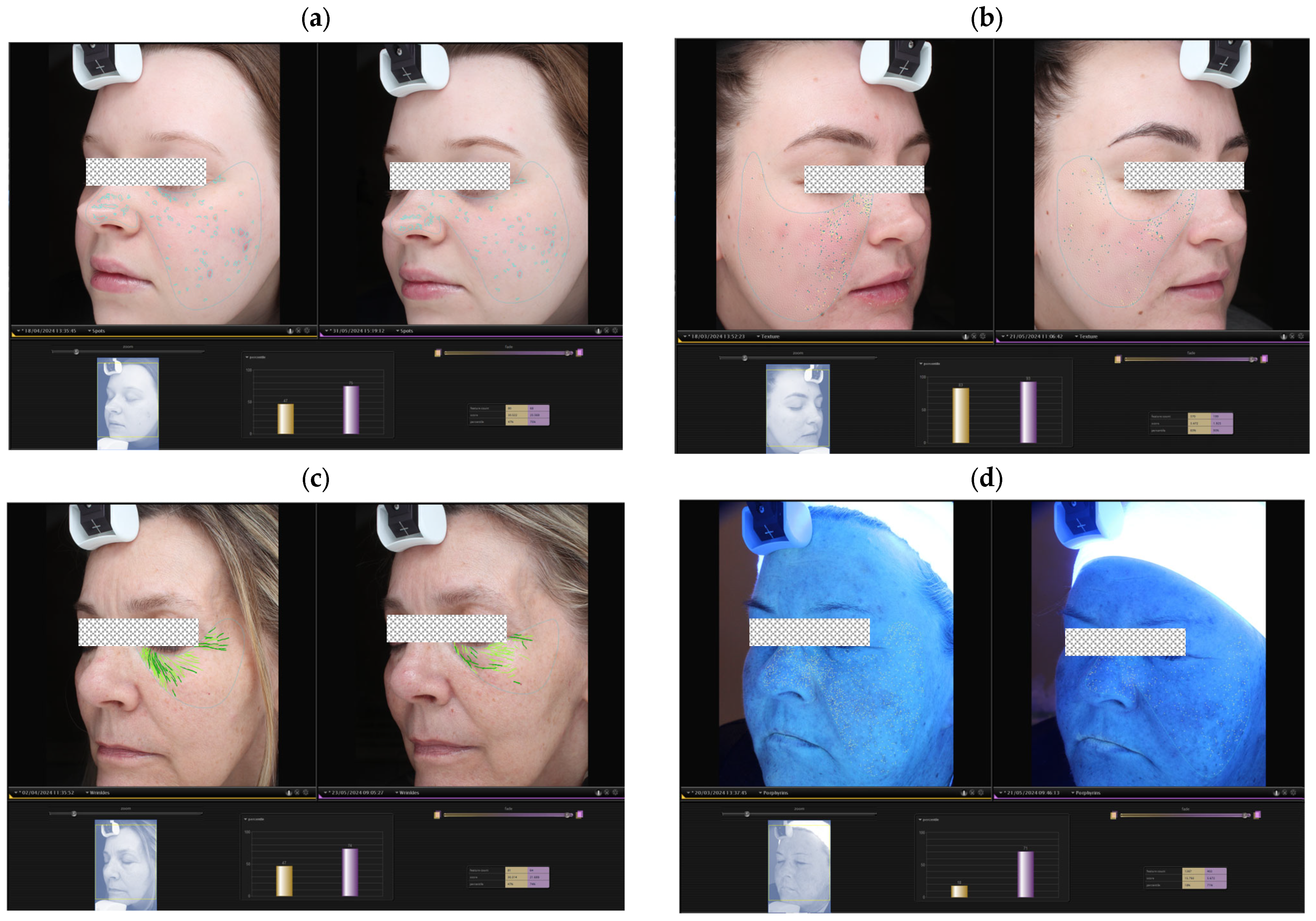
The Canfield VISIA-CR Imaging System (Parsippany, NJ, USA) provides a quantitative analysis of various skin parameters by capturing standardised digital images using white light, cross-polarised light, and UV fluorescence. The parameters analysed in this study include skin texture, brown spots (e.g., pigmentation), pore size, UV spots, red areas (e.g., acne, inflammation, rosacea), and wrinkle depth. All evaluations were conducted on the forehead and both cheek regions. Figure 2 illustrates the changes in selected parameters over time, with baseline (yellow bars) and post-treatment (purple bars) measurements indicating improvement percentages relative to average skin type and age-matched controls.
  • Cortex DermaLab
The Cortex DermaLab Combo 4 Skin Analysis System (Aalborg, Denmark) was used to assess skin hydration, elasticity, density (via ultrasound), and transepidermal water loss (TEWL) using dedicated probes for each parameter. Measurements were conducted on the cheek area.
  • Self-assessment
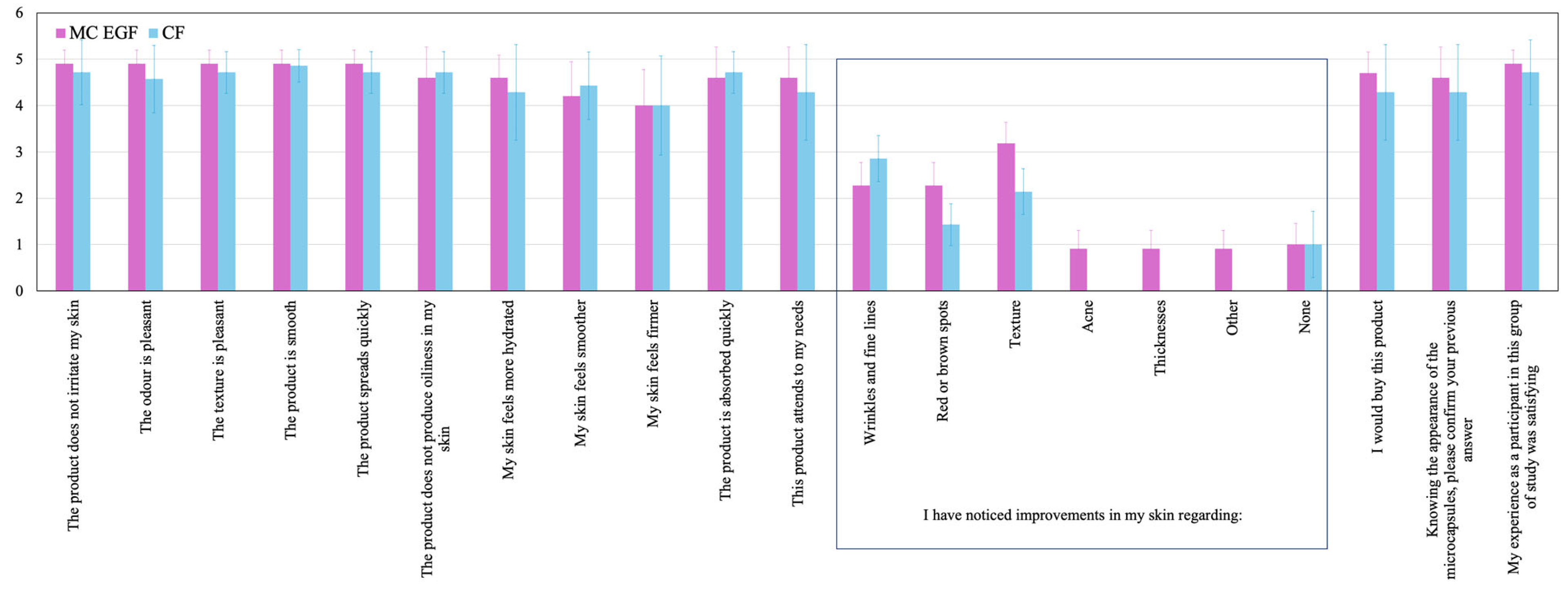
A self-assessment questionnaire was electronically completed by the participants after 60 days, capturing perceptions of their skin’s appearance, product effectiveness, and overall satisfaction, through 16 structured questions. A 5-point scale was used (1 = disagree, 5 = completely agree). Any adverse events were also reported throughout the study.

3. Results and Discussion

3.1. The Resulting Microcapsules

The AG/GE microcapsules containing EGF were produced with an initial EGF concentration of 116 µg·mL−1. The resulting microcapsules exhibited a mean particle size of approximately 62.6 µm and a SC of 8.4 ± 0.2%. The EE reached 78.8 + 1.0%, indicating effective incorporation of EGF within the polymeric matrix through electrostatic interactions and hydrogen bonding [9]. This value is comparable to encapsulation efficiencies reported for gelatin-alginate coacervate systems, which achieved EE values of 81.3% [9].
The coacervation process was originally developed to encapsulate hydrophobic compounds [10]. An earlier work reported EE exceeding 98% for α-tocopherol [12]. In the present study, the same formulation was applied to a hydrophilic bioactive, EGF. At pH values below its isoelectric point (IpEGF = 4.6), the protein carries a net positive charge [18], which interacts with the anionic carboxyl groups of the AG matrix, facilitating coacervation [10]. These electrostatic interactions contribute to enhancing EE and may protect the EGF from proteolytic degradation, improving its stability [9].
A previously patented study on the coacervation of SA/GE explored the encapsulation of EGF to ensure stabilisation of the protein while applied to a wound surface [11]. The invention referred to a higher EE at a lower pH after interpretation of Western blotting assays.
Figure 3 shows the optical images of the MC EGF produced in this study. The microcapsules exhibit a spherical, multinucleated honeycomb-like structure, reflecting similar morphological characteristics to those previously reported in an optimisation study using confocal and cryoSEM analyses [10].

3.2. Formulation Skin Compatibility

The hydrating cream formulation containing 0.3% MC EGF demonstrated excellent skin tolerance in all tested volunteers. No erythema, edema, or other visible reactions were observed in any of the subjects. The calculated Mean Irritation Index (M.I.I.) remained ≤0.2, confirming the absence of irritant potential. Accordingly, the formulation was classified as non-irritant under dermatological evaluation.

3.3. The Performance of the Formulation

The performance of the formulation is summarised in Table 1. Skin compatibility testing confirmed that the enriched MC EGF hydrating cream is non-irritant. Stability was validated through spin and temperature cycle tests, with only slight textural changes observed after exposure to 40 °C. High temperatures destabilise the oil–water interface in emulsions, causing colour variations and phase separation [19]. Although the observed changes were minor, the stability test results suggest that prolonged storage at high temperatures could lead to instability. According to ISO/TR 18811:2018 guidelines [17], temperatures around 40 °C are used as accelerated stress conditions rather than realistic storage conditions, and stability under typical storage temperatures (e.g., 4–25 °C) is considered more representative of product performance.
The formulation developed in this study showed no significant differences in viscosity or pH compared to the CF. Viscosity remained at 1.2 to 1.3 Pa·s, while the pH stayed within the safe range of 5.9 to 6.1. These parameters confirm the formulation’s safety, non-irritancy, and biocompatibility, making it suitable for use as a leave-on cosmetic. Furthermore, sensory perceptions described through the self-assessment questionnaire revealed no noticeable difference in odour, further supporting its acceptability for skin application.

3.4. The Effects on the Skin

The hydrating creams containing MC EGF and CF were tested under controlled conditions, with data normalised by CF to minimise interference from variations in skin type and initial skin conditions.
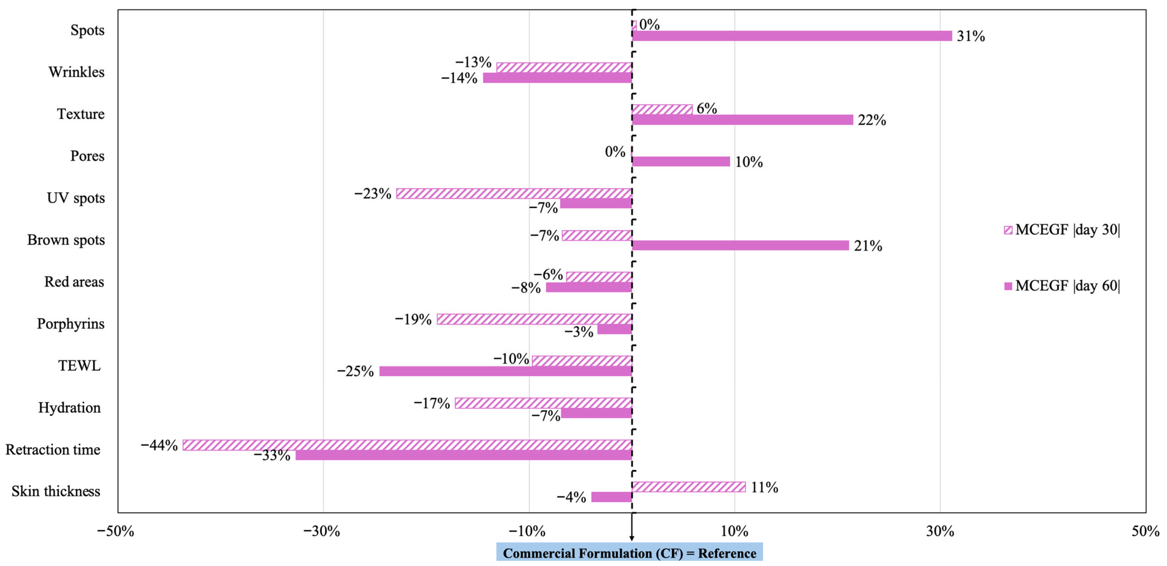
Figure 4 presents the results from Canfield VISIA-CR and Cortex DermaLab assessments, based on data collected over 30-day and 60-day intervals with 20 volunteers, using CF as the control. After the whole testing period, the cream with MC EGF showed superior performance, reducing spots by 31%, pores by 10%, brown spots by 21%, improving texture by 22%, and retraction time by 33%. These results indicate that volunteers using the microencapsulated formula achieved significantly enhanced skin quality compared to CF. Specifically, reducing surface spots and brown spots is associated with improving skin lesions such as freckles, acne scars, hyperpigmentation, and vascular lesions. Fewer visible pores suggest reduced visibility of sweat gland ducts, while improvements in texture enhance skin smoothness and colour consistency [20]. The decrease in retraction time indicates improved skin elasticity and a better ability to regain shape after stretching or deformation. These are key indicators of youthful and resilient skin, largely dependent on collagen level [21].
This study challenges established notions linking skin elasticity, wrinkle reduction, and hydration. Previous research often suggests that improvements in skin elasticity are associated with fewer wrinkles and enhanced hydration levels [22,23]. However, these skin properties are influenced by different biological mechanisms and may evolve at different time scales. In the present study, the application of MC EGF resulted in a noticeable improvement in skin elasticity, while the wrinkle parameter was 14% higher than in CF. One possible explanation is that elasticity changes may occur relatively quickly due to alterations in skin mechanical properties and barrier function, whereas visible wrinkle reduction is more closely associated with longer-term collagen remodeling processes. In addition, wrinkle detection using imaging systems such as VISIA may be influenced by factors including skin hydration, lighting conditions, facial micro-expressions, and natural variability among participants. Considering the limited number of volunteers included in this exploratory study, these results should be interpreted cautiously and may reflect short-term physiological responses rather than long-term structural skin changes.
Additionally, the MC EGF group gave rise to a 25% reduction in TEWL, a 7% decrease in hydration, and a 4% decrease in skin thickness relative to CF. The lower TEWL values suggest that MC EGF strengthened the skin’s barrier function, effectively reducing moisture loss. However, the barrier function and elasticity improvement did not correspond to increased hydration levels or reduced deep wrinkles. This suggests that multiple interacting factors play a role in skin health. The data underscores the need for comprehensive approaches in skincare formulations that consider the independent influences of elasticity, wrinkle reduction, and hydration to achieve more predictable outcomes in skin appearance. Minor differences were also observed in UV spots (7%), red areas (8%), and porphyrins (3%), parameters that indicate potential melanin accumulation beneath the skin surface, inflammation, and bacterial excretions, respectively [20].
The volunteer study included a relatively small number of participants (n = 20, divided into two groups), which limits the statistical strength of the observations. While the results show consistent trends across several skin parameters, the sample size and the natural variability among participants, including differences in age and gender, may influence the measured outcomes. For this reason, the findings should be interpreted as an initial assessment of formulation performance under real-use conditions. Additional studies involving larger groups of volunteers would be required to confirm these effects with greater statistical robustness.
The global acceptance of the novel-designed hydration cream and the CF was evaluated after 60 days of use through a 16-question survey, with responses scored from 1 (disagree) to 5 (completely agree), as shown in Figure 5. Both products received consistently positive scores for non-irritancy, texture, odour, smoothness, and spreadability. These results should be interpreted as indicative user perceptions rather than statistically conclusive outcomes due to the limited number of participants included in the study. Participant feedback was also collected during supervised evaluation visits, where qualitative observations and comments were recorded alongside the questionnaire responses. Participants reported perceived improvements in skin appearance after a brief adaptation period, potentially influenced by the harsh weather conditions experienced over the evaluation period. Iceland local forecast records indicate temperature variations from −1.6 °C to 6.9 °C and air humidity ranging from 59% to 78% between March and May 2024 [24]. Overall satisfaction and purchase intent were high for both formulations, and the presence of microcapsules in the MC EGF formulation did not negatively influence user perceptions.

4. Conclusions

This study demonstrated the effective encapsulation of EGF within AG/GE coacervates, achieving an encapsulation efficiency of 78.8 + 1.0%. The encapsulation system is expected to stabilise EGF through electrostatic and hydrogen-bond interactions commonly reported for gelatin-polysaccharide coacervate systems, which may contribute to protecting the protein and enabling controlled release into the skin.
The incorporation of MC EGF into a hydrating cream formulation revealed benefits in key skin parameters, such as reductions in surface spots, brown spots, pore visibility, as well as improvements in skin texture and elasticity. The observed 25% reduction in TEWL and 33% increase in retraction time indicate that MC EGF enhanced the skin’s barrier function and resilience, promoting moisture retention, reducing transepidermal water loss, and improving the skin’s capacity to regain its shape after stretching or deformation. However, the 14% increase in wrinkle presence and 7% decrease in hydration compared to CF suggest that these properties may not be directly correlated. This highlights a complex relationship between TEWL, elasticity, hydration, and wrinkle formation that requires further investigation. Although the results demonstrate promising effects on several skin parameters, the limited number of volunteers included in the study means that these findings should be considered preliminary and would benefit from validation in larger-scale studies.
User feedback indicated high acceptance for MC EGF and CF products, with positive ratings for non-irritancy, texture, and overall sensory experience. Participants reported visible improvements in skin appearance, even under challenging environmental conditions, supporting the formulations’ robustness and efficacy.
In conclusion, the AG/GE coacervate encapsulation system provides an effective and stable delivery method for EGF in skincare formulations. These findings highlight the potential of this approach to enhance specific skin quality parameters and emphasise the importance of developing skincare products that address unique parameters. This study underscores the value of advanced encapsulation techniques in achieving targeted and sustained improvements in skin health and appearance.

Author Contributions

Conceptualization, J.C.K. and M.M.D.; Methodology, J.C.K., I.M.M. and S.D.G.; Validation, J.C.K., I.M.M. and S.D.G.; Formal Analysis, J.C.K., I.M.M. and S.D.G.; Investigation, J.C.K., I.M.M. and Y.A.M.; Resources, S.D.G. and M.M.D.; Data Curation, Y.A.M.; Writing—Original Draft Preparation, J.C.K. and M.M.D.; Writing—Review and Editing, I.M.M., A.E.R. and M.F.B.; Visualization, S.D.G.; Supervision, A.E.R., M.F.B. and M.M.D.; Project Administration, M.F.B. and M.M.D.; Funding Acquisition, A.E.R., M.F.B. and M.M.D. All authors have read and agreed to the published version of the manuscript.

Funding

This work was financially supported by Fundação para a Ciência e a Tecnologia, I.P./MCTES through national funds: LSRE-LCM, UID/50020/2025 (https://doi.org/10.54499/UID/50020/2025); ALiCE, LA/P/0045/2020 (https://doi.org/10.54499/LA/P/0045/2020); CIMO, UIDB/00690/2025 (https://doi.org/10.54499/UID/00690/2025) and UID/PRR/00690/2025 (https://doi.org/10.54499/UID/PRR/00690/2025); and SusTEC, LA/P/0007/2020 (https://doi.org/10.54499/LA/P/0007/2020).

Institutional Review Board Statement

The national bioethics committee—Vísindasiðanefnd. BIOEFFECT) did not need an ethical approval as we were not treating a vulnerable population (patients) but people with no competing skin diseases, and these participants signed an informed consent form that we needed to have according to the data protection act.

Informed Consent Statement

Informed consent was obtained from all subjects involved in the study. Written informed consent has been obtained from the participants to publish this paper.

Data Availability Statement

The raw data supporting the conclusion of this article will be made available by the authors on request.

Acknowledgments

Júlia Cristiê Kessler acknowledges her PhD scholarship (ref. 2020.06656.BD, (https://doi.org/10.54499/2020.06656.BD) from FCT. All experimentation from cosmetic production to volunteer skin assessments were conducted at BIOEFFECT ehf. (https://www.bioeffect.com, Reykjavík, Iceland) inspired by the in-house methodologies under an agreement established between BIOEFFECT and the Faculty of Engineering of the University of Porto. The authors extend their gratitude to BIOEFFECT for their invaluable support during J.C. Kessler’s stay at their facility. During the preparation of this manuscript/study, the author(s) used AI (ChatGPT-5) for the purposes of text editing (e.g., grammar, structure, spelling, punctuation, and formatting). The authors have reviewed and edited the output and take full responsibility for the content of this publication.

Conflicts of Interest

Sigrún Dögg Gudjónsdóttir is an employee of BIOEFFECT ehf. The authors declare that the research was conducted in the absence of any commercial or financial relationships that could be construed as a potential conflict of interest. The funders had no role in the design of the study; in the collection, analyses or interpretation of data; in the writing of the manuscript; or in the decision to publish the results.

Abbreviations

AGArabic gum
α-tocα-tocopherol
ANOVAAnalysis of Variance
CAPCaprylic triglyceride
CFControl formulation
DTTDithiothreitol
ECEuropean Commission
EDC1-Ethyl-3-(3-dimethylaminopropyl) carbodiimide
EE (%)Encapsulation Efficiency
EGFEpidermal Growth Factor
GEGelatine A
GMPGood Manufacturing Practice
HClHydrogen chloride
HLBHydrophilic-Lipophilic Balance
IpEGFIsoelectric point of Epidermal Growth Factor
ISOInternational Organization for Standardization
JESSCapillary-based immunoassay platform (ProteinSimple, Bio-Techne)
KOHPotassium hydroxide
MC EGFMicrocapsules containing Epidermal Growth Factor
MCCMicroencapsulation by Complex Coacervation
M.I.I.Mean Irritation Index
MoMoringa oleifera Lam.
NHSN-hydroxy succinimide
P80Polysorbate 80
pHPotential of Hydrogen
PEG 8Polyoxyethylene glycol 8 ester
SA/GESodium alginate and gelatine complex
SC (%)Solid Content
TEWLTransepidermal Water Loss
UVUltraviolet

References

  1. Hwang, H.J.; Han, S.; Jeon, S.; Seo, J.; Oh, D.; Cho, S.-W.; Choi, Y.W.; Lee, S. Recombinant human epidermal growth factor (rhEGF)-loaded solid lipid nanoparticles: Fabrication and their skin accumulation properties for topical rhEGF delivery. Bull. Korean Chem. Soc. 2014, 35, 2290–2294. [Google Scholar] [CrossRef]
  2. Jeon, Y.J.; Kim, Y.H.; Jeon, Y.J.J.; Lee, W.W.; Bae, I.G.; Yi, K.W.; Hong, S.H. Increased synthesis of hyaluronic acid by enhanced penetration of CTP-EGF recombinant in human keratinocytes. J. Cosmet. Dermatol. 2019, 18, 1539–1545. [Google Scholar] [CrossRef] [PubMed]
  3. Heo, S.G.; Hong, W.K.; Kim, E.M.; Kim, M.S.; Song, S.Y.; Choi, S.M.; Park, J.H.; Kim, J.H.; Choi, G.S. Development of an epidermal Growth Factor (EGF) loaded elastosome formulation for enhanced skin penetration and anti-aging effects. Cosmetics 2026, 13, 10. [Google Scholar] [CrossRef]
  4. Shin, S.H.; Koh, Y.G.; Lee, W.G.; Seok, J.; Park, K.Y. The use of epidermal growth factor in dermatological practice. Int. Wound J. 2023, 20, 2414–2423. [Google Scholar] [CrossRef] [PubMed]
  5. Yang, Y.; Luo, R.; Chao, S.; Xue, J.; Jiang, D.; Feng, Y.H.; Guo, X.D.; Lu, D.; Zhang, J.; Li, Z.; et al. Improved pharmacodynamics of epidermal growth factor via microneedles-based self-powered transcutaneous electrical stimulation. Nat. Commun. 2022, 13, 6908. [Google Scholar] [CrossRef] [PubMed]
  6. Surini, S.; Leonyza, A.; Suh, C.W. Formulation and in vitro penetration study of recombinant human epidermal growth factor-loaded transfersomal emulgel. Adv. Pharm. Bull. 2020, 10, 586–594. [Google Scholar] [CrossRef] [PubMed]
  7. Bhat, M.; Tharmatt, A.; Bhargava, S.; Kumeria, T.; Mishra, A.; Mittal, A.; Chitkara, D. Can breakthroughs in dermal transdermal macromolecule delivery surmount existing barriers revolutionize future therapeutics? J. Transl. Med. 2025, 23, 513. [Google Scholar] [CrossRef] [PubMed]
  8. Zhang, L.; Dong, Z.; Liu, W.; Wu, X.; He, H.; Lu, Y.; Wu, W.; Qi, J. Novel pharmaceutical strategies for enhancing skin penetration of biomacromolecules. Pharmaceutics 2022, 15, 887. [Google Scholar] [CrossRef] [PubMed]
  9. Jeong, S.; Kim, B.-W.; Lau, H.-C.; Kim, A. Gelatin–alginate complexes for EGF encapsulation: Effects of H-bonding and electrostatic interactions. Pharmaceutics 2019, 11, 530. [Google Scholar] [CrossRef] [PubMed]
  10. Kessler, J.C.; Martins, I.M.; Manrique, Y.A.; Lopes, J.C.B.; Rodrigues, A.E.; Barreiro, M.F.; Dias, M.M. Optimised model microcapsules of Arabic gum and gelatin A for functional cosmetic applications: From formulation to scale-up using a mesostructured reactor. Chem. Eng. Res. Des. 2025, 215, 108–121. [Google Scholar] [CrossRef]
  11. Kim, A.; Jeong, S.H. Coacervate Composition Containing Protein Drug and Wound Healing Agent Comprising Same. U.S. Patent 11,541,151, 1 March 2023. Available online: https://www.freepatentsonline.com/11541151.html (accessed on 25 November 2024).
  12. Kessler, J.C.; Martins, I.M.; Manrique, Y.A.; Gudjónsdóttir, S.D.G.; Rodrigues, A.E.; Barreiro, M.F.; Dias, M.M. Microencapsulated α-tocopherol and Moringa extract for improved skin protection: Insights from human skin assessment in cosmetic formulations. J. Cosmet. Dermatol. 2025, 24, e70486. [Google Scholar] [CrossRef] [PubMed]
  13. Shaddel, R.; Hesari, J.; Damirchi, S.A.; Hamishehkar, H.; Achachlouei, B.F.; Hung, Q. Use of gelatin and gum Arabic for encapsulation of black raspberry anthocyanins by complex coacervation. Int. J. Biol. Macromol. 2018, 107, 1800–1810. [Google Scholar] [CrossRef] [PubMed]
  14. Hernandez-Nava, R.; López-Malo, A.; Palou, E.; Ramírez-Corona, N.; Jiménez-Munguía, M.T. Encapsulation of oregano essential oil (Origanum vulgare) by complex coacervation between gelatin and chia mucilage and its properties after spray drying. Food Hydrocoll. 2020, 109, 106077. [Google Scholar] [CrossRef]
  15. International Organization for Standardization. Cosmetics—Good Manufacturing Practices (GMP)—Guidelines on ISO/TR 22716:2007; ISO: Geneva, Switzerland, 2007; Available online: https://www.iso.org/standard/36437.html (accessed on 12 May 2024).
  16. The European Parliament and the Council of the European Union. Regulation (EC) No 1223/2009 of the European Parliament and of the Council of 30 November 2009 on cosmetic products. Off. J. Eur. Union L 2009, 342, 59–209. [Google Scholar]
  17. International Organization for Standardization. Guidelines for Stability Testing of Cosmetic Products ISO/TR 18811:2018; ISO: Geneva, Switzerland, 2018; Available online: https://www.iso.org/standard/63465.html (accessed on 12 May 2024).
  18. Campion, S.R.; Tadaki, D.K.; Niyogi, S.K. Evaluation of the role of electrostatic residues in human epidermal growth factor by site-directed mutagenesis and chemical modification. J. Cell Biochem. 1992, 50, 35–42. [Google Scholar] [CrossRef] [PubMed]
  19. Venkataramani, D.; Tsulaia, A.; Amin, S. Fundamentals and applications of particle stabilized emulsions in cosmetic formulations. Adv. Colloid Interface Sci. 2020, 283, 102234. [Google Scholar] [CrossRef] [PubMed]
  20. Canfield Scientific. VISIA Complexion Analysis. Available online: https://www.canfieldsci.com/imaging-systems/visia-complexion-analysis/ (accessed on 7 November 2024).
  21. Everett, J.S.; Sommers, M.S. Skin viscoelasticity: Physiologic mechanisms, measurement issues, and application to nursing science. Biol. Res. Nurs. 2013, 15, 338–346. [Google Scholar] [CrossRef] [PubMed]
  22. Fujimura, T.; Haketa, K.; Hotta, M.; Kitahara, T. Loss of skin elasticity precedes rapid increase of wrinkle levels. J. Dermatol. Sci. 2007, 47, 233–239. [Google Scholar] [CrossRef] [PubMed]
  23. Choi, J.W.; Kwon, S.H.; Huh, C.H.; Park, K.C.; Youn, S.W. The influences of skin visco-elasticity, hydration level and aging on the formation of wrinkles: A comprehensive and objective approach. Skin. Res. Technol. 2013, 19, e349–e355. [Google Scholar] [CrossRef] [PubMed]
  24. Icelandic Meteorology Office. Climatological Data. Available online: https://en.vedur.is/climatology/data/ (accessed on 25 August 2024).
Figure 1. Schematic setup for the development of the cosmetic formulations. Notation in order of appearance: RT, room temperature; P80, polysorbate 80; α-toc, α-tocopherol; PEG 8, polyoxyethylene glycol; CAP, caprylic triglyceride; KOH, potassium hydroxide; MC EGF, microcapsules containing epidermal growth factor.
Cosmetics 13 00089 g001
Figure 2. Examples of skin measurements performed over the 30 and 60-day study period, evaluating (a) spots and (b) texture in volunteers using MC EGF and (c) wrinkles and (d) brown spots in volunteers using commercial formulation (CF). Baseline and post-treatment measurements are represented in yellow and purple bars, respectively.
Cosmetics 13 00089 g002
Figure 3. Optical microscope images of microcapsules containing EGF in magnifications of 10× and 40×, respectively.
Cosmetics 13 00089 g003
Figure 4. Performance of the hydrating creams containing MC EGF, normalized by the commercial formulation (CF). Skin measurements performed at 30 and 60-day product use with 20 volunteers.
Cosmetics 13 00089 g004
Figure 5. Cosmetic acceptance obtained by the self-assessment study of the hydrating cream containing MC EGF and commercial formulation (CF) after 60-day product use. Scores: 1 = disagree; 5 = completely agree.
Cosmetics 13 00089 g005
Table 1. Stability of the hydrating cream formulation containing MC EGF, and comparison of viscosity and pH to CF.
TestCompatibilitySpinTemperature CycleLong-Term and Heat StabilityViscosity (Pa⋅s)pH (20 °C)
EGFNon-irritantStableStable with slightly different textureStable at 4 °C and 20 °C; unstable at 40 °C1.3 a5.9–6.0 a
CF----1.2 a5.9–6.1 a
Criteria: Stable if no phase separation is observed after the stability test; unstable if phase separation is observed after the stability test. Different letters in the same column represent significant differences (α = 0.05).
Disclaimer/Publisher’s Note: The statements, opinions and data contained in all publications are solely those of the individual author(s) and contributor(s) and not of MDPI and/or the editor(s). MDPI and/or the editor(s) disclaim responsibility for any injury to people or property resulting from any ideas, methods, instructions or products referred to in the content.