Article

1064 nm Q-Switched Fractional Laser for Transcutaneous Delivery of a Biostimulator: Efficacy and Safety Outcomes of a Split-Face Study

1
Dermatology Unit, IDI-IRCCS, 00167 Rome, Italy
2
Clinical Psychology Unit, Clinical Epidemiology Unit, IDI-IRCCS, 00167 Rome, Italy
3
Clinical Epidemiology Unit, IDI-IRCCS, 00167 Rome, Italy
4
El.En. Group, 50041 Calenzano, Italy
*
Author to whom correspondence should be addressed.
These authors contributed equally to this work.
Cosmetics 2024, 11(1), 14; https://doi.org/10.3390/cosmetics11010014
Submission received: 5 December 2023 / Revised: 5 January 2024 / Accepted: 15 January 2024 / Published: 23 January 2024
(This article belongs to the Special Issue Treatment for Anti-aging and Rejuvenation)

Abstract

:
Background: Laser-assisted drug delivery is a promising strategy that enhances topical treatment by increasing cutaneous permeation and reducing side effects. In cosmetic settings, the efficacy and safety profiles of a treatment must meet the need of a painless procedure with fast recovery. In this context, Q-switched laser appears promising as it can open cutaneous pores without creating a carbonisation barrier. Methods: A split-face study on patients presenting for cosmetic procedures at IDI-IRCCS, Rome (30 September–18 October 2023), was conducted. Pan-facial Q-Switched laser was followed by a topical biostimulator applied on half of the face. Post-procedure local reactions were recorded together with patients’ perceptions. Cutaneous elasticity and hydration were assessed at baseline and three-week follow-up. The Skindex17 questionnaire evaluated the procedure’s impact on patients’ life quality. Also, participants and physicians expressed satisfaction with the treatment. Results: The procedure was well tolerated by patients; local reactions include transitory erythema, superficial bleeding, and oedema, none of which had an impact on daily life. An improvement in skin quality was documented objectively and subjectively by patients and physicians. Conclusion: Q-switched lasers emerge as promising devices for drug delivery, especially for cosmetic reasons. Indeed, the low risk of local reactions together with a remarkable increase in cutaneous permeation make this a suitable strategy for cosmetic procedures.

1. Introduction

Topical drug delivery is favourable since it avoids the first hepatic metabolism that occurs with oral administration. Nonetheless, it is more convenient and comfortable than an injectable application. The dense structure of the stratum corneum (SC), however, limits transcutaneous absorption [1].
A variety of techniques are used to enhance drug delivery, such as chemical or physical methods or combinations of them [2]. Particularly, physical methods include those that damage the SC barrier and those that work by an external force on the active components in the skin. These techniques include ultrasound (cavitational and non-cavitational), electroporation, iontophoresis, sonophoresis, microdermabrasion, external pressure, magnetoporation, and thermal ablation (laser, microneedle, and radio frequency) [3,4]. In order to deliver a drug through the skin, topical creams and hypodermic needles have been increasingly employed. However, patients are less compliant with needles due to the pain associated with them, and also topical treatments have low bioavailability. Various researchers have investigated microneedles (MNs) for distributing drugs via the transdermal route and for overcoming the limitations of the current techniques [5]. However, there are some issues concerning this technique related to the device itself, the procedure, and the experience being painful, which may limit its use in clinical practice [6].
In this setting, among the possible techniques for drug delivery, lasers have gained a promising role, being not only less invasive and painful but also faster than manual needle injection and, above all, not implying direct contact with human materials.
In recent years, laser-assisted drug delivery (LADD) has received significant attention due to its use for clinical and academic purposes, with increasing application to human subjects [7].
Different types of lasers have been used in the context of LADD. Each of them presents a higher or lower effectiveness and also a specific safety profile. For practical purposes, the lasers used in LADD can be divided into up to four subgroups: (1) fully ablative lasers, such as CO2 (10,600 nm) or Er:YAG (2940 nm), which have water as their main chromophore and produce total heating and vaporisation of the tissue; (2) fractional ablative lasers, which are the same lasers of the previous subgroup but used in fractional mode in order to produce thermal damage columns (the so-called microthermal zones (MTZ)); (3) non-ablative fractional lasers such as the erbium fibre laser 1550 nm, which, analogously to the previous ones, produce MTZ but, by contrast, in those columns, only tissue heating occurs without determining dermo-epidermal ablation; and (4) non-ablative lasers for dermal remodelling; this group includes the remainder of lasers with chromophores different from water that have been used to try to induce the greater absorption of molecules. These include vascular lasers, such as the 585/595 nm pulsed dye laser or the Nd:YAG of 1064 nm [8].
LADD parameters must be adapted to the patient, the skin condition and area, and medication. As for the latter, LADD has been used in conjunction with a variety of topical treatments, including corticosteroids, photosensitisers, and immunotherapy drugs (imiquimod or 5-fluorouracil) to treat several disorders, including scarring, nonmelanoma skin cancer, and photodamage. LADD is a promising approach that increases the absorption of topical compounds by taking advantage of the laser’s synergistic effect.
Moreover, as for safety, side effects related to the procedure are comparable to those produced by the laser treatment itself and mainly consist of local transitory reactions which are well tolerated by patients. Hence, safety being equal, LADD provides a significant improvement in procedure outcome [9].
Of note, Han and colleagues compared the laser-induced microjet injection versus needle injection of polylactic acid/hyaluronic acid filler for skin enhancement and rejuvenation [2]. They proved that using a laser-induced microjet injector allows not only for the delivery of a controlled dose and filler depth but also an even distribution, enhanced clinical efficacy, reduced discomfort and side effects, and enough time for doctors to perform treatment [2].
In comparison to other approaches, lasers have been shown to offer significant advantages in improving transdermal drug delivery (TDD), avoiding all the potential complications associated with the use of microneedling. Earlier studies demonstrate that the mechanisms responsible for this enhanced mechanism may rely on the photomechanical action that results in SC damage [10].
However, further investigations are warranted in the standardisation of the energy and density required in laser applications, as well as in determining the concentration of each substance, according to their characteristics, to be applied via drug release processes. Interestingly, already several articles have demonstrated that laser-assisted drug delivery is associated with lower concentrations of active ingredients due to increased permeability [11,12,13,14]. Indeed, lower concentrations of active principles increase procedure security and reduce the risk of adverse events. Nevertheless, the role of lesional skin must be taken into account since it may determine side effects for lower concentrations of the product. Tissue–laser contact may promote skin permeability, increasing the assimilation of substances applied topically to the body’s surfaces.
The use of ablative fractional lasers for drug delivery, such as the CO2 10,600 nm or Er:YAG 2940 nm lasers, is well known, and its efficacy has been assessed and confirmed in many clinical trials [11,15,16]. Fractional CO2 lasers determine tissue heating with consequent water vaporisation and skin ablation. Thermal ablation devices selectively heat the skin surface in a fractionated way, generating micro-perforations in the SC that are surrounded by coagulated tissue, the so-called MTZ; these micro-channels increase the hydrophilic and lipophilic substance permeability, allowing a deeper drug penetration into the skin. [17]. The CO2 10,600 nm laser penetrates deeper and produces higher heat quantity [16]. However, the main downsides of CO2 laser-assisted drug delivery include long downtime, significant pain related to the procedure, and high rates of side effects, such as hyperpigmentation (especially in dark phototypes) and scarring [18]. In addition, the thick coagulation area that surrounds the MTZ may represent a serious obstacle to the transportation of molecules [19]. Of note, recent evidence suggests that non-ablative fractional photothermolysis can increase topical drug absorption less invasively than ablative fractional lasers, potentially overcoming such issue [20].
Indeed, non-ablative lasers cause targeted thermal damage while leaving the skin surface intact, as histological examinations demonstrated that there is no epidermal damage or real ablation as compared to fractional ablative lasers.
However, contrarily to ablative fractional lasers, few data are available on the use of the non-ablative counterpart for drug delivery. Interestingly, the Q-switched (QS) laser is a promising opportunity that is gaining ground in this setting with the aim to increase skin penetration. Indeed, non-ablative QS lasers emit ultrashort waves with high energy peaks in the range of nanoseconds that create micropores and microcraters in the SC that have two advantages: they are not surrounded by coagulated tissue and are smaller than those generated with fractional CO2 lasers [10,14].
Interestingly, Liu et al. studied real-time fluctuations in the skin surface temperature after Q-switched 1064 nm laser therapy, recording a peak of only 1.01 ± 0.23 °C in the skin temperature as soon as 1 min after the procedure [10]. Interestingly, the same study revealed that the QS laser treatment determined a 12-fold increase in cutaneous absorption of glycerol, a hydrophilic agent that slowly penetrates into intact SC. Also, the authors histologically proved that the microchannels of the SC progressively close up with complete restitutio ad integrum after 14 days [10]. All these findings confirm the efficacy and the minimally invasive nature of QS lasers in increasing cutaneous permeability of substances and the short downtime of the procedure both on the clinical and molecular levels.
A comprehensive representation of the three technologies discussed (microneedles, QS, and fractional CO2 laser) for drug delivery is shown in Figure 1.
Other key points for the use of Q-switched lasers in LADD are the painless nature of the treatment and the short downtime that increase patients’ adherence to treatment.
To date, few studies assessing the efficacy of Q-switched 1064 nm Nd:YAG laser to promote and potentiate drug penetration for facial rejuvenation, onychomycosis, and melasma have been published in the scientific literature [21,22,23,24].
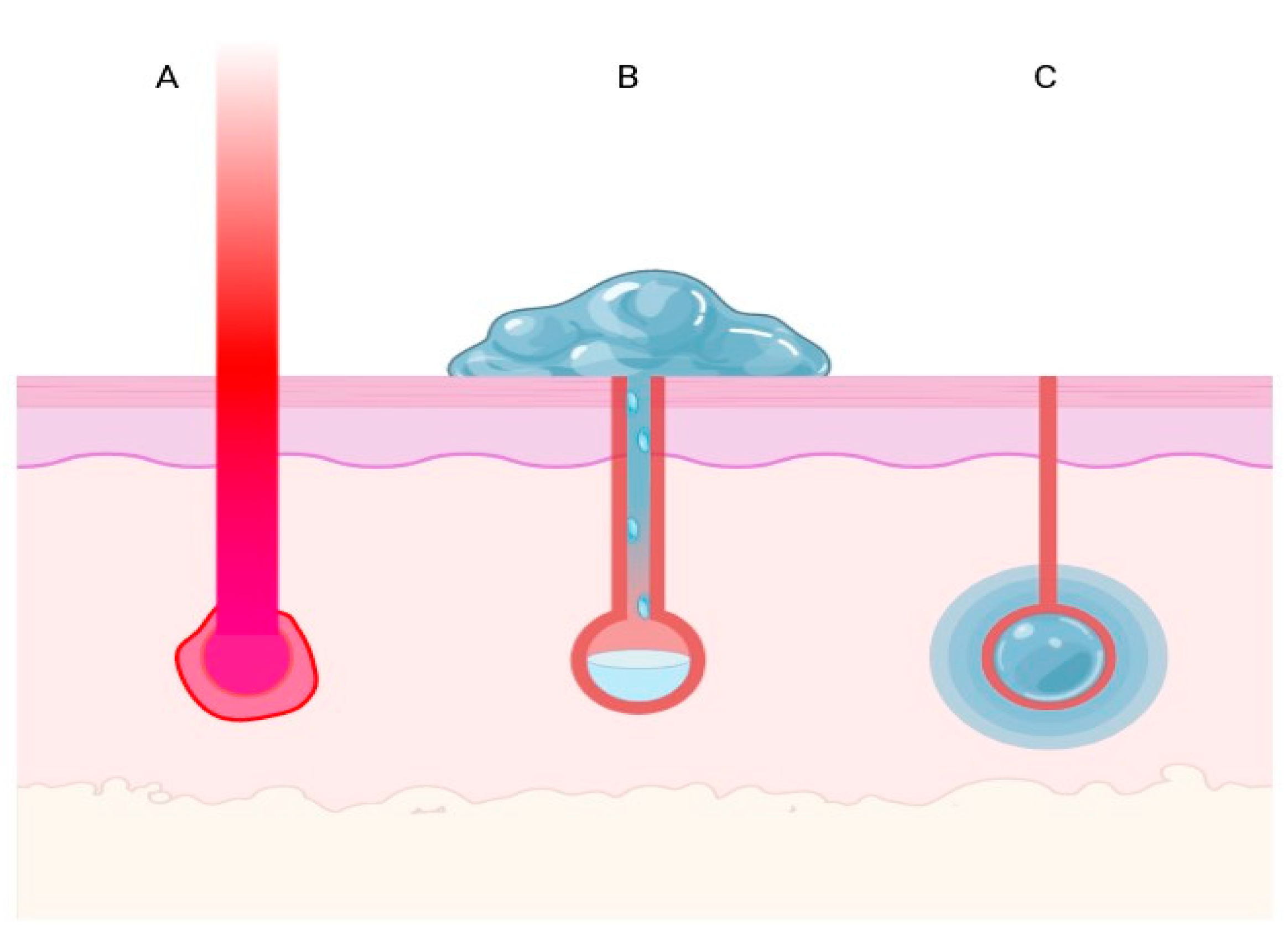
Based on these scientific findings, a split-face study was conducted to evaluate the efficacy and safety of the combination of treatments with a 1064 nm Q-switched fractional Nd:YAG system (SmartPico, Deka M.E.L.A, Calenzano, Italy) and a topical bio-restructuring drug with amino acid composition for facial rejuvenation. The mechanism of action of this technology is graphically represented in Figure 2.

2. Materials and Methods

A split-face, case series study was conducted on participants presenting for the first time at the dermatological reference centre IDI-IRCCS in Rome, Italy, between 30 September 2023 and 18 October 2023, for cosmetic bio-restructuring procedures.
Informed consent was obtained from all patients at the enrolment stage. At baseline, demographic, anamnestic, and epidemiologic data of each subject were collected. In particular, the anamnestic collection was focused on factors relevant to skin aging, such as cigarette smoking habits and insomnia. In fact, insomnia is known to affect the body’s stress response and hormonal balance, which, in turn, can accelerate skin aging processes [25]; likewise, cigarette smoke is a well-documented source of oxidative stress, known to accelerate the aging process in skin cells, being associated with collagen degradation, wound healing impairment, and change in cutaneous elasticity and texture [26].
The study protocol is detailed in Figure 3.
It consisted in the following phases (Figure 3): first, assessment of facial cutaneous elasticity and hydration at baseline through the device Checkup (Multi Skin Center® MC 750, Courage + Khazaka electronic GmbH, Koln, Germany); second, pan-facial laser treatment in the absence of local anaesthesia, using a 1064 nm Q-switched fractional Nd:YAG laser (SmartPico, Deka M.E.L.A, Calenzano, Italy), with an 8 mm spot size, at a fluence of 0.5 J/cm2 performed by expert health professionals (F.M., L.C., and L.P.). Three passes were performed over the entire face, except for the periocular and perioral areas. Subsequently, a topical bio-restructuring product containing glycine, L-proline, L-lysine monohydrochloride, L-leucine, sodium hyaluronate, and water for injection (Jalupro, Professional Derma, Lugano, Switzerland) was applied and massaged until absorption on half of the treated area. This product is usually employed for cutaneous biostimulation through injections over the entire facial skin surface.
Immediately after the procedure, participants were asked to score the discomfort experienced using the Visual Analogue Scale (VAS) (range: 0–10, indicating: no pain–extreme pain). Moreover, local reactions following the laser treatment were recorded. After 7 days, patients completed the Skindex 17 questionnaire, a reliable and validated tool that evaluates the physical and psychosocial impact of a procedure or a disease on patients’ health-related quality of life [27]. The questionnaire consists of 17 items about two domains, i.e., the physical (mild-to-moderate <50 and severe ≥50) and psychosocial functions (mild <20.82, moderate 20.83–37.50, and severe ≥37.51) with their severity [27]. The higher the scores, the higher the severity of the reduction in life quality [27,28].
At 3-week follow-up, a second evaluation of cutaneous hydration and elasticity was performed in the area that received the combination treatment (laser + topical bio-restructuring agent) as well as in the one receiving the laser-only treatment. Moreover, at the same timepoint, patients as well as the physicians that performed the laser treatment were asked to assess the subjective improvement in skin quality.
This study was conducted in accordance with the principles of the Declaration of Helsinki and later amendments.

Statistical Analysis

All the analyses were performed with the Statistical Package for Social Science (SPSS) (version 26.0) (IBM SPSS Statistics for Windows 26.0, 2019) [29]. For descriptive statistics, either means (and standard deviation (SD)) or percentage for continuous and categorical variables, respectively, were calculated as descriptive statistics. Finally, in order to determine whether there was statistical evidence that the mean difference between paired observations is significantly different from zero, paired t tests were performed between mean score of hydration and elasticity at baseline and after a three-week follow-up for the two different areas.

3. Results

Ten females with a mean age of 44.87 (±7.73) years were enrolled. The average Body Mass Index (BMI) among participants was 21.58 (±3.86), indicating an overall healthy weight range. Also, 66.7% of the sample were smokers. More than half of the subjects (55.6%) reported suffering from insomnia (Table 1).
The whole sample completed the study.
Concerning the procedure, the average VAS score registered soon after the laser treatment was 2.5 (±1.35), indicating a moderate level of pain or discomfort.
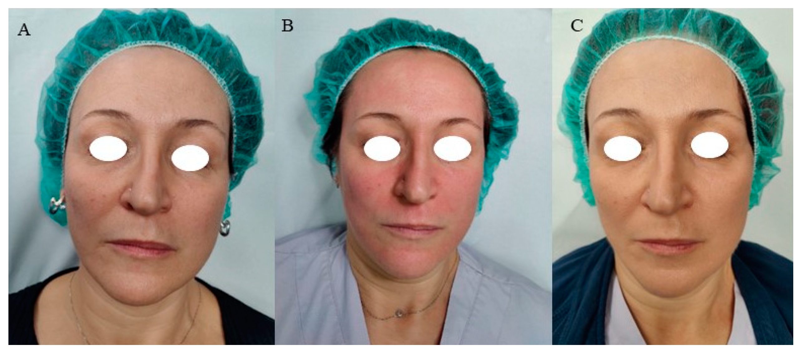
Also, procedure-related local reactions included erythema, oedema, and superficial bleeding with transitory aspect (lasting from 4 to 72 h) (Figure 4).
As for cutaneous hydration, the mean skin hydration score at baseline was 59.56 (±8.71). After 3 weeks of treatment with the laser + drug, there was a notable increase in hydration, reaching a mean value of 67.30 (±5.45) (Table 2). The paired mean difference of −7.740 (95% lower confidence interval (L.C.I.) = −10.745, upper confidence interval (U.C.I.) = −4.734, p < 0.001) indicates a statistically significant improvement (Table 3).
Regarding cutaneous elasticity, the mean value for skin elasticity was 66.10 (±13.51) at baseline, slightly decreasing to 67.10 (SD = 8.59) after three weeks from the combination treatment (Table 2). However, the paired mean difference of −1.000 (95% L.C.I. = −8.994, U.C.I. = 6.994, p = 0.784) suggests that this improvement was not statistically significant (Table 3).
In the comparison between laser + drug and laser-only treatments for skin hydration, the area treated with the combination approach displayed a higher mean hydration level (67.30, ±5.45) compared to that receiving monotherapy (59.60, ±7.47). The paired mean difference was 7.700 (95% L.C.I. = 4.360, U.C.I. = 11.039, p < 0.001), confirming the superiority of the combined treatment over laser alone in improving skin hydration (Table 3).
Likewise, comparing skin elasticity between the laser + drug group (mean = 67.10, SD = 8.59) and the laser-only group (mean = 65.20, SD = 7.84), the paired mean difference accounted for 1.900 (95% L.C.I. = −0.135, U.C.I. = 3.935, p = 0.064), indicating a trend towards improved elasticity with the combined treatment, although not statistically significant (Table 3).
The results of the Skindex17 questionnaire (Table 4) showed a low impact of the procedure on psychosocial and physical aspects with no interference on patients’ daily routine.
At three-week follow-up, overall, participants scored an improvement in skin quality as mild to moderate, with only one subject not displaying a visible change. Likewise, a positive assessment was given by the physicians who realised the procedure.

4. Discussion

LADD is an emerging technique that enhances topical treatment penetration, reducing dosage and side effects [9,30].
Its use is extremely wide in dermatology, finding application in medical as well as cosmetic fields. Indeed, such a method has been used for the treatment of pigmentary disorders, scars, oncological lesions, and cosmetics [30,31].
The outcomes of the procedure depend on one side on the type of lasers and, on the other side, on the type of topical agent [32]. Concerning the first, there are ablative, non-ablative, and fractional lasers that, through different strategies, overcome the cutaneous barrier allowing a deeper penetration [33]. Of them, ablative devices are those with higher risk of procedure-related side effects such as scarring and hyperpigmentation, while non-ablative and fractional ones are usually associated with transitory erythema and crusting [7]. Such aspects represent an important point to be considered when deciding the best approach to treat a specific dermatological condition, since the post-procedure may impact on patients’ quality of life. In particular, in the setting of cosmetic procedure, the pain and the downtime must be proportional to the wanted outcome [34]. That is to say that an aesthetic treatment would be more acceptable in the case of a pleasant procedure and fast recovery. It is commonly known that the primary mechanism underlying non-ablative treatments may include the production of dermal collagen [35]. Published studies have suggested that non-ablative laser rejuvenation is associated with new collagen deposition at the histological level [36,37]. Particularly, scientific literature findings demonstrated that the 1064 nm Q-switched laser affects epidermal barrier function by upregulating proinflammatory cytokines and enhancing the production of aquaporins, filaggrin, TGase, and HSP70. A decrease in matrix metalloproteinases type 1 (MMP-1) and an increase in procollagen, type I collagen, and elastin were observed in fibroblasts stimulated with keratinocyte conditioned medium (KCM) and exposed to a 1064 nm Q-switched Nd:YAG laser, suggesting that Q-switched 1064 nm laser treatment could be a useful tool in protecting against skin aging [38,39,40]. In addition, histological examinations have documented that the 1064 nm Q-switched laser induced collagen production [41], being more effective than the 532 nm laser in skin rejuvenation [42].
In this context, the possibility of improving skin quality by using biostimulating products delivered through a minimally invasive system, such as the Q-switched laser, represents a great strategy both on the safety and efficacy aspects [43,44,45].
Indeed, our study showed that the procedure was well tolerated by patients with mild discomfort as demonstrated by a mean VAS score of 2.5, remarkably in the absence of local anaesthesia.
On the other hand, from the scientific literature, higher levels of pain and discomfort than those revealed in our study have been found with the application of MN in the management of skin disorders such as hypertrophic and burn scars, as documented in the study of Aust et al., where an average VAS score that increased from 4.5 to 8.5 following one to four sessions of MN treatment was reported [46].
Furthermore, the most feared adverse event of MN treatment is the so-called “railroad” or “tramtrack” scarring that can occur with aggressive treatment, as reported in some studies [47,48].
Moreover, in our study, local reactions such as erythema and superficial bleeding were transitory, lasting from 4 to 72 h after treatment (Figure 4). Such findings define the procedure as suitable and safe.
Concerning topicals’ characteristics, Labadie et al. provided recommendations to maximise LADD reducing side effects; in detail, they advise relying only on products approved for parenteral use to avoid risks of granuloma formation, sensitivity reactions, or infections [7]. Also, viscosity and solubility are important parameters. In detail, a low-viscosity product may penetrate faster into the skin but, at the same time, a high-viscosity one may be more effective thanks to the prolonged time of contact, occlusion, and hydration. Also, the hydro soluble or phobic property of an agent may influence the grade of penetration into the skin as well as particles’ size with respect to channel dimensions. Finally, to increase permeability, accompanying strategies such as heat, occlusion, and pressure may be of interest, together with the application time, soon after the procedure [7].
The topical agent used in our study, with a formulation rich in amino acids, is meant to be employed in cutaneous bio-revitalisation procedures though needle injections, donating hydration, elasticity, and skin replenishment to treated areas. Anyway, the treatment may be painful and risky for vessel compression and haematomas that may follow the injections. Hence, the delivery of the product through laser is revolutionising as it allows keeping the same outcome, reducing discomfort and side effects.
Indeed, as for skin quality outcomes, our study showed that, as soon as three weeks post-treatment, there was an objectively documented increase in skin elasticity and, in particular, hydration of treated areas, especially in those receiving the combination treatment. Thus, the addition of a biostimulating agent after a laser procedure has been shown to increase the aesthetic outcome, with no further side effects. Also, the fast visible betterments encourage the development of longer and more complex treatment protocols. Moreover, patients themselves as well as the physicians that performed the procedure noticed an overall mild-to-moderate improvement in skin quality. Lastly, the minimal downtime following the procedure, as shown by the result of the Skindex 17 questionnaire, meets the need of patients that require fast recovery.
Hence, such preliminary results provide promising findings concerning the use of Q-switched laser in the setting of LADD techniques. The strength points are few and transient side effects and short downtime with consequent increased patient compliance and satisfaction.

5. Conclusions

In conclusion, this study has shown that Q-switched lasers may occupy an important position in the setting of LADD, given their painless feature, short downtime, and fast recovery, without renouncing high efficacy. Properties that are especially suitable in the setting of cosmetic procedures.
This study has some limitations related to the number of participants enrolled and sessions realised but provides promising results for further investigations on bigger samples with more complex treatment protocols. Indeed, randomised and head-to-head clinical trials are required to determine the benefit of different lasers in pre-treating dermatological conditions, highlighting the role that ethnicity, age, and gender may have on the general outcome.
Also, the absence of guidelines that aim to standardise the procedures (type of laser, fluence, density, number of sessions, and treatment duration) as well as the most appropriate topical agents (vehicle and ingredients) to be used warrants the definition of the proper treatment strategies, maximising efficacy, and reducing costs and side effects.

Author Contributions

Conceptualisation, F.M., T.S., L.C., E.C., T.Z. and I.F.; methodology, T.S., T.Z., I.F., F.M. and L.P.; validation, F.M., L.P., L.C., E.C. and M.B.P.; formal analysis, T.Z., I.F., F.M., T.S. and E.C.; investigation, F.M., T.S., L.C. and E.C.; resources, L.P., M.B.P. and T.S.; data curation, F.M., E.C., T.S. and L.C.; writing—original draft preparation, F.M., T.S., E.C., I.F., T.Z. and M.B.P.; writing—review and editing, F.M., E.C., T.S., L.C., T.Z. and I.F.; visualisation, L.C. and L.P.; supervision, F.M., T.S. and E.C.; project administration, I.F. and T.Z. All authors have read and agreed to the published version of the manuscript.

Funding

This research received no external funding.

Institutional Review Board Statement

The study was conducted in accordance with the Declaration of Helsinki and later amendments. Ethical review and approval were waived for this study given that the study protocol is part of our daily routine in clinical practice.

Informed Consent Statement

Informed consent was obtained from all subjects involved in the study.

Data Availability Statement

Data are available upon reasonable request.

Conflicts of Interest

T.Z. and I.F. are employed at El.En. Group. The other authors declare that the research was conducted in the absence of any commercial or financial relationships that could be construed as a potential conflict of interest.

References

  1. Schoellhammer, C.M.; Blankschtein, D.; Langer, R. Skin permeabilization for transdermal drug delivery: Recent advances and future prospects. Expert Opin. Drug Deliv. 2014, 11, 393–407. [Google Scholar] [CrossRef] [PubMed]
  2. Brown, M.B.; Martin, G.P.; Jones, S.A.; Akomeah, F.K. Dermal and transdermal drug delivery systems: Current and future prospects. Drug Deliv. 2006, 13, 175–187. [Google Scholar] [CrossRef] [PubMed]
  3. Sivamani, R.K.; Liepmann, D.; Maibach, H.I. Microneedles and transdermal applications. Expert Opin. Drug Deliv. 2007, 4, 19–25. [Google Scholar] [CrossRef]
  4. Alkilani, A.Z.; McCrudden, M.T.C.; Donnelly, R.F. Transdermal Drug Delivery: Innovative Pharmaceutical Developments Based on Disruption of the Barrier Properties of the stratum corneum. Pharmaceutics 2015, 7, 438–470. [Google Scholar] [CrossRef] [PubMed]
  5. Waghule, T.; Singhvi, G.; Dubey, S.K.; Pandey, M.M.; Gupta, G.; Singh, M.; Dua, K. Microneedles: A smart approach and increasing potential for transdermal drug delivery system. Biomed. Pharmacother. 2019, 109, 1249–1258. [Google Scholar] [CrossRef] [PubMed]
  6. Williams, A.C.; Barry, B.W. Penetration enhancers. Adv. Drug Deliv. Rev. 2004, 56, 603–618. [Google Scholar] [CrossRef] [PubMed]
  7. Labadie, J.G.; Ibrahim, S.A.; Worley, B.; Kang, B.Y.; Rakita, U.; Rigali, S.; Arndt, K.A.; Bernstein, E.; Brauer, J.A.; Chandra, S.; et al. Evidence-Based Clinical Practice Guidelines for Laser-Assisted Drug Delivery. JAMA Dermatol. 2022, 158, 1193–1201. [Google Scholar] [CrossRef]
  8. Manstein, D.; Herron, G.S.; Sink, R.K.; Tanner, H.; Anderson, R.R. Fractional photothermolysis: A new concept for cutaneous remodeling using microscopic patterns of thermal injury. Lasers Surg. Med. 2004, 34, 426–438. [Google Scholar] [CrossRef]
  9. Ng, W.H.S.; Smith, S.D. Laser-Assisted Drug Delivery: A Systematic Review of Safety and Adverse Events. Pharmaceutics 2022, 14, 2738. [Google Scholar] [CrossRef]
  10. Liu, C.; Zhang, J.; Yue, Y.; Luo, Q.; Zhu, D. 1064 nm-Nd:YAG lasers with different output modes enhancing transdermal delivery: Physical and physiological mechanisms. J. Biomed. Opt. 2013, 18, 61228. [Google Scholar] [CrossRef]
  11. Haedersdal, M.; Erlendsson, A.M.; Paasch, U.; Anderson, R.R. Translational medicine in the field of ablative fractional laser (AFXL)-assisted drug delivery: A critical review from basics to current clinical status. J. Am. Acad. Dermatol. 2016, 74, 981–1004. [Google Scholar] [CrossRef] [PubMed]
  12. Waibel, J.S.; Rudnick, A.; Nousari, C.; Bhanusali, D.G. Fractional Ablative Laser Followed by Transdermal Acoustic Pressure Wave Device to Enhance the Drug Delivery of Aminolevulinic Acid: In Vivo Fluorescence Microscopy Study. J. Drugs Dermatol. 2016, 15, 14–21. [Google Scholar] [PubMed]
  13. Sklar, L.R.; Burnett, C.T.; Waibel, J.S.; Moy, R.L.; Ozog, D.M. Laser assisted drug delivery: A review of an evolving technology. Lasers Surg. Med. 2014, 46, 249–262. [Google Scholar] [CrossRef] [PubMed]
  14. Lin, C.-H.; Aljuffali, I.A.; Fang, J.-Y. Lasers as an approach for promoting drug delivery via skin. Expert Opin. Drug Deliv. 2014, 11, 599–614. [Google Scholar] [CrossRef] [PubMed]
  15. Wenande, E.; Anderson, R.R.; Haedersdal, M. Fundamentals of fractional laser-assisted drug delivery: An in-depth guide to experimental methodology and data interpretation. Adv. Drug Deliv. Rev. 2020, 153, 169–184. [Google Scholar] [CrossRef] [PubMed]
  16. Haedersdal, M.; Sakamoto, F.H.; Farinelli, W.A.; Doukas, A.G.; Tam, J.; Anderson, R.R. Fractional CO(2) laser-assisted drug delivery. Lasers Surg. Med. 2010, 42, 113–122. [Google Scholar] [CrossRef]
  17. Prausnitz, M.R.; Langer, R. Transdermal drug delivery. Nat. Biotechnol. 2008, 26, 1261–1268. [Google Scholar] [CrossRef]
  18. Favaro, J.; Loureiro, V.B. Fractional Non-ablative Laser and Drug Delivery. In Drug Delivery in Dermatology: Fundamental and Practical Applications; Kalil, C.L.P.V., Campos, V., Eds.; Springer International Publishing: Cham, Switzerland, 2021; pp. 75–82. [Google Scholar] [CrossRef]
  19. Taudorf, E.H.; Haak, C.S.; Erlendsson, A.M.; Philipsen, P.A.; Anderson, R.R.; Paasch, U.; Haedersdal, M. Fractional ablative erbium YAG laser: Histological characterization of relationships between laser settings and micropore dimensions. Lasers Surg. Med. 2014, 46, 281–289. [Google Scholar] [CrossRef]
  20. Lim, H.K.; Jeong, K.H.; Kim, N.I.; Shin, M.K. Nonablative fractional laser as a tool to facilitate skin penetration of 5-aminolaevulinic acid with minimal skin disruption: A preliminary study. Br. J. Dermatol. 2014, 170, 1336–1340. [Google Scholar] [CrossRef]
  21. Kalil, C.; Campos, V.B.; Reinehr, C.P.H.; Chaves, C.R.P. Laser toning and drug delivery: A pilot study using laser Q-switched laser 1064nm. Surg. Cosmet. Dermatol. 2016, 8. Available online: https://api.semanticscholar.org/CorpusID:113503096 (accessed on 10 November 2023). [CrossRef]
  22. Park, S.J.; Park, J.W.; Seo, S.J.; Park, K.Y. Evaluating the tolerance and efficacy of laser-assisted delivery of tranexamic acid, niacinamide, and kojic acid for melasma: A single center, prospective, split-face trial. Dermatol. Ther. 2022, 35, e15287. [Google Scholar] [CrossRef] [PubMed]
  23. Park, K.Y.; Kim, D.H.; Kim, H.K.; Li, K.; Seo, S.J.; Hong, C.K. A randomized, observer-blinded, comparison of combined 1064-nm Q-switched neodymium-doped yttrium-aluminium-garnet laser plus 30% glycolic acid peel vs. laser monotherapy to treat melasma. Clin. Exp. Dermatol. 2011, 36, 864–870. [Google Scholar] [CrossRef] [PubMed]
  24. Kim, T.I.; Shin, M.K.; Jeong, K.-H.; Suh, D.H.; Lee, S.J.; Oh, I.-H.; Lee, M.-H. A randomised comparative study of 1064 nm Neodymium-doped yttrium aluminium garnet (Nd:YAG) laser and topical antifungal treatment of onychomycosis. Mycoses 2016, 59, 803–810. [Google Scholar] [CrossRef] [PubMed]
  25. Oyetakin-White, P.; Suggs, A.; Koo, B.; Matsui, M.S.; Yarosh, D.; Cooper, K.D.; Baron, E.D. Does poor sleep quality affect skin ageing? Clin. Exp. Dermatol. 2015, 40, 17–22. [Google Scholar] [CrossRef] [PubMed]
  26. Wong, Q.Y.A.; Chew, F.T. Defining skin aging and its risk factors: A systematic review and meta-analysis. Sci. Rep. 2021, 11, 22075. [Google Scholar] [CrossRef] [PubMed]
  27. Nijsten, T.E.C.; Sampogna, F.; Chren, M.-M.; Abeni, D.D. Testing and reducing skindex-29 using Rasch analysis: Skindex-17. J. Investig. Dermatol. 2006, 126, 1244–1250. [Google Scholar] [CrossRef]
  28. Chren, M.M.; Lasek, R.J.; Quinn, L.M.; Mostow, E.N.; Zyzanski, S.J. Skindex, a quality-of-life measure for patients with skin disease: Reliability, validity, and responsiveness. J. Investig. Dermatol. 1996, 107, 707–713. [Google Scholar] [CrossRef] [PubMed]
  29. IBM SPSS Statistics for Windows 25.0 IBM Corp. (n.d.). IBM SPSS Statistics for Windows, Version 25.0; IBM: Armonk, NY, USA, 2007; p. 335. [Google Scholar]
  30. Zaleski-Larsen, L.A.; Fabi, S.G. Laser-Assisted Drug Delivery. Dermatol. Surg. Off. Publ. Am. Soc. Dermatol. Surg. 2016, 42, 919–931. [Google Scholar] [CrossRef]
  31. Alegre-Sánchez, A.; Jiménez-Gómez, N.; Boixeda, P. Laser-Assisted Drug Delivery. Actas Dermosifiliogr. 2018, 109, 858–867. [Google Scholar] [CrossRef]
  32. Zhao, Y.; Voyer, J.; Li, Y.; Kang, X.; Chen, X. Laser microporation facilitates topical drug delivery: A comprehensive review about preclinical development and clinical application. Expert Opin. Drug Deliv. 2023, 20, 31–54. [Google Scholar] [CrossRef]
  33. Searle, T.; Ali, F.R.; Al-Niaimi, F. Lessons Learned from the First Decade of Laser-Assisted Drug Delivery. Dermatol. Ther. 2021, 11, 93–104. [Google Scholar] [CrossRef] [PubMed]
  34. Hu, X.; He, H. A review of cosmetic skin delivery. J. Cosmet. Dermatol. 2021, 20, 2020–2030. [Google Scholar] [CrossRef] [PubMed]
  35. Alam, M.; Hsu, T.-S.; Dover, J.S.; Wrone, D.A.; Arndt, K.A. Nonablative laser and light treatments: Histology and tissue effects—A review. Lasers Surg. Med. 2003, 33, 30–39. [Google Scholar] [CrossRef] [PubMed]
  36. Fatemi, A.; Weiss, M.A.; Weiss, R.A. Short-term histologic effects of nonablative resurfacing: Results with a dynamically cooled millisecond-domain 1320 nm Nd:YAG laser. Dermatol. Surg. Off. Publ. Am. Soc. Dermatol. Surg. 2002, 28, 172–176. [Google Scholar] [CrossRef] [PubMed]
  37. Tanzi, E.L.; Alster, T.S. Comparison of a 1450-nm diode laser and a 1320-nm Nd:YAG laser in the treatment of atrophic facial scars: A prospective clinical and histologic study. Dermatol. Surg. Off. Publ. Am. Soc. Dermatol. Surg. 2004, 30, 152–157. [Google Scholar] [CrossRef]
  38. De Filippis, A.; Perfetto, B.; Guerrera, L.P.; Oliviero, G.; Baroni, A. Q-switched 1064 nm Nd-Yag nanosecond laser effects on skin barrier function and on molecular rejuvenation markers in keratinocyte-fibroblasts interaction. Lasers Med. Sci. 2019, 34, 595–605. [Google Scholar] [CrossRef] [PubMed]
  39. Yang, Z.; Duan, X.; Wang, X.; Xu, Q.; Guo, B.; Xiang, S.; Jia, X.; He, L. The effect of Q-switched 1064-nm Nd: YAG laser on skin barrier and collagen synthesis via miR-663a to regulate TGFβ1/smad3/p38MAPK pathway. Photodermatol. Photoimmunol. Photomed. 2021, 37, 412–421. [Google Scholar] [CrossRef]
  40. Ifrach, H. Non-ablative laser treatment improves lip volume, texture, and color. J. Cosmet. Laser Ther. Off. Publ. Eur. Soc. Laser Dermatol. 2022, 24, 98–102. [Google Scholar] [CrossRef]
  41. Goldberg, D.J.; Silapunt, S. Histologic evaluation of a Q-switched Nd:YAG laser in the nonablative treatment of wrinkles. Dermatol. Surg. Off. Publ. Am. Soc. Dermatol. Surg. 2001, 27, 744–746. [Google Scholar] [CrossRef]
  42. Dang, Y.; Ye, X.; Weng, Y.; Tong, Z.; Ren, Q. Effects of the 532-nm and 1,064-nm Q-switched Nd:YAG lasers on collagen turnover of cultured human skin fibroblasts: A comparative study. Lasers Med. Sci. 2010, 25, 719–726. [Google Scholar] [CrossRef]
  43. Watchmaker, L.E.; Watchmaker, J.D.; Callaghan, D.; Arndt, K.A.; Dover, J.S. The Unhappy Cosmetic Patient: Lessons From Unfavorable Online Reviews of Minimally and Noninvasive Cosmetic Procedures. Dermatol. Surg. Off. Publ. Am. Soc. Dermatol. Surg. 2020, 46, 1191–1194. [Google Scholar] [CrossRef] [PubMed]
  44. Waldman, A.; Maisel, A.; Weil, A.; Iyengar, S.; Sacotte, K.; Lazaroff, J.M.; Kurumety, S.; Shaunfield, S.L.; Reynolds, K.A.; Poon, E.; et al. Patients believe that cosmetic procedures affect their quality of life: An interview study of patient-reported motivations. J. Am. Acad. Dermatol. 2019, 80, 1671–1681. [Google Scholar] [CrossRef] [PubMed]
  45. Carroll, L.; Humphreys, T.R. LASER-tissue interactions. Clin. Dermatol. 2006, 24, 2–7. [Google Scholar] [CrossRef] [PubMed]
  46. Aust, M.C.; Knobloch, K.; Reimers, K.; Redeker, J.; Ipaktchi, R.; Altintas, M.A.; Gohritz, A.; Schwaiger, N.; Vogt, P.M. Percutaneous collagen induction therapy: An alternative treatment for burn scars. Burns 2010, 36, 836–843. [Google Scholar] [CrossRef]
  47. Šuca, H.; Zajíček, R.; Vodsloň, Z. Microneedling—A form of collagen induction therapy—Our first experiences. Acta Chir. Plast. 2017, 59, 33–36. [Google Scholar]
  48. Moftah, N.H.; El Khayyat, M.A.M.; Ragai, M.H.; Alaa, H. Carboxytherapy Versus Skin Microneedling in Treatment of Atrophic Postacne Scars: A Comparative Clinical, Histopathological, and Histometrical Study. Dermatol. Surg. Off. Publ. Am. Soc. Dermatol. Surg. 2018, 44, 1332–1341. [Google Scholar] [CrossRef]
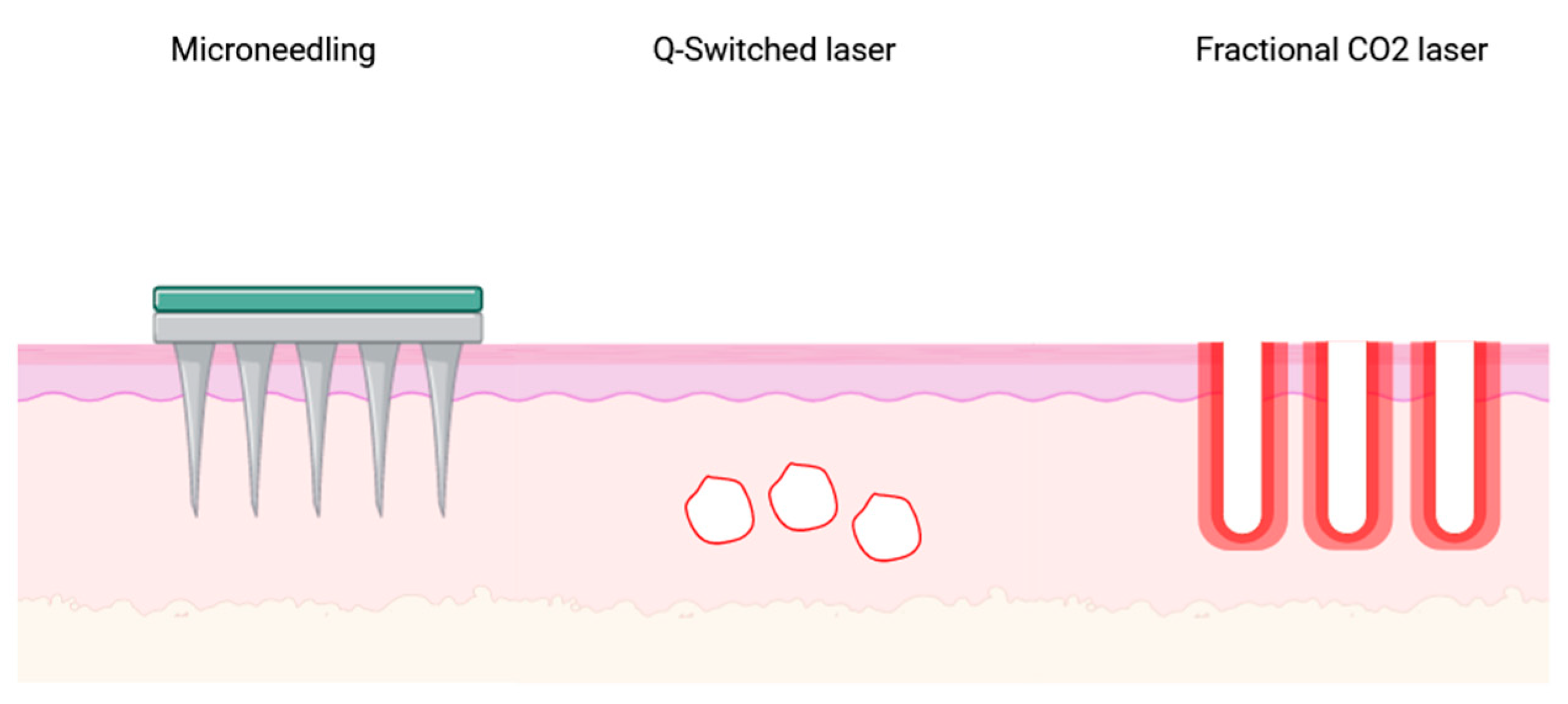
Figure 1. Graphic representation of the mechanism of action of the three techniques (microneedling, QS, and fractional CO2 laser) used for drug delivery.
Cosmetics 11 00014 g001
Figure 2. (A) The 1064 nm fractional Q-switched laser beam is used to reach the dermal layer and create a glomerular-shaped microlesion. (B) The drug in a gel/cream solution is topically applied to the skin surface with a slight pressure and massage. The solution reaches the dermis thanks to the microchannels and micropores generated by the laser activity. (C) The canal closes as part of the wound-healing process and the drug is released into the surrounding area.
Cosmetics 11 00014 g002
Figure 3. Graphic representation of the study protocol.
Cosmetics 11 00014 g003
Figure 4. Clinical pictures of a patient before and after the procedure: (A) before treatment; (B) soon after treatment with 1064 nm Q-switched laser and biostimulation product where erythema and superficial bleeding are present; (C) 72 h post-treatment: disappearance of erythema and superficial bleeding.
Cosmetics 11 00014 g004
Table 1. Sociodemographic features of the sample.
%m (SD)
Age 44.87 (7.73)
Female sex100
BMI 21.58 (3.86)
Smoking habit
Yes
66.7
Insomnia
Yes
55.6
Legend. BMI, Body Mass Index; m, mean; SD, standard deviation.
Table 2. Cutaneous characteristics (hydration and elasticity) at baseline and 3-week follow-up from treatment assessed through Checkup.
MeanSD
1Skin Hydration baseline59.568.71
Skin Hydration Laser + Drug 3-week FU67.305.45
2Skin Elasticity baseline66.1013.51
Skin Elasticity Laser + Drug 3-week FU67.108.59
3Skin Hydration Laser + Drug 3-week FU67.305.45
Skin Hydration Only Laser 3-week FU59.607.47
4Skin Elasticity Laser + Drug 3-week FU67.108.59
Skin Elasticity Laser 3-week FU65.207.84
Legend. SD, standard deviation; FU, follow-up.
Table 3. Comparison of skin elasticity and hydration between laser and drug versus laser-only treated areas.
MeansSD95% L.C.I. (df9)95% U.C.I. (df9)p
Skin Hydration baseline vs.
Skin Hydration Laser + Drug 3-week FU
−7.7404.201−10.745−4.734<0.001
Skin Elasticity baseline vs.
Skin Elasticity Laser + Drug 3-week FU
−1.00011.175−8.9946.9940.784
Skin Hydration Laser + Drug 3-week FU
vs.
Skin Hydration Only Laser 3-week FU
7.7004.6674.36011.039<0.001
Skin Elasticity Laser + Drug 3-week FU
vs.
Skin Elasticity Laser 3-week FU
1.9002.846−0.1353.9350.064
Legend. SD, standard deviation; C.I., confidence intervals; U., upper; L., lower; FU, follow-up.
Table 4. Results from the Skindex17 questionnaire.
m (SD)
Physical domain10.00 (12.472)
Psychosocial domain0.417 (1.318)
Legend. M, mean; SD, standard deviation.
Disclaimer/Publisher’s Note: The statements, opinions and data contained in all publications are solely those of the individual author(s) and contributor(s) and not of MDPI and/or the editor(s). MDPI and/or the editor(s) disclaim responsibility for any injury to people or property resulting from any ideas, methods, instructions or products referred to in the content.