Case Report

Using 3D Imaging to Preoperatively Plan Facelift Procedures for the Lower Third of the Face and Neck

by
Anastasiya S. Borisenko
*,
Valentin I. Sharobaro
,
Alexey E. Avdeev
,
Nigora S. Burkhonova
and
Anastasiya O. Fisun
Department of Plastic Surgery, I.M. Sechenov First Moscow State Medical University, Moscow 119435, Russia
*
Author to whom correspondence should be addressed.
Cosmetics 2024, 11(1), 28; https://doi.org/10.3390/cosmetics11010028
Submission received: 30 November 2023 / Revised: 24 January 2024 / Accepted: 4 February 2024 / Published: 11 February 2024
(This article belongs to the Special Issue Treatment for Anti-aging and Rejuvenation)

Abstract

:
The pursuit of youth and attractiveness is a fundamental desire in contemporary society. For many individuals, the appearance of visible signs of aging, such as skin laxity, gravitational changes in facial and neck soft tissues, and loss of harmonious proportions, can be psychologically distressing. The choice of effective methods for addressing these changes depends on their specific characteristics, the patient’s anatomical features, and the type of facial aging. The introduction of 3D imaging to determine the hyoid bone level, the volume of soft tissue structures in the cervico-mental area, ptosis of subplatysmal structures, and the volume of submandibular glands, in addition to ultrasound study of the submental projection at the preoperative stage, are key for making the best decisions regarding preoperative planning. This case report aims to illustrate how modern imaging techniques are a fundamental part of the preoperative assessment of the mid and lower third of the face to ensure the most favorable aesthetic outcomes for each patient.

1. Introduction

Over the past decades, numerous techniques have emerged for correcting the contours of the lower third of the face [1]. Modern patients are well-informed about the capabilities of plastic surgery and demonstrate an increased interest in rejuvenation methods. According to statistics from our practice, approximately 400 patients seek facial rejuvenation, including the cervico-mental area, at our clinic annually, and approximately 20% of them are over 60 years old.
The term “age-related changes in the lower third of the face” commonly refers to the deepening of perioral wrinkles, “marionette lines”, sagging of the cervico-mental region, and the apparition of jowls, secondary to the redistribution of the fat in the neck, leading to the formation of an obtuse cervical-mandibular angle that frequent occurs in the elderly [2]. Aging manifests as ptosis, skin laxity in the anterior neck region, deformation of the “witch’s chin,” and weakness of the platysma muscle [3]. Anatomical studies have described significant supraplatysmal and subplatysmal fat-increased volumes, which means that a superficial approach is not sufficient; multiplanar surgical intervention is usually required in older patients [4]. Also, controversial information about the correlation between age and body mass with hypertrophy and ptosis of submandibular salivary glands has been described, which affect the mandibular and cervical-mandibular angles, and require submandibular gland reduction to improve the contour of the neck [4,5,6,7,8].
A beautiful, youthful neckline also dramatically depends on the anatomy of the platysma muscle. Age-related changes in this muscle include shortening and thinning, with loss of anatomical architecture and, therefore, effacement of the cervical-mandibular angle [9].
This study aimed to employ a comprehensive and individualized approach in addressing age-related changes in the lower third of the face using diagnostic imaging as a key component of preoperative assessments.

2. Preoperative Planning

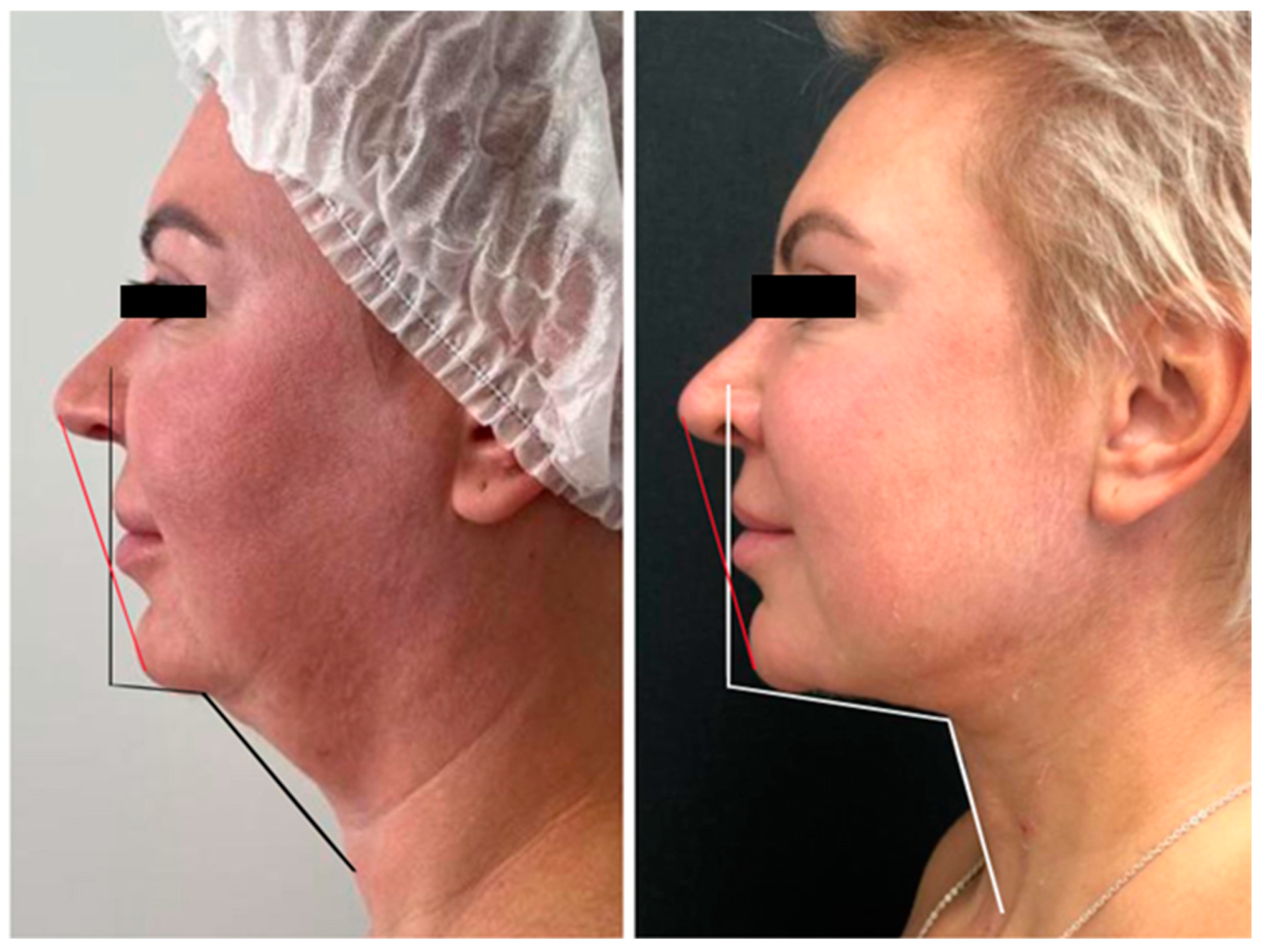
2.1. Methods of Evaluation for Cervico-Mental Area Correction

The initial assessment of the face begins with a visual examination. The facial shape of an individual is primarily determined by the underlying bone structure, which is covered by soft tissues that possess distinct characteristics. Bone tissue generally undergoes minimal changes with age, such as a decrease in density and, in some cases, volume. Specifically in the face, the reduction in volume is significant in the maxilla and the mandible, with decreased projection of the soft tissue above [10]. These changes can be addressed through the use of implants or, to a certain extent, by augmenting the soft tissues [11,12]. However, facial aesthetic surgery primarily focuses on the manipulation of soft tissues.
Contemporary correction methods necessitate a comprehensive and individualized approach for each patient. Factors such as the size and shape of the lower jaw, neck fat distribution, the position of the hyoid bone, and the thickness and anatomical variations of the platysma muscle fibers change due to aging and need to be assessed individually. To achieve optimal aesthetic results, a meticulous analysis of a patient’s parameters is essential [13].
The angle of the submental region, which can be estimated in a lateral projection, refers to the angle formed between the submentum (the area under the chin) and the neckline. This angle is an important factor in determining the youthfulness of the face, as a 90 degree angle between the submentum and neckline is considered the standard of beauty and follows the principles of the “Golden Ratio”. This angle can be affected by genetic predisposition, aging, excess weight, and gravitational changes that cause wrinkles in the neck area [10]. The ideal angle for the submental region is between 105 and 120 degrees. In our surgical procedures, we take into account the individual anatomical features of the patient (both soft tissue and bone) to select an appropriate surgical approach that aims to achieve maximum results. By choosing appropriate surgical methods based on individual anatomy, we strive to achieve a youthful appearance with a 90 degree submental angle.
A notable advantage of selecting a correction method is that aesthetic surgery is not standardized. Similar to all forms of art, it is contingent upon the subjective judgments of both the patient and the surgeon. Consequently, existing approaches for treating the aging neck, including neck liposuction, bilateral platysma plication, midline platysma plication with distal fiber crossing, neck lift with skin and soft tissue excision, and botulinum toxin injections for platysma relaxation, cater to specific patient categories. The surgeon must possess a thorough understanding of all approaches mentioned above to ensure the most favorable aesthetic outcome for each patient.

2.2. Modern Imaging Techniques in Plastic Surgery
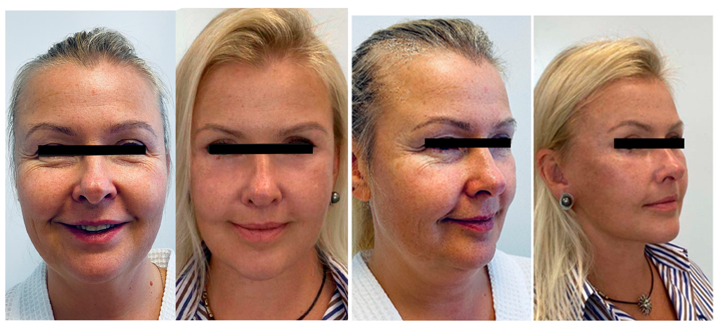
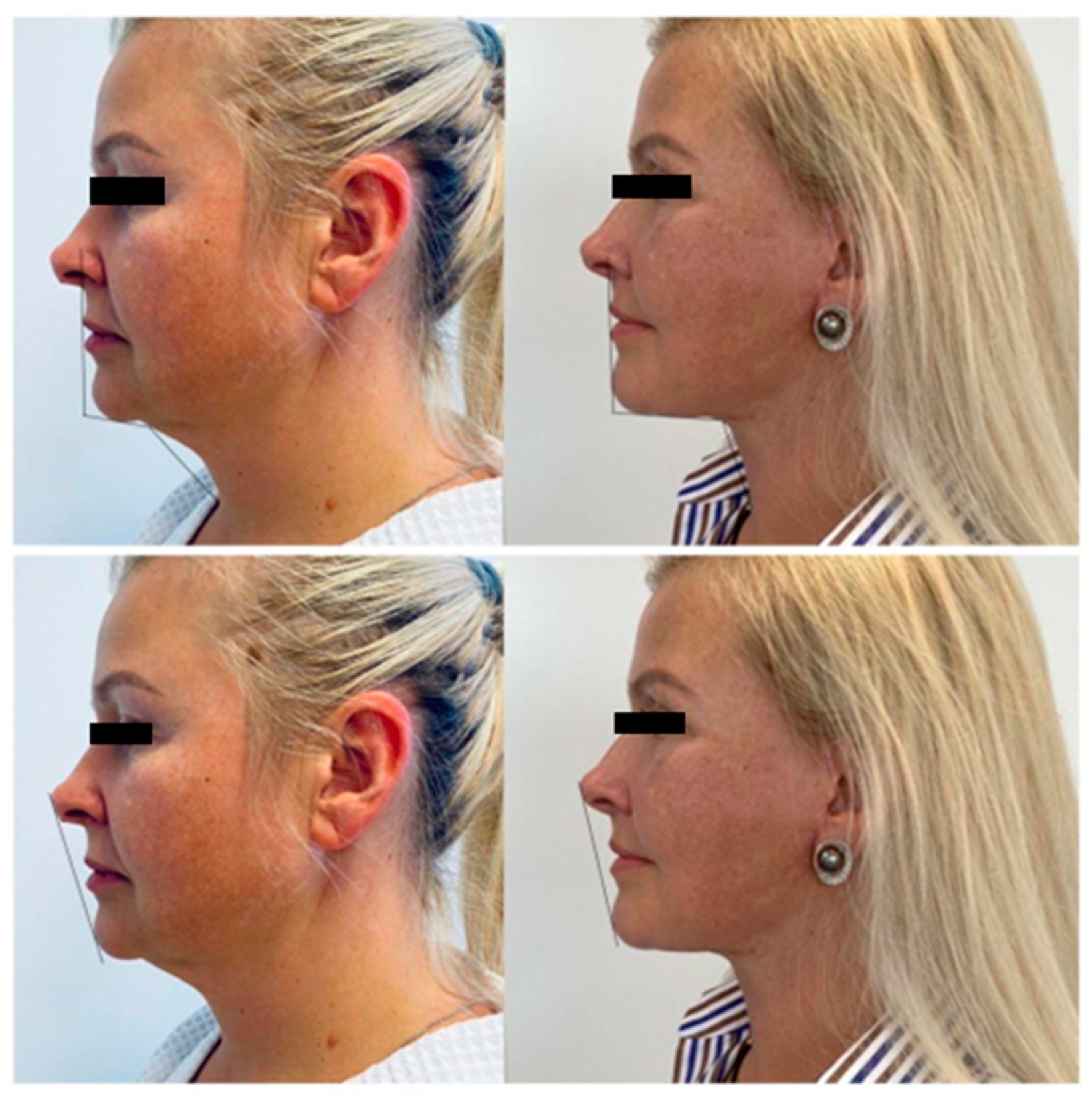
Three-dimensional imaging has played a fundamental role in the evolution of plastic surgery. Computed tomography (CT), magnetic resonance imaging (MRI), and ultrasound (US) have improved the evaluation and preoperative planning processes by providing precise and detailed information about the tissues, which helps the surgeon visualize a model that is useful during intraoperative navigation [14,15].
These tools have been used for different goals in the field of reconstructive surgery, including assessment of the microvascular anatomy for microsurgical reconstructions, diagnosis, preoperative evaluation, virtual planning, modeling of body parts, 3D printing, intraoperative evaluation, and postoperative follow-up [16,17,18,19,20,21,22].
In craniofacial reconstructive surgery, the use of 3D imaging has been proven to reduce operating room time, increase the accuracy of reconstructions, facilitate preoperative planning, and even simulate a surgery before it is performed [21]. A CT scan can also be employed in the preoperative planning of facial feminization in transgender patients, in which the surgeon aims to address the osseous aspects of the whole anatomy of the face [23]. This approach has been fundamentally popular in the field of plastic reconstructive surgery regarding skeletal features, but we believe that the role of 3D imaging can also be valuable in the aesthetic facial surgery of soft tissues.
Regarding rejuvenation of the lower third, a CT scan is performed to determine the hyoid bone elevation level, the volume of soft tissue structures in the cervico-mental area, ptosis of subplatysmal structures, and the volume of submandibular salivary glands [4,24]. MRI studies of the submandibular glands have reported a logarithmic increase in the fat component over the years, to the ninth decade of life [25]. Ultrasound study of the submental projection at the preoperative stage can reveal insufficient projection in this area, and postoperative assessment can determine the degree of fat subplatysmal autograft survival, the use of which increases chin projection and harmonizes the contours of the lower third [13].

3. Clinical Cases

3.1. Case 1

A 47-year-old woman with no concomitant diseases presented with slight gravitational changes in the lower third, violation of the cervico-mental angle, and deep nasolabial and jaw folds. Along with this, she presented drooping of the eyebrow tails, overhanging skin in the upper eyelid area, bags in the lower eyelid area, and fine mimic wrinkles. When looking at frontal view images, the lack of proportions of the lower third was evident (Figure 1).
In the lateral view, according to the Riedel–Rikkitz lines, the projection of the chin protrusion was missing [26] (Figure 2). This highlighted the lack of support for the midface.
Using the CT scan, we assessed the level of the hyoid bone so we could guarantee the formation of a beautiful line of the chin–neck angle. In this case, the hyoid bone was quite high. Also, determining the presence of submandibular gland ptosis/hypertrophy was key to achieving an optimal result. For stable results, we viewed the angle on the sagittal CT view and planned a slight extension to harmonize the face (Figure 3).
Surgery plan: complex rejuvenation in the volume of DEEP SMAS; temporal lifting; upper + lower blepharoplasty; medial and lateral platysmaplasty with resection of the subplatysmal volume of adipose tissues; a slight extension of the chin with fat autografting; and a bullhorn lip lift.

3.2. Case 2

A 45-year-old normasthenic female patient (height 165, weight 67, BMI 24, smoker) presented with a dysmorphic–edematous type of aging. The lower third was unsatisfactory; when analyzing images, a straight line was drawn showing the insufficiency of the lower third. In the lateral projection, it was evident that the chin protrusion was not sufficient, and there was malocclusion with microgenia and micrognathia (Figure 4). From the anamnesis, chin liposuction + cryotherapy–cryolipolysis were performed twice with unsatisfactory results. The patient’s main complaint was her chin–neck contour.
Some cephalometric measures used in orthodontics, such as the Björk and Snellman parameters, were applied to the images in Case 2. However, it should be noted that although these types of analyses may be useful for evaluating dental and skeletal relationships, they are typically performed using radiographs, specifically cephalometric radiographs. Therefore, although these measurements may provide some insight into a patient’s dental and skeletal relationships based on the images, they should be interpreted with caution, as they were not obtained using traditional cephalometric radiography. In general, it is recommended to use traditional radiographic methods for accurate cephalometric analysis in orthodontic treatment planning. Here, a lateral projection analysis with the use of the Rickets line is presented to demonstrate the harmony of the submental projection with the tip of the nose and lips. The Rickets line is commonly used in orthodontics, and its analysis helps to suggest suitable methods to restore aesthetic harmony in the submental region relative to the nose and lips. As orthognathic surgeries are relatively traumatic multistage corrective methods, we proposed lipofilling or intraoperative use of subplatysmal autologous fat grafting as alternative methods for restoring the projection of the submental region.
According to the CT scan, the glands were lower than the level of the lower jaw and showed how hypertrophied in volume the anterior abdomens of the digastric muscles were (Figure 5). The level of the hyoid bone was low, the cervico-mental angle was 145 degrees, and the bone level might have shifted during the resection of the subplatysmal tissues.
Surgery plan: medial platysmaplasty with extended resection of subplatysmal tissues; resection of fat; bilateral digastric muscles by squamous resection, as well as submental glands; closure with medial platysmaplasty with suturing behind the hyoid bone, fixations to raise it to an aesthetically advantageous position; lateral platysmaplasty and vertical midface lifting with minimal access; upper lower blepharoplasty; bullhorn lip lift; and chin augmentation using subplatysmal fat autografting to strengthen the support of the middle zone (Figure 6).

4. Discussion

Facial and neck rejuvenation has been, and continues to be, one of the most critical aspects of plastic surgery. Age-related changes in the cervical submental area are characterized by the development of an obtuse cervico-mental angle, the disappearance of well-defined lower jaw borders, the presence of jowls, the emergence of vertical platysmal bands, localized submental fat deposits, and excess skin [27]. An “ideal” surgical rejuvenation method for the face and neck should address all aforementioned age-related changes with minimal risk of complications and a short recovery period [28]. A satisfactory result is considered to be one that allows the patient to perceive a reduction in gravitational changes on their face, a biological age younger than their actual age, attractiveness, and a desire to radiate positive energy.
Attractiveness, or the patient’s biological (external) age, is closely linked to various factors, such as skin quality, which is influenced by parameters like harmful habits, improper nutrition, sleep patterns, stress, and more [29]. Our comprehensive rehabilitation measures include promoting a healthy lifestyle and cosmetological skin care.
In our practice, we pay attention to the following significant criteria for age-related changes:
  • Decreased skin turgor and tone and the appearance of excess skin;
  • Involutional changes in facial skeletal structures;
  • Excess volume in the lower third of the face;
  • Eyebrow ptosis of the eyebrow tail and displacement of soft tissues in the temporal area;
  • Age-related changes in periocular soft tissues;
  • Hypertonicity or hypotonicity of the chin area;
  • Presence of jowls;
  • Presence of an obtuse cervical-mental angle;
  • Ptosis of the upper lip;
  • Manifestation of deep facial wrinkles.
These age-related changes must be approached individually, assessing their specific anatomic characteristics; therefore, we proposed a CT scan as a mandatory measure for preoperative evaluation and surgical planning, especially in the rejuvenation of the lower third of the face and neck. In one study, patients were divided by age, type of aging, and BMI. Depending on the individual characteristics of patients and the understanding of the mechanisms underlying the processes of tissue involution and age-related changes in the face, the choice of effective methods for their elimination was determined [12].
Cases presented in this article showed gravitational changes that were sufficiently pronounced that minimally invasive surgical procedures were not sufficient. In this age group of 55–65 years, medial platysmaplasty, lateral platysmaplasty, liposuction of the neck and submental area, and a small correction of the submental projection were performed with the aim of advancing the projection and creating harmonious proportions. In our practice, we analyzed 100 patients aged 35–65+ to determine the volume of surgical intervention. Patients were divided according to BMI, age, skin type, and type of aging in the lower third of the face. The results are presented in Table 1.
The choice of techniques for lower third rejuvenation depends on anatomical features, patient age, BMI, and the severity of aging changes in the lower third. For patients over 65 years old, a comprehensive approach is employed for lower third rejuvenation, including medial platysmaplasty, lateral platysmaplasty, and submental projection advancement. In cases of significant localized fat deposits in the neck and submental area, liposuction is also utilized. For patients between 35 and 45 years old with less pronounced aging changes, medial platysmaplasty is combined with liposuction of the neck and submental area. The positioning of the hyoid bone is assessed to determine whether additional soft tissue fixation is necessary to create a more defined neck angle.
Concerns about radiation-induced malignancy can be mitigated by introducing low-dose computed tomography during aesthetic preoperative evaluation; we believe this is an option that deserves further research [30]. Also, advanced CT techniques like dual-energy, which is frequently used in the assessment of head and neck oncological pathologies, could have a role in aesthetic plastic surgery in the future [31]. However, the radiation exposure after a usual CT scan is not significant when performed only once.
In this study, we used a 640-slice spiral computed tomography (CT) scanner, specifically the Toshiba Aquilion ONE, for our preoperative 3D modeling of the neck and submental region. This CT scanner allows us to perform preoperative 3D modeling of a patient’s individual anatomical features, taking into account the favorable and unfavorable positions of the mandibular bone, micrognathia and microgenia, as well as in certain modes, the examination of soft tissue compartments above and below the platysma muscle and the position of submandibular glands. All these factors are considered in a comprehensive analysis, which allows us to plan an appropriate surgical volume. The slice thickness during scanning is 1.0–1.5 mm, which is sufficient to construct an accurate 3D model that demonstrates the patient’s anatomical features to them and improves our preoperative understanding of the surgical intervention volume. The minimum volume of the scanning area extends from the midline of the skull to the thyroid cartilage.
An important requirement is that a patient’s head should be in a physiological position (not tilted or brought forward to the chest) during the scan, as this is important for assessing the condition of soft tissues in the frontal region of the neck. In other words, the headrest should be low.
Currently, there are various advanced non-invasive technological methods employed for the rejuvenation of the soft tissues in the face and neck that can complement surgical results to achieve an optimal aesthetic effect. For instance, the use of radiofrequency and ultrasound have shown good results in terms of improvement of facial wrinkles and jaw sagging [32].
To integrate lifestyle changes with medical procedures for long-lasting improvement, it is essential to address unhealthy habits such as smoking, excessive alcohol consumption, a sedentary lifestyle, and unhealthy eating patterns. These habits affect not only the hormonal balance, but also the quality of the skin and the ratio of collagen and elastin in the tissues, which ultimately impacts the longevity of surgical results. Patients who lead an unhealthy lifestyle are more prone to swelling and lymphatic issues during the recovery period. Based on our observations, patients who followed a healthy sleep–wake cycle, an active lifestyle, and a well-balanced diet had better skin quality than those who did not follow a healthy lifestyle. Therefore, it is crucial to educate patients about the importance of adopting a healthy lifestyle to achieve long-lasting results from medical procedures.

5. Conclusions

To obtain a harmonious result from the surgical correction of age-related changes in the face and neck, it is necessary to consider the anatomical features of the patient and the severity of the ptosis. In addition, with the help of 3D imaging, a comprehensive analysis of the face, including the hyoid bone level, the volume of soft tissue structures in the cervico-mental area, the ptosis of subplatysmal structures, and the volume of submandibular glands, is key to making the best decisions regarding preoperative planning. Modern imaging techniques are a fundamental component of the preoperative assessment of the mid and lower third of the face to ensure the most favorable aesthetic outcomes for each patient. Type of aging, BMI, concomitant diseases, patient expectations, and anatomical characteristics are key for preoperative planning and achieving harmony and golden proportions, which not only prevent aging, but also enhance beauty. Further investigation with a larger sample size and a comparative group is required to assess long-term outcomes, patient satisfaction, and potential limitations, such as the accessibility and cost-effectiveness, of the implementation of 3D imaging as part of the routine assessment for this type of patient.

Author Contributions

Conceptualization, A.S.B. and V.I.S.; methodology, A.S.B. and N.S.B.; software, 3D modeling, and analysis, N.S.B. and A.E.A.; investigation, A.E.A. and A.O.F.; resources, V.I.S.; writing—original draft preparation, A.S.B.; writing—review and editing, V.I.S.; visualization, N.S.B. and A.E.A.; supervision, V.I.S. All authors have read and agreed to the published version of the manuscript.

Funding

This research received no external funding.

Institutional Review Board Statement

Ethical approval was not required since anonymity is assured in this study and there are no risks associated.

Informed Consent Statement

Written informed consent was obtained from patients to publish this paper.

Data Availability Statement

The data presented in this study are available on request from the corresponding author.

Conflicts of Interest

The authors declare no conflicts of interest.

References

  1. Sinno, S.; Thorne, C. Cervical Branch of Facial Nerve: An Explanation for Recurrent Platysma Bands Following Necklift and Platysmaplasty. Aesthetic Surg. J. 2019, 39, 1–7. [Google Scholar] [CrossRef]
  2. Orra, S.; Tadisina, K.; Charafeddine, A.; Derakhshan, A.; Halliburton, S.; Hashem, A.; Doumit, G.; Zins, J.E. The Effect of Age on Fat Distribution in the Neck Using Volumetric Computed Tomography. Plast. Reconstr. Surg. 2021, 147, 49. [Google Scholar] [CrossRef] [PubMed]
  3. Frisenda, J.L.; Nassif, P.S. Correction of the Lower Face and Neck. Facial Plast. Surg. 2018, 34, 480–487. [Google Scholar] [CrossRef] [PubMed]
  4. Raveendran, S.S.; Anthony, D.J.; Ion, L. An Anatomic Basis for Volumetric Evaluation of the Neck. Aesthetic Surg. J. 2012, 32, 685–691. [Google Scholar] [CrossRef] [PubMed]
  5. Mendelson, B.C.; Tutino, R. Submandibular Gland Reduction in Aesthetic Surgery of the Neck: Review of 112 Consecutive Cases. Plast. Reconstr. Surg. 2015, 136, 463–471. [Google Scholar] [CrossRef] [PubMed]
  6. Mahne, A.; El-Haddad, G.; Alavi, A.; Houseni, M.; Moonis, G.; Mong, A.; Hernandez-Pampaloni, M.; Torigian, D.A. Assessment of Age-Related Morphological and Functional Changes of Selected Structures of the Head and Neck by Computed Tomography, Magnetic Resonance Imaging, and Positron Emission Tomography. Semin. Nucl. Med. 2007, 37, 88–102. [Google Scholar] [CrossRef] [PubMed]
  7. Lee, M.; Sepahdari, A.; Cohen, M. Radiologic Measurement of Submandibular Gland Ptosis. Facial Plast. Surg. 2013, 29, 316–320. [Google Scholar] [CrossRef] [PubMed]
  8. Heo, M.-S.; Lee, S.-C.; Lee, S.-S.; Choi, H.-M.; Choi, S.-C.; Park, T.-W. Quantitative Analysis of Normal Major Salivary Glands Using Computed Tomography. Oral Surg. Oral Med. Oral Pathol. Oral Radiol. Endod. 2001, 92, 240–244. [Google Scholar] [CrossRef]
  9. Hwang, K.; Kim, J.Y.; Lim, J.H. Anatomy of the Platysma Muscle. J. Craniofac. Surg. 2017, 28, 539. [Google Scholar] [CrossRef]
  10. Shaw, R.B.; Katzel, E.B.; Koltz, P.F.; Kahn, D.M.; Puzas, E.J.; Langstein, H.N. Facial Bone Density: Effects of Aging and Impact on Facial Rejuvenation. Aesthetic Surg. J. 2012, 32, 937–942. [Google Scholar] [CrossRef]
  11. Mendelson, B.; Wong, C.-H. Changes in the Facial Skeleton With Aging: Implications and Clinical Applications in Facial Rejuvenation. Aesthetic Plast. Surg. 2012, 36, 753–760. [Google Scholar] [CrossRef]
  12. Dikarev, A.; Mantardzhiev, D.; Tsinenko, D. Facial Skeleton Augmentation with Porous Implants for Long-Term Stability of Surgical Rejuvenation. Plast. Surg. Aesthetic Med. 2021, 1, 36–43. [Google Scholar] [CrossRef]
  13. Alimova, S.M.; Sharobaro, V.I.; Telnova, A.V.; Stepanyan, E.E. Planning of methods of surgical correction of soft tissues of the face and neck. Med. Vis. 2021, 25, 47–52. [Google Scholar] [CrossRef]
  14. Tarassoli, S.P.; Shield, M.E.; Allen, R.S.; Jessop, Z.M.; Dobbs, T.D.; Whitaker, I.S. Facial Reconstruction: A Systematic Review of Current Image Acquisition and Processing Techniques. Front. Surg. 2020, 7, 537616. [Google Scholar] [CrossRef]
  15. Shaye, D.A.; Tollefson, T.T.; Strong, E.B. Use of Intraoperative Computed Tomography for Maxillofacial Reconstructive Surgery. JAMA Facial Plast. Surg. 2015, 17, 113–119. [Google Scholar] [CrossRef]
  16. Laungani, A.; Ritman, E.; Lachman, N.; Christner, J.; Vercnocke, A.; Jorgensen, S.; Anderson, J.; Regnier, T.; Saint-Cyr, M. The Use of Micro Computed Tomography in Plastic Surgery: Towards a Better Understanding of Flaps Microvascular Architecture. Plast. Reconstr. Surg. 2013, 132, 173. [Google Scholar] [CrossRef]
  17. Kumar, T.S.; Vijai, A. 3D Reconstruction of Face from 2D CT Scan Images. Procedia Eng. 2012, 30, 970–977. [Google Scholar] [CrossRef]
  18. Krakowczyk, Ł.; Piotrowska-Seweryn, A.; Szymczyk, C.; Wierzgoń, J.; Oleś, K.; Ulczok, R.; Donocik, K.; Dowgierd, K.; Maciejewski, A. Virtual Surgical Planning and Cone Beamcomputed Tomography in Reconstruction Ofhead and Neck Tumors—Pilot Study. Otolaryngol. Pol. 2020, 75, 28–33. [Google Scholar] [CrossRef] [PubMed]
  19. Chen, Y.-W.; Yen, J.-H.; Chen, W.-H.; Chen, I.-C.; Lai, C.-S.; Lu, C.-T.; Song, D.-Y. Preoperative Computed Tomography Angiography for Evaluation of Feasibility of Free Flaps in Difficult Reconstruction of Head and Neck. Ann. Plast. Surg. 2016, 76, S19. [Google Scholar] [CrossRef] [PubMed]
  20. Xu, S.-Q.; Jia, X.-L.; Choi, Y.; Zhu, Z.-W.; Xu, Y.-B.; Velamuri, R.; Liu, X.-X. Three-Dimensional Scanning Technique in the Congenital Microtia Reconstruction with Tissue Expander. Chin. Med. J. 2021, 134, 842–844. [Google Scholar] [CrossRef] [PubMed]
  21. Markiewicz, M.R.; Bell, R.B. The Use of 3D Imaging Tools in Facial Plastic Surgery. Facial Plast. Surg. Clin. N. Am. 2011, 19, 655–682. [Google Scholar] [CrossRef]
  22. Lynn, A.Q.; Pflibsen, L.R.; Smith, A.A.; Rebecca, A.M.; Teven, C.M. Three-Dimensional Printing in Plastic Surgery: Current Applications, Future Directions, and Ethical Implications. Plast. Reconstr. Surg. Glob. Open 2021, 9, e3465. [Google Scholar] [CrossRef] [PubMed]
  23. Callen, A.L.; Badiee, R.K.; Phelps, A.; Potigailo, V.; Wang, E.; Lee, S.; Talbott, J.; Glastonbury, C.; Pomerantz, J.H.; Narvid, J. Facial Feminization Surgery: Key CT Findings for Preoperative Planning and Postoperative Evaluation. Am. J. Roentgenol. 2021, 217, 709–717. [Google Scholar] [CrossRef]
  24. Alimova, S.M.; Sharobaro, V.I.; Avdeev, A.E.; Sidorenkov, D.A.; Guseva, T.S. Cone-Beam Computed Tomography for Objective Diagnosis of Age-Related Soft Tissue Changes in Lower Face and Neck. Aesthetic Plast. Surg. 2023, 47, 2370–2377. [Google Scholar] [CrossRef]
  25. Saito, N.; Sakai, O.; Bauer, C.M.; Norbash, A.M.; Jara, H. Age-Related Relaxo-Volumetric Quantitative Magnetic Resonance Imaging of the Major Salivary Glands. J. Comput. Assist. Tomogr. 2013, 37, 272. [Google Scholar] [CrossRef] [PubMed]
  26. Naini, F.B.; Garagiola, U.; Wertheim, D. Analysing Chin Prominence in Relation to the Lower Lip: The Lower Lip-Chin Prominence Angle. J. Cranio-Maxillofac. Surg. 2019, 47, 1310–1316. [Google Scholar] [CrossRef] [PubMed]
  27. Langsdon, P.R.; Velargo, P.A.; Rodwell, D.W.; Douglas, D. Submental Muscular Medialization and Suspension. Aesthetic Surg. J. 2013, 33, 953–966. [Google Scholar] [CrossRef] [PubMed]
  28. Giampapa, V.C.; Mesa, J.M. Neck Rejuvenation with Suture Suspension Platysmaplasty Technique. Clin. Plast. Surg. 2014, 41, 109–124. [Google Scholar] [CrossRef]
  29. Sveikata, K.; Balciuniene, I.; Tutkuviene, J. Factors Influencing Face Aging. Literature Review. Stomatologija 2011, 13, 113–116. [Google Scholar]
  30. Lala, B.; Shah, J.; Salvador, T.M.; Ricci, J.A. Expanding the Utilization of Low-Dose Computed Tomography in Plastic and Reconstructive Surgery Based on Validated Practices Among Surgical Specialties. Ann. Plast. Surg. 2021, 87, e163. [Google Scholar] [CrossRef]
  31. Forghani, R.; Mukherji, S.K. Advanced Dual-Energy CT Applications for the Evaluation of the Soft Tissues of the Neck. Clin. Radiol. 2018, 73, 70–80. [Google Scholar] [CrossRef] [PubMed]
  32. Hugul, H.; Cigdem, M.; Kirisci, M.; Kutlubay, Z. Focused Radiofrequency and Ultrasound for Face and Neck Rejuvenation: A Retrospective Evaluation of 158 Patients. J. Cosmet. Dermatol. 2022, 21, 290–295. [Google Scholar] [CrossRef] [PubMed]
Figure 1. Patient before and six months after complete rejuvenation of the face.
Cosmetics 11 00028 g001
Figure 2. Patient before and six months after complete rejuvenation of the face. The Riedel plane and Rickett’s line show improvement in the chin projection.
Cosmetics 11 00028 g002
Figure 3. (A) Computed tomography; the Rocabado plane shows a high hyoid bone compared to the position of the cervical vertebrae, which is a favorable situation to achieve excellent results with platysmaplasty. (B) The soft tissues of the neck show an obtuse angle of 134°, far from the 90° that is considered aesthetically ideal.
Cosmetics 11 00028 g003
Figure 4. The Riedel plane and Rickett’s line show insufficiency in the lower third of the face, poor chin projection, and an obtuse angle of the soft tissues. After surgery, there was improvement in the lower third of the face.
Cosmetics 11 00028 g004
Figure 5. CT scan before surgery. In this projection, the ptosis of the submandibular glands and the hypertrophic digastric muscles are evident.
Cosmetics 11 00028 g005
Figure 6. Before and after surgery. It is evident how the proportions of the face were harmonized; the result was possible thanks to the integral management of soft tissues in the neck, identified in the CT scan.
Cosmetics 11 00028 g006
Table 1. Patients classified by age and type of procedure in our clinic.
AgeCombination of Surgical MethodsMean Value (%)
35–45 years oldLiposuction of the neck and submental area in combination with medial platysmaplasty, as well as isolated corset platysmaplasty.20%
45–55 years oldLiposuction of the neck and submental area, medial platysmaplasty; lateral platysmaplasty with and without this procedure. 55%
55–65+ years oldLiposuction of the neck and submental area, medial platysmaplasty; lateral platysmaplasty, additional fixation of soft tissues to the mylohyoid bone, elimination of submental muscle hypertrophy, and harmonization of the projection of the submental prominence.25%
BMICombination of Surgical MethodsMean Value (%)
19–21Isolated corset platysmoplasty.10%
21–23Neck and submental area liposuction, medial platysmaplasty; lateral platysmaplasty and without it; additional fixation of soft tissues to the mylohyoid bone.63%
23–25Neck and submental area liposuction, medial platysmaplasty; lateral platysmaplasty.23%
Type of AgingCombined Surgical MethodsMean Value (%)
Fine wrinkling type of facial agingIsolated corset plasticy; lateral plasticy (referring to a surgical procedure called plasticy, which is used to reshape or contour the body).20%
Deformational type of facial agingIn simpler terms, this procedure involves liposuction in the neck and submental area, as well as medial platysmoplasty, lateral platysmoplasty, or a combination of both, with or without additional fixation of soft tissues to the mandibular bone for added support.55%
Mixed type of facial agingIn simpler terms, this procedure involves liposuction in the neck and submental area, as well as medial platysmoplasty or isolated corset platysmoplasty, along with lateral platysmoplasty, with or without additional fixation of soft tissues to the mandibular bone for added support.35%
Disclaimer/Publisher’s Note: The statements, opinions and data contained in all publications are solely those of the individual author(s) and contributor(s) and not of MDPI and/or the editor(s). MDPI and/or the editor(s) disclaim responsibility for any injury to people or property resulting from any ideas, methods, instructions or products referred to in the content.

