Review

Factors Participating in the Occurrence of Inflammation of the Lips (Cheilitis) and Perioral Skin

1
Department of Dermatovenereology, University Hospital Center Sestre Milosrdnice, 10000 Zagreb, Croatia
2
School of Dental Medicine, University of Zagreb, 10000 Zagreb, Croatia
*
Author to whom correspondence should be addressed.
Cosmetics 2023, 10(1), 9; https://doi.org/10.3390/cosmetics10010009
Submission received: 22 November 2022 / Revised: 18 December 2022 / Accepted: 28 December 2022 / Published: 3 January 2023
(This article belongs to the Special Issue Feature Papers in Cosmetics in 2022)

Abstract

:
Lip inflammation may manifest as mainly reversible cheilitis, mainly irreversible, or cheilitis connected to dermatoses or systemic diseases. Therefore, knowing a patient’s medical history is important, especially whether their lip lesions are temporary, recurrent, or persistent. Sometimes temporary contributing factors, such as climate and weather conditions, can be identified and avoided—exposure to extreme weather conditions (e.g., dry, hot, or windy climates) may cause or trigger lip inflammation. Emotional and psychological stress are also mentioned in the etiology of some lip inflammations (e.g., exfoliative cheilitis) and may be associated with nervous habits such as lip licking. To better manage cheilitis, it is also helpful to look for potential concomitant comorbidities and the presence of related diseases/conditions. Some forms of cheilitis accompany dermatologic or systemic diseases (lichen, pemphigus or pemphigoid, erythema multiforme, lupus, angioedema, xerostomia, etc.) that should be uncovered. Occasionally, lip lesions are persistent and involve histological changes: actinic cheilitis, granulomatous cheilitis, glandular cheilitis, and plasmacellular cheilitis. Perioral skin inflammation with simultaneous perioral dermatitis can have various causes: the use of corticosteroids and cosmetics, dysfunction of the skin’s epidermal barrier, a contact reaction to allergens or irritants (e.g., toothpaste, dental fillings), microorganisms (e.g., Demodex spp., Candida albicans, fusiform bacteria), hormonal changes, or an atopic predisposition. Epidermal barrier dysfunction can worsen perioral dermatitis lesions and can also be related to secondary vitamin or mineral deficiencies (e.g., zinc deficiency), occlusive emollient use, sunscreen use, or excessive exposure to environmental factors such as heat, wind, and ultraviolet light. Current trends in research are uncovering valuable information concerning the skin microbiome and disruption of the epidermal barrier of persons suffering from perioral dermatitis. Ultimately, an effective approach to patient management must take all these factors and new research into account.

1. Background

Visible skin lesions present significant problems for the majority of cheilitis patients, especially since face-to-face communication is the most common way of interacting with others. Therefore, the proper recognition and treatment of inflammation of the lips and perioral skin are important (and common) challenges in dermatology and for cosmetic and aesthetic clinics. Various etiologies and contributing factors may cause lip inflammation [1]. Therefore, knowing a patient’s medical history is important, especially whether their lesions are temporary, recurrent, or persistent—sometimes temporary factors, such as climate and weather conditions, can be identified and avoided.
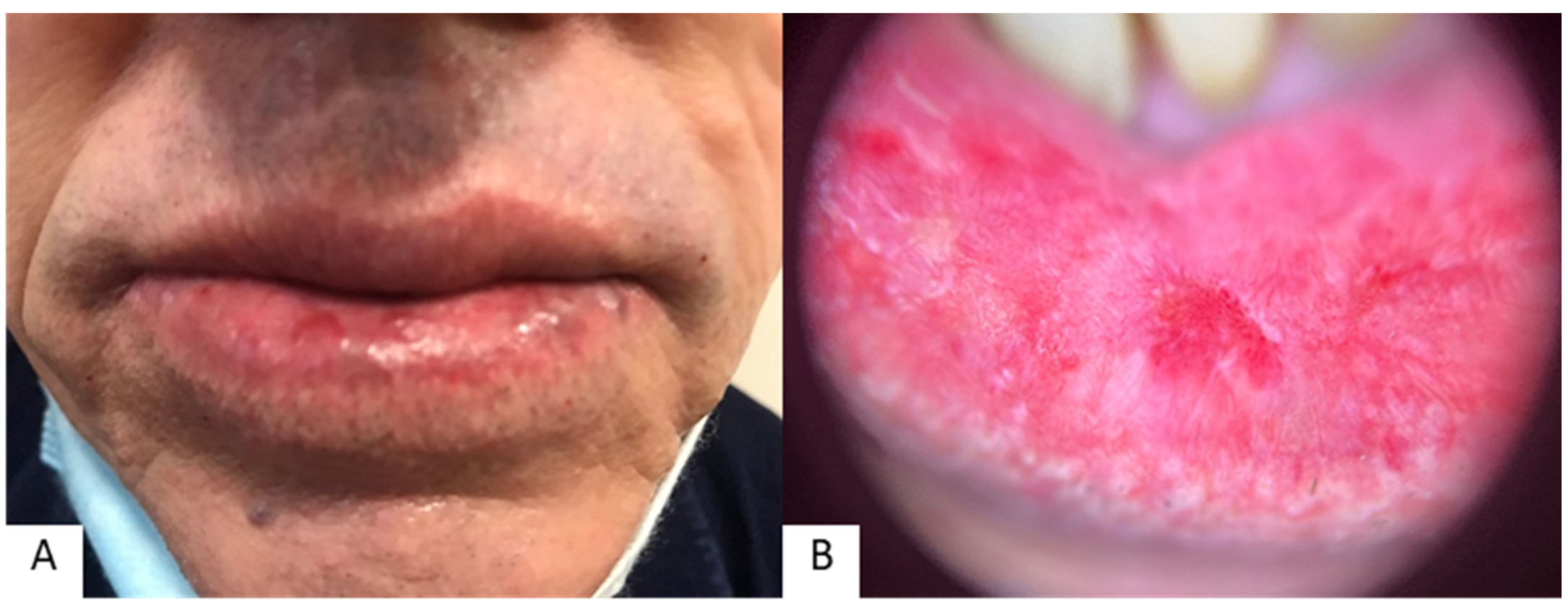
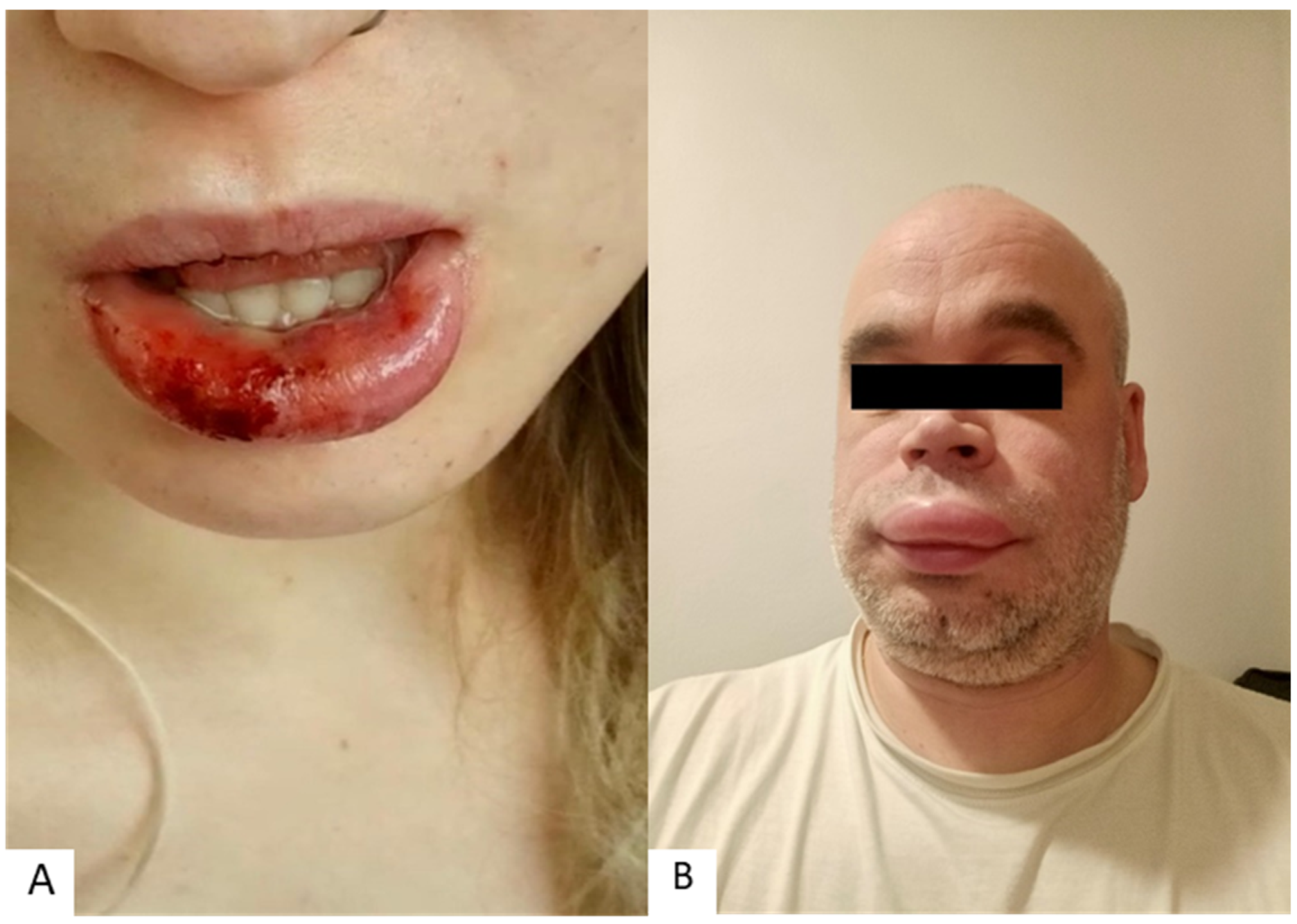
Based on disease features and course, three different forms (subtypes) of lip inflammation are mentioned in the literature: mainly reversible cheilitis, mainly irreversible, and cheilitis connected to dermatoses and systemic diseases (Figure 1, Figure 2 and Figure 3) [2,3]. Inflammatory lip lesions are sometimes permanent; in these lesions, histological changes exist that are unique to certain subtypes of cheilitis (actinic cheilitis, granulomatous cheilitis, glandular cheilitis, and plasma cell cheilitis). In addition, some forms of cheilitis are related to or associated with certain disorders or conditions, and this needs to be taken into account when managing a patient’s therapy.
Thus, in this narrative review we will present the numerous factors participating in the occurrence of inflammation of the lips and perioral skin (Table 1). All these factors can make recognizing and diagnosing cheilitis/the different types of cheilitis difficult, affecting the approach to patient management.

2. Most Common Causes of Lip and Perioral Inflammation

Inflammation of the lips and perioral skin can also be related to various other factors and triggers, such as a person’s habits or concomitant diseases, among others.
Lip inflammation may be acute or chronic and mostly occurs in the area of the vermilion, but it can also spread to the surrounding skin and, less often, to the oral mucosa. It can be caused by numerous factors, including infections, exposure to, or contact with, certain substances (irritants and allergens), chronic sun exposure, nutritional deficiencies, and certain skin or systemic illnesses. Common clinical manifestations are scaling, erythema, fissuring, itching, dryness, burning, and edema [4].
A very common cause of lip inflammation is herpes simplex virus (HSV) infection, which causes herpetic cheilitis, a cheilitis that mostly manifests on the vermilion and the vermilion boundary [1,5,6] (Figure 1A). Herpes labialis frequently recurs and can be related to different factors, such as stress, fever, menstruation, systemic infection, and others [5,7]. Aside from herpetic cheilitis, another common type is cheilitis simplex, which may be triggered by mechanical irritation, such as wearing surgical masks, scarves, niqabs, etc., or due to frequent lip chewing (cheilophagia), licking, or biting [1,8,9,10]. Exposure to extreme weather conditions, such as dry, hot, or windy climates, is also recognized as an important factor [2,11]. Emotional and psychological stress are also mentioned in the etiology of some lip inflammations, such as exfoliative cheilitis, because it is commonly caused by nervous habits such as lip licking [11,12].
Some types of cheilitis are recognizable by their typical clinical features (e.g., angular, exfoliative, and herpetic), while many forms often do not have a specific picture and thus need adequate diagnostic management to reach a correct diagnosis [3]. Based on disease duration and etiology, three types of cheilitis are mentioned in the research literature: (1) mainly reversible (simplex, angular/infective, contact/eczematous, exfoliative, and drug-related); (2) mainly irreversible (actinic, granulomatous, glandular, and plasma cell); and (3) cheilitis connected to dermatologic/systemic diseases (lupus, lichen planus, pemphigus/pemphigoid group, angioedema, xerostomia, etc.) (Figure 1, Figure 2 and Figure 3) [2]. Concerning cheilitis frequency/occurrence in general, inflammatory lip lesions often run a temporary course; persistent lesions are less common. Therefore, in the case of long-lasting, persistent cheilitis, a biopsy of the lip lesion is often necessary to identify the specific type of irreversible cheilitis (actinic cheilitis, granulomatous cheilitis, glandular cheilitis, or plasmacellular cheilitis). With persistent lesions (such as those seen in actinic cheilitis), it is especially important to check for malignant disorders, the timely recognition of which is sometimes crucial for disease prognosis [3]. In addition, for many patients, clinicians need to check for a potential concomitant systemic or skin disease, which can also indicate a specific cheilitis type. Thus, an effective diagnostic approach takes into account many factors that are also important for the outcome, especially in cases where lesional abnormalities (or potentially malignant disorders) need to be detected early to stop the spread of the disease.
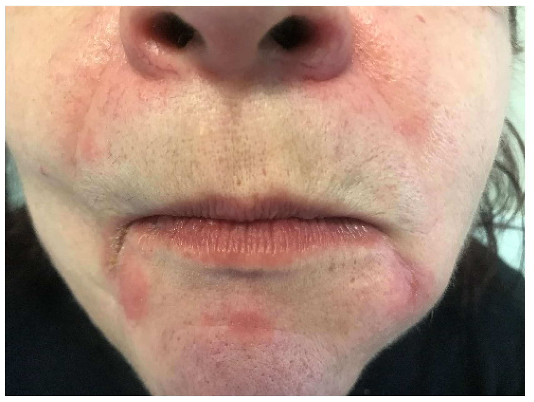
Perioral dermatitis is characterized by erythematous lesions (papules, pustules, or vesicles), which typically occur in the perioral and perinasal regions, often with concomitant itching or burning sensations [13,14] (Figure 4). Some authors use the term “periorificial dermatitis” for perioral dermatitis, although the less exact “periorificial dermatitis” is a more widely used term that encompasses lesions of various localizations, such as periocular dermatitis and perianal dermatitis. Thus, the term “perioral dermatitis” is more precise and suitable for perioral inflammatory lesions. The pathophysiology of perioral dermatitis is not clearly known, but it is considered a variant of rosacea (due to a similar histopathology). Although perioral dermatitis typically occurs in young adult women, it may also occur in children. Inflammation of the perioral skin with the occurrence of perioral dermatitis can be triggered by different factors, most commonly as a result of previous continuous use of topical or systemic corticosteroids, though there is also an association with fluorinated mouthwashes and toothpastes [13]. The risk factors associated with perioral dermatitis include the use of corticosteroids (topical, nasal, and inhaled); the use of cosmetic products; dysfunction of the skin’s epidermal barrier (such as a contact reaction); potential allergens and irritants (such as toothpaste and dental fillings); microorganisms (such as Demodex spp., Candida albicans, and fusiform bacteria); hormonal changes (due to oral contraceptive use, pregnancy, and premenstrual flares); and atopic predisposition [15,16]. In perioral dermatitis, Demodex folliculorum, a saprophytic mite that resides in sebaceous follicles, may play a potential role. These mites can be found on normal skin in a majority of adults, but in some facial diseases/lesions there is an increased density of these mites, e.g., an association with Demodex infestation has been confirmed in patients with rosacea (where the degree of infestation is more important than the infestation rate). Furthermore, perioral dermatitis lesions can worsen due to dysfunction of the skin’s epidermal barrier and can also be related to secondary mineral or vitamin deficiencies (e.g., zinc deficiency); occlusive emollient use (which leads to overhydration); use of sunscreen; or prolonged exposure to ultraviolet radiation or other environmental factors, such as heat or wind [15,17].
The current trends in perioral dermatitis research of the skin microbiome and disruption of the epidermal barrier of persons suffering from perioral dermatitis are promising and valuable. Some studies on skin microbiome features of the periorificial region, among which is research by Zheng et al., have shown that bacteria of the genera Streptococcus and Rothia predominate on the skin of the perioral region of healthy infants [18,19]. In addition, skin sensitivity is influenced by seasonal changes, living habits, social customs, and stress [20,21]. Other studies have looked at how changes in the microbiome of healthy skin may be related to mask wearing, as this action can have profound implications for various dermatological diseases, such as atopic dermatitis, acne, and perioral dermatitis, in which bacterial dysbiosis is involved in pathogenesis [19,22,23,24]. In addition, in the pathogenesis of perioral dermatitis, we now know that Fusobacteria may be involved, though it can successfully be treated (e.g., with beta-lactam antibiotics in those who are tetracycline-resistant or intolerant) [25,26].
Due to the recent COVID-19 pandemic, it is important to mention here the association between prolonged use of protective equipment (goggles and masks) and lip licker’s dermatitis [10]. A recently published study confirmed that constant lip licking is not only an indirect result of limited fluid intake due to protective equipment usage, but also as a result of the subjective feeling of discomfort that comes with poorly fitting face protection and excessive sweating during its use. Whole-face erythema and lip licker’s dermatitis are affected by factors such as duration and fit of personal protective equipment use and amount of sweating. For example, excessive sweating and poorly fitting masks were clearly related to an increased feeling of irritation in both diseases [10].

3. Accompanying Nonpsychiatric Diseases and Conditions in Patients with Lip and Perioral Inflammation

The inflammation of the lips and perioral skin may be associated with different skin and nonskin diseases and conditions. According to literature data, cheilitis is associated with gastrointestinal diseases, metabolic diseases such as type II diabetes mellitus, oral diseases, respiratory diseases, and other conditions [2,27,28,29,30,31,32,33,34,35]. For example, angular cheilitis is more frequent in patients with type II diabetes mellitus than in healthy persons (26.4% versus 8%) [34]. Furthermore, they are more likely to have frequent fungal and bacterial infections with prolonged wound healing (especially in uncontrolled diabetes). Concerning gastrointestinal diseases, granulomatous cheilitis is also a potential manifestation of Crohn’s disease [32,36]. Moreover, gastroesophageal reflux is a possible cause of lip inflammation [33].
According to recent research data, the most common concomitant conditions in patients with cheilitis are skin conditions; diseases of the thyroid gland and gastrointestinal tract; and other conditions, such as diabetes, respiratory diseases, oral diseases, anemia, and psychiatric conditions and illnesses. In addition, the most frequent dermatologic conditions were atopic dermatitis, contact dermatitis, and urticaria [37]. Of the other skin diseases that can be associated with cheilitis, acne is mentioned as a common skin condition with concomitant lip inflammation. In one study of 400 patients diagnosed with acne vulgaris, it was recorded that they commonly had lip inflammation (irritation), i.e., 34% of the subjects had clinically diagnosed cheilitis of an irritative nature, and lesions were more frequent on the lower lip [38]. In addition, patients with acne excoriée (a subtype of acne vulgaris) commonly had cheilitis. Its occurrence was caused by undesirable habits linked to psychological factors such as anxiety, stress, and personality disorders (this research was conducted before the use of treatment with systemic isotrethionine, which is known to cause cheilitis).
Sometimes, lip inflammation is caused or triggered by infectious agents. There is a possible association between infections such as Mycobacterium tuberculosis, for instance, and granulomatous cheilitis (Miescher’s syndrome) [35]. In a study by Liu et al., patients with granulomatous cheilitis had greater and more diverse microbial flora than healthy controls [39]. Patients with granulomatous cheilitis had significantly more Prevotella, Porphyromonas, Alloprevotella, Actinomyces, Fusobacterium, Rothia, Haemophilus, and Aggregatibacter. It has been proven that they coexist with other bacteria [39]. Another possible cause of lip inflammation associated with infection is syphilis, primarily the first stage. Respiratory factors may also contribute to the occurrence of cheilitis, e.g., oral respiration may promote the development of cheilitis [2]. In addition, it is necessary to be aware of other conditions including systemic and skin conditions that may cause lip lesions, such as pemphigus or pemphigoid, lichen, erythema multiforme, angioedema, and other diseases (Figure 3). Thus, a thorough physical examination and detailed medical history are needed to reach a correct diagnosis.

4. Psychiatric Diseases and Conditions and Common Behavioral Attitudes in Patients with Lip and Perioral Inflammation

The inflammation of the lips and perioral skin may be related to, or triggered by, some patients’ habits or psychological condition. Older literature data have already confirmed that licking of the lips is a crucial trigger for irritant contact cheilitis [40,41]. Now, our recent research results concerning undesirable habits has found positive associations between lip licking or biting and exfoliative cheilitis [37]. According to other literature data, among subjects with exfoliative cheilitis, mainly female, the main undesirable habit was lip licking (53%), and 40% of the patients had a history of a mental disorder [42].
Of other habits, one of the most unfavorable habits related to lip inflammation is frequent or prolonged sun exposure [43]. Actinic cheilitis is considered a premalignant lesion to carcinoma of the lip and has a high probability of developing into invasive squamous cell carcinoma (SCC) [43,44,45,46,47] (Figure 2). Almost all SCCs (95%) of the lower lip develop on pre-existing actinic cheilitis [45,46]. Other risk factors include fair skin, aging, occupation or daily activities which involve intense sun exposure (more than four hours daily), male gender, immunosuppression, latitude of residence, genetic predisposition, and others [43,48]. Actinic cheilitis often has asymptomatic lesions, but some patients report dryness, cracking, burning, stinging, pain, or even abnormal mobility of the lips [49]. In recent research, patients with actinic cheilitis reported that they were often or very often exposed to the sun, never or almost never use a hat or cap during sun exposure, and never/almost never use protective creams [37].
Aside from behavior, inflammation of the lips and perioral skin may also be associated with some psychiatric diseases and conditions [30]. For example, an association between exfoliative cheilitis and anxious conditions (le tic des lèvres) has been recorded in the research literature [30]. In addition, some researchers have singled out factitial cheilitis as a special type of cheilitis caused by habits patients are unaware of [30,50]. According to recent research on different types of cheilitis and psychiatric diseases associated with cheilitis, anxiety was more commonly recorded in patients with exfoliative cheilitis [37]. Previous research on the occurrence of oral lesions in patients with eating disorders (anorexia, bulimia, and nonspecific eating disorders) showed that oral lesions were observed in most of these patients (94%) [31]. Furthermore, the occurrence of exfoliative cheilitis caused by undesirable habits is associated with a higher incidence of stress and anxiety [38,51]. Other research on cheilitis patients showed that the largest percentage of them exhibiting high stress were those with exfoliative cheilitis [37]. In cheilitis simplex, low stress is somewhat more frequent in those who do not have recurrent lesions than in those who do. Moreover, mental stress and psychiatric disorders (e.g., personality disorders) can lead to undesirable habits and self-harm (biting, sucking, pulling, and licking) in factitial cheilitis, as in exfoliative cheilitis [52]. In addition, it is known that patients with atopic dermatitis (in which cheilitis is a typical manifestation) are prone to anxiety and depression [53,54].
In addition, patients with herpetic cheilitis often mention its association with stress. Recently, psychological stress levels were compared between patients with herpetic cheilitis and a healthy group, and significantly higher stress levels were seen in the patients with herpetic cheilitis [37]. In an older study on herpetic cheilitis, eighteen subjects completed a questionnaire on stressful events at two different times—during the latent phase of viral infection when they had no symptoms and in the active phase of disease. The results showed that, in the active phase of viral infection, patients experienced higher numbers of stressful events, as well as anxiety and daily falls, all of which differed significantly from the results recorded during the latent phase of the disease [55]. Despite these psychological factors related to lip inflammation, generally, psychiatrists and mental health professionals are rarely involved in a patient’s treatment for cheilitis, which could make a great difference in treatment efficacy. For example, one study showed that the use of an antidepressant, a selective serotonin reuptake inhibitor (fluoxetine), led to the improvement of lip inflammation [30].

5. Allergies Associated with Lip and Perioral Inflammation

Inflammation of the lips and perioral skin can be triggered by different allergens. Allergens associated with cheilitis are predominantly cosmetic and decorative products (e.g., lipstick, toothpaste, nail polish, and lip balm), dental materials, metals found in musical instruments, food, drugs, etc. [1,2,6,56,57]. Allergic contact cheilitis typically manifests with dryness, erythema, peeling, and fissures, most often on the lips, with occasional spreading to the perioral skin. Lip inflammation caused by food oftentimes spreads to the perioral skin [1,2]. Although contact allergic cheilitis is frequently caused by a reaction to balms and lipsticks, other items can also cause an allergic reaction (e.g., pencils or hairpins) [6]. Allergic contact cheilitis can occasionally leave residual lip hyperpigmentation that resolves over time [3,6].
Inflammation of the lips or surrounding skin may reflect gender-specific behavior—for instance, allergic contact cheilitis is more frequent among women because they use cosmetics more often, are more aware of changes in their appearance, and seek medical attention more often than men [58]. The prevalence of this type of cheilitis increases with age, given the more frequent use of hygienic and cosmetic products [57,59].
The prevalence of allergic reactions is generally more frequent in patients with cheilitis compared to patients with perioral diseases. Allergies to contact allergens more commonly manifest as cheilitis, while other allergy-related oral manifestations include gingivitis, stomatitis, perioral dermatitis, lichenoid reaction, etc. However, contact allergic reactions have been most frequently found in patients with cheilitis [3,60,61]. Previous studies in patients with oral and perioral manifestations have shown that 60% of patients with cheilitis had a positive patch test reaction, more often than in other conditions involving this area (angioedema, oral lichen planus, gingivostomatitis, and perioral dermatitis) [62]. A study of 121 dermatological patients also showed that contact allergies are relatively common in patients with cheilitis and perioral dermatitis (41.9% of the subjects had a positive patch test to allergens from the dental series), ranking them third, after orofacial granulomatosis and burning mouth syndrome [63].
Inflammation of the lips and perioral skin is relatively often caused by contact irritation and allergic reactions, especially to metals such as nickel, mercury, cobalt, chromium, impression materials, eugenol, and others. When a patient presents with lesions in this area, it is important to take a history of the patient’s use of these and other substances used in dentistry, as they can cause an allergic reaction [1,56]. In addition, allergies to cinnamon and benzoates are a potential predisposing factor for granulomatous cheilitis [2,57,64]. The prevalence of allergies to contact allergens ranges from 25.9% to 75% (according to previous studies, at least one positive patch test reaction was reported by Torgerson et al. in 25.9%, Budimir et al. in 26.7%, Khamaysi et al. in 41.9%, and Kim et al. in 75% of cases) [61,62,63,65]. Overall, the most common allergens confirmed by patch test were cobalt chloride, nickel sulfate, and mercury precipitate [61,62]. In the study conducted by Zoli et al., which performed patch tests with the standard series of allergens, nickel sulfate was the most common allergen, followed by thimerosal and cobalt chloride [59]. Other frequently mentioned contact allergens include balsam of Peru, fragrances, benzophenone, gold, and other allergens [66]. Cobalt chloride is widely used in the chemical and pharmaceutical industries (e.g., in dyes and vitamin products and as an additive to animal feed). It is important to note that cobalt hypersensitivity is often associated with nickel hypersensitivity, and patients should be informed of this [67]. Nickel is a metal found in drinking water, jewelry, fertilizer, food, crockery, paints, and cutlery, among other products. Thimerosal is used as a preservative in a variety of cosmetic and ophthalmic products as well as vaccines [57,67]. In older articles, reactions to gold have been reported in patients with oral diseases; however, this is not common nowadays.
Inflammation of the lips and perioral skin can also be caused by other, noncontact allergens [57]. According to research by Budimir et al., atopy-related reactions to inhalant allergens were found in 30% of patients with cheilitis, most commonly weed pollen, grass pollen, and dust [62]. An association between cheilitis and food allergies is also possible. According to literature data, 13.3% of patients with cheilitis showed evidence of a food allergy, most commonly to fruit, vegetables, and preservatives (e.g., glutaraldehyde, glutamate, and citric acid) [62]. Recent studies in patients with various oral and perioral skin diseases have shown that immediate-type allergic reactions (positive prick test) to food and additives were more frequently confirmed in patients with cheilitis (33.3%), whereas they were less frequent in patients with perioral dermatitis (10%) [68]. Patients with atopic dermatitis are also prone to lip inflammation (Figure 5). The percentage of atopic patients tested with allergy tests ranges from 19% to 34.9% in the research literature. In a study by Freeman et al., atopy was found in 19% of patients, while studies by Lim et al. and Zoli et al. showed that approximately one-third of subjects had atopy [40,41,59]. However, the number of atopic patients in studies depends on the populations tested and the allergen sets used in the tests. According to research by Blagec et al. on patients with lip inflammation, the percentage of atopic patients was even higher—84% (positive prick test result to at least one allergen) [57].
A patch test for contact allergens, performed with standard contact allergens, is essential in the assessment of contact cheilitis; however, possible allergens to personal products not included in the standard allergen series should also be considered [1]. In addition to patch tests, prick tests are also essential to confirm other mechanisms of IgE-mediated reaction (e.g., contact urticaria) and should be performed when indicated by the patient’s medical history [40,41,57,58,59,61,65,69]. Contact urticaria with lip manifestations is also possible on this part of the skin and can be triggered by food, preservatives, odors, and other factors (e.g., mint in toothpaste—with a positive prick test to mint leaves and negative patch test) [69]. Performing prick tests to assess and confirm food allergens is important for the diagnosis of so-called food cheilitis or lip manifestations in food allergic reactions [56]. In addition to patch tests, prick tests are sometimes needed to diagnose cheilitis, as they may be decisive in determining the cause of the lesions [40,41,57,58,59,61,65].
However, the clinical significance of positive allergy tests in patients remains to be established [57,62]. For this reason, patients are advised to avoid potential allergens in order to determine the relevance of the allergen to the disease itself [57].

6. Nutritional Deficiencies and Microbiome Changes in Patients with Lip and Perioral Inflammation

Inflammation of the lips and perioral skin can be related to various nutritional factors. Cheilitis can be linked to various vitamin and mineral deficiencies, such as iron (sideropenic anemia) [15,27,70]. Angular cheilitis can be caused by deficiencies of vitamin B2 (riboflavin), vitamin B3 (niacin), vitamin B6 (pyridoxine), vitamin B7 (biotin), vitamin B9 (folic acid), vitamin B12 (cyanocobalamin), and zinc, as well as protein deficiencies [1,6,11,15,27,70]. Additionally, studies have shown that angular cheilitis is a common clinical manifestation of sideropenic and megaloblastic anemia [70]. Exfoliative cheilitis can also be associated with iron deficiency (sideropenic anemia) and vitamin B12 deficiency [2].
Individuals with excess skin or wrinkles at the corner of the mouth may be prone to Candida overgrowth, leading to angular cheilitis [71]. Patients with Plummer–Vinson syndrome commonly present with angular cheilitis (along with anemia and dysphagia) mostly due to iron deficiency; therefore, iron supplementation is essential in the treatment of these patients [28]. A study analyzing children with vitamin B12 deficiency reported clinical features of cheilitis in 7.01% of patients. These patients were given cobalamin and saw complete resolution of lip inflammation [29]. A similar study analyzed dermatological signs of vitamin B12 deficiency in infants and found that 6.07% of participants presented with angular cheilitis. The most common sign of vitamin B12 deficiency on the skin, observed in almost all patients, was cutaneous hyperpigmentation [72]. Recent studies carried out in patients with cheilitis, analyzing levels of vitamin B9, vitamin B12, and iron, showed that only a small number of patients had abnormalities [37]. However, nutritional deficiencies should be considered in resistant cases. Abnormalities in zinc metabolism can cause disorders that usually manifest themselves on the skin, such as acrodermatitis enteropathica, an autosomal recessive genetic disorder of zinc deficiency. Acrodermatitis enteropathica presents with the triad of periorificial dermatitis, alopecia, and diarrhea. Oral zinc replacement therapy usually leads to a rapid clinical remission of this disorder [73]. In patients with perioral dermatitis and proven zinc deficiency, zinc supplementation can effectively improve the skin’s condition [74].
These data support measuring vitamin and mineral levels in patients with inflammation of the lips and perioral skin, especially in resistant cases.

7. Conclusions

Inflammation of the lips (cheilitis) and perioral skin (perioral dermatitis) can be caused by various factors; thus, clinicians must consider many possible etiologies and triggers. Some cheilitis types are identifiable by their typical clinical features, but many forms have a nonspecific picture, and thorough diagnostic management is needed to set a correct diagnosis. Key etiological factors and triggers associated with lip inflammation include patient habits (e.g., lip licking), weather conditions, infectious agents, immune and nutritional deficiencies, atopy, contact allergens and irritants, psychological stress, sun exposure, and other factors. Therefore, patients’ general medical histories and comorbidities must be established before making a final diagnosis. Persistent inflammatory lip lesions (mainly irreversible cheilitis forms) are rare but involve histological lip changes and require a biopsy to set a correct diagnosis. Furthermore, in perioral dermatitis, the factors associated with the disease are numerous: corticosteroid use (topical and inhaled), infectious agents, dysfunction of the skin’s epidermal barrier, nutritional deficiencies, atopy, contact allergens or irritants, cosmetics, occlusive emollients, sunscreen, and hormonal imbalances, as well as other factors. Currently, one research trend looks at the skin microbiome and disruption of the epidermal barrier of persons with perioral dermatitis; so far, studies have been promising. Thus, numerous factors also need to be taken into account in the approach to patient management. Knowledge of the various factors that influence the onset or persistence of inflammation of the lips and perioral skin is crucial for patient management and disease prognosis and outcome.

Author Contributions

Conceptualization, L.L.-M., B.Š. and T.B.; methodology, L.L.-M., B.Š. and T.B.; software, B.Š., T.B. and M.D.A.; validation, L.L.-M., B.Š., M.D.A. and N.F.; formal analysis, L.L.-M., B.Š. and T.B.; investigation, L.L.-M., B.Š., T.B. and M.D.A.; resources, L.L.-M. and T.B.; data curation, L.L.-M., B.Š. and T.B.; writing—original draft preparation, L.L.-M., B.Š., T.B., M.D.A., A.G. and E.P.; writing—review and editing, L.L.-M., B.Š., M.D.A. and E.P.; visualization, M.D.A. and N.F.; supervision, L.L.-M.; project administration, E.P. All authors have read and agreed to the published version of the manuscript.

Funding

This research received no external funding.

Institutional Review Board Statement

Not applicable.

Informed Consent Statement

Not applicable.

Data Availability Statement

Not applicable.

Conflicts of Interest

The authors declare no conflict of interest.

References

  1. Greenberg, S.A.; Schlosser, B.J.; Mirowski, G.W. Diseases of the lips. Clin. Dermatol. 2017, 35, e1–e14. [Google Scholar] [CrossRef] [PubMed] [Green Version]
  2. Lugović-Mihić, L.; Pilipović, K.; Crnarić, I.; Šitum, M.; Duvančić, T. Differential diagnosis od cheilitis: How to classify cheilitis? Acta Clin. Croat. 2018, 57, 342–351. [Google Scholar] [PubMed]
  3. Lugović-Mihić, L.; Blagec, T.; Japundžić, I.; Skroza, N.; Delaš Adžajić, M.; Mravak-Stipetić, M. Diagnostic management of cheilitis: An approach based on a recent proposal for cheilitis classification. Acta Dermatovenerol. Alp. Pannonica Adriat. 2020, 29, 67–72. [Google Scholar] [CrossRef] [PubMed]
  4. Litaiem, N.; Ben Slimane, M.; Bacha, T.; Rammeh, S.; Zeglaoui, F. Cheilitis with Hemorrhagic Crusts of the Vermilion Lips. Int. J. Dermatol. 2020, 59, e234–e236. [Google Scholar] [CrossRef]
  5. Opstelten, W.; Neven, A.K.; Eekhof, J. Treatment and prevention of herpes labialis. Can. Fam. Phys. 2008, 54, 1683–1687. [Google Scholar]
  6. Scully, C. Dermatoses of the Oral Cavity and Lips. In Rook’s Textbook of Dermatology; Griffiths, C., Barker, J., Bleiker, T., Chalmers, R., Creamer, D., Eds.; Wiley Blackwell: Chichester, UK, 2016; pp. 110.1–110.94. [Google Scholar]
  7. Orion, E.; Wolf, R. Psychologic factors in the development of facial dermatoses. Clin. Dermatol. 2014, 32, 763–766. [Google Scholar] [CrossRef]
  8. Hitz Lindenmüller, I.; Itin, P.H.; Fistarol, S.K. Dermatology of the lips: Inflammatory diseases. Quintessence Int. 2014, 45, 875–883. [Google Scholar]
  9. Samimi, M. Cheilitis: Diagnosis and treatment. Presse Med. 2016, 45, 240–250. [Google Scholar] [CrossRef]
  10. Singh, M.; Pawar, M.; Bothra, A.; Maheshwari, A.; Dubey, V.; Tiwari, A.; Kelati, A. Personal protective equipment induced facial dermatoses in healthcare workers managing Coronavirus disease 2019. J. Eur. Acad. Dermatol. Venereol. 2020, 34, e378–e380. [Google Scholar] [CrossRef]
  11. Bhutta, B.S.; Hafsi, W. Cheilitis. Available online: https://www.statpearls.com/articlelibrary/viewarticle/37546/ (accessed on 17 November 2022).
  12. Thongprasom, K. Glycerin borax treatment of exfoliative cheilitis induced by sodium lauryl sulfate: A case report. Acta Stomatol. Croat. 2016, 50, 158–161. [Google Scholar] [CrossRef]
  13. Parker, J.; Neill, B.; Whitsitt, J.; Rajpara, A.; Aires, D. Exacerbation of Pediatric Periorificial Dermatitis: A Novel Adverse Reaction. J. Drugs Dermatol. 2020, 19, 428. [Google Scholar] [PubMed]
  14. Maarouf, M.; Saberian, C.; Lio, P.A.; Shi, V.Y. Head-and-neck dermatitis: Diagnostic difficulties and management pearls. Pediatr. Dermatol. 2018, 35, 748–753. [Google Scholar] [CrossRef] [PubMed]
  15. Oakley, A. Cheilitis. Avaliable online: https://www.dermnetnz.org/topics/cheilitis/ (accessed on 17 November 2022).
  16. Tolaymat, L.; Hall, M.R. Perioral Dermatitis. Available online: https://www.ncbi.nlm.nih.gov/books/NBK525968/ (accessed on 17 November 2022).
  17. Contento, M.; Maher, J.; Cline, A.; Rose, S. Why Does Facial Eczema Differ from Body Eczema? J. Drugs Dermatol. 2022, 21, 1119–1123. [Google Scholar] [CrossRef] [PubMed]
  18. Zheng, Y.; Wang, Q.; Ma, L.; Chen, Y.; Gao, Y.; Zhang, G.; Cui, S.; Liang, H.; He, C.; Song, L. Alterations in the skin microbiome are associated with disease severity and treatment in the perioral zone of the skin of infants with atopic dermatitis. Eur. J. Clin. Microbiol. Infect. Dis. 2019, 38, 1677–1685. [Google Scholar] [CrossRef] [PubMed]
  19. Ferček, I.; Lugović-Mihić, L.; Tambić-Andrašević, A.; Ćesić, D.; Grginić, A.G.; Bešlić, I.; Mravak-Stipetić, M.; Mihatov-Štefanović, I.; Buntić, A.-M.; Čivljak, R. Features of the Skin Microbiota in Common Inflammatory Skin Diseases. Life 2021, 11, 962. [Google Scholar] [CrossRef]
  20. Ye, C.; Chen, J.; Yang, S.; Yi, J.; Chen, H.; Li, M.; Yin, S.; Lai, W.; Zheng, Y. Skin sensitivity evaluation: What could impact the assessment results? J. Cosmet. Dermatol. 2020, 19, 1231–1238. [Google Scholar] [CrossRef]
  21. Ehnis-Pérez, A.; Torres-Álvarez, B.; Cortés-García, D.; Hernández-Blanco, D.; Fuentes-Ahumada, C.; Castanedo-Cázares, J.P. Relationship between transient receptor potential vanilloid-1 expression and the intensity of sensitive skin symptoms. J. Cosmet. Dermatol. 2016, 15, 231–237. [Google Scholar] [CrossRef]
  22. Blicharz, L.; Rudnicka, L.; Samochocki, Z. Staphylococcus aureus: An Underestimated Factor in the Pathogenesis of Atopic Dermatitis? Postepy Dermatol. Alergol. 2019, 36, 11–17. [Google Scholar] [CrossRef]
  23. Abdi, F.; Kashani, H.; Naeini, F.; Narimani, T.; Khorvash, F. Staphylococcus aureus in Acne Pathogenesis: A Case-Control Study. N. Am. J. Med. Sci. 2012, 4, 573. [Google Scholar] [CrossRef] [Green Version]
  24. Takiwaki, H.; Tsuda, H.; Arase, S.; Takeichi, H. Differences between Intrafollicular Microorganism Profiles in Perioral and Seborrhoeic Dermatitis. Clin. Exp. Dermatol. 2003, 28, 531–534. [Google Scholar] [CrossRef]
  25. Ishiguro, N.; Maeda, A.; Suzuki, K.; Yamana, Y.; Fukuya, Y.; Kawashima, M. Three Cases of Perioral Dermatitis Related to Fusobacteria Treated with β-Lactam Antibiotics. J. Dermatol. Treat. 2013, 25, 507–509. [Google Scholar] [CrossRef] [PubMed]
  26. Teo, W. The “Maskne” Microbiome—Pathophysiology and Therapeutics. Int. J. Dermatol. 2021, 60, 799–809. [Google Scholar] [CrossRef] [PubMed]
  27. Ayesh, M.H. Angular cheilitis induced by iron deficiency anemia. Cleve Clin. J. Med. 2018, 85, 581–582. [Google Scholar] [CrossRef] [PubMed]
  28. Phatak, S.; Redkar, N.; Patil, M.A.; Kuwar, A. Plummer-Vinson Syndrome. Case Rep. 2012, 2012, bcr2012006403. [Google Scholar] [CrossRef] [PubMed] [Green Version]
  29. Demir, N.; Doğan, M.; Koç, A.; Kaba, S.; Bulan, K.; Ozkol, H.U.; Doğan, S.Z. Dermatological findings of vitamin B12 deficiency and resolving time of these symptoms. Cutan. Ocul. Toxicol. 2014, 33, 70–73. [Google Scholar] [CrossRef]
  30. Nico, M.M.S.; Dwan, A.J.; Lourenço, S.V. Ointment pseudo-cheilitis: A disease distinct from factitial cheilitis. A series of 13 patients from São Paolo, Brazil. J. Cutan. Med. Surg. 2019, 23, 277–281. [Google Scholar] [CrossRef]
  31. Panico, R.; Piemonte, E.; Lazos, J.; Gilligan, G.; Zampini, A.; Lanfranchi, H. Oral mucosal lesions in anorexia nervosa, bulimia nervosa and EDNOS. J. Psychiatr. Res. 2018, 96, 178–182. [Google Scholar] [CrossRef]
  32. Seghers, A.K.; Grosber, M.; Urbain, D.; Mana, F. Cheilitis granulomatosa and Crohn’s disease: A case report. Acta Gastroenterol. Belg. 2019, 82, 326–328. [Google Scholar]
  33. Mathelier-Fusade, P. Cheilitis: A new manifestation of gastro-oesophageal reflux? Ann. Dermatol. Venereol. 2009, 136, 887–889. [Google Scholar] [CrossRef]
  34. Dorocka-Bobkowska, B.; Zozulinska-Ziolkiewicz, D.; Wierusz-Wysocka, B.; Hedzelek, W.; Szumala-Kakol, A.; Budtz-Jörgensen, E. Candida-associated denture stomatitis in type 2 diabetes mellitus. Diabetes Res. Clin. Pract. 2010, 90, 81–86. [Google Scholar] [CrossRef]
  35. Charpentier, C.; Kottler, D.; Fite, C.; Pelletier, A.L.; Deschamps, L.; Descamps, V. A surprising granulomatous cheilitis. Gastroenterology 2018, 154, 1239–1240. [Google Scholar] [CrossRef] [PubMed] [Green Version]
  36. Jellali, K.; Mellouki, I.; Ibrahimi, A. Cheilitis granulomatosa revealing Crohn’s disease. Pan Afr. Med. J. 2018, 30, 147. [Google Scholar] [PubMed]
  37. Blagec, T.; Glavina, A.; Špiljak, B.; Bešlić, I.; Bulat, V.; Lugović-Mihić, L. Cheilitis: A Cross-Sectional Study—Multiple Factors Involved in the Aetiology and Clinical Features. Oral Dis. 2022. ahead of print. [Google Scholar] [CrossRef] [PubMed]
  38. Balighi, K.; Daneshpazhooh, M.; Lajevardi, V.; Talebi, S.; Azizpour, A. Cheilitis in acne vulgaris patients with no previous use of systemic retionoid products. Australas. J. Dermatol. 2017, 58, 211–213. [Google Scholar] [CrossRef]
  39. Liu, Y.; Zhang, Q.; Hu, X.; Chen, F.; Hua, H. Characteristics of the Salivary Microbiota in Cheilitis Granulomatosa. Med. Oral Patol. Oral Cir. Bucal 2019, 24, 719–725. [Google Scholar] [CrossRef]
  40. Freeman, S.; Stephens, R. Cheilitis: Analysis of 75 cases referred to a contact dermatitis clinic. Am. J. Contact Dermat. 1999, 10, 198–200. [Google Scholar]
  41. Lim, S.W.; Goh, C.L. Epidemiology of eczematous cheilitis at a tertiary dermatological refferal centre in Singapore. Contact Dermat. 2000, 43, 322–326. [Google Scholar] [CrossRef]
  42. Almazrooa, S.A.; Woo, S.B.; Mawardi, H.; Treister, N. Characterization and mangement of exfoliative cheilitis: A single-centre experience. Oral Surg. Oral Med. Oral Pathol. Oral Radiol. 2013, 116, 485–489. [Google Scholar] [CrossRef]
  43. Rodríguez-Blanco, I.; Flórez; Paredes-Suárez, C.; Rodríguez-Lojo, R.; González-Vilas, D.; Ramírez-Santos, A.; Paradela, S.; Conde, I.; Pereiro-Ferreirós, M. Actinic Cheilitis Prevalence and Risk Factors: A Cross-Sectional, Multicentre Study in a Population Aged 45 Years and over in North-West Spain. Acta Derm. Venereol. 2018, 98, 970–974. [Google Scholar] [CrossRef] [Green Version]
  44. Gheno, J.N.; Martins, M.A.T.; Munerato, M.C.; Hugo, F.N.; Sant’ana Filho, M.; Weissheimer, C.; Carrard, V.C.; Martins, M.D. Oral Mucosal Lesions and Their Association with Sociodemographic, Behavioral, and Health Status Factors. Braz. Oral Res. 2015, 29, S1806-83242015000100289. [Google Scholar] [CrossRef]
  45. Lopes, M.L.D.d.S.; da Silva Júnior, F.L.; Lima, K.C.; de Oliveira, P.T.; da Silveira, É.J.D. Clinicopathological Profile and Management of 161 Cases of Actinic Cheilitis. An. Bras. Dermatol. 2015, 90, 505–512. [Google Scholar] [CrossRef] [PubMed]
  46. Bakirtzi, K.; Papadimitriou, I.; Andreadis, D.; Sotiriou, E. Treatment Options and Post-Treatment Malignant Transformation Rate of Actinic Cheilitis: A Systematic Review. Cancers 2021, 13, 3354. [Google Scholar] [CrossRef] [PubMed]
  47. Vasilovici, A.; Ungureanu, L.; Grigore, L.; Cojocaru, E.; Şenilă, S. Actinic Cheilitis—From Risk Factors to Therapy. Front. Med. 2022, 9, 805425. [Google Scholar] [CrossRef] [PubMed]
  48. De Lucena, I.M.; Santos, I.d.S.; Daroit, N.B.; Salgueiro, A.P.; Cavagni, J.; Haas, A.N.; Rados, P.V. Sun Protection as a Protective Factor for Actinic Cheilitis: Cross-Sectional Population-Based Study. Oral Dis. 2021, 28, 1802–1810. [Google Scholar] [CrossRef] [PubMed]
  49. Lupu, M.; Caruntu, A.; Caruntu, C.; Boda, D.; Moraru, L.; Voiculescu, V.; Bastian, A. Non-Invasive Imaging of Actinic Cheilitis and Squamous Cell Carcinoma of the Lip. Mol. Clin. Oncol. 2018, 8, 640–646. [Google Scholar] [CrossRef] [PubMed] [Green Version]
  50. Brown, G.E.; Malakouti, M.; Sorenson, E.; Gupta, R.; Koo, J.Y. Psychodermatology. Adv. Psychosom. Med. 2015, 34, 123–134. [Google Scholar] [PubMed]
  51. Daley, T.D.; Gupta, A.K. Exfoliative cheilitis. J. Oral Pathol. Med. 1995, 24, 177–179. [Google Scholar] [CrossRef]
  52. Girijala, R.L.; Falkner, L.; Dalton, S.R.; Martin, B.D. Exfoliative cheilitis as a manifestation of factitial cheilitis. Cureus 2018, 10, 2565. [Google Scholar] [CrossRef] [Green Version]
  53. Lugović-Mihić, L.; Meštrović-Štefekov, J.; Ferček, I.; Pondeljak, N.; Lazić-Mosler, E.; Gašić, A. Atopic Dermatitis Severity, Patient Perception of the Disease, and Personality Characteristics: How Are They Related to Quality of Life? Life 2021, 11, 1434. [Google Scholar] [CrossRef]
  54. Fishbein, A.B.; Silverberg, J.I.; Wilson, E.J.; Ong, P.Y. Update on Atopic Dermatitis: Diagnosis, Severity Assessment, and Treatment Selection. J. Allergy Clin. Immunol. Pract. 2020, 8, 91–101. [Google Scholar] [CrossRef]
  55. Schmidt, D.D.; Zyzansky, S.; Ellner, J.; Kumar, M.L.; Arno, J. Stress as a precipitating factor in subjects with recurrent herpes labialis. J. Fam. Pract. 1985, 20, 359–366. [Google Scholar]
  56. Collet, E.; Jeudy, G.; Dalac, S. Cheilitis, perioral dermatitis and contact allergy. Eur. J. Dermatol. 2013, 23, 303–307. [Google Scholar] [CrossRef] [PubMed]
  57. Blagec, T.; Crnarić, I.; Homolak, D.; Pondeljak, N.; Buljan, M.; Lugović-Mihić, L. The association between allergic reactions and lip inflammatory lesions (cheilitis). Acta Clin. Croat. 2022, in press. [Google Scholar]
  58. O’Gorman, S.M.; Torgerson, R.R. Contact allergy in cheilitis. Int. J. Dermatol. 2016, 55, 386–391. [Google Scholar] [CrossRef] [PubMed]
  59. Zoli, V.; Silvani, S.; Vincenzi, C.; Tosti, A. Allergic contact cheilitis. Contact Dermat. 2006, 54, 296–297. [Google Scholar] [CrossRef]
  60. Bakula, A.; Lugović-Mihić, L.; Šitum, M.; Turčin, J.; Sinković, A. Contact allergy in the mouth: Diversity of clinical presentations and diagnosis of common allergens relevant to dental practice. Acta Clin. Croat. 2011, 50, 553–561. [Google Scholar]
  61. Kim, T.W.; Kim, W.I.; Mun, J.H.; Song, M.; Kim, H.S.; Kim, B.S.; Kim, M.B.; Ko, H.C. Patch testing with dental screening series in oral disease. Ann. Dermatol. 2015, 27, 389–393. [Google Scholar] [CrossRef]
  62. Budimir, J.; Mravak-Stipetić, M.; Bulat, V.; Ferček, I.; Japundžić, I.; Lugović-Mihić, L. Allergic reactions in oral and perioral diseases- what do allergy skin test results show? Oral Surg. Oral Med. Oral Pathol. Oral Radiol. 2019, 127, 40–48. [Google Scholar] [CrossRef]
  63. Khamaysi, Z.; Bergman, R.; Weltfriend, S. Positive patch test reactions to allergens of the dental series and the relation to the clinical presentations. Contact Dermat. 2006, 55, 216–218. [Google Scholar] [CrossRef]
  64. Critchlow, W.A.; Chang, D. Cheilitis granulomatosa: A review. Head Neck Pathol. 2014, 8, 209–213. [Google Scholar] [CrossRef]
  65. Torgerson, R.R.; Davis, M.D.P.; Bruce, A.J.; Farmer, S.A.; Rogers, R.S., 3rd. Contact allergy in oral disease. J. Am. Acad. Dermatol. 2007, 57, 315–321. [Google Scholar] [CrossRef] [PubMed]
  66. Cheng, H.S.; Konya, J.; Lobel, E.; Fernandez-Penas, P. Patch testing for cheilitis: A 10-year series. Dermatitis 2019, 30, 347–351. [Google Scholar] [CrossRef] [PubMed]
  67. Tomljanović-Veselski, M.; Jovanović, I. Najčešći kontaktni alergeni u bolesnika s kontaktnim dermatitisima u području Slavonskog Broda. Med. Jadertina 2006, 36, 45–52. [Google Scholar]
  68. Domić, I.; Budimir, J.; Novak, I.; Mravak-Stipetić, M.; Lugović-Mihić, L. Assessment of allergies to food and additives in patients with angioedema, burning mouth syndrome, cheilitis, gingivostomatitis, oral lichenoid reactions, and perioral dermatitis. Acta Clin. Croat. 2021, 60, 276–281. [Google Scholar] [CrossRef]
  69. Holmes, G.; Freeman, S. Cheilitis caused by contact urticaria to mint flavoured toothpaste. Australas J. Dermatol. 2001, 42, 43–45. [Google Scholar] [CrossRef] [PubMed]
  70. Schlosser, B.J.; Pirigyi, M.; Mirowski, G.W. Oral manifestations of hematologic and nutritional diseases. Otolaryngol. Clin. N. Am. 2011, 44, 183–203. [Google Scholar] [CrossRef] [PubMed]
  71. Baumgardner, D.J. Oral Fungal Microbiota: To Thrush and Beyond. J. Patient Cent. Res. Rev. 2019, 6, 252–261. [Google Scholar] [CrossRef] [Green Version]
  72. Kaur, S.; Goraya, J.S. Dermatologic findings of vitamin B(12) deiciency in infants. Pediatr. Dermatol. 2018, 35, 796–799. [Google Scholar] [CrossRef]
  73. Glutsch, V.; Hamm, H.; Goebeler, M. Zinc and Skin: An Update. J. Dtsch. Dermatol. Ges. 2019, 17, 589–596. [Google Scholar] [CrossRef] [Green Version]
  74. Gürtler, A.; Laurenz, S. The Impact of Clinical Nutrition on Inflammatory Skin Diseases. J. Dtsch. Dermatol. 2022, 20, 185–202. [Google Scholar] [CrossRef]
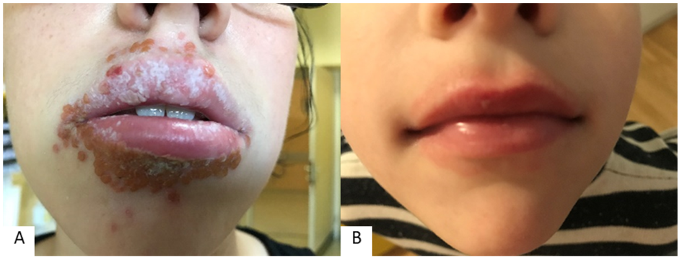
Figure 1. Herpetic cheilitis (extreme clinical picture) (A) and contact cheilitis (in a child) (B); (mainly reversible forms).
Cosmetics 10 00009 g001
Figure 2. Actinic cheilitis: clinical finding (A) and dermoscopic finding (B); (mainly irreversible form).
Cosmetics 10 00009 g002
Figure 3. Erythema multiforme on the lip (A) and angioedema of the upper lip (B); (cheilitis connected to dermatologic/systemic diseases).
Cosmetics 10 00009 g003
Figure 4. Perioral dermatitis.
Cosmetics 10 00009 g004
Figure 5. Cheilitis and periorificial lesions in patient with confirmed allergies (atopic dermatitis) (A), positive prick test to inhalant allergens (B), and positive patch test to contact allergens (C).
Cosmetics 10 00009 g005
Table 1. Key factors participating in the occurrence of inflammation of the lips (cheilitis) and perioral skin.
Lip InflammationPerioral Dermatitis
Involved factors
-
Habits (e.g., lip licking)
-
Weather conditions
-
Infective agents
-
Deficiencies (immune, nutritional)
-
Atopy
-
Contact allergens or irritants
-
Psychological distress
-
Sunlight exposure
-
Corticosteroid use (topical, nasal insufflation, inhaled)
-
Infective agents
-
Nutritional deficiency
-
Atopy
-
Contact allergens or irritants
-
Cosmetics, occlusive emollients, sunscreen use
-
Hormonal factors and changes
Disclaimer/Publisher’s Note: The statements, opinions and data contained in all publications are solely those of the individual author(s) and contributor(s) and not of MDPI and/or the editor(s). MDPI and/or the editor(s) disclaim responsibility for any injury to people or property resulting from any ideas, methods, instructions or products referred to in the content.