Review

Surface Modification and Coating for Titanium Dental Implants: A Review on Advances in Techniques, Biological Performance, and Clinical Applications


by
Amantle Balang
1,*,
Gordon Blunn
2,
Marta Roldo
2,
Katerina Karali
1 and
Roxane Bonithon
1
1
School of Electrical and Mechanical Engineering, University of Portsmouth, Anglesea Road, Portsmouth PO1 3DJ, UK
2
School of Medicine, Pharmacy and Biomedical Sciences, University of Portsmouth, White Swan Road, Portsmouth PO1 2DT, UK
*
Author to whom correspondence should be addressed.
Coatings 2026, 16(4), 423; https://doi.org/10.3390/coatings16040423
Submission received: 6 March 2026 / Revised: 25 March 2026 / Accepted: 30 March 2026 / Published: 2 April 2026
(This article belongs to the Special Issue Surface Properties and Modification of Implanted Materials)

Highlights

What are the main findings?
  • Surface modifications significantly enhance titanium dental implant properties.
  • Micro/nano changes improve surface properties such as roughness, wettability and drug delivery.
  • Emerging smart coatings increase osseointegration and reduce bacterial colonisation.
What are the implications of the main findings
  • Multifunctional coatings can improve long-term titanium dental implant success.
  • Drug-delivery surfaces enable localised antimicrobial and targeted regenerative therapy.
  • Advanced coating strategies support next-generation bioactive dental implants.

Abstract

Dental implants have become common for restoring function and aesthetics after edentulism, with titanium (Ti) remaining the most widely used material due to its excellent mechanical properties and biocompatibility. Despite their clinical success, long-term performance is strongly influenced by surface characteristics, which regulate osseointegration and susceptibility to bacterial colonisation. Consequently, surface modification approaches have become critical strategies to enhance implant stability, bioactivity and longevity. This review critically evaluates conventional, advanced, and hybrid surface modification strategies. Subtractive methods, such as sandblasting and acid etching, increase microroughness (Ra 1.5–3 μm), enhancing osteoblast attachment and differentiation, but may promote bacterial adhesion and surface contamination. Combined treatments like SLA and SLActive generate hierarchical micro–nano topographies, improving protein adsorption, early-stage osteoblast proliferation (up to 2-fold), and clinical stability. Laser ablation and photofunctionalisation further modulate surface chemistry and wettability, accelerating osseointegration and epithelial cell adhesion. Coating approaches, including layer-by-layer self-assembly, nanospray drying, plasma spraying, and piezoelectric nanocomposites, introduce antimicrobial activity (>95% reduction in Escherichia coli or Staphylococcus aureus) and enhanced osteogenic differentiation with mechanical stability, with adhesion values reaching 49 MPa. Hybrid techniques such as sol–gel, hydrothermal, and anodisation provide controlled topography, chemical composition, and bioactivity, promoting early bone-to-implant contact (BIC increase of 10%–25%) in preclinical models. Notwithstanding promising in vitro and in vivo outcomes, variability in processing parameters and limited standardisation restrict large-scale clinical translation. Overall, contemporary Ti surface engineering emphasises a synergistic balance of topography, chemistry, wettability, and hierarchical structuring to optimise biological performance for dental implant applications.

1. Introduction

Biomedical implants represent a major milestone in modern medicine and have been extensively applied to alleviate the complications caused by tissue degeneration or loss, particularly in hard-tissue repair and replacement [1,2,3]. The successful development of implantable devices relies heavily on the selection of materials that must exhibit excellent biocompatibility, appropriate mechanical compatibility with host tissues, and long-term stability [4,5,6]. Currently, three main types of metallic materials are commonly used in clinical implants: medical-grade stainless steel, cobalt–chromium alloys, and titanium (Ti)-based alloys [7,8,9,10,11]. The mechanical mismatch between stainless steel and natural hard tissue, which can result in stress concentration, combined with inferior corrosion resistance compared with Ti in physiological environments, increases the risk of implant failure [12,13,14]. Although cobalt-based alloys offer superior strength, wear and corrosion resistance, the potential release of metal ions such as cobalt and nickel may induce cytotoxic effects and tissue damage [15]. In contrast, Ti and its alloys are widely regarded as favourable implant materials owing to their outstanding biocompatibility, excellent corrosion resistance, and relatively low stiffness compared with other metallic implant materials [16]. While the elastic modulus of Ti is still more than an order of magnitude higher than that of cancellous bone, it is considerably lower than that of stainless steel and cobalt–chromium alloys, which helps to mitigate stress shielding effects [11,17].
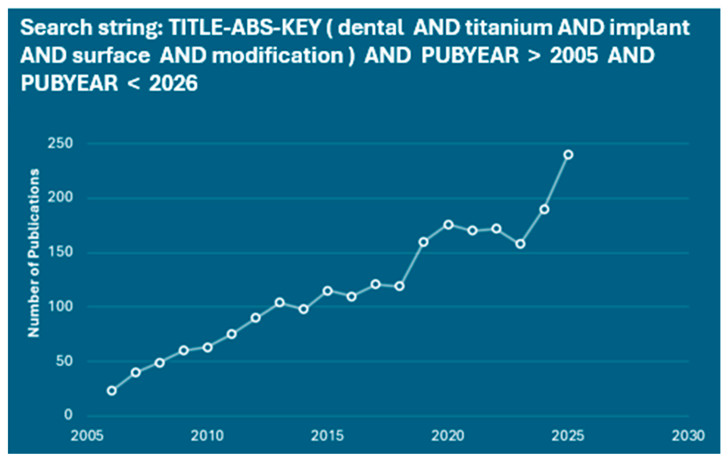
Ti implants are extensively utilised across different medical applications and tissues, including bone scaffolds, stents [18], skull restoration [19,20], dental implants [21,22,23], knees [24], hip [25,26], and other joint replacements [27]. Research on Ti implants remains a prominent and rapidly expanding area. Analyses of publication trends on Scopus-indexed journals indicate that research on surface modification of Ti dental implants has grown significantly over recent years (Figure 1) [28,29].
The success rate of Ti dental implant procedures can exceed 90%, yet approximately 10% of implants still fail [21]. The critical factor influencing implant success is the achievement of effective bone integration [30,31,32,33]. Additionally, enhancing the antibacterial properties of the implant and mitigating peri-implant inflammation are crucial for favourable surgical outcomes, as inflammation is often associated with bacterial colonisation and biofilm formation on the implant surface [34,35,36,37]. In addition to bone integration, stable soft-tissue adhesion at the transmucosal region is essential for long-term clinical outcomes. Unlike natural teeth, which are anchored by periodontal ligaments and hemidesmosome-mediated epithelial attachment, dental implants rely on a weaker peri-implant epithelial seal directly formed on the implant surface [38]. These processes are strongly influenced by the surface characteristics of Ti implants. However, altering the bulk material to improve surface properties can compromise the overall performance of the implant, as well as increase manufacturing complexity and cost. By contrast, surface modification represents an efficient and practical strategy, enabling control over surface morphology and composition without negatively affecting the implant’s overall properties [39,40,41,42]. Surface modification can enhance the micro- and nano topography as well as the chemical composition of implant surfaces, improving surface roughness [43] and hydrophilicity [14] which in turn promotes osteoblast adhesion. Additionally, incorporating antibacterial elements onto Ti implant surfaces can suppress bacterial growth and reduce the risk of inflammation [44,45,46]. When properly applied, surface modification techniques can effectively achieve these objectives.
This review provides a comprehensive and critical overview of both conventional and cutting-edge surface modification strategies for Ti dental implants, examining how variations in surface microstructure, chemistry, and hierarchical architecture influence biological performance. Beyond summarising individual techniques, the manuscript systematically compares surface treatments, coating technologies, and hybrid approaches in terms of osteointegration, antibacterial efficacy, drug delivery capability, manufacturability, and clinical outcomes. By identifying current limitations, reproducibility challenges, and translational gaps, the review highlights opportunities for integrated and multifunctional dental implant surface enhancement, offering a clear framework to guide future research, innovation, and clinical implementation. Its novelty lies in highlighting the comparison between conventional and emerging surface modification and functionalisation techniques to support the rational development of next-generation dental implants, while also providing a holistic, performance-oriented perspective that bridges laboratory findings with clinical relevance.

2. Literature Review

This paper was conducted as a narrative literature review to synthesise current knowledge on surface coating strategies used to enhance the performance of Ti dental implants. The aim was to integrate findings from experimental, clinical, and materials science studies describing advances in coating technologies, biological responses, and clinical implications.
A structured literature search was performed using the databases PubMed, Scopus, and Web of Science, covering publications from January 2015 to December 2025. Searches were conducted using combinations of keywords such as “titanium dental implants,” “surface coating,” “surface modification,” “osseointegration,” “bioactive coatings,” and “implant surface treatment.”
Relevant peer-reviewed articles written in English were considered, including in vitro studies, in vivo experiments, clinical investigations, and review articles focusing on surface modification approaches for Ti implants. Because the objective of this review was to provide a conceptual overview of technological developments rather than a quantitative comparison of treatment outcomes, formal systematic review procedures and methodological quality scoring were not applied. Instead, studies were selected based on their relevance to coating technologies, biological performance, and clinical applicability.

3. Functional Significance of Surface Treatments and Modifications

3.1. Enhanced Osseointegration

Osseointegration is a dynamic, multistage process that begins with mechanical interlocking between the implant surface and bone, followed by biological fixation driven by continuous bone apposition and remodelling at the implant interface [47,48]. The rate and quality of anchorage of Ti dental implants are strongly influenced by surface-related parameters, including material composition, surface roughness, chemistry and wettability, all of which govern early implant–tissue interactions [23,49]. Among these factors, surface roughness plays a pivotal role, as moderately roughened surfaces (Ra or Sa values between 0.5–1 μm refer to minimally rough, 1–2 μm to moderately rough, and >2 μm to rough) have consistently demonstrated superior early stabilisation and long-term clinical performance compared with smooth surfaces (Ra or Sa values <0.5 μm) [50,51].
Osman et al. [52] investigated the influence of industrially machined Ti surfaces with varying roughness levels on human gingival fibroblasts (HGFB) and keratinocytes (HGKC). Surfaces ranging from smooth (0.08–0.1 µm) to rough (3.3–3.7 µm) were characterised using scanning electron microscopy (SEM) and profilometry, as well as cell proliferation and cytotoxicity over 10 days. Their findings revealed significantly higher HGFB proliferation on minimally rough and rough surfaces, highlighting the importance of surface topography in promoting favourable soft-tissue responses and peri-implant tissue stability [52].
Bose et al. [53] demonstrated that doped calcium phosphate (CaP) coatings applied to 3D-printed porous Ti implants significantly improved bone ingrowth. Using Laser Engineered Net Shaping (LENS™), Ti implants were fabricated and subsequently modified with TiO2 nanotubes, followed by Sr2+- and Si4+-doped hydroxyapatite (HA) coatings. In vivo studies using a rat femoral model revealed that HA-coated implants exhibited increased new bone formation, stronger bone–implant bonding, and enhanced mineralisation compared with uncoated or nanotube-only surfaces [53]. These findings indicate that bioactive HA coatings accelerate and improve mechanical interlocking at the bone–implant interface, potentially promoting faster healing and long-term implant stability. However, plasma-sprayed HA coatings faced rejection in dental use partly because conventional plasma sprays often produce low crystalline and mixed phases that can degrade or dissolve over time, compromising long-term stability, and bare HA surfaces may support bacterial adhesion relative to Ti, prompting research into antimicrobial modifications [54].
Surface hydrophilicity has also emerged as a critical determinant of early biological events such as protein adsorption and cell adhesion [55]. Hydrophilic surfaces enhance the adsorption of key serum proteins and support integrin-mediated osteoblast attachment and differentiation, processes that are essential for bone healing at the interface. More specifically, animal and clinical studies have shown that implants with more hydrophilic surfaces exhibit significantly greater bone-to-implant contact (BIC) during early healing compared with hydrophobic surfaces [55,56,57]. Pae et al. [58] compared conventional sandblasted, large-grit, acid-etched (SLA) Ti surfaces with hydrophilic variants stored in calcium chloride solution or coated with a pH-buffering agent. Enhanced hydrophilicity was associated with increased protein adsorption, improved osteoblast adhesion, proliferation, differentiation, and mineralisation, as well as greater platelet adhesion and activation [58]. Notably, the pH-buffered hydrophilic surface demonstrated superior wettability and biological performance compared with conventional SLA surfaces, emphasising the importance of surface chemistry in establishing a biologically favourable implant interface.
Synergistic surface functionalisation strategies incorporating osteoinductive biomolecules have further advanced implant bioactivity [49,59]. Yang et al. [60] reported Ti surfaces chemically modified using imidazole carbamate with HA, heparin (Hep), and bone morphogenetic protein-2 (BMP-2) significantly enhanced bone formation both in vitro and in vivo. The Ti/HAp/Hep/BMP-2 coating exhibited increased osteocyte size and density at the bone–implant interface, indicative of active bone remodelling and improved early implant stability. These results highlight the synergistic effects of bioactive coatings in promoting osteogenesis and accelerating osseointegration. Notably, the pH-buffered hydrophilic surface demonstrated superior wettability and biological performance compared with conventional SLA surfaces, emphasising the importance of surface chemistry in establishing a biologically favourable implant interface.

3.2. Antimicrobial Performance

Microbial infection remains one of the leading causes of dental implant failure [61]. Bacterial colonisation occurs through an initial reversible adhesion phase, followed by irreversible attachment mediated by specific and nonspecific interactions between bacterial surface proteins and implant-associated molecules [62]. Once biofilms form, the penetration of antibiotics into the biofilm is retarded, and bacteria can potentially become highly resistant to antibiotics and to host immune responses, making eradication of infection extremely challenging and often resulting in implant loosening or failure [29].
Surface modification strategies have therefore been widely explored to impart antimicrobial functionality to dental implants. Antibiotic-based coatings represent one of the most extensively studied approaches. Jose et al. [63] developed a vancomycin-functionalised Ti surface via covalent coupling to an aminopropyltriethoxysilane-modified Ti substrate. Their results showed that this vancomycin-functionalized surface reduced Staphylococcus aureus colony-forming units by 88% ± 16% within 2 h and maintained antibacterial efficacy thereafter. Similarly, Lv et al. [64] fabricated multilayer coatings incorporating minocycline, which demonstrated strong antibacterial activity through both drug release and electrostatic repulsion mechanisms. While antibiotic-based coatings are effective, they are limited by restricted drug loading capacity, short-term release, and potential cytotoxicity. Consequently, non-antibiotic antimicrobial strategies have gained increasing attention. Silver-based coatings, in particular, have demonstrated broad-spectrum antibacterial efficacy, including activity against antibiotic-resistant strains [65]. Li et al. [66] incorporated silver nanoparticles into TiO2 nanomembranes, creating a dual antibacterial mechanism involving electrostatic repulsion and oxidative damage. The negatively charged surface inhibited initial bacterial adhesion, while oxidative stress and lipid peroxidation effectively inactivated adherent microorganisms [67,68]. Chitosan-based coatings also contributed to bacterial membrane disruption, increased permeability, and inhibition of biofilm formation due to their intrinsic antimicrobial and hydrophilic properties [69,70,71,72]. Collectively, these surface modification strategies significantly reduce bacterial colonisation and biofilm formation, thereby enhancing the long-term success and clinical reliability of dental implants.

4. Surface Modification Approaches for Dental Implant Applications

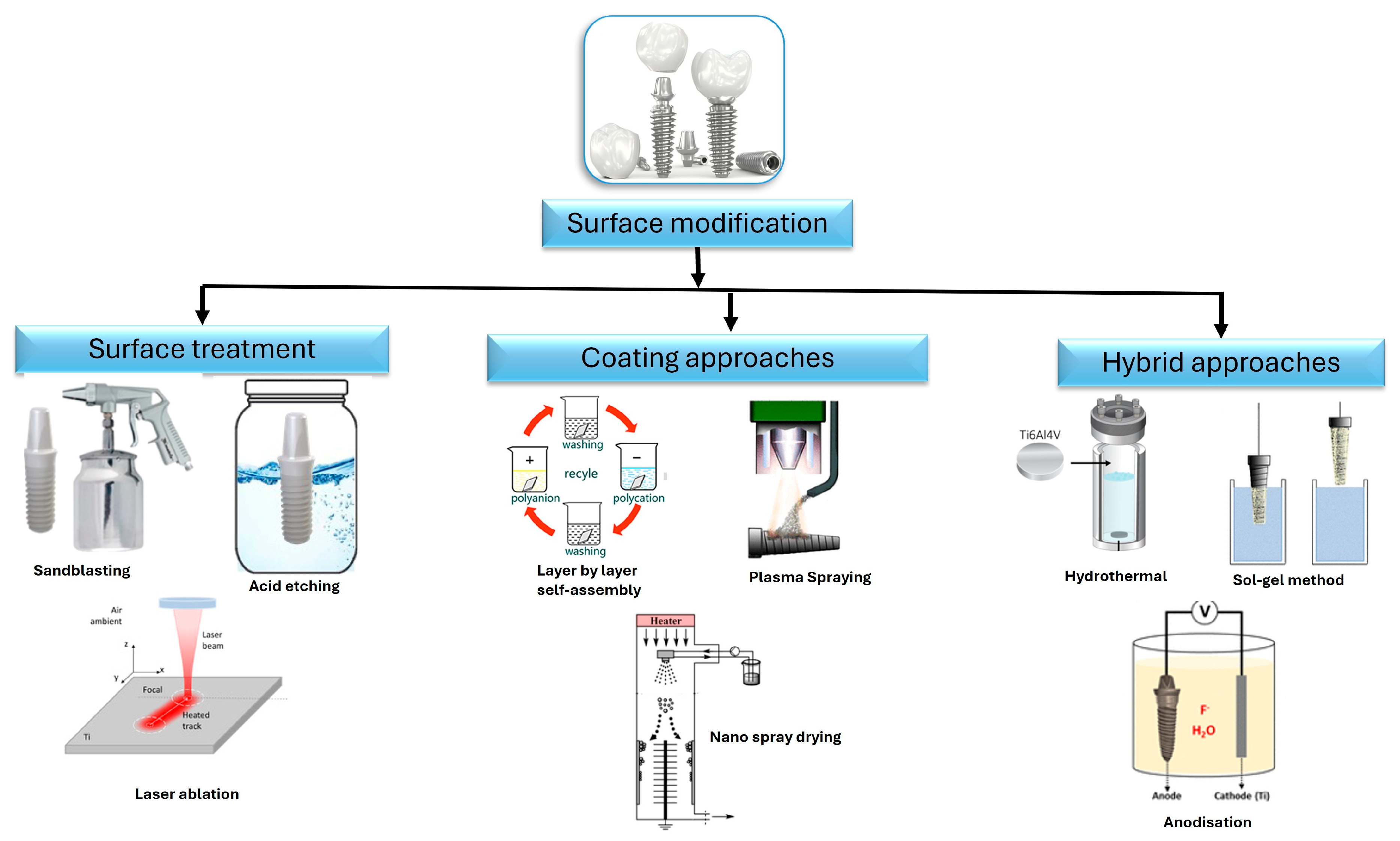
A wide range of surface modification strategies has been proposed to improve the performance of Ti dental implants, as summarised in Figure 2. These approaches are generally divided into two main categories. The first category involves surface treatment methods, including acid etching [73,74], sandblasting [75,76], anodization [67,70,77], and laser irradiation [78,79], which primarily tailor surface topography, mechanical properties, and oxide layer characteristics of Ti substrates. The second category comprises coating-based techniques, in which an additional material layer is deposited onto the Ti surface. Such coatings enable more substantial alteration of surface chemistry and are often employed to impart enhanced biological functionalities. Coating methods include nanospray drying [80], plasma spraying [81,82], hydrothermal treatment [83,84], sol–gel processing [85,86], and layer-by-layer self-assembly [87,88]. These methods were selected based on their widespread use and proven effectiveness in modifying implant surfaces for improved osseointegration and long-term clinical performance. Furthermore, this review specifically focuses on these modifications and coating strategies because they represent the most extensively studied and clinically relevant approaches, offering a balance between established techniques and emerging innovations. By concentrating on these well-documented methods, the review aims to provide a coherent and comparative analysis of their mechanisms, advantages, and limitations, while also highlighting their translational potential in clinical practice.

4.1. Surface Treatment Approaches

4.1.1. Sandblasting

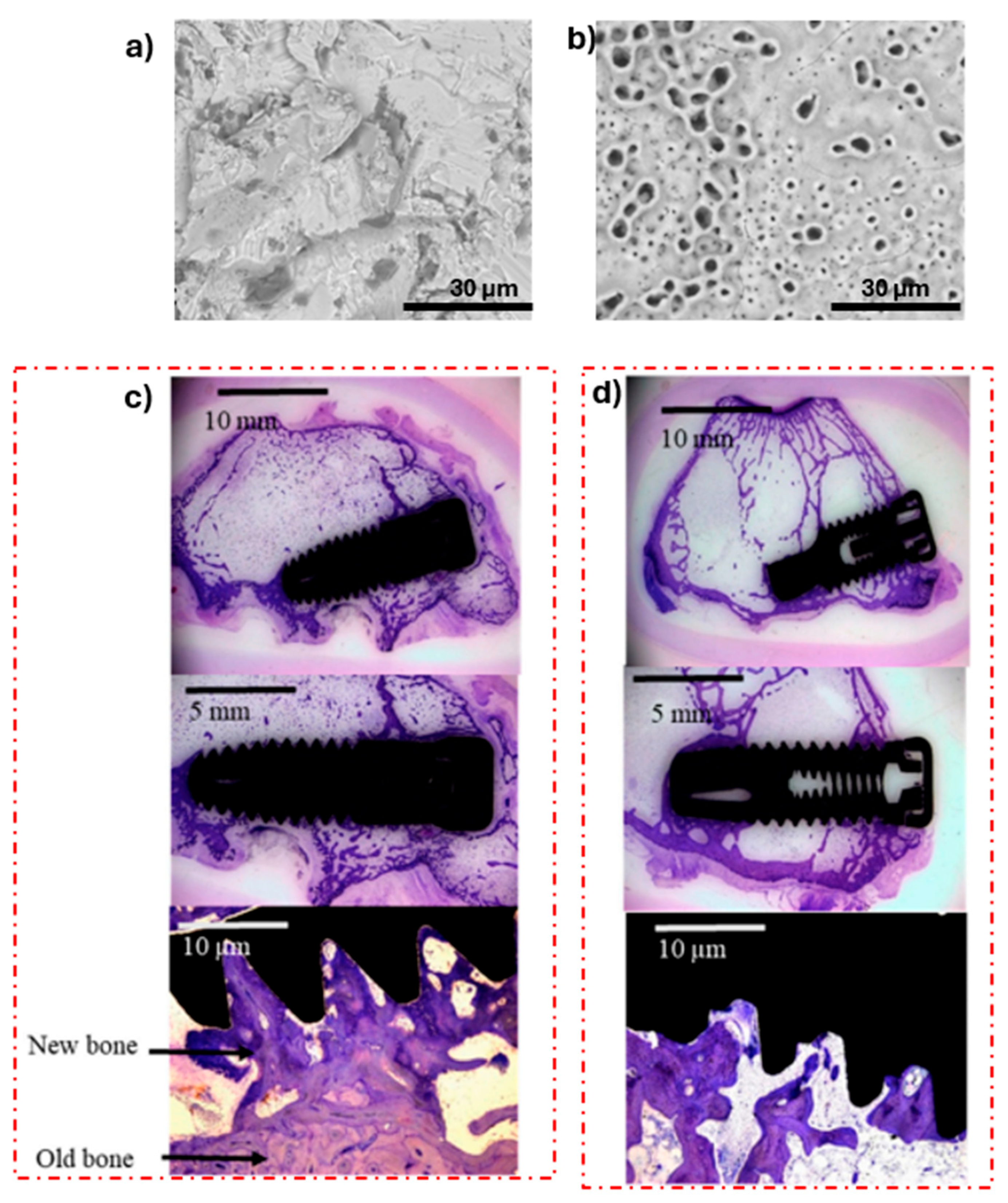
Sandblasting is among the most extensively employed subtractive surface modification techniques for Ti implants due to its technical simplicity, scalability, and commercial availability [21]. The process involves using compressed gas to propel abrasive microscale particles, most commonly aluminium oxide (Al2O3), Ti dioxide (TiO2), silicon dioxide (SiO2), or HA onto the implant surface, thereby inducing plastic deformation and generating a micro-rough topography [96]. Compared to oxidised Ti surfaces, which typically exhibit an average roughness (Ra) of approximately 0.5–1.5 μm, sandblasted surfaces generally achieve Ra values in the range of 1.5–3 μm, as illustrated in Figure 3a,b [50]. This range is frequently cited as optimal for bone anchorage; however, it is important to recognise that such generalisations often overlook the complex interplay between topography, chemistry, and biological response.
While increased roughness is widely associated with improved osseointegration, the recent findings present a more nuanced picture. For instance, although microrough surfaces enhance osteoblast attachment and early differentiation largely due to increased surface area and surface free energy facilitating protein adsorption [75,97], they may simultaneously promote bacterial adhesion. Surfaces with roughness exceeding ~2 μm have been shown to provide protective niches for microbial colonisation, thereby increasing the risk of biofilm formation [98]. This introduces a critical trade-off: the same features that support bone integration may also predispose the implant to infection. Importantly, these effects are not universal but depend heavily on bacterial species, surface chemistry, and wettability, suggesting that roughness alone is an insufficient predictor of biological performance [98].
A further limitation, often underemphasised, lies in the variability introduced by process parameters. The final surface characteristics are highly sensitive to factors such as abrasive particle size, hardness, and velocity, as well as spray angle, stand-off distance, and treatment duration [99]. This raises concerns regarding reproducibility, particularly in clinical-grade manufacturing. Moreover, the embedding of residual abrasive particles, especially when using harder materials like Al2O3, can lead to unintended chemical contamination of the Ti substrate [59]. Such contamination may alter surface chemistry in ways that are not always beneficial, and in some cases could impair long-term biocompatibility. In defiance of this, some studies continue to prioritise topographical outcomes [50,51] which, in our view, represents a significant oversight in the optimisation of implant surfaces.
Additionally, the morphological features generated by sandblasting are often highly irregular, characterised by sharp asperities and poorly controlled surface geometry. This typically necessitates subsequent post-processing steps, including ultrasonic cleaning, acid etching, or plasma treatment, to remove contaminants and refine surface properties [100]. From a practical standpoint, this undermines the perceived simplicity of sandblasting as a standalone technique. Its frequent integration into multi-step protocols suggests that, while effective in generating microroughness, it is insufficient in isolation to achieve the desired combination of topographical, chemical, and biological properties.
Another critical issue is the effect of sandblasting on surface wettability. Evidence indicates that sandblasted Ti surfaces often exhibit increased hydrophobicity, which may negatively impact early-stage protein adsorption and cell attachment [56,99]. For example, Osak et al. [56] demonstrated that Ti grade 4 surfaces treated with Al2O3 particles showed increased roughness, but reduced wettability compared to polished controls, despite improvements in tribological performance under simulated oral conditions. This again highlights the multifaceted nature of surface optimisation; improvements in one domain (e.g., mechanical behaviour) may come at the expense of another (e.g., biological response).
Taken together, these studies consistently support sandblasting for effectively enhancing microroughness, but do not adequately optimise surface chemistry or biological performance when used alone [96,99,100]. Consequently, contemporary surface engineering strategies tend to employ sandblasting as an initial step within more complex treatment sequences, aimed at mitigating contamination, improving hydrophilicity, and enhancing osteoblastic activity [73,96]. In this context, its role has shifted from a definitive solution to a preparatory technique. Therefore, while sandblasting continues to hold value as a foundational and accessible method for implant surface modification, its limitations, particularly regarding reproducibility, contamination, and biological trade-offs, necessitate a more critical and integrated approach. Its declining use as a standalone treatment in clinical practice reflects a broader recognition that successful implant performance depends on carefully balancing multiple interdependent surface properties, rather than on roughness alone.

4.1.2. Acid Etching

Acid etching is extensively utilised as a surface modification technique for Ti dental implants, primarily to generate controlled microroughness and enhance biological response. The process typically involves immersion in strong inorganic acids such as hydrofluoric (HF), nitric (HNO3), hydrochloric (HCl), or sulfuric acid (H2SO4), used individually or in combination to dissolve the native oxide layer and parts of the Ti substrate [99,101]. This produces relatively uniform micro-pits (0.5–2 μm), often cited as favourable for bone–implant interactions [102]. However, while such topographies are widely reported as beneficial, the assumption that uniform microroughness alone governs biological success remains overly simplistic.
Acid etching is also effective in removing contaminants and smoothing sharp asperities from prior mechanical treatments, resulting in a more homogeneous surface [96]. This is frequently linked to improved fibrin stabilisation and osteoblast attachment, supporting early-stage implant attachment [99,103,104]. Recent in vitro and in vivo studies continue to support increased bone–implant contact, as illustrated in Figure 3c,d), and reduced peri-implant bone loss on etched surfaces further reinforce these claims [74,105,106]. Nevertheless, the literature often underplays the sensitivity of outcomes to etching parameters. Excessive exposure can induce micro-cracks and compromise fatigue strength [107]. In clinical use, Sterngold Dental LLC, Massachusetts, United States, manufactures Stern EX acid-etched Ti implants that show predictable integration due to micro-roughened surfaces that enhance bone contact, supporting fixed or removable prostheses across maxillary and mandibular sites with favourable early stability and long-term survival rates. Therefore, while acid etching is clearly effective, its benefits are highly parameter-dependent, and its standalone optimisation remains insufficient without considering broader surface chemistry and mechanical trade-offs.
Figure 3. (a) SEM image of a dental implant exhibiting a sandblasted then acid-treated surface (SA), (b) dental implant with oxidised surface. Microscopic evaluation and quantitative histological analysis of (c) sandblasted and acid-etched surface implant (SA), and (d) oxidised surface. Adapted with permission from [105] (Copyright 2019, MDPI).
Coatings 16 00423 g003

4.1.3. Sandblasted, Large-Grit, Acid-Etched

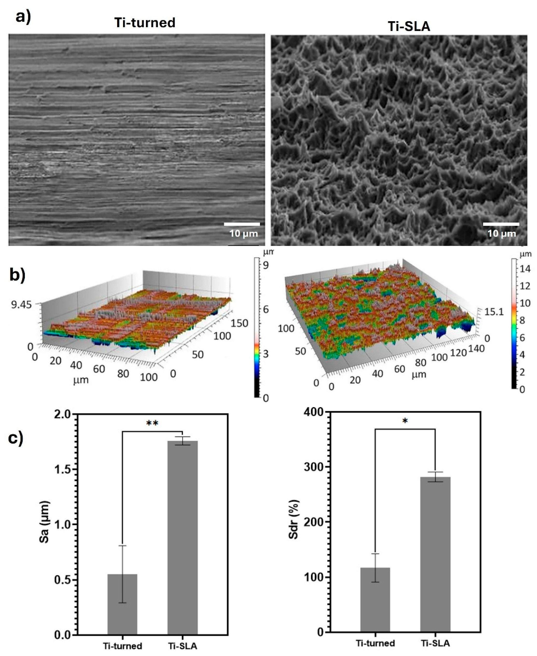
Sandblasted, large grit, acid etched (SLA) surfaces represent one of the most extensively investigated and clinically established surface modification strategies for Ti dental implants, combining macro roughening via abrasive blasting with subsequent acid etching to generate a hierarchical micro–nano topography. The process produces a dual-scale surface in which large-grit blasting creates pronounced macroroughness, while acid etching refines the morphology by introducing superimposed micro pits, collectively enhancing surface complexity. Figure 4a shows a comparative field-emission scanning electron microscopy (FE-SEM) analysis between machined (Ti-turned) and SLA-treated surfaces, consistently demonstrating a markedly more heterogeneous and irregular microstructure on SLA implants [96]. Similarly, three-dimensional surface reconstructions and quantitative parameters such as Sa and Sdr confirm significantly increased roughness and expanded interfacial surface area following SLA processing, reinforcing its capacity to substantially modify Ti surface architecture (Figure 4b,c). However, while these metrics are often interpreted as inherently beneficial, their biological relevance is not strictly linear and must be considered in conjunction with chemistry, wettability, and mechanical stability.
In vitro studies have shown that SLA surfaces enhance osteoblast adhesion, proliferation, and osteogenic differentiation relative to smoother or minimally treated Ti surfaces, largely attributed to improved protein adsorption and cell anchorage within the hierarchical topography [96]. The studies discussed above prove that SLA is a well-established modification technique with clinical evidence, which is well-documented for consistently high long-term survival and minimal peri-implant bone loss, establishing its performance as reliably comparable to other contemporary surface technologies [108]. Notably, randomised controlled clinical trials have reported that moderate variations in roughness (Ra 3.09 μm vs. 2.50 μm) produced by SLA do not significantly influence clinical outcomes, including primary stability, secondary stability, or marginal bone loss over a 12-year follow-up period [109]. This suggests that beyond a certain threshold, further increases in roughness may not yield proportional biological benefit, challenging the assumption that “rougher is better”. Nevertheless, SLA performance is highly dependent on strict control of blasting and etching parameters, which influence not only topography but also residual stress, surface chemistry, and potential contamination [57]. In this context, SLA should not be seen as one fixed “best” condition, but rather as a delicate process that needs careful and precise control during manufacturing. Overall, while SLA remains a gold standard due to its reproducible clinical success and favourable biological response, evidence indicates that its effectiveness arises from a balanced interplay of hierarchical structure rather than roughness magnitude alone.
Figure 4. Surface characterisation of Ti implants: Ti-turned (machined Ti implant) and Ti-SLA (sandblasted, large-grit, acid-etched Ti implant). (a) FE-SEM micrographs of the implant surfaces obtained at 5000× magnification (scale bar = 10 μm). (b) Confocal Laser Scanning Microscopy (CLSM) analysis showing surface topography, presented as three-dimensional colour-coded height maps with texture overlays. (c) Quantitative surface roughness measurements (Sa and Sdr) for each implant type. Results are expressed as mean ± SD (n = 3). Statistical significance is indicated as * p < 0.05 and ** p < 0.01. This figure incorporates material adapted from [110] (Copyright 2023, Nature Publishing Group, distributed under the terms of CC BY licence).
Coatings 16 00423 g004

4.1.4. Chemically Modified Sandblasted and Acid Etched Hydrophilic Surfaces

Chemically modified sandblasted and acid-etched hydrophilic surfaces (SLActive) represent a further evolution of conventional SLA implant technology, primarily aimed at preserving high surface energy and preventing post-processing contamination [21]. Following sandblasting with large grit particles and acid etching, the implant surface is rinsed and stored under controlled conditions to minimise exposure to atmospheric hydrocarbons, thereby maintaining a chemically activated, high-energy state [111]. This approach shifts the focus from purely topographical modification to combined control of surface chemistry and wettability, which is increasingly recognised as critical in early biological interactions.
Enhanced hydrophilicity has been consistently associated with improved early-stage cellular responses, including increased osteoblast adhesion, proliferation, and differentiation [49,55]. Compared with less hydrophilic or hydrophobic roughened surfaces, SLActive implants demonstrate lower contact angles and more favourable protein adsorption kinetics, indicating that wettability strongly governs the initial biological cascade [23]. However, this apparent advantage warrants a more cautious interpretation, as some studies indicate that such effects are largely confined to the initial healing phase, with long-term outcomes converging toward those of conventional SLA surfaces [112]. While clinical data report favourable survival and stability, these findings are not unequivocally superior, raising questions about the extent of their added clinical value [113]. The commercial SLActive® system (Straumann®, Switzerland) exemplifies efforts to preserve surface energy through controlled processing and storage conditions [48]; nevertheless, the broader literature remains inconclusive regarding the persistence of these benefits in vivo. Collectively, this suggests that although wettability and chemical state play important roles, their relative contribution may be context-dependent, reinforcing the need for more critical, long-term comparative studies rather than attributing performance gains predominantly to surface energy modifications alone.

4.1.5. Laser Ablation

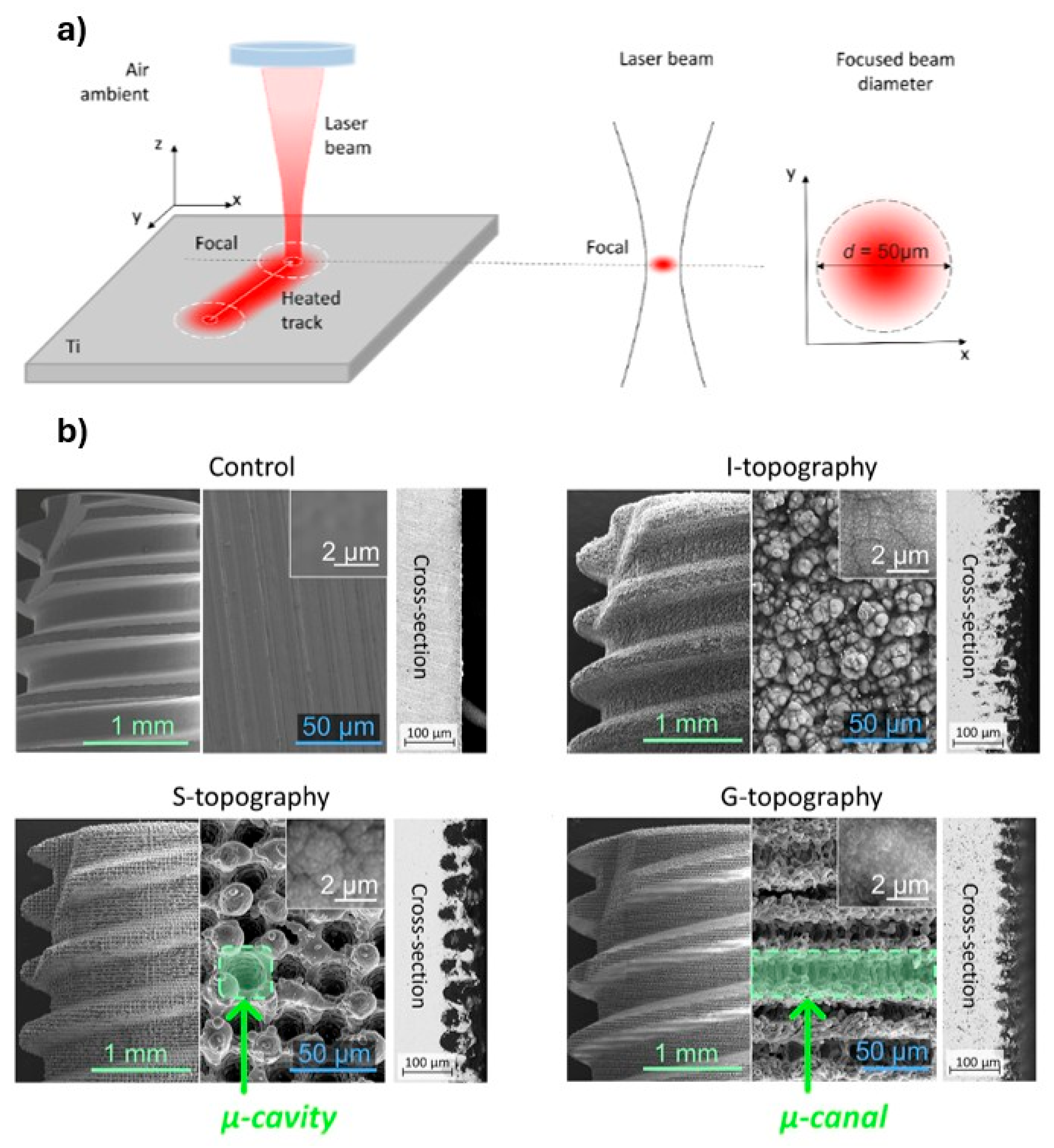
The principle of laser surface modification is schematically illustrated in Figure 5a. In this process, a highly focused laser beam is directed onto the Ti implant surface, where intense localised energy input induces rapid melting, ablation, and/or vaporisation of material [114]. While laser-based surface processing originated several decades ago, early “laser spraying” approaches were largely limited by coarse energy control, thermal damage, and inconsistent surface outcomes. In contrast, recent advances in laser physics, beam shaping, and scanning systems have repositioned laser ablation as a high-precision surface engineering tool for biomedical applications [114].
Contemporary laser systems enable controlled micro- and nanoscale structuring of Ti surfaces with high reproducibility, allowing deliberate tuning of roughness, wettability, and hierarchical architecture without altering bulk mechanical properties. The literature increasingly frames this as a “multi-functional surface design” capability; however, this narrative can be overstated, as the reproducibility of biological outcomes still depends strongly on subtle variations in processing parameters and post-treatment surface oxidation states. Nevertheless, the technique remains uniquely capable of generating surface features that are difficult or impossible to achieve via conventional subtractive or coating methods.
The final surface morphology is governed by laser parameters, including wavelength, pulse duration, fluence, scanning strategy, and incidence angle [114]. Through systematic adjustment of these variables, laser processing can produce controlled hierarchical structures spanning micro- to nanoscale dimensions. Recent studies reinforce its renewed relevance, demonstrating that laser-assisted nanotexturing and surface alloying can generate nanoporous architectures that enhance osteoblast activity, promote mineralisation, and simultaneously suppress bacterial colonisation while preserving bulk implant integrity [90,115].
Veiko et al. [91] provide a clear example of how the laser scanning strategy directly dictates biological function. Using a pulsed ytterbium fibre laser (λ = 1.064 μm, pulse duration 100 ns, spot diameter 50 μm), they fabricated distinct Ti surface topographies by varying scanning trajectories. Integration with a rotary system enabled uniform treatment of complex threaded geometries. Continuous μ-grooves (G-topography) were produced via parallel scanning, discrete μ-cavities (S-topography) via point irradiation, and irregular patterns (I-topography) via stochastic scanning. SEM analysis (Figure 5b) confirmed highly reproducible, well-defined structures. Importantly, in vivo evaluation demonstrated that G-type surfaces provided superior osseointegration, attributed to continuous microgrooves (20–50 μm) that guided cell alignment, facilitated bone in-growth, reduced fibrous tissue formation, and accelerated lamellar bone maturation [91,114]. This highlights a critical insight often underemphasised in the literature: not all “increased roughness” is beneficial; topographical continuity and spatial organisation are equally decisive.
Dorcioman et al. [116] reports fabrication of marine-derived HA (MdHA) thin films via pulsed laser deposition (PLD), positioning the approach as a sustainable and cost-effective alternative for implant surface modification. The authors demonstrate that PLD-produced MdHA films exhibit desirable surface characteristics, including pronounced roughness and high hydrophilicity (contact angles of 15–18°), which are linked to enhanced cell adhesion and for improved implant anchorage. Mechanical testing indicates adhesion strength (~49 MPa), suggesting adequate interfacial stability. Biologically, the synthesised MdHA show low cytotoxicity across multiple cell lines and promotes apatite layer formation in simulated body fluid solutions, supporting their mineralisation potential [116]. Additionally, moderate antimicrobial activity (1–3 log reduction) against bacterial and fungal strains is reported, indicating a degree of protective functionality. The authors present a technically sound and environmentally motivated application of PLD for MdHA coatings, but the work would benefit from deeper process optimisation, comparative analysis, and in vivo validation to fully establish its novelty and translational significance.
At the nanoscale, Gonçalves et al. [117] demonstrated that femtosecond laser processing can generate laser-induced periodic surface structures (LIPSS), significantly increase surface roughness and disrupt bacterial adhesion of Staphylococcus aureus and Escherichia coli through membrane instability mechanisms. This dual antibacterial and bioactive behaviour illustrates the multifunctionality achievable through ultrafast laser–matter interactions. Owing to its precision, scalability, and reproducibility, laser ablation has progressed from experimental methodology to industrial implementation. Commercial Ti implant systems, such as BioHorizons Laser-Lok and CSM Korean implants, already incorporate laser-engineered surface features [118], demonstrating successful translation into clinically deployed technologies.
Collectively, the literature presents laser ablation as a highly versatile and precise surface engineering approach; however, its perceived superiority warrants a more critical appraisal. While studies enhanced osteogenic response and antibacterial effects, these outcomes are often derived from controlled in vitro or short-term in vivo models, limiting their direct clinical extrapolation. Moreover, the strong dependence of surface morphology and biological performance on tightly controlled processing parameters raises concerns regarding standardisation and reproducibility across different systems and manufacturing environments. Although the ability to generate complex hierarchical structures is frequently highlighted as a distinct advantage, the relative contribution of specific features (e.g., groove geometry versus nanoscale texturing) remains insufficiently isolated, complicating mechanistic interpretation. Additionally, despite emerging commercial applications, long-term clinical evidence directly comparing laser-modified surfaces with established treatments remains relatively limited. We can safely conclude that while laser ablation demonstrates clear potential, current evidence suggests that its benefits may be context-dependent, and further systematic, comparative, and longitudinal studies are required to substantiate its claimed multifunctionality and clinical superiority.

4.1.6. Photofunctionalisation

Photofunctionalisation is one of the strategies that have been developed to improve implant success. It involves ultraviolet irradiation of Ti implant surfaces to modify their surface characteristics [119,120]. Photofunctionalisation itself is usually applied clinically using specialised UV light devices that treat the implant surface immediately before placement. This process is added during treatment rather than incorporated by the implant manufacturer. This process alters physicochemical properties, increases biological activity, improves surface wettability, and removes accumulated hydrocarbon contaminants associated with biological ageing [119]. As a result, photofunctionalisation enhances cell migration, adhesion, and proliferation, contributing to improved formation of a stable coronal soft-tissue seal [120].
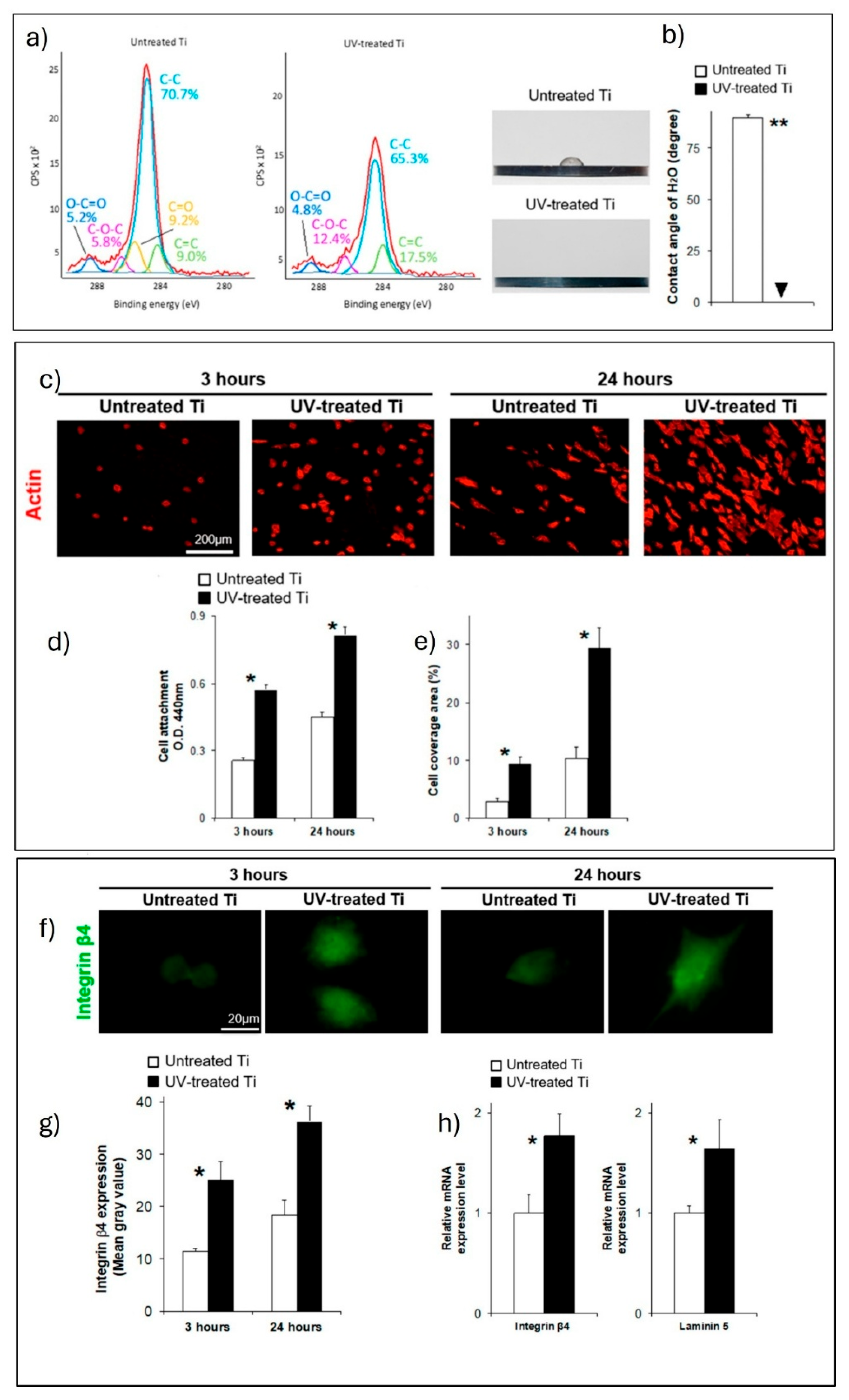
In a study by Nakhaei et al. [121] UV treatment significantly reduced surface carbon contamination, decreasing the atomic carbon content from approximately 40%, as demonstrated by X-ray photoelectron spectroscopy and C1s peak fitting analysis (Figure 6a). This decarbonization converted the Ti surface from hydrophobic to superhydrophilic, as evidenced by near-zero water contact angles (Figure 6b) [122]. Biologically, UV-treated Ti exhibited significantly enhanced initial attachment of human oral epithelial cells, with 1.8 to 2.2-fold increases in cell numbers at 3 and 24 h compared with untreated surfaces (Figure 6c,d). Confocal microscopy further revealed substantially greater cell coverage on photofunctionalised surfaces (Figure 6e) [121]. Early cell spreading was accelerated, with cells displaying well-developed lamellipodia and filopodia on UV-treated Ti, while cells on untreated surfaces remained rounded at early time points. Quantitative morphometric analysis confirmed larger cell area, perimeter, and diameter following UV treatment [121].
At the molecular level, photofunctionalisation upregulated hemidesmosome-associated adhesion markers, including integrin β4 and laminin-5, at both protein and gene expression levels (Figure 6f,g). Functionally, this translated into significantly greater epithelial cell retention under mechanical and enzymatic detachment, with approximately double the number of cells remaining adherent on UV-treated surfaces [121]. Notably, the enhancement achieved by photofunctionalisation was comparable to that obtained by laminin-5 coating (Figure 6h), highlighting UV treatment as a simple yet effective strategy to improve soft-tissue integration of Ti implants. The available clinical evidence consistently demonstrates favourable outcomes for UV-treated Ti implants across prospective [123], retrospective [124], and case–controlled studies [125]. Photofunctionalisation was associated with higher implant stability and shortened healing periods, particularly in compromised bone conditions [125]. Several studies reported improved early implant stability, enabling immediate or early loading protocols with high success rates. Enhanced clinical performance was observed even in sites with poor bone quality or low primary stability, where photofunctionalised implants showed outcomes comparable to or better than conventional implants in healthy bone [126].
Notwithstanding the consistently reported benefits of photofunctionalisation, the literature warrants a more critical interpretation. Much of the evidence emphasises short-term biological enhancements, particularly in early cell attachment and surface wettability, yet the durability of these effects remains uncertain, as recontamination and hydrophobic recovery may occur rapidly after treatment. Furthermore, many studies rely heavily on in vitro or early-stage clinical outcomes, with limited robust long-term comparative data demonstrating sustained superiority over conventional surfaces. The clinical protocols themselves also introduce variability, as the effectiveness of photofunctionalisation is highly dependent on timing, handling, and device-specific parameters, raising concerns regarding standardisation and reproducibility in routine practice. Additionally, while improvements in compromised conditions are frequently highlighted, the extent to which these outcomes translate into meaningful long-term clinical advantages remains insufficiently established. Collectively, while photofunctionalisation represents a promising and clinically accessible adjunct, current evidence suggests its benefits may be transient and context-dependent, underscoring the need for more rigorous, standardised, and longitudinal investigations.

4.2. Coating Modification Approaches

4.2.1. Self-Assembly Method Layer-by-Layer

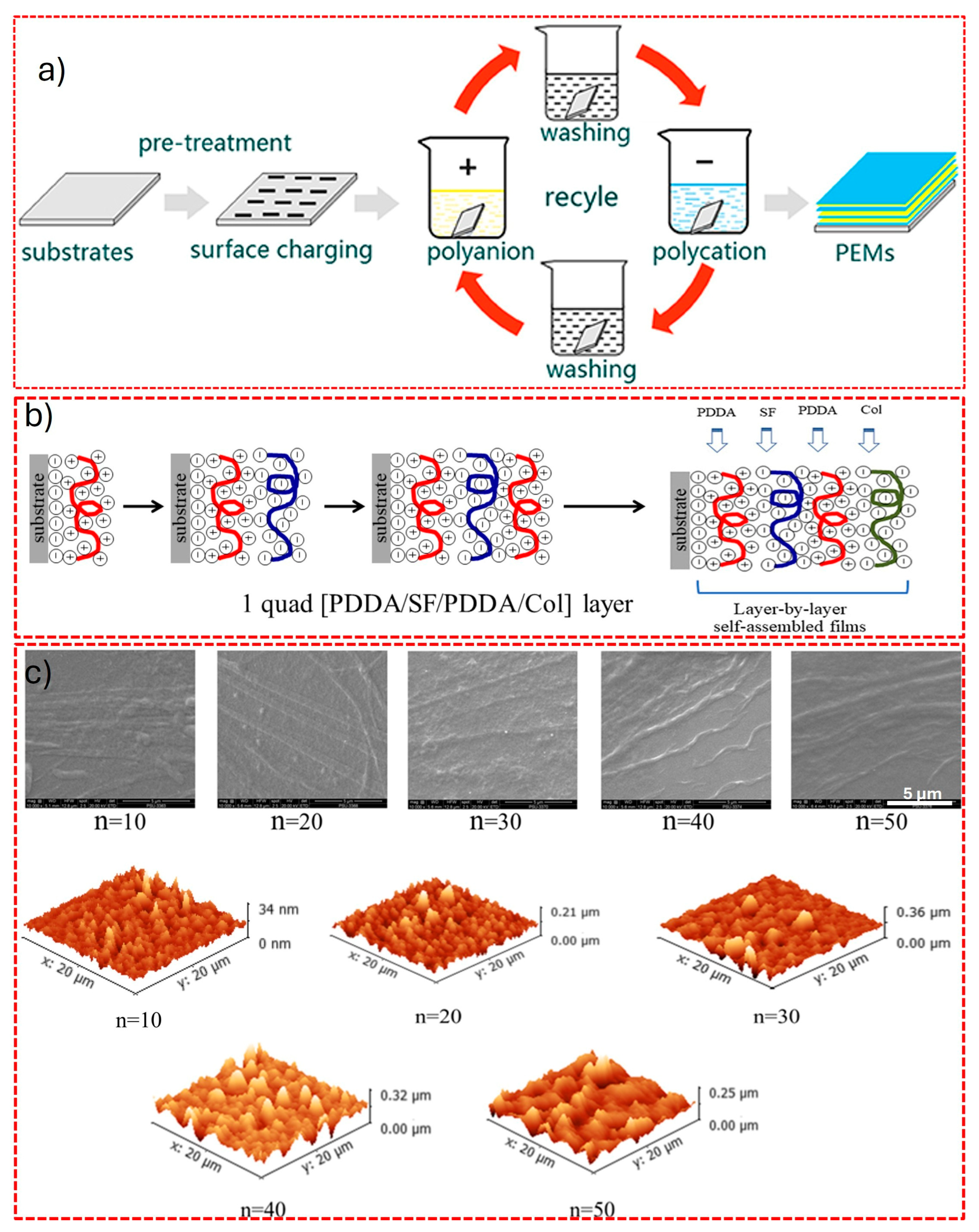
The self-assembly method Layer-by-layer (LbL) has become a widely used surface-modification strategy because it is straightforward, versatile, and broadly applicable to different materials. As illustrated in Figure 7a, in this approach, functional coatings are constructed through the spontaneous organisation of building blocks driven by physicochemical interactions, rather than by direct external manipulation [88]. In general, self-assembly refers to a process in which molecules, atoms, or larger structural units autonomously organise into ordered and functional architectures. Similar principles operate in natural systems; for example, during biomineralisation, polymeric templates regulate the nucleation and growth of inorganic phases [127]. Monomolecular films found in nature represent a classic manifestation of such self-organised behaviour. In self-assembled monolayers, one terminal group exhibits strong affinity toward the substrate, while the opposite end can be tailored with different functional moieties. LbL assembly creates multilayers through electrostatic interactions, introducing functional amino groups (–NH3+) and carboxyl groups (–COO) onto the surface, as well as bioactive peptides. By modifying the chemical nature of these terminal groups, the surface properties of the underlying matrix, such as wettability, roughness, and biological response, can be precisely tuned.
Using this concept, Nawae et al. [128] fabricated multilayered poly (diallyl dimethylammonium chloride)–silk fibroin–collagen (PDDA/SF/PDDA/Col) coatings via an LbL self-assembly technique, as schematically illustrated in Figure 7b. In this system, negatively charged biomacromolecules (SF and Col) were alternately deposited with the positively charged PDDA through electrostatic interactions. The buildup of the multilayer film proceeds through multilayer architectures that were fabricated with varying numbers of deposition cycles, including 0 (untreated control), 10, 20, 30, 40, and 50 layers, enabling controlled growth of the coating. As shown in Figure 7c, the n = 10 deposition cycle exhibits a relatively smooth surface, whereas the n = 50 deposition sample displays a distinct granular morphology [128]. With increasing numbers of deposited layers, both surface roughness and film thickness increase progressively, as confirmed by SEM and AFM analyses. These results indicate that the resulting bio-based coatings possess enhanced surface roughness and hydrophilicity, which are favourable for cell adhesion and proliferation.
Similarly, de Avila et al. [129] reported the fabrication of poly (acrylic acid) (PAA)/polyline multilayer coatings on Ti substrates using the LbL technique, demonstrating improved antibacterial performance. The LbL self-assembled coating on Ti showed excellent biocompatibility, with enhanced hydrophilicity, cytocompatibility, and hemocompatibility. It supported murine (mouse-derived) macrophage cell line (macrophage M2) polarisation, promoted Human Umbilical Vein Endothelial Cells (HUVEC) angiogenic activity, and enhanced MC3T3-E1 osteoblast adhesion, proliferation, and differentiation, while demonstrating good in vivo biosafety and minimal inflammatory response [130].
In spite of its versatility and capacity for precise surface functionalisation, the literature on LbL self-assembly reports that biological and antibacterial enhancements are derived from controlled laboratory conditions, with limited validation under clinically relevant mechanical and physiological environments. Issues related to long-term coating stability, delamination, and durability during implant insertion remain insufficiently addressed. Furthermore, the complexity of multistep fabrication and sensitivity to assembly parameters challenge scalability and reproducibility. Consequently, despite promising multifunctionality, clinical translation remains uncertain and underdeveloped.

4.2.2. Nanospray Drying

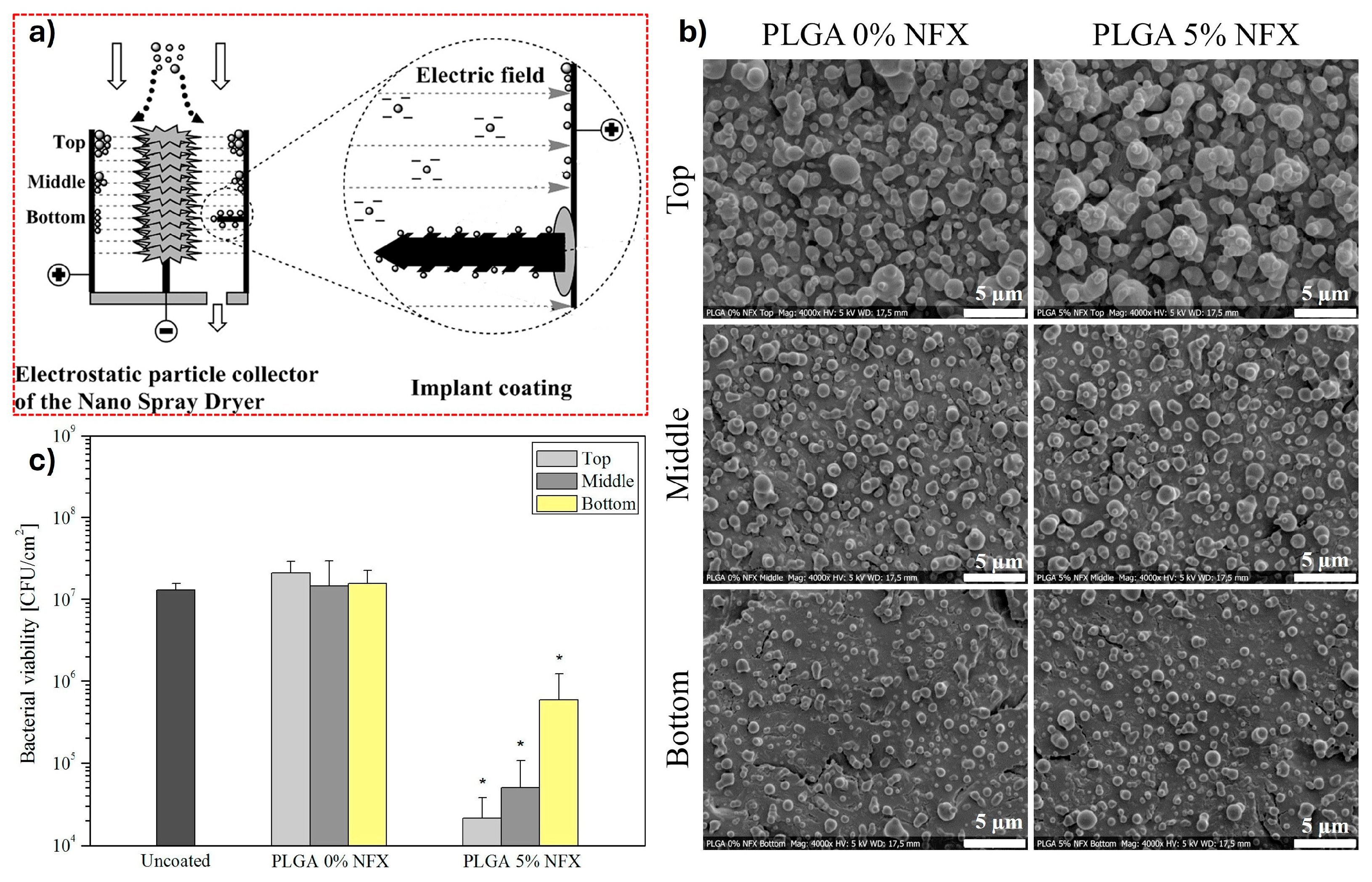
Nanospray drying is an emerging technique for depositing nanoparticle-based coatings composed of bioactive materials onto implant surfaces. As illustrated in Figure 8a, the process operates by generating ultrafine aerosol droplets that rapidly dry within a confined chamber to form solid nanoparticles. These charged particles are subsequently guided towards the implant substrate by an applied electric field between an electrode and collector, resulting in electrostatically driven deposition and surface coating formation [95]. Nanostructured coatings produced via nanospray drying offer several functional advantages, including reduced risk of peri-implant infection, improved early-stage healing, and enhanced biointerface integration. Importantly, these nanoparticles can also serve as drug carriers, enabling localised therapeutic delivery directly at the implant site, thereby reducing systemic exposure and improving pharmacological efficiency [131]. This dual functionality positions nanospray drying not merely as a coating method, but as a potential drug–device integration platform.
Baghdan et al. [80] demonstrated this potential by fabricating Ti discs from high-purity Ti foil and coating them using a Nanospray Dryer B-90 system. They developed poly(lactic-co-glycolic acid) (PLGA)-based nano coatings, both drug-free and loaded with norfloxacin (NFX) at 0%–10% w/w, using acetonitrile water feed solutions. Deposition was carried out under tightly controlled parameters: a 4.0 µm mesh spray cap angled at 45°, gas flow rate of 100 L/min, spray intensity of 50%, and inlet temperature of 70 °C in closed-loop nitrogen mode. The feed solution contained PLGA and NFX dissolved in a 95:5 acetonitrile–water solvent system with 0.005% w/v sodium acetate, ensuring stable aerosol formation. Their results showed that deposition position within the collector significantly influenced coating morphology (Figure 8b). SEM analysis confirmed reproducible spherical nanoparticles with narrow size distribution for both PLGA (0% NFX) and PLGA (5% NFX) formulations. However, coatings collected at the top region exhibited multilayered structures with larger aggregates, whereas those at the bottom formed more uniform monolayers composed of smaller particles, highlighting a key limitation in achieving spatially consistent coatings at scale. Biological evaluation demonstrated that PLGA 5% NFX coatings exhibited the lowest Escherichia coli colony-forming units after 24 h, confirming enhanced antibacterial efficacy as illustrated in Figure 8c [80]. This indicates that controlled antibiotic incorporation can significantly improve anti-infective performance without compromising coating formation.
From a critical perspective, nanospray drying is often presented as a highly uniform and controllable deposition strategy; however, the recent findings reveals that coating homogeneity is strongly influenced by particle trajectory, chamber geometry, and electrostatic field distribution. This introduces spatial variability, particularly in complex collector configurations, which can limit reproducibility across different implant positions. Nevertheless, the technique remains attractive due to its ability to generate nanoscale coatings with high surface area and tunable physicochemical properties. In spite of these multifunctional outcomes, nanospray drying remains largely restricted to laboratory-scale research. Challenges related to scale-up uniformity, collector design, and process standardisation further limit clinical translation. Nonetheless, the approach offers important conceptual and technological insights for next-generation antimicrobial and drug-eluting dental implant coatings.

4.2.3. Plasma Spraying

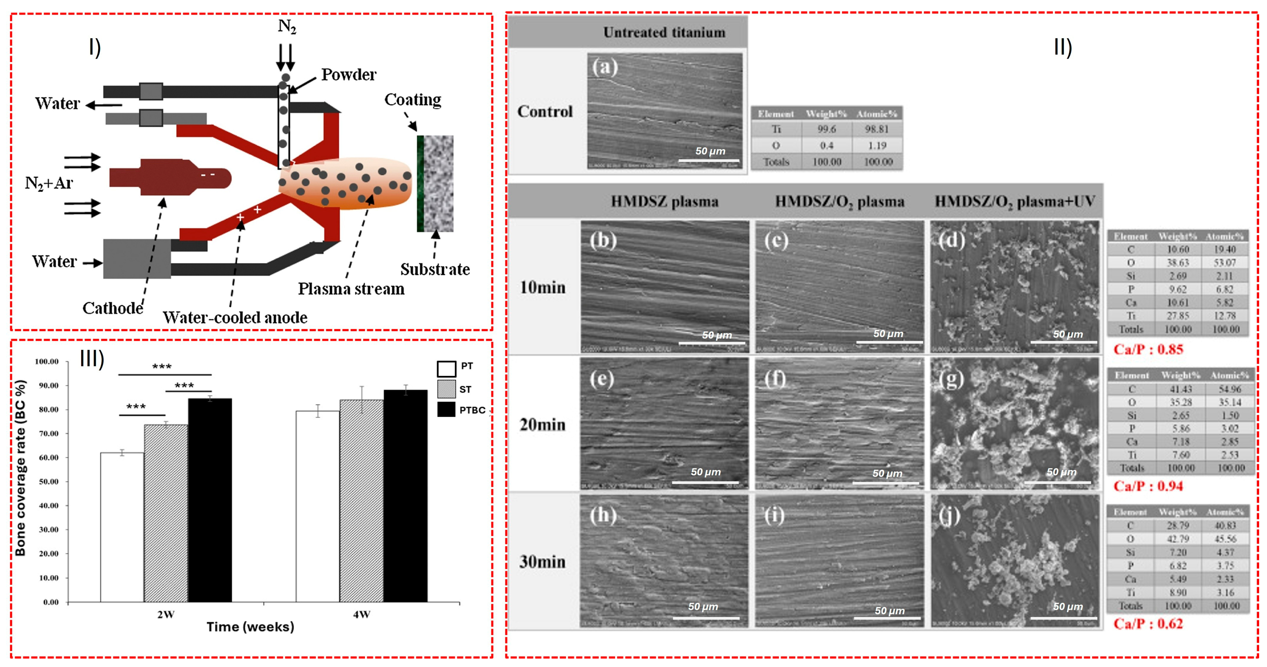
Plasma spraying is a thermal deposition technique in which coating powders are injected into a high-temperature plasma jet and propelled onto a substrate, where rapid solidification leads to coating formation. A schematic of the experimental setup is illustrated in Figure 9I. Although historically one of the earliest industrial methods for Ti implant coating, plasma spraying has undergone substantial refinement, maintaining its relevance in modern dental implant engineering. The literature reflects a shift in perception: plasma spraying is simultaneously described as both a legacy technique and a modern high-performance platform. This duality is justified by significant technological improvements in plasma torch design, feedstock engineering, and process control (plasma power, particle velocity, and stand-off distance), which have enhanced coating uniformity, phase stability, and adhesion strength compared with early systems [132].
Chuang et al. [133] demonstrated a multifunctional plasma-based modification strategy for Ti dental implants. Untreated Ti exhibited a smooth morphology (Figure 9II(a)). Plasma polymerisation of hexamethyldisilazane (HMDSZ) for 10–30 min progressively altered surface morphology, indicating organo-silane film formation (Figure 9II(b,e,h)). Oxygen plasma treatment introduced reactive functional groups while preserving morphology (Figure 9II(e,f,i)), enabling subsequent UV-induced grafting of a dibasic calcium phosphate (DCPA)-containing thermoresponsive hydrogel (Figure 9II(d,g,j)), producing a bioactive granular coating enriched in calcium–phosphate phases.
In vivo evaluation using a Lee-Sung miniature swine model showed that implants incorporating bone morphogenetic protein-2 and chlorhexidine (PTBC) achieved significantly higher bone coverage at 2 weeks compared with untreated (PT) and sandblasted (ST) implants, indicating accelerated early osseointegration. By 4 weeks, PTBC implants reached bone coverage comparable to commercial implants while outperforming untreated Ti, highlighting improved early-stage healing dynamics. Antibacterial testing confirmed activity against Enterococcus faecalis and Escherichia coli, whereas untreated Ti showed no effect [133].
Critically, research demonstrates that plasma spraying, when integrated with plasma-assisted surface chemistry and post-functionalisation, enables multifunctional coatings with clinically relevant biological performance. A 5-year prospective study involving 200 patients and 626 non-submerged Ti plasma-sprayed implants reported high survival (99.4%) and success (92.5%) rates, documenting clinical predictability and patient satisfaction. Importantly, plasma-sprayed Ti implants are already widely used in dental practice [134]. However, modern dental implants more frequently use blasted/acid-etched or SLA surfaces; pure plasma-sprayed Ti implants are less common today but still part of clinical and historical surface-modification options.
Figure 9. (I) Schematic of the plasma spraying process. Reproduced with permission from ref. [135]. (II) Surface morphology of Ti substrates, (a) untreated; (bd) after 10 min, (eg) after 20 min and (hj) after 30 min of HMDSZ plasma deposition, oxygen plasma treatment and UV-grafted DCPA-containing hydrogel. (III) Bone coverage rate at 2 and 4 weeks for PT (untreated), ST (sandblasted), and PTBC (BMP2/CHX-modified) implants, *** p < 0.001. Reproduced with permission from [133] (Copyright 2025, Elsevier).
Coatings 16 00423 g009

4.2.4. Smart Surface Functionalisation via Piezoelectric Nanocomposites

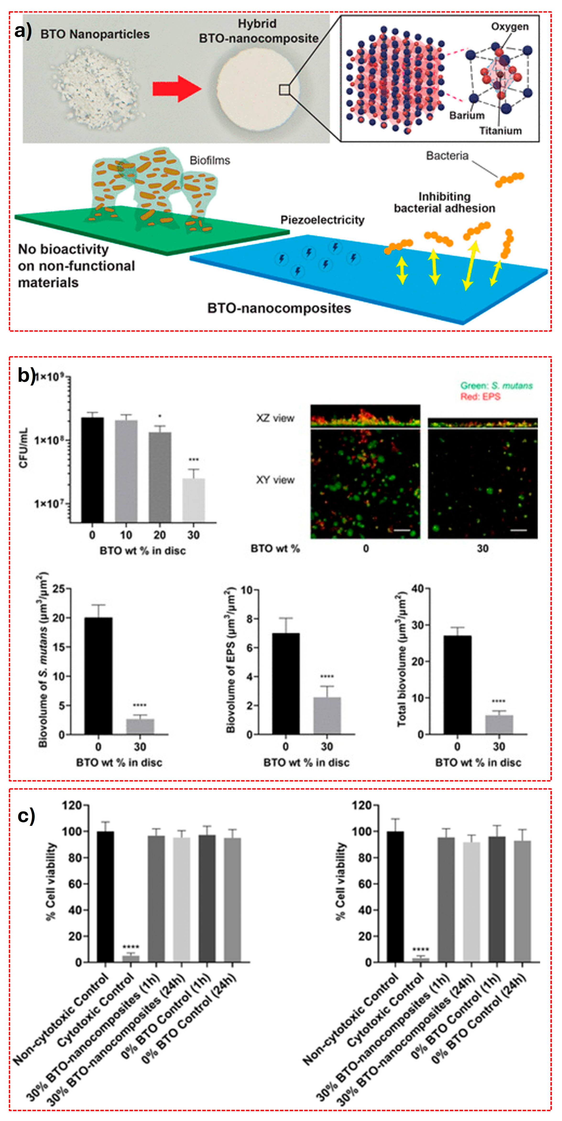
Piezoelectric nanocomposites involve integrating piezoelectric nanoparticles (e.g., barium titanate, zinc oxide, or potassium sodium niobate) into implant coatings or polymer matrices. These nanoparticles generate localised electrical charges when mechanically stimulated, such as during chewing or physiological loading [136]. Dhall et al. [137] introduced a multifunctional surface concept based on barium titanate (BaTiO3, BTO) nanoparticle-embedded polymer nanocomposites, offering a non-bactericidal, anti-biofilm approach combined with self-powering capability (Figure 10a).
In this system, piezoelectrically active BTO nanoparticles (~400 nm) were embedded at varying weight fractions (up to 30 wt%) within a UV-curable polymer matrix. The resulting surfaces demonstrated a significantly increased repulsive interaction energy between the composite surfaces and Streptococcus mutans, a key oral pathogen. This stronger repulsive interaction made it energetically unfavourable for the bacteria to attach, which led to much lower adhesion and biofilm formation compared with composites with less or no BTO. Direct binding-force measurements using atomic force microscopy (AFM) also confirmed this reduced adhesion at higher BTO content, explaining the significant decrease in biofilm at 30 wt% BTO (Figure 10b) [137]. This is particularly significant for dental implants, as non-lethal anti-adhesive strategies reduce the risk of antimicrobial resistance and preserve the surrounding oral microbiome [138]. Mechanistically, the antibiofilm effect was attributed to unfavourable physicochemical interactions at the bacteria–surface interface, rather than surface roughness or nanoparticle size. AFM analysis revealed that BTO-nanocomposite surfaces generated repulsive interaction energy barriers and significantly lower bacterial adhesion forces [139].
A unique aspect relevant to smart implant design is the piezoelectric poling of BTO nanocomposites, which creates asymmetrically charged surfaces [140]. After poling, one surface became relatively positive while the opposite surface remained negative. This asymmetry enabled dual functionality: the negatively charged surface retained strong antibiofilm properties, while the positively charged surface promoted cell adhesion and is hypothesised to support improved tissue integration [137]. Importantly, BTO nanocomposite cytotoxicity assays using human gingival fibroblasts and keratinocytes confirmed excellent biocompatibility, supporting their translational relevance for oral applications (Figure 10c) [137]. Such spatially controlled surface charge distribution is highly relevant for dental implants, where opposing requirements exist at the bone-implant and trans-mucosal interfaces.
Regardless of the innovative concept of piezoelectric nanocomposites, the current literature remains largely exploratory and requires cautious interpretation. Reported antibiofilm and bioactive effects are predominantly based on in vitro models, with limited evidence under dynamic in vivo conditions where mechanical loading and long-term stability are critical. The durability of piezoelectric responses and consistency of charge generation over time are insufficiently characterised. Additionally, fabrication complexity and material integration challenges may hinder scalability. Consequently, while promising, their clinical applicability remains preliminary.

4.3. Hybrid Approaches in Surface Treatment and Coating Technologies

Hybrid surface modification techniques challenge the conventional distinction between surface treatments and coatings by integrating both functionalities within a single process. The authors argue that such approaches offer superior control over surface chemistry and structure; however, inconsistent classification and limited standardisation continue to hinder comparative evaluation and broader clinical translation.

4.3.1. Sol–Gel Technique

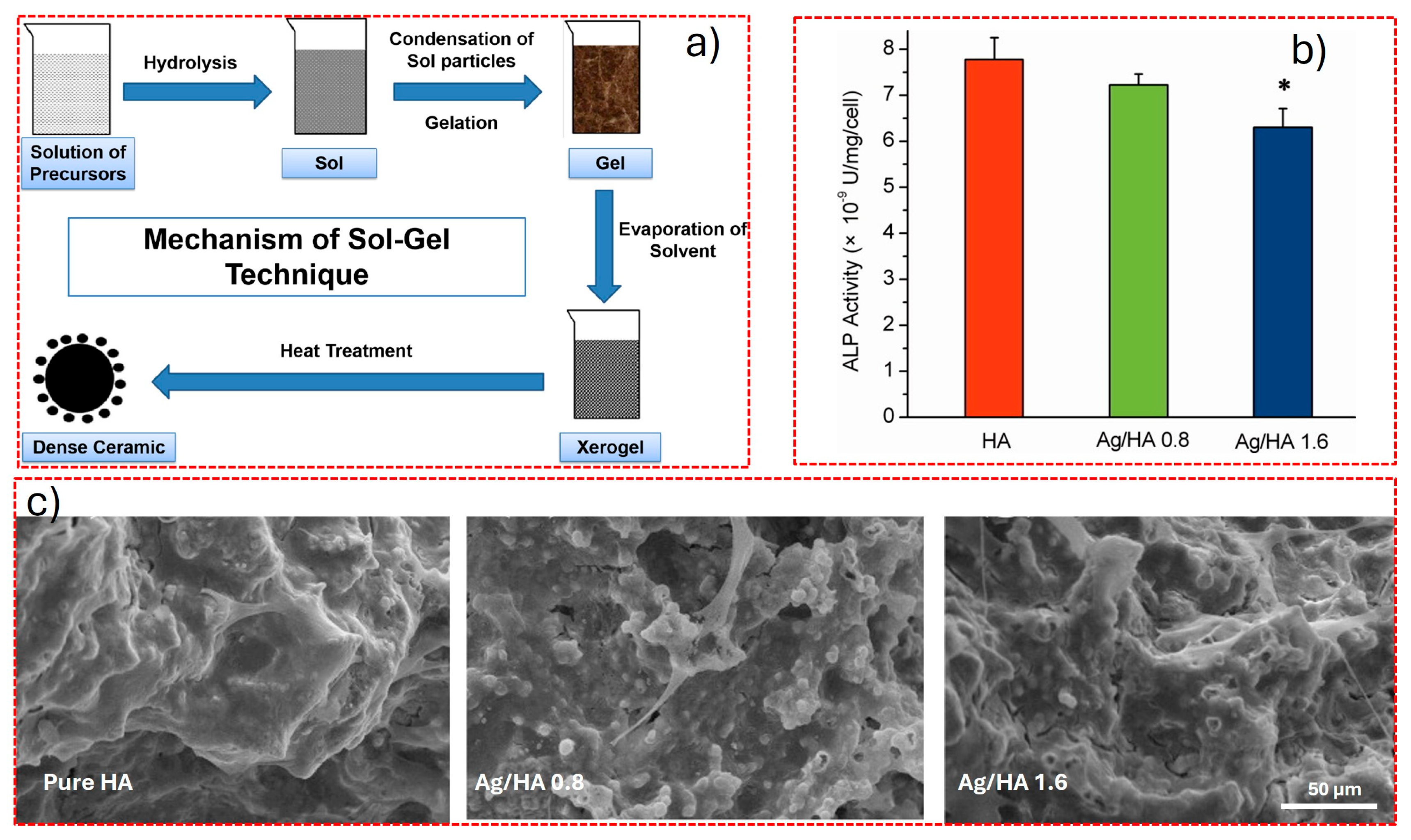
Sol–gel processing is a hybrid surface modification technique that forms thin functional coatings while enabling chemical interaction with the substrate, combining coating deposition and surface modification to improve bioactivity, adhesion, and surface properties [141]. The sol–gel process represents a versatile chemical route for fabricating oxide-based thin films, enabling both inorganic and organic–inorganic hybrid materials to be synthesised at relatively low processing temperatures As conceptually illustrated in Figure 11a, a “sol” describes a stable colloidal dispersion of solid particles in a liquid medium, while a “gel” denotes a semi-solid network in which a continuous inorganic framework immobilises the liquid phase. Unlike interfacial reaction-driven coating methods, sol–gel chemistry is governed entirely by solution-phase hydrolysis and condensation reactions, which progressively construct the oxide network [142]. During synthesis, hydrolysis and condensation reactions generate metal–oxygen–metal networks, enabling deposition of bioactive coatings such as HA, TiO2, and SiO2 on metallic implant surfaces [142].
Qu et al. [143] exemplify this translational potential by fabricating silver-doped HA (Ag/HA) coatings on porous Ti using an ethanol-based sol–gel dip-coating approach. Their system achieved uniform infiltration of Ag/HA (0.8 and 1.6 wt%) throughout interconnected porosity, with structural analysis confirming crystalline HA and well-dispersed Ag nanoparticles. Biologically, Figure 11b reveals a concentration-dependent decline in osteoblast ALP activity, indicating that higher silver loading suppresses osteogenic differentiation, despite its antibacterial benefits. While 0.8 wt% preserved ALP activity comparable to pure HA, 1.6 wt% significantly impaired it. SEM analysis (Figure 11c), however, showed enhanced cell spreading with increasing Ag content, highlighting a disconnect between morphological adhesion and functional differentiation. Antibacterial testing demonstrated more than 95% efficacy against Escherichia coli and Staphylococcus aureus, whereas pure HA exhibited no antimicrobial activity. This reinforces the necessity of balancing antibacterial potency with osteogenic compatibility in clinically relevant coatings rather than maximising dopant concentration.
From a critical perspective, the sol–gel method is frequently positioned as a near-universal coating strategy due to its simplicity, compositional flexibility, and ability to uniformly coat complex geometries [142,144]. However, this favourable narrative often downplays practical constraints such as drying-induced shrinkage, cracking, and reproducibility challenges, particularly when transitioning from laboratory-scale dip-coating to industrial or clinical manufacturing. Despite these limitations, its low-temperature processing remains a decisive advantage for biomedical substrates, especially thermally sensitive alloys and polymer-containing implant systems.
Importantly, these coatings are not merely experimental constructs but represent clinically relevant surface-engineering strategies aimed at enhancing bioactive surface to ensure implant longevity in vivo. Notably, the sol–gel method has advanced beyond laboratory validation to clinical application. Nanosprayed implant systems such as Endopore® implants, developed in Ontario, Canada, demonstrated that porous and bioactive implant surfaces can achieve reliable bone ingrowth and good clinical performance, particularly in compromised bone [145]. However, while these earlier systems are supported by longer clinical experience, newer nanocrystalline carbonated hydroxyapatite coatings still lack extensive long-term clinical data. In this context, sol–gel-derived coatings function not only as surface modifications but as clinically deployed technologies that directly support improved early bone anchorage and long-term success in dental patients.

4.3.2. Hydrothermal Approach

Hydrothermal treatment is another hybrid surface modification method that alters substrate chemistry while enabling in situ growth of crystalline coatings, combining surface treatment and coating to enhance bioactivity, adhesion, and functionality. In this approach, Ti implants are immersed in a reactive aqueous solution and exposed to elevated temperature and pressure, enabling in situ chemical reactions that directly generate a surface coating on the substrate [146]. This method promotes nucleation and crystallisation of HA or beta-tricalcium phosphate (β-TCP), yielding hydroxyl-rich surfaces that enhance osteoconductivity and early-stage bone response. A key conceptual strength of this approach lies in its “growth-from-substrate” mechanism: because the coating forms through direct interfacial reactions rather than post-deposition application, it typically exhibits superior adhesion to the underlying Ti surface [147]. However, the literature often overstates its simplicity, as subtle variations in precursor chemistry and autoclave conditions can significantly alter phase composition and reproducibility. From a process perspective, coating characteristics are highly sensitive to parameters such as initial Ti surface condition, temperature, pressure, reaction time, and ionic composition of the solution [147]. This sensitivity can be interpreted both as an advantage, allowing fine chemical tuning and as a limitation, since it introduces challenges for process standardisation and scale-up.
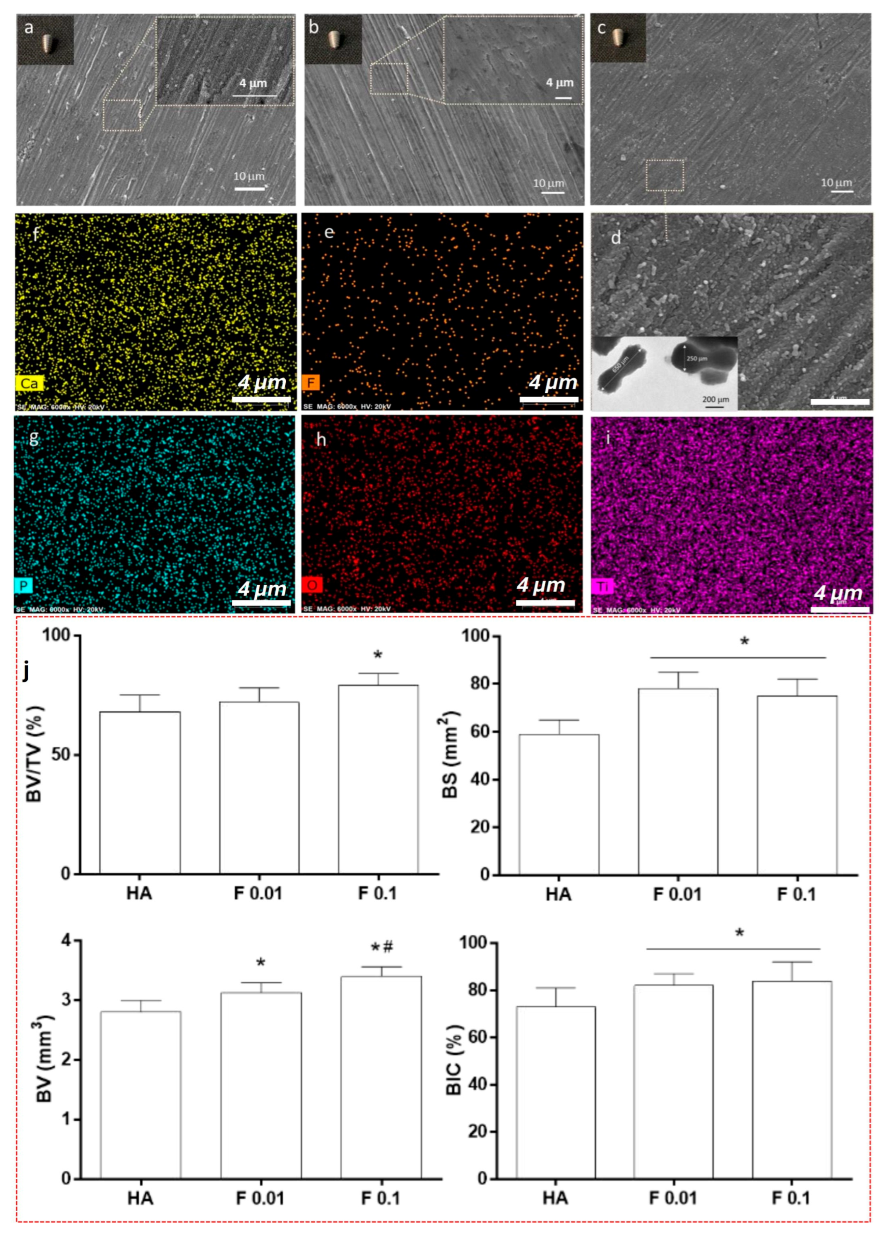
Santiago et al. [148] demonstrated the capacity of hydrothermal processing to precisely engineer fluorapatite coatings (HA, F0.1, and F0.01) on Ti implants. Notably, their system preserved citrate species on the coating surface, which influenced crystal growth kinetics and produced homogeneous nanoscale morphologies distinct from conventional plasma-sprayed coatings (Figure 12a–d). Through controlled hydroxyl-to-fluoride substitution within the HA lattice, stoichiometric fluorapatite phases were achieved (Figure 12e–i), highlighting the chemical tunability of the method. In vivo evaluation using a rabbit tibia model showed that fluoride incorporation significantly enhanced peri-implant bone formation. Histomorphometric and micro-CT analyses at 4 weeks revealed increased bone volume (BV), bone surface (BS), and bone-to-implant contact (BIC), particularly for F0.1, with sustained improvements observed at 8 weeks (Figure 12j) [148]. These findings underscore a dose-dependent biological response, where moderate fluoride substitution optimises early osseointegration outcomes.
Similarly, Tang et al. [149] developed zinc-containing nanocluster architectures on Ti via a two-step hydrothermal process. By varying reaction time, they achieved distinct nanocluster morphologies that consistently increased surface hydrophilicity and energy while enabling sustained Zn ion release. Across all variants, enhanced cell adhesion, proliferation, and osteogenic differentiation were observed, indicating that nanoscale morphological control can translate into robust biological performance. Further extending the versatility of this approach, Cheng et al. [150] employed a low-temperature microwave-assisted hydrothermal oxidation technique to modify microrough Ti surfaces. This hybrid strategy preserved microscale topography while introducing nanoscale features, producing synergistic hierarchical surfaces with markedly improved wettability.
Hydrothermal approaches are frequently presented as a straightforward route to highly adherent, bioactive coatings; however, the literature reveals a more complex reality. While strong interfacial bonding and chemical tunability are clear advantages, the process remains highly sensitive to subtle variations in reaction conditions, often leading to inconsistencies in phase composition and coating uniformity. Many reported biological benefits are derived from controlled experimental settings, with limited long-term comparative clinical validation. Additionally, scalability and process standardisation remain significant challenges. Although the ability to tailor ionic composition and nanostructure is compelling, the relative contribution of these modifications to sustained clinical performance is not yet fully resolved, indicating that further systematic and translational studies are required.

4.3.3. Anodisation

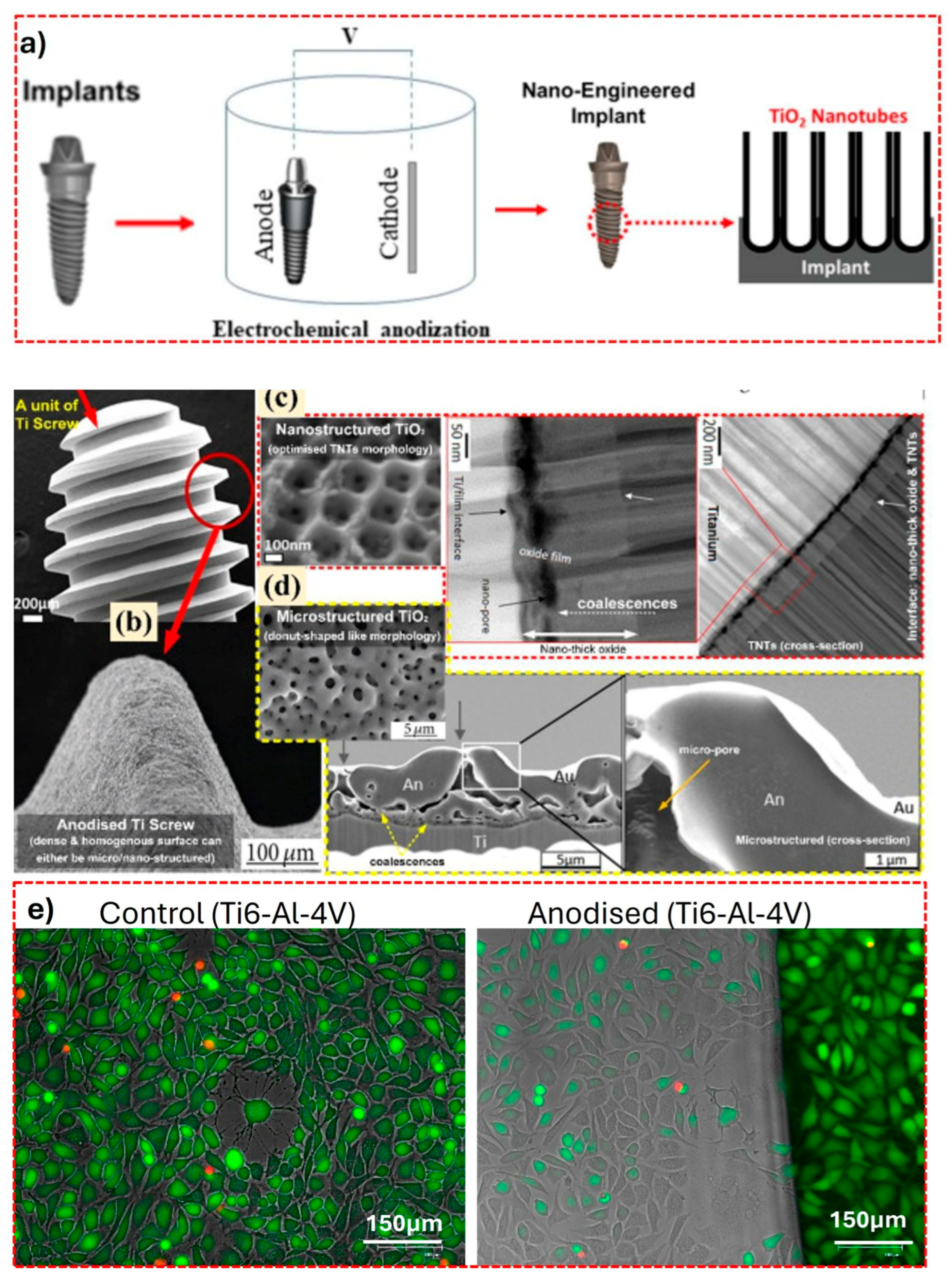
Anodisation is a hybrid surface modification technique that simultaneously alters surface topography and forms a controlled oxide layer, providing both surface treatment and coating functions for enhanced biological and mechanical performance. Anodisation is widely regarded as a foundational electrochemical approach for modifying Ti dental implant surfaces, enabling the formation of controlled oxide architectures with tunable biological functionality [151]. As shown in Figure 13a, the process involves Ti acting as the anode and an inert counter-electrode as the cathode within an electrolyte under an applied potential [152]. Within the broader framework of electrochemical oxidation (ECO), anodisation represents a relatively low-energy and highly controlled regime, in which oxide growth proceeds primarily through field-assisted ion migration. This distinguishes it from more aggressive variants such as plasma electrolytic oxidation (PEO), where dielectric breakdown and plasma discharge fundamentally alter the growth mechanism and resulting surface characteristics [153].
Under these conditions, electrochemical oxidation produces a stable TiO2 layer, which can be engineered into highly ordered nanoporous or nanotubular morphologies by adjusting anodization parameters such as voltage, electrolyte composition, and treatment duration [154]. SEM observations of anodised Ti screws (Figure 13b–d) demonstrate the formation of dense, homogeneous TiO2 layers with distinct nano- and micro-scale features, including a vertically aligned nanotubular TiO2 architecture (TNT), as shown in Figure 13c, which significantly increases the surface area and surface energy. In addition to morphology, anodisation can produce distinct TiO2 crystalline phases such as anatase and rutile, which differ in surface chemistry and biological interactions [155].
The characteristic TiO2 nanotubes are typically generated via electrochemical anodisation, where Ti acts as the anode in the electrolyte, commonly ethylene glycol with NH4F and a controlled water content [156,157]. Under an applied voltage, field-assisted oxide growth and simultaneous chemical dissolution occur: an initial compact TiO2 layer forms, followed by localised pore nucleation and etching, which drives the self-organisation and vertical elongation of nanotubes at the metal/oxide and oxide/electrolyte interfaces [158]. The resulting nanotube diameter, wall thickness, and length are strongly governed by anodisation parameters, including applied potential, electrolyte composition, water content, pH, and treatment duration [157,158]. Subsequent thermal annealing enables crystallisation of the initially amorphous nanotubes into anatase or rutile phases, thereby altering surface chemistry and modulating biological responses [159]. Recent studies show that nanocrystalline anatase coatings significantly enhance osteoblast adhesion, proliferation, and early mineralisation compared with untreated surfaces and promote favourable in vitro biological responses pertinent to osseointegration [160]. Computational analyses further suggest that anatase exhibits greater biomolecular binding affinity than rutile, potentially underpinning improved cell attachment at the nano-bio interface [155].
Hadzik et al. [154] observed that nanotubular anodised surfaces facilitate cell spreading and cytoskeletal organisation compared with untreated Ti, which is consistent with the improved cell viability observed in live–dead fluorescence imaging (Figure 13e) [154]. In addition to osteogenic benefits, the intrinsic voids of anodised TiO2 nanotubes provide an effective platform for functionalisation, enabling the incorporation of antibacterial agents or bioactive ions [44,161]. For example, nanocomposite coatings combining anodised TiO2 nanotubes with silver or calcium-phosphate-based phases have demonstrated synergistic antibacterial activity and enhanced bioactivity, while maintaining cytocompatibility [162,163]. Moreover, elemental mapping of anodised surfaces reveals a homogeneous distribution of the constituent elements across the modified surface. The anodization process generates intermediate oxide layers enriched with hydroxyl (–OH) and Ti–O functional groups, which can promote strong electrostatic attractions and hydrogen-bonding interactions with subsequently deposited layers or biological molecules, further supporting their clinical relevance [164,165].
From the authors’ perspective, anodisation provides a high degree of control over surface nano- and micro-topography and enables the development of biologically active and multifunctional implant surfaces. Anodization has been employed in Nobel Biocare TiUnite™ and TiUltra™ implants, Zürich, Switzerland, and it generates a controlled, porous TiO2 layer that enhances early bone anchorage. This surface modification translates directly into clinical benefits, including predictable implant stability, accelerated healing, and long-term success in dental implant therapy [166]. However, its limitations in terms of coating thickness, mechanical robustness, and scalability must be critically acknowledged. Future work should therefore focus not only on optimising anodisation parameters, but also on integrating it with other strategies to achieve more robust, multifunctional, and clinically translatable surface designs.
Figure 13. (a) Schematic of a conventional anodization system employed to produce titania (TiO2) nanotube structures on implant surfaces. Reproduced with permission from ref [92] (Copyright © 2021 American Chemical Society). (b) SEM images of Anodised Ti screw, and (c) nanostructure and (d) microstructures of TiO2. Adapted with permission from [164] (Copyright © 2021 Elsevier), adapted from ref [167] (Copyright © 2021 Taylor & Francis, under CC BY 4.0 licence) and ref [168] (Copyright © 2015 Wiley, under CC BY 3.0 licence), [169] (Copyright © 2018 with permission from Elsevier), [170], (Copyright © 2017 with permission Elsevier), (e) live–dead fluorescence micrographs of cells cultured with Anodised Ti and control. Viable cells appear green and nonviable cells red. Images acquired at 20× magnification (EVOS FL). Reproduced from [154] (Copyright 2023, MDPI, distributed under the terms of CC BY licence).
Coatings 16 00423 g013

5. Comparative Analysis of Surface Modification Strategies

The preceding sections highlight the breadth of available surface modification strategies for Ti implants; however, when considered collectively, it becomes evident that no single approach simultaneously optimises all functional requirements. Rather, each technique represents a distinct compromise across interdependent parameters, including surface roughness control, chemical functionalisation, antibacterial performance, drug delivery capability, mechanical stability, manufacturability, and clinical maturity [39,40,41].
Surface roughness control varies substantially across techniques. Subtractive methods such as sandblasting and acid etching offer relatively simple and scalable routes to microroughness [96]; however, they lack precise control over feature uniformity and spatial organisation. SLA improves this by introducing hierarchical structuring yet still operates within a stochastic roughness regime. In contrast, laser ablation and anodisation provide significantly higher precision, enabling controlled micro- and nanoscale architectures, including ordered grooves and nanotubes [91,164]. Hybrid techniques such as hydrothermal treatment and sol–gel processing further extend this control by enabling nanoscale crystallite growth, although often at the expense of reproducibility. Thus, while traditional methods dominate in consistency at the microscale, advanced techniques offer superior design flexibility at smaller length scales.
Chemical functionalisation represents a key differentiator often underemphasised in earlier-generation methods. Sandblasting and SLA primarily modify topography, with no control over surface chemistry and a risk of contamination (e.g., residual Al2O3). Acid etching partially addresses this by cleaning and activating the surface, but it remains chemically non-specific. In contrast, anodisation, plasma treatments, and hydrothermal approaches enable the formation of chemically active oxide layers enriched with functional groups (e.g., –OH), significantly enhancing protein adsorption and bioactivity [164]. Coating-based strategies, particularly sol–gel and layer-by-layer (LbL) assembly, offer the highest degree of chemical tunability, allowing incorporation of ions, biomolecules, and polymers [128]. However, this flexibility introduces additional complexity in maintaining coating stability and reproducibility.
Antibacterial performance reveals a fundamental divergence in design philosophy. Conventional roughening techniques (e.g., sandblasting, SLA) may inadvertently increase bacterial adhesion due to micro-scale surface features, highlighting a critical biological trade-off [43]. In contrast, advanced strategies increasingly adopt either bactericidal (e.g., ion-doped coatings, antibiotic-loaded nanospray systems) or anti-adhesive (e.g., piezoelectric nanocomposites) approaches [136]. Laser-induced nanostructures and anodised nanotubes also demonstrate intrinsic antibacterial effects through membrane disruption or surface energy modulation [90,115]. Notably, non-toxic coating strategies may offer longer-term advantages by reducing the risk of antimicrobial resistance, although their clinical validation remains limited.
Drug delivery capability is largely absent in conventional surface treatments but becomes a defining advantage of coating-based and hybrid systems. Nanospray drying, LbL assembly, anodised nanotubes, and sol–gel coatings enable the incorporation and controlled release of therapeutic agents, including antibiotics, growth factors, and ions [80,85,129]. While this multifunctionality is frequently highlighted as a major advancement, the authors note that release kinetics remain difficult to standardise and burst release or premature depletion continues to challenge clinical translation. Consequently, drug-eluting systems should be viewed as promising but not yet fully mature solutions.
Mechanical stability and adhesion strength remain critical constraints, particularly for coating-based approaches. Subtractive methods (sandblasting, SLA) and anodisation benefit from strong substrate integration, resulting in excellent mechanical reliability [99,156]. Plasma-sprayed coatings, although historically limited by delamination risks, have improved significantly with modern process control and remain clinically viable [171]. In contrast, LbL and nanospray coatings often suffer from limited mechanical robustness and long-term durability. Hybrid techniques such as hydrothermal processing offer improved interfacial bonding through in situ growth mechanisms yet still require further validation under cyclic loading conditions representative of the oral environment.
Manufacturability and scalability further differentiate established and emerging technologies. Sandblasting, acid etching, and SLA remain dominant in clinical practice largely due to their simplicity, cost-effectiveness, and compatibility with industrial-scale production [73]. Plasma spraying also retains relevance due to its established manufacturing infrastructure. Conversely, techniques such as laser ablation, LbL assembly, and nanospray drying, while technologically advanced, face challenges related to throughput, cost, and process standardisation [80,88]. Hydrothermal and sol–gel methods occupy an intermediate position, offering scalability potential but requiring tighter control over processing conditions to ensure reproducibility.
Clinical maturity ultimately reflects the cumulative impact of these factors. SLA and its derivatives (e.g., SLActive) remain the clinical benchmark, supported by extensive long-term data and consistent outcomes [57,111]. Plasma-sprayed coatings and anodised surfaces also demonstrate strong clinical track records, although their use is more selective [54,172]. In contrast, LbL systems, nanospray coatings, piezoelectric nanocomposites, and many drug-eluting platforms remain largely experimental, with limited in vivo validation and minimal regulatory translation. This disparity underscores a persistent gap between laboratory innovation and clinical implementation.
Taken together, this comparative analysis reinforces a central argument: implant surface optimisation cannot be reduced to a single dominant parameter such as roughness. Instead, successful design requires a balanced integration of topographical, chemical, biological, and mechanical factors. Future progress will depend less on the development of entirely new techniques and more on the intelligent integration of existing ones into hybrid, multifunctional systems. However, this must be accompanied by improved standardisation, reproducibility, and clinically relevant validation frameworks to ensure that technological advances translate into tangible patient benefit.

6. Prospects for Future Research

Looking forward, the next phase of development in Ti implant surface engineering should move decisively toward multifunctional and adaptive systems. Rather than serving as passive substrates, future implant surfaces should be designed to actively modulate the peri-implant environment by simultaneously promoting osteogenesis, preventing bacterial colonisation, and regulating host immune responses. The integration of antimicrobial agents, immunomodulatory cues, and controlled drug delivery mechanisms represents a path in the right direction, but these features must be engineered in a way that ensures durability and predictable performance over time.
Another key priority is the development of patient-specific implant surfaces. The variability in patients’ systemic health, bone quality, and oral microbiome suggests that a “one-size-fits-all” approach is inherently limited. Advances in digital dentistry, additive manufacturing, and biomaterials design provide a realistic pathway toward customised surface architectures tailored to individual clinical conditions. From the authors’ perspective, this shift toward personalisation represents one of the most impactful yet underexplored opportunities in implantology.
At the same time, translational challenges must be addressed more explicitly. Many emerging surface technologies remain confined to in vitro or preclinical stages, with insufficient long-term clinical evidence to support widespread adoption. Regulatory constraints, cost-effectiveness, and manufacturing reproducibility are not peripheral concerns but central determinants of whether a technology can realistically reach clinical practice. Future research must therefore prioritise not only biological performance but also scalability, standardisation, and regulatory compatibility.
In addition, there is a growing opportunity to explore stimuli-responsive and dynamic surfaces enabled by advances in nanotechnology and smart materials. Surfaces capable of responding to environmental changes such as pH, mechanical loading, or inflammatory signals could offer adaptive functionality during different stages of healing. While conceptually compelling, these systems require careful validation to ensure safety and long-term stability in the complex oral environment.
The dental implantology field is at a transitional stage; the foundational principles of surface modification are well established, but their evolution into clinically transformative technologies will depend on a shift toward integrative, patient-centred, and translationally viable solutions. Continued multidisciplinary collaboration between materials scientists, biologists, engineers, and clinicians will be essential to realise the full potential of next-generation Ti dental implants.

7. Conclusions

This review highlights that surface modification of Ti dental implants has progressed from relatively simple topographical alterations to increasingly sophisticated strategies that integrate chemical, biological, and functional design principles. Collectively, the evidence demonstrates that modifying surface roughness, wettability, and surface chemistry plays a decisive role in enhancing osseointegration, mitigating early inflammatory responses, and improving overall clinical performance. Techniques such as sandblasting and acid etching remain foundational due to their reproducibility and long-standing clinical validation, while hydrophilic surface treatments have further refined early healing outcomes.
However, a critical evaluation of the field reveals that many currently adopted approaches are still rooted in decades-old technologies, with limited capacity for further functional advancement. While newer techniques, including laser structuring, anodisation, and nano-engineered coatings, offer greater precision and biological tunability, their clinical translation remains inconsistent. In particular, coating-based strategies, although they promise bioactive or antimicrobial agents, continue to face unresolved challenges related to long-term stability, interfacial adhesion, and manufacturing scalability. Overall, while substantial progress has been achieved, the gap between laboratory findings and reliable long-term clinical outcomes remains significant. Bridging this gap will require not only technological innovation but also more rigorous in vivo validation and a stronger alignment between material design and clinical realities.

Author Contributions

Conceptualisation, A.B. and R.B.; Data curation: A.B.; validation, G.B. and R.B.; investigation, A.B.; formal analysis, A.B.; writing—original draft preparation, A.B.; writing—review and editing, G.B., R.B. and K.K.; supervision, R.B., M.R. and K.K.; visualisation, A.B. All authors have read and agreed to the published version of the manuscript.

Funding

This study was supported by the Faculty of Technology at the University of Portsmouth.

Institutional Review Board Statement

Not applicable.

Informed Consent Statement

Not applicable.

Data Availability Statement

No new data were created or analysed in this study.

Acknowledgments

During the preparation of this manuscript, the author used DALL·E, an image generation model developed by OpenAI (accessed via ChatGPT, 2026), for the purpose of generating the graphical abstract. The generated content was subsequently reviewed, modified, and validated by the authors, who take full responsibility for the final content of this publication. The graphical abstract also incorporates elements reproduced or adapted from previously published works. Appropriate copyright acknowledgements are provided for the following sources: [173] (Copyright © 2024 permissions from American Chemical Society), [88] (Copyright 2017, Frontiers Media S.A. distributed under the terms CC BY licence, [174] (Copyright © 2024, Spinger Nature, distributed under the terms of the CC BY licence), [175] (Copyright © 2025, Springer Nature, distributed under the terms of the CC BY licence) and [94] (Copyright © 2020, Springer Nature, distributed under the terms of the CC BY licence).

Conflicts of Interest

The authors declare no competing interests.

References

  1. Li, H.; Zheng, Y.; Ji, X. Synthesis and in vitro evaluation of Ca-P coating on biodegradable Zn alloys. J. Mater. Sci. Technol. 2023, 141, 124–134. [Google Scholar] [CrossRef] [Scilit]
  2. Pandey, L.M. Design of biocompatible and self-antibacterial titanium surfaces for biomedical applications. Curr. Opin. Biomed. Eng. 2023, 25, 100423. [Google Scholar] [CrossRef] [Scilit]
  3. Wang, M.; Yan, T.; Gao, M.; Han, C.; Yan, Z.; Gao, Y.; Zhang, W.; Yi, Z. A review of the advances in implant technology: Accomplishments and challenges for the design of functionalized surface structures. Biomed. Mater. 2025, 20, 032003. [Google Scholar] [CrossRef] [Scilit] [PubMed]
  4. Ding, Y.; Tao, B.; Ma, R.; Zhao, X.; Liu, P.; Cai, K. Surface modification of titanium implant for repairing/improving microenvironment of bone injury and promoting osseointegration. J. Mater. Sci. Technol. 2023, 143, 1–11. [Google Scholar] [CrossRef] [Scilit]
  5. Bartolomeu, F.; Carvalho, O.; Gasik, M.; Silva, F.S. Multi-functional Ti6Al4V-CoCrMo implants fabricated by multi-material laser powder bed fusion technology: A disruptive material’s design and manufacturing philosophy. J. Mech. Behav. Biomed. Mater. 2023, 138, 105583. [Google Scholar] [CrossRef] [Scilit]
  6. Kalia, G.; Mishra, S.; Singh, P.; Sharma, R. A review study on biomechanical properties of biocompatible materials. Mater. Today Proc. 2022, 56, 3160–3164. [Google Scholar] [CrossRef] [Scilit]
  7. Narayanan, D.; Liu, M.; Kuttolamadom, M.; Castaneda, H. Identification and development of a new local corrosion mechanism in a Laser Engineered Net Shaped (LENS) biomedical Co-Cr-Mo alloy in Hank’s solution. Corros. Sci. 2022, 207, 110599. [Google Scholar] [CrossRef] [Scilit]
  8. Prasad, K.; Bazaka, O.; Chua, M.; Rochford, M.; Fedrick, L.; Spoor, J.; Symes, R.; Tieppo, M.; Collins, C.; Cao, A.; et al. Metallic Biomaterials: Current Challenges and Opportunities. Materials 2017, 10, 884. [Google Scholar] [CrossRef] [Scilit]
  9. Karabulut, G.; Beköz Üllen, N.; Akyüz, E.; Karakuş, S. Surface modification of 316L stainless steel with multifunctional locust gum/polyethylene glycol-silver nanoparticles using different coating methods. Prog. Org. Coat. 2023, 174, 107291. [Google Scholar] [CrossRef] [Scilit]
  10. Yang, J.; Liu, C.; Sun, H.; Liu, Y.; Liu, Z.; Zhang, D.; Zhao, G.; Wang, Q.; Yang, D. The progress in titanium alloys used as biomedical implants: From the view of reactive oxygen species. Front. Bioeng. Biotechnol. 2022, 10, 1092916. [Google Scholar] [CrossRef] [Scilit]
  11. Yan, X.; Cao, W.; Li, H. Biomedical Alloys and Physical Surface Modifications: A Mini-Review. Materials 2021, 15, 66. [Google Scholar] [CrossRef] [Scilit]
  12. Benčina, M.; Junkar, I.; Vesel, A.; Mozetič, M.; Iglič, A. Nanoporous Stainless Steel Materials for Body Implants—Review of Synthesizing Procedures. Nanomaterials 2022, 12, 2924. [Google Scholar] [CrossRef] [Scilit] [PubMed]
  13. Chacón, J.M.; Núñez, P.J.; Caminero, M.A.; García-Plaza, E.; Vallejo, J.; Blanco, M. 3D printing of patient-specific 316L–stainless–steel medical implants using fused filament fabrication technology: Two veterinary case studies. Bio-Des. Manuf. 2022, 5, 808–815. [Google Scholar] [CrossRef] [Scilit]
  14. Parau, A.C.; Juravlea, G.A.; Raczkowska, J.; Vitelaru, C.; Dinu, M.; Awsiuk, K.; Vranceanu, D.M.; Ungureanu, E.; Cotrut, C.M.; Vladescu, A. Comparison of 316L and Ti6Al4V biomaterial coated by ZrCu-based thin films metallic glasses: Structure, morphology, wettability, protein adsorption, corrosion resistance, biomineralization. Appl. Surf. Sci. 2023, 612, 155800. [Google Scholar] [CrossRef] [Scilit]
  15. Mace, A.; Khullar, P.; Bouknight, C.; Gilbert, J.L. Corrosion properties of low carbon CoCrMo and additively manufactured CoCr alloys for dental applications. Dent. Mater. 2022, 38, 1184–1193. [Google Scholar] [CrossRef] [Scilit]
  16. Akshaya, S.; Rowlo, P.K.; Dukle, A.; Nathanael, A.J. Antibacterial Coatings for Titanium Implants: Recent Trends and Future Perspectives. Antibiotics 2022, 11, 1719. [Google Scholar] [CrossRef] [Scilit]
  17. Laskowska, D.; Bałasz, B.; Kaczorowski, W.; Grabarczyk, J.; Svobodova, L.; Szatkiewicz, T.; Mitura, K. The DLC Coating on 316L Stainless Steel Stochastic Voronoi Tessellation Structures Obtained by Binder Jetting Additive Manufacturing for Potential Biomedical Applications. Coatings 2022, 12, 1373. [Google Scholar] [CrossRef] [Scilit]
  18. Benčina, M.; Rawat, N.; Lakota, K.; Sodin-Šemrl, S.; Iglič, A.; Junkar, I. Bio-Performance of Hydrothermally and Plasma-Treated Titanium: The New Generation of Vascular Stents. Int. J. Mol. Sci. 2021, 22, 11858. [Google Scholar] [CrossRef] [Scilit]
  19. Yang, M.; Chen, B.; Zhang, Y.; Zhao, Z. Feasibility of customizing titanium implant with three-dimensional CT imaging of low dose in skull. Medicine 2020, 99, e21009. [Google Scholar] [CrossRef] [Scilit]
  20. Zhang, J.; Su, Y.; Rao, X.; Pang, H.; Zhu, H.; Liu, L.; Chen, L.; Li, D.; He, J.; Peng, J.; et al. Additively manufactured polyether ether ketone (PEEK) skull implant as an alternative to titanium mesh in cranioplasty. Int. J. Bioprint. 2022, 9, 634. [Google Scholar] [CrossRef] [Scilit]
  21. Accioni, F.; Vázquez, J.; Merinero, M.; Begines, B.; Alcudia, A. Latest Trends in Surface Modification for Dental Implantology: Innovative Developments and Analytical Applications. Pharmaceutics 2022, 14, 455. [Google Scholar] [CrossRef] [Scilit]
  22. Sánchez-Bodón, J.; Andrade del Olmo, J.; Alonso, J.M.; Moreno-Benítez, I.; Vilas-Vilela, J.L.; Pérez-Álvarez, L. Bioactive Coatings on Titanium: A Review on Hydroxylation, Self-Assembled Monolayers (SAMs) and Surface Modification Strategies. Polymers 2021, 14, 165. [Google Scholar] [CrossRef] [Scilit]
  23. Čivljak, T.; Ban, T.; Kopić, V.; Petrović, V.; Morelato, L.; Vuletić, M.; Gabrić, D. Comparison of Hydrophilic Properties of Titanium and Zirconia Dental Implants’ Surfaces. Materials 2025, 18, 1724. [Google Scholar] [CrossRef] [Scilit] [PubMed]
  24. Terranova, M.L. Key Challenges in Diamond Coating of Titanium Implants: Current Status and Future Prospects. Biomedicines 2022, 10, 3149. [Google Scholar] [CrossRef] [Scilit] [PubMed]
  25. Posiyano, K.; Prasad, R.V.S.; Dzogbewu, T.C.; Olakanmi, E.O.; Leso, T.P.; Setswalo, K.; Sello, A.T. The potential of Ti-6Al-7Nb, and design for manufacturing considerations in mitigating failure of hip implants in service. Biomed. Eng. Adv. 2024, 8, 100136. [Google Scholar] [CrossRef] [Scilit]
  26. Fernández Silva, B.; Suárez Fernández, D.; Green, I.; Jackson, M. Digital fingerprints of microstructural variation in titanium alloy hip joint forgings via machining force feedback analysis. Mater. Charact. 2022, 192, 112198. [Google Scholar] [CrossRef] [Scilit]
  27. Yang, W.; Diao, H.; Xin, H.; Chen, W.; Wang, S.; Xue, L.; Jin, Z.; Li, H.; He, X. Wear assessment of a Ti–6Al–4V motion-preserving porous artificial-cervical-joint fabricated by SLM after surface carburization. Ceram. Int. 2022, 48, 26137–26146. [Google Scholar] [CrossRef] [Scilit]
  28. Joshi, V.M.; Kandaswamy, E. Bibliometric Analysis of Dental Implantology Journals from 1991–2023. Int. J. Oral Maxillofac. Implant. 2023, 39, e193–e202. [Google Scholar] [CrossRef] [Scilit]
  29. Sun, X.-D.; Liu, T.-T.; Wang, Q.-Q.; Zhang, J.; Cao, M.-S. Surface Modification and Functionalities for Titanium Dental Implants. ACS Biomater. Sci. Eng. 2023, 9, 4442–4461. [Google Scholar] [CrossRef] [Scilit]
  30. López-Valverde, N.; Aragoneses, J.; López-Valverde, A.; Quispe-López, N.; Rodríguez, C.; Aragoneses, J.M. Effectiveness of biomolecule-based bioactive surfaces, on os-seointegration of titanium dental implants: A systematic review and meta-analysis of in vivo studies. Front. Bioeng. Biotechnol. 2022, 10, 986112. [Google Scholar] [CrossRef] [Scilit]
  31. Zhang, Y.; Fan, Z.; Xing, Y.; Jia, S.; Mo, Z.; Gong, H. Effect of microtopography on osseointegration of implantable biomaterials and its modification strategies. Front. Bioeng. Biotechnol. 2022, 10, 981062. [Google Scholar] [CrossRef] [Scilit] [PubMed]
  32. Wang, Z.; Zhang, J.; Hu, J.; Yang, G. Gene-activated titanium implants for gene delivery to enhance osseointegration. Biomater. Adv. 2022, 143, 213176. [Google Scholar] [CrossRef] [Scilit] [PubMed]
  33. Neto, J.V.C.; Teixeira, A.B.V.; Cândido dos Reis, A. Hydroxyapatite coatings versus osseointegration in dental implants: A systematic review. J. Prosthet. Dent. 2023, 134, 92–99. [Google Scholar] [CrossRef] [Scilit] [PubMed]
  34. Kadirvelu, L.; Sivaramalingam, S.S.; Jothivel, D.; Chithiraiselvan, D.D.; Karaiyagowder Govindarajan, D.; Kandaswamy, K. A review on antimicrobial strategies in mitigating biofilm-associated infections on medical implants. Curr. Res. Microb. Sci. 2024, 6, 100231. [Google Scholar] [CrossRef] [Scilit]
  35. Labis, V.; Bazikyan, E.; Sizova, S.; Oleinikov, V.; Trulioff, A.; Serebriakova, M.; Kudryavtsev, I.; Zhigalina, O.; Khmelenin, D.; Dyachkova, I.; et al. Immunopathological Inflammation in the Evolution of Mucositis and Peri-Implantitis. Int. J. Mol. Sci. 2022, 23, 15797. [Google Scholar] [CrossRef] [Scilit]
  36. Wang, Y.-C.; Lin, S.-H.; Chien, C.-S.; Kung, J.-C.; Shih, C.-J. In Vitro Bioactivity and Antibacterial Effects of a Silver-Containing Mesoporous Bioactive Glass Film on the Surface of Titanium Implants. Int. J. Mol. Sci. 2022, 23, 9291. [Google Scholar] [CrossRef] [Scilit]
  37. Rakic, M.; Radunovic, M.; Petkovic-Curcin, A.; Tatic, Z.; Basta-Jovanovic, G.; Sanz, M. Study on the immunopathological effect of titanium particles in peri-implantitis granulation tissue: A case–control study. Clin. Oral Implant. Res. 2022, 33, 656–666. [Google Scholar] [CrossRef] [Scilit]
  38. Jin, S.; Yu, Y.; Zhang, T.; Xie, D.; Zheng, Y.; Wang, C.; Liu, Y.; Xia, D. Surface modification strategies to reinforce the soft tissue seal at transmucosal region of dental implants. Bioact. Mater. 2024, 42, 404–432. [Google Scholar] [CrossRef] [Scilit]
  39. Jayasree, A.; Gómez-Cerezo, M.N.; Verron, E.; Ivanovski, S.; Gulati, K. Gallium-doped dual micro-nano titanium dental implants towards soft-tissue integration and bactericidal functions. Mater. Today Adv. 2022, 16, 100297. [Google Scholar] [CrossRef] [Scilit]
  40. Costa, R.C.; Nagay, B.E.; Dini, C.; Borges, M.H.R.; Miranda, L.F.B.; Cordeiro, J.M.; Souza, J.G.S.; Sukotjo, C.; Cruz, N.C.; Barão, V.A.R. The race for the optimal antimicrobial surface: Perspectives and challenges related to plasma electrolytic oxidation coating for titanium-based implants. Adv. Colloid Interface Sci. 2023, 311, 102805. [Google Scholar] [CrossRef] [Scilit]
  41. Trueba, P.; Navarro, C.; Giner, M.; Rodríguez-Ortiz, J.A.; Montoya-García, M.J.; Delgado-Pujol, E.J.; Rodríguez-Albelo, L.M.; Torres, Y. Approach to the Fatigue and Cellular Behavior of Superficially Modified Porous Titanium Dental Implants. Materials 2022, 15, 3903. [Google Scholar] [CrossRef] [Scilit] [PubMed]
  42. Lim, B.-S.; Cho, H.-R.; Choe, H.-C. Corrosion behaviors of macro/micro/nano-scale surface modification on Ti-6Al-4V alloy for bio-implant. Thin Solid Films 2022, 754, 139314. [Google Scholar] [CrossRef] [Scilit]
  43. Romero-Serrano, M.; Romero-Ruiz, M.-M.; Herrero-Climent, M.; Rios-Carrasco, B.; Gil-Mur, J. Correlation between Implant Surface Roughness and Implant Stability: A Systematic Review. Dent. J. 2024, 12, 276. [Google Scholar] [CrossRef] [Scilit] [PubMed]
  44. Thukkaram, M.; Coryn, R.; Asadian, M.; Esbah Tabaei, P.S.; Rigole, P.; Rajendhran, N.; Nikiforov, A.; Sukumaran, J.; Coenye, T.; Van Der Voort, P.; et al. Fabrication of Microporous Coatings on Titanium Implants with Improved Mechanical, Antibacterial, and Cell-Interactive Properties. ACS Appl. Mater. Interfaces 2020, 12, 30155–30169. [Google Scholar] [CrossRef] [Scilit] [PubMed]
  45. Teulé-Trull, M.; Altuna, P.; Arregui, M.; Rodriguez-Ciurana, X.; Aparicio, C. Antibacterial coatings for dental implants: A systematic review. Dent. Mater. 2025, 41, 229–247. [Google Scholar] [CrossRef] [Scilit]
  46. Huang, H.; Chang, Y.; Weng, J.; Chen, Y.; Lai, C.; Shieh, T. Anti-bacterial performance of Zirconia coatings on Titanium implants. Thin Solid Films 2013, 528, 151–156. [Google Scholar] [CrossRef] [Scilit]
  47. Li, J.; Jansen, J.A.; Walboomers, X.F.; van den Beucken, J.J.J.P. Mechanical aspects of dental implants and osseointegration: A narrative review. J. Mech. Behav. Biomed. Mater. 2020, 103, 103574. [Google Scholar] [CrossRef] [Scilit]
  48. Kligman, S.; Ren, Z.; Chung, C.-H.; Perillo, M.A.; Chang, Y.-C.; Koo, H.; Zheng, Z.; Li, C. The Impact of Dental Implant Surface Modifications on Osseointegration and Biofilm Formation. J. Clin. Med. 2021, 10, 1641. [Google Scholar] [CrossRef] [Scilit]
  49. Zheng, Z.; Gan, S.; Yang, S.; Hou, C.; Zhu, Z.; Wang, H.; Yu, D.; Qian, Z.; Xu, H.H.K.; Chen, W. Enhanced surface hydrophilicity improves osseointegration of titanium implants via integrin-mediated osteoimmunomodulation. J. Mater. Chem. B 2025, 13, 496–510. [Google Scholar] [CrossRef] [Scilit]
  50. Dehghanpour, A.A.; Rouhi, G. A Critical Review on the Effects of Dental Implants Micro-roughness on Osseointegration. Biomed. Mater. Devices 2025. [Google Scholar] [CrossRef] [Scilit]
  51. Osman, M.A.; Alamoush, R.A.; Kushnerev, E.; Seymour, K.G.; Shawcross, S.; Yates, J.M. In-Vitro Phenotypic Response of Human Osteoblasts to Different Degrees of Titanium Surface Roughness. Dent. J. 2022, 10, 140. [Google Scholar] [CrossRef] [Scilit]
  52. Osman, M.A.; Alamoush, R.A.; Kushnerev, E.; Seymour, K.G.; Watts, D.C.; Yates, J.M. Biological response of epithelial and connective tissue cells to titanium surfaces with different ranges of roughness: An in-vitro study. Dent. Mater. 2022, 38, 1777–1788. [Google Scholar] [CrossRef] [Scilit] [PubMed]
  53. Bose, S.; Banerjee, D.; Shivaram, A.; Tarafder, S.; Bandyopadhyay, A. Calcium phosphate coated 3D printed porous titanium with nanoscale surface modification for orthopedic and dental applications. Mater. Des. 2018, 151, 102–112. [Google Scholar] [CrossRef] [Scilit] [PubMed]
  54. Kowalski, S.; Gonciarz, W.; Belka, R.; Góral, A.; Chmiela, M.; Lechowicz, Ł.; Kaca, W.; Żórawski, W. Plasma-Sprayed Hydroxyapatite Coatings and Their Biological Properties. Coatings 2022, 12, 1317. [Google Scholar] [CrossRef] [Scilit]
  55. Gabric, D.; Reiser, Y.; Barišić, B.; Čivljak, T.; Ban, T.; Marković, L. Hydrophilic Properties of Dental Implant Surfaces: Literature Review. J. Dent. 2025, 13, 26–34. [Google Scholar] [CrossRef] [Scilit]
  56. Osak, P.; Maszybrocka, J.; Zubko, M.; Rak, J.; Bogunia, S.; Łosiewicz, B. Influence of Sandblasting Process on Tribological Properties of Titanium Grade 4 in Artificial Saliva for Dentistry Applications. Materials 2021, 14, 7536. [Google Scholar] [CrossRef] [Scilit]
  57. Adhitya, K.; Mustika, T.; Manawan, M.; Ulfah, I.M.; Hanafi, R.; Setyadi, I.; Arif Hidayat, S.; Wibisono, M.; Sah, J. Optimizing surface properties in pure titanium for dental implants: A crystallographic analysis of sandblasting and acid-etching techniques. Powder Diffr. 2024, 39, 206–216. [Google Scholar] [CrossRef] [Scilit]
  58. Pae, H.-C.; Kim, S.-K.; Park, J.-Y.; Song, Y.W.; Cha, J.-K.; Paik, J.-W.; Choi, S.H. Bioactive characteristics of an implant surface coated with a pH buffering agent: An in vitro study. J. Periodontal Implant. Sci. 2019, 49, 366–381. [Google Scholar] [CrossRef] [Scilit]
  59. Wennerberg, A.; Jimbo, R.; Stübinger, S.; Obrecht, M.; Dard, M.; Berner, S. Nanostructures and hydrophilicity influence osseointegration: A biomechanical study in the rabbit tibia. Clin. Oral Implant. Res. 2014, 25, 1041–1050. [Google Scholar] [CrossRef] [Scilit]
  60. Yang, D.H.; Lee, D.-W.; Kwon, Y.-D.; Kim, H.J.; Chun, H.J.; Jang, J.W.; Khang, G. Surface modification of titanium with hydroxyapatite–heparin–BMP-2 enhances the efficacy of bone formation and osseointegration in vitro and in vivo. J. Tissue Eng. Regen. Med. 2015, 9, 1067–1077. [Google Scholar] [CrossRef] [Scilit]
  61. Wiśniewska, B.; Spychała, S.; Piekarski, K.; Golusińska-Kardach, E.; Stelmachowska-Banaś, M.; Wyganowska, M. Dental Implantology in Acromegaly: Pathophysiological Challenges, Biomaterial Interactions, and Future Directions—A Narrative Review. J. Funct. Biomater. 2025, 16, 411. [Google Scholar] [CrossRef] [Scilit] [PubMed]
  62. Braem, A.; Chaudhari, A.; Vivan Cardoso, M.; Schrooten, J.; Duyck, J.; Vleugels, J. Peri- and intra-implant bone response to microporous Ti coatings with surface modification. Acta Biomater. 2014, 10, 986–995. [Google Scholar] [CrossRef] [Scilit] [PubMed]
  63. Jose, B.; Antoci, V.; Zeiger, A.R.; Wickstrom, E.; Hickok, N.J. Vancomycin Covalently Bonded to Titanium Beads Kills Staphylococcus aureus. Chem. Biol. 2005, 12, 1041–1048. [Google Scholar] [CrossRef] [Scilit] [PubMed]
  64. Lv, H.; Chen, Z.; Yang, X.; Cen, L.; Zhang, X.; Gao, P. Layer-by-layer self-assembly of minocycline-loaded chitosan/alginate multilayer on titanium substrates to inhibit biofilm formation. J. Dent. 2014, 42, 1464–1472. [Google Scholar] [CrossRef] [Scilit]
  65. Stuart, B.W.; Stan, G.E.; Popa, A.C.; Carrington, M.J.; Zgura, I.; Necsulescu, M.; Grant, D.M. New solutions for combatting implant bacterial infection based on silver nano-dispersed and gallium incorporated phosphate bioactive glass sputtered films: A preliminary study. Bioact. Mater. 2022, 8, 325–340. [Google Scholar] [CrossRef] [Scilit]
  66. Li, J.; Liu, X.; Qiao, Y.; Zhu, H.; Ding, C. Antimicrobial activity and cytocompatibility of Ag plasma-modified hierarchical TiO2 film on titanium surface. Colloids Surf. B Biointerfaces 2014, 113, 134–145. [Google Scholar] [CrossRef] [Scilit]
  67. Oleshko, O.; Liubchak, I.; Husak, Y.; Korniienko, V.; Yusupova, A.; Oleshko, T.; Banasiuk, R.; Szkodo, M.; Matros-Taranets, I.; Kazek-Kęsik, A.; et al. In vitro biological characterization of silver-doped anodic oxide coating on titanium. Materials 2020, 13, 4359. [Google Scholar] [CrossRef] [Scilit]
  68. van Hengel, I.A.J.; Tierolf, M.W.A.M.; Fratila-Apachitei, L.E.; Apachitei, I.; Zadpoor, A.A. Antibacterial Titanium Implants Biofunctionalized by Plasma Electrolytic Oxidation with Silver, Zinc, and Copper: A Systematic Review. Int. J. Mol. Sci. 2021, 22, 3800. [Google Scholar] [CrossRef] [Scilit]
  69. Keegan, G.M.; Smart, J.D.; Ingram, M.J.; Barnes, L.-M.; Burnett, G.R.; Rees, G.D. Chitosan microparticles for the controlled delivery of fluoride. J. Dent. 2012, 40, 229–240. [Google Scholar] [CrossRef] [Scilit]
  70. Pawlik, A.; Rehman, M.A.U.; Nawaz, Q.; Bastan, F.E.; Sulka, G.D.; Boccaccini, A.R. Fabrication and characterization of electrophoretically deposited chitosan-hydroxyapatite composite coatings on anodic titanium dioxide layers. Electrochim. Acta 2019, 307, 465–473. [Google Scholar] [CrossRef] [Scilit]
  71. Zhang, T.; Zhang, X.; Mao, M.; Li, J.; Wei, T.; Sun, H. Chitosan/hydroxyapatite composite coatings on porous Ti6Al4V titanium implants: In vitro and in vivo studies. J. Periodontal Implant. Sci. 2020, 50, 392–405. [Google Scholar] [CrossRef] [Scilit] [PubMed]
  72. Zhang, T.; Qin, X.; Gao, Y.; Kong, D.; Jiang, Y.; Cui, X.; Guo, M.; Chen, J.; Chang, F.; Zhang, M.; et al. Functional chitosan gel coating enhances antimicrobial properties and osteogenesis of titanium alloy under persistent chronic inflammation. Front. Bioeng. Biotechnol. 2023, 11, 1118487. [Google Scholar] [CrossRef] [Scilit] [PubMed]
  73. Bayrak, M.; Kocak-Oztug, N.A.; Gulati, K.; Cintan, S.; Cifcibasi, E. Influence of Clinical Decontamination Techniques on the Surface Characteristics of SLA Titanium Implant. Nanomaterials 2022, 12, 4481. [Google Scholar] [CrossRef] [Scilit] [PubMed]
  74. Mangano, F.G.; Pires, J.T.; Shibli, J.A.; Mijiritsky, E.; Iezzi, G.; Piattelli, A.; Mangano, C. Early Bone Response to Dual Acid-Etched and Machined Dental Implants Placed in the Posterior Maxilla. Implant. Dent. 2017, 26, 24–29. [Google Scholar] [CrossRef] [Scilit]
  75. Fang, K.; Shen, Y.; Ru Yie, K.H.; Zhou, Z.; Cai, L.; Wu, S.; Al-Bishari, A.M.; Al-Baadani, M.A.; Shen, X.; Ma, P.; et al. Preparation of Zirconium Hydrogen Phosphate Coatings on Sandblasted/Acid-Etched Titanium for Enhancing Its Osteoinductivity and Friction/Corrosion Resistance. Int. J. Nanomed. 2021, 16, 8265–8277. [Google Scholar] [CrossRef] [Scilit]
  76. Buser, D.; Janner, S.F.M.; Wittneben, J.; Brägger, U.; Ramseier, C.A.; Salvi, G.E. 10-Year Survival and Success Rates of 511 Titanium Implants with a Sandblasted and Acid-Etched Surface: A Retrospective Study in 303 Partially Edentulous Patients. Clin. Implant. Dent. Relat. Res. 2012, 14, 839–851. [Google Scholar] [CrossRef] [Scilit]
  77. Abdul Rani, R.; Jamil, N.S.; Fadzilah Suhaimi, M.H.; Zoolfakar, A.S.; Khusaimi, Z.; Mahmood, M.R. Influence of Zinc Acetate Concentration on ZnO Growth on Anodized Nb2O5 Nanoporous Films and Photocatalytic Dye Degradation. Arab. J. Sci. Eng. 2023, 48, 9009–9022. [Google Scholar] [CrossRef] [Scilit]
  78. Carek, A.; Slokar Benić, L.; Buković, D.; Šlaj, M. Investigation of Laser-Welded Co-Cr Dental Alloys by Microscopy and Mechanical Testing. Metals 2023, 13, 1323. [Google Scholar] [CrossRef] [Scilit]
  79. Kedia, S.; Das, A.; Patro, B.S.; Nilaya, J.P. Potential tribological and antibacterial benefits of pulsed laser-deposited zirconia thin film on Ti6Al4V bio-alloy. Appl. Phys. A 2022, 128, 670. [Google Scholar] [CrossRef] [Scilit]
  80. Baghdan, E.; Raschpichler, M.; Lutfi, W.; Pinnapireddy, S.R.; Pourasghar, M.; Schäfer, J.; Schneider, M.; Bakowsky, U. Nano spray dried antibacterial coatings for dental implants. Eur. J. Pharm. Biopharm. 2019, 139, 59–67. [Google Scholar] [CrossRef] [Scilit]
  81. Salehi, S.; Kharaziha, M.; Salehi, M. Multifunctional plasma-sprayed nanocomposite coating based on FA-ZnO-GO with improved bioactivity and wear behaviour. Surf. Coat. Technol. 2020, 404, 126472. [Google Scholar] [CrossRef] [Scilit]
  82. Udduttula, A.; Li, J.; Ma, Z.; Teng, B.; Zhang, J.V.; Ferreira, A.M.; Gentile, P.; Wang, G.; Zhao, X.; Ren, P. A novel apatite-inspired Sr5(PO4)2SiO4 plasma-sprayed coating on Ti alloy promoting biomineralization, osteogenesis and angiogenesis. Ceram. Int. 2022, 48, 10979–10989. [Google Scholar] [CrossRef] [Scilit]
  83. Xie, M.; Zhong, Y.; Wang, S.; Tian, L.; Wang, X.; Hu, Y.; Zeng, R. EGCG/Zn coating on titanium implants by one-step hydrothermal method for improving anticorrosion, antibacterial and osteogenesis properties. Mater. Chem. Phys. 2022, 292, 126872. [Google Scholar] [CrossRef] [Scilit]
  84. Fu, Z.; Deng, X.; Fang, X. Effect of addition of Ca2+ to titanium by a hydrothermal method on soft tissue sealing. Microsc. Res. Tech. 2022, 85, 3050–3055. [Google Scholar] [CrossRef] [Scilit] [PubMed]
  85. García-Arnáez, I.; Cerqueira, A.; Romero-Gavilán, F.; Elortza, F.; Azkargorta, M.; Iloro, I.; Suay, J.; Goñi, I.; Gurruchaga, M. Development and characterisation of strontium-doped sol-gel coatings to optimise the initial bone regeneration processes. Mater. Today Commun. 2022, 33, 104674. [Google Scholar] [CrossRef] [Scilit]
  86. Azari, R.; Rezaie, H.R.; Khavandi, A. Effect of titanium dioxide intermediate layer on scratch and corrosion resistance of sol–gel-derived HA coating applied on Ti-6Al-4V substrate. Prog. Biomater. 2021, 10, 259–269. [Google Scholar] [CrossRef] [Scilit]
  87. Zhao, J.; Jin, S.; Delgado, A.H.; Chen, Z.; Matinlinna, J.P.; Tsoi, J.K.-H. Self-Assembled PHMB Titanium Coating Enables Anti-Fusobacterium nucleatum Strategy. Coatings 2021, 11, 1190. [Google Scholar] [CrossRef] [Scilit]
  88. Shi, Q.; Qian, Z.; Liu, D.; Liu, H. Surface Modification of Dental Titanium Implant by Layer-by-Layer Electrostatic Self-Assembly. Front. Physiol. 2017, 8, 574. [Google Scholar] [CrossRef] [Scilit]
  89. Schünemann, F.H.; Galárraga-Vinueza, M.E.; Magini, R.; Fredel, M.; Silva, F.; Souza, J.C.M.; Zhang, Y.; Henriques, B. Zirconia surface modifications for implant dentistry. Mater. Sci. Eng. C 2019, 98, 1294–1305. [Google Scholar] [CrossRef] [Scilit]
  90. Civantos, A.; Martínez-Campos, E.; Ramos, V.; Elvira, C.; Gallardo, A.; Abarrategi, A. Titanium Coatings and Surface Modifications: Toward Clinically Useful Bioactive Implants. ACS Biomater. Sci. Eng. 2017, 3, 1245–1261. [Google Scholar] [CrossRef] [Scilit]
  91. Veiko, V.; Karlagina, Y.; Zernitckaia, E.; Egorova, E.; Radaev, M.; Yaremenko, A.; Chernenko, G.; Romanov, V.; Shchedrina, N.; Ivanova, E.; et al. Laser-Induced µ-Rooms for Osteocytes on Implant Surface: An In Vivo Study. Nanomaterials 2022, 12, 4229. [Google Scholar] [CrossRef] [Scilit] [PubMed]
  92. Gulati, K.; Zhang, Y.; Di, P.; Liu, Y.; Ivanovski, S. Research to Clinics: Clinical Translation Considerations for Anodized Nano-Engineered Titanium Implants. ACS Biomater. Sci. Eng. 2022, 8, 4077–4091. [Google Scholar] [CrossRef] [Scilit]
  93. Bright, R.; Hayles, A.; Wood, J.; Ninan, N.; Palms, D.; Visalakshan, R.M.; Burzava, A.; Brown, T.; Barker, D.; Vasilev, K. Bio-Inspired Nanostructured Ti-6Al-4V Alloy: The Role of Two Alkaline Etchants and the Hydrothermal Processing Duration on Antibacterial Activity. Nanomaterials 2022, 12, 1140. [Google Scholar] [CrossRef] [Scilit] [PubMed]
  94. Wu, Y.-L.; Tsai, M.-H.; Chen, H.-S.; Chang, Y.-T.; Lin, T.-T.; Wu, A.Y.-J. Biomechanical effects of original equipment manufacturer and aftermarket abutment screws in zirconia abutment on dental implant assembly. Sci. Rep. 2020, 10, 18406. [Google Scholar] [CrossRef] [Scilit] [PubMed]
  95. Baghdan, E.; Pinnapireddy, S.R.; Vögeling, H.; Schäfer, J.; Eckert, A.W.; Bakowsky, U. Nano spray drying: A novel technique to prepare well-defined surface coatings for medical implants. J. Drug Deliv. Sci. Technol. 2018, 48, 145–151. [Google Scholar] [CrossRef] [Scilit]
  96. Liu, Q.; Zhao, Y.; Hao, J.; Qiu, L.; Su, H.; Wang, X.; Su, Y. Preparation of high-quality pure titanium SLA surface and evaluation of its physicochemical and biological properties—In vitro experiments. BMC Oral. Health 2025, 25, 1220. [Google Scholar] [CrossRef] [Scilit]
  97. Villapun Puzas, V.M.; Carter, L.N.; Schröder, C.; Colavita, P.E.; Hoey, D.A.; Webber, M.A.; Addison, O.; Shepherd, D.E.T.; Attallah, M.M.; Grover, L.M.; et al. Surface Free Energy Dominates the Biological Interactions of Postprocessed Additively Manufactured Ti-6Al-4V. ACS Biomater. Sci. Eng. 2022, 8, 4311–4326. [Google Scholar] [CrossRef] [Scilit]
  98. Romero-Serrano, M.; Romero-Ruiz, M.M.; Ríos-Santos, J.V.; Ríos-Carrasco, B.; Gil, J. Influence of alumina shot blasting induced roughness on bacterial adhesion to titanium. Clin. Oral. Investig. 2025, 29, 497. [Google Scholar] [CrossRef] [Scilit]
  99. Akbas, O.; Gaikwad, A.; Reck, L.; Ehlert, N.; Jahn, A.; Hermsdorf, J.; Winkel, A.; Stiesch, M.; Greuling, A. Effects of sandblasting and acid etching on the surface properties of additively manufactured and machined titanium and their consequences for osteoblast adhesion under different storage conditions. Front. Bioeng. Biotechnol. 2025, 13, 1640122. [Google Scholar] [CrossRef] [Scilit]
  100. Ayub, F.A.; Sunarso, S.; Dewi, R.S. Influence of Acid Composition on Morphology and Physical Properties of Titanium Alloy. F1000Research 2025, 14, 216. [Google Scholar] [CrossRef] [Scilit]
  101. Mandracci, P.; Mussano, F.; Rivolo, P.; Carossa, S. Surface Treatments and Functional Coatings for Biocompatibility Improvement and Bacterial Adhesion Reduction in Dental Implantology. Coatings 2016, 6, 7. [Google Scholar] [CrossRef] [Scilit]
  102. Gautam, H.; Yadav, G.; Bansal, R. Implant surface modification as a basis of osseointegration: A narrative review. J. Dent. Def. Sect. 2022, 16, 139. [Google Scholar] [CrossRef] [Scilit]
  103. Herrero-Climent, M.; Lázaro, P.; Vicente Rios, J.; Lluch, S.; Marqués, M.; Guillem-Martí, J.; Gil, F.J. Influence of acid-etching after grit-blasted on osseointegration of titanium dental implants: In vitro and in vivo studies. J. Mater. Sci. Mater. Med. 2013, 24, 2047–2055. [Google Scholar] [CrossRef] [Scilit] [PubMed]
  104. Faverani, L.P.; Assunção, W.G.; de Carvalho, P.S.P.; Yuan, J.C.-C.; Sukotjo, C.; Mathew, M.T.; Barao, V.A. Effects of Dextrose and Lipopolysaccharide on the Corrosion Behavior of a Ti-6Al-4V Alloy with a Smooth Surface or Treated with Double-Acid-Etching. PLoS ONE 2014, 9, e93377. [Google Scholar] [CrossRef] [Scilit]
  105. Velasco-Ortega, E.; Ortiz-García, I.; Jiménez-Guerra, A.; Monsalve-Guil, L.; Muñoz-Guzón, F.; Perez, R.A.; Gil, F.J. Comparison between Sandblasted Acid-Etched and Oxidized Titanium Dental Implants: In Vivo Study. Int. J. Mol. Sci. 2019, 20, 3267. [Google Scholar] [CrossRef] [Scilit]
  106. Gil, J.; Sanz, M. Bactericidal nanotopography of titanium dental implants: In vitro and in vivo studies. Clin. Oral Investig. 2025, 29, 351. [Google Scholar] [CrossRef] [Scilit]
  107. Wu, Y.; Wan, K.; Lu, J.; Yuan, C.; Cui, Y.; Duan, R.; Yu, J. Research Progress on Surface Modification of Titanium Implants. Coatings 2025, 15, 229. [Google Scholar] [CrossRef] [Scilit]
  108. Velasco-Ortega, E.; Pato-Mourelo, J.; López-López, B.; Monsalve-Guil, L.; Moreno-Muñoz, J.; López-López, J.; Núñez-Márquez, E.; Matos Garrido, N.; Rondón-Romero, J.L.; Jiménez-Guerra, Á.; et al. Long-Term Clinical Study on Sandblasted–Acid-Etched Surface Dental Implants: 12-Year Follow-Up. Materials 2025, 18, 183. [Google Scholar] [CrossRef] [Scilit]
  109. Jeon, J.-H.; Kim, M.-J.; Yun, P.-Y.; Jo, D.-W.; Kim, Y.-K. Randomized clinical trial to evaluate the efficacy and safety of two types of sandblasted with large-grit and acid-etched surface implants with different surface roughness. J. Korean Assoc. Oral Maxillofac. Surg. 2022, 48, 225–231. [Google Scholar] [CrossRef] [Scilit]
  110. Thu, M.K.; Kang, Y.S.; Kwak, J.M.; Jo, Y.-H.; Han, J.-S.; Yeo, I.-S.L. Comparison between bone–implant interfaces of microtopographically modified zirconia and titanium implants. Sci. Rep. 2023, 13, 11142. [Google Scholar] [CrossRef] [Scilit]
  111. Łosiewicz, B.; Osak, P.; Nowińska, D.; Maszybrocka, J. Developments in Dental Implant Surface Modification. Coatings 2025, 15, 109. [Google Scholar] [CrossRef] [Scilit]
  112. Parisi, L.; Ghezzi, B.; Bianchi, M.G.; Toffoli, A.; Rossi, F.; Bussolati, O.; Macaluso, G.M. Titanium dental implants hydrophilicity promotes preferential serum fibronectin over albumin competitive adsorption modulating early cell response. Mater. Sci. Eng. C 2020, 117, 111307. [Google Scholar] [CrossRef] [Scilit] [PubMed]
  113. Kim, M.-J.; Kim, I.; Chang, N.-H.; Kim, Y.-K. Long-term evaluation of the prognosis of super hydrophilic surface treated CA implants: A retrospective clinical study. BMC Oral. Health 2022, 22, 97. [Google Scholar] [CrossRef] [Scilit] [PubMed]
  114. Saran, R.; Ginjupalli, K.; George, S.D.; Chidangil, S.; Unnikrishnan, V.K. LASER as a tool for surface modification of dental biomaterials: A review. Heliyon 2023, 9, e17457. [Google Scholar] [CrossRef] [Scilit] [PubMed]
  115. Maruszczyk, A.; Dudek, A.; Szala, M. Research into Morphology and Properties of TiO2—NiAl Atmospheric Plasma Sprayed Coating. Adv. Sci. Technol. Res. J. 2017, 11, 204–210. [Google Scholar] [CrossRef] [Scilit]
  116. Dorcioman, G.; Grumezescu, V.; Stan, G.E.; Chifiriuc, M.C.; Gradisteanu, G.P.; Miculescu, F.; Matei, E.; Popescu-Pelin, G.; Zgura, I.; Craciun, V.; et al. Hydroxyapatite Thin Films of Marine Origin as Sustainable Candidates for Dental Implants. Pharmaceutics 2023, 15, 1294. [Google Scholar] [CrossRef] [Scilit]
  117. Gonçalves, I.M.R.; Herrero, E.R.; Carvalho, O.; Henriques, B.; Silva, F.S.; Teughels, W.; Souza, J.C.M. Antibiofilm effects of titanium surfaces modified by laser texturing and hot-pressing sintering with silver. J. Biomed. Mater. Res. B Appl. Biomater. 2021, 109, 1588–1600. [Google Scholar] [CrossRef] [Scilit]
  118. Kolarovszki, B.; Ficsor, S.; Frank, D.; Katona, K.; Soos, B.; Turzo, K. Unlocking the potential: Laser surface modifications for titanium dental implants. Lasers Med. Sci. 2024, 39, 162. [Google Scholar] [CrossRef] [Scilit]
  119. Mehl, C.; Kern, M.; Neumann, F.; Bähr, T.; Wiltfang, J.; Gassling, V. Effect of ultraviolet photofunctionalization of dental titanium implants on osseointegration. J. Zhejiang Univ.-Sci. B 2018, 19, 525–534. [Google Scholar] [CrossRef] [Scilit]
  120. Dini, C.; Nagay, B.E.; Cordeiro, J.M.; da Cruz, N.C.; Rangel, E.C.; Ricomini-Filho, A.P.; de Avila, E.D.; Barão, V.A.R. UV-photofunctionalization of a biomimetic coating for dental implants application. Mater. Sci. Eng. C 2020, 110, 110657. [Google Scholar] [CrossRef] [Scilit]
  121. Nakhaei, K.; Ishijima, M.; Ikeda, T.; Ghassemi, A.; Saruta, J.; Ogawa, T. Ultraviolet Light Treatment of Titanium Enhances Attachment, Adhesion, and Retention of Human Oral Epithelial Cells via Decarbonization. Materials 2020, 14, 151. [Google Scholar] [CrossRef] [Scilit] [PubMed]
  122. Hayashi, R.; Ueno, T.; Migita, S.; Tsutsumi, Y.; Doi, H.; Ogawa, T.; Hanawa, T.; Wakabayashi, N. Hydrocarbon Deposition Attenuates Osteoblast Activity on Titanium. J. Dent. Res. 2014, 93, 698–703. [Google Scholar] [CrossRef] [Scilit] [PubMed]
  123. Hirota, M.; Ozawa, T.; Iwai, T.; Mitsudo, K.; Ogawa, T. UV-Mediated Photofunctionalization of Dental Implant: A Seven-Year Results of a Prospective Study. J. Clin. Med. 2020, 9, 2733. [Google Scholar] [CrossRef] [Scilit] [PubMed]
  124. Suzuki, S.; Kobayashi, H.; Ogawa, T. Implant Stability Change and Osseointegration Speed of Immediately Loaded Photofunctionalized Implants. Implant. Dent. 2013, 22, 481–490. [Google Scholar] [CrossRef] [Scilit]
  125. Funato, A.; Ogawa, T. Photofunctionalized Dental Implants: A Case Series in Compromised Bone. Int. J. Oral Maxillofac. Implant. 2013, 28, 1589–1601. [Google Scholar] [CrossRef] [Scilit]
  126. Hirota, M.; Ozawa, T.; Iwai, T.; Ogawa, T.; Tohnai, I. Implant Stability Development of Photofunctionalized Implants Placed in Regular and Complex Cases: A Case-Control Study. Int. J. Oral Maxillofac. Implant. 2016, 31, 676–686. [Google Scholar] [CrossRef] [Scilit]
  127. Stephanopoulos, N.; Ortony, J.H.; Stupp, S.I. Self-assembly for the synthesis of functional biomaterials. Acta Mater. 2013, 61, 912–930. [Google Scholar] [CrossRef] [Scilit]
  128. Nawae, S.; Meesane, J.; Muensit, N.; Daengngam, C. Layer-by-layer self-assembled films of silk fibroin/collagen/poly (diallyldimethylammonium chloride) as nucleating surface for osseointegration to design coated dental implant materials. Mater. Des. 2018, 160, 1158–1167. [Google Scholar] [CrossRef] [Scilit]
  129. de Avila, E.D.; Castro, A.G.B.; Tagit, O.; Krom, B.P.; Löwik, D.; van Well, A.A.; Bannenberg, L.J.; Vergani, C.E.; van den Beucken, J.J.J.P. Anti-bacterial efficacy via drug-delivery system from layer-by-layer coating for percutaneous dental implant components. Appl. Surf. Sci. 2019, 488, 194–204. [Google Scholar] [CrossRef] [Scilit]
  130. Guan, J.; Wang, J.; Jia, F.; Jiang, W.; Song, L.; Xie, L.; Yang, H.; Han, P.; Lin, H.; Wu, Z.; et al. Layer-by-layer self-assembly coatings on strontium titanate nanotubes with antimicrobial and anti-inflammatory properties to prevent implant-related infections. Colloids Surf. B Biointerfaces 2024, 244, 114183. [Google Scholar] [CrossRef] [Scilit]
  131. Marasli, C.; Katifelis, H.; Gazouli, M.; Lagopati, N. Nano-Based Approaches in Surface Modifications of Dental Implants: A Literature Review. Molecules 2024, 29, 3061. [Google Scholar] [CrossRef] [Scilit] [PubMed]
  132. Nadian, N.; Nourouzi, S.; Jamshidi Aval, H. Plasma spray coating of HA-Ti-MgO composite on Ti6Al4V alloy substrate. Results Phys. 2025, 76, 108387. [Google Scholar] [CrossRef] [Scilit]
  133. Chuang, T.-L.; Yeh, C.-L.; Chen, C.-Y.; Chang, H.-H.; Liao, S.-C.; Lin, C.-P. Development of Dual-Functional Titanium Implant for Osseointegration and Antimicrobial Effects via Plasma Modification. Int. Dent. J. 2025, 75, 103871. [Google Scholar] [CrossRef] [Scilit]
  134. Cochran, D.L.; Jackson, J.M.; Jones, A.A.; Jones, J.D.; Kaiser, D.A.; Taylor, T.D.; Weber, H.P.; Higginbottom, F.L.; Richardson, J.R.; Oates, T. A 5-Year Prospective Multicenter Clinical Trial of Non-Submerged Dental Implants With a Titanium Plasma-Sprayed Surface in 200 Patients. J. Periodontol. 2011, 82, 990–999. [Google Scholar] [CrossRef] [Scilit] [PubMed]
  135. He, J.; He, Z.; Qin, Y.; Zhao, H.; Bi, Y. A Review of TiCN Coating Prepared by Reaction Plasma Spraying. J. Therm. Spray Technol. 2022, 31, 2280–2299. [Google Scholar] [CrossRef] [Scilit]
  136. Huang, H.; Wang, K.; Liu, X.; Liu, X.; Wang, J.; Suo, M.; Wang, H.; Chen, S.; Chen, X.; Li, Z. Piezoelectric biomaterials for providing electrical stimulation in bone tissue engineering: Barium titanate. J. Orthop. Transl. 2025, 51, 94–107. [Google Scholar] [CrossRef] [Scilit]
  137. Dhall, A.; Islam, S.; Park, M.; Zhang, Y.; Kim, A.; Hwang, G. Bimodal Nanocomposite Platform with Antibiofilm and Self-Powering Functionalities for Biomedical Applications. ACS Appl. Mater. Interfaces 2021, 13, 40379–40391. [Google Scholar] [CrossRef] [Scilit]
  138. Parau, A.C.; Büyüksungur, S.; Li, G.; Liu, Q.; Badillo, E.; Blum, L.; Schmidt, J.; Pana, I.; Vitelaru, C.; Marinescu, I.M.; et al. Zn-doped CaP coating equips Ti implants with corrosion resistance, biomineralization, antibacterial and immunotolerant activities. J. Adv. Res. 2025, 77, 689–704. [Google Scholar] [CrossRef] [Scilit]
  139. Hwang, G.; Ahn, I.-S.; Mhin, B.J.; Kim, J.-Y. Adhesion of nano-sized particles to the surface of bacteria: Mechanistic study with the extended DLVO theory. Colloids Surf. B Biointerfaces 2012, 97, 138–144. [Google Scholar] [CrossRef] [Scilit]
  140. Metwally, S.; Stachewicz, U. Surface potential and charges impact on cell responses on biomaterials interfaces for medical applications. Mater. Sci. Eng. C 2019, 104, 109883. [Google Scholar] [CrossRef] [Scilit]
  141. Gautam, S.; Bhatnagar, D.; Bansal, D.; Batra, H.; Goyal, N. Recent advancements in nanomaterials for biomedical implants. Biomed. Eng. Adv. 2022, 3, 100029. [Google Scholar] [CrossRef] [Scilit]
  142. Jaafar, A.; Hecker, C.; Árki, P.; Joseph, Y. Sol-Gel Derived Hydroxyapatite Coatings for Titanium Implants: A Review. Bioengineering 2020, 7, 127. [Google Scholar] [CrossRef] [Scilit]
  143. Qu, J.; Lu, X.; Li, D.; Ding, Y.; Leng, Y.; Weng, J.; Qu, S.; Feng, B.; Watari, F. Silver/hydroxyapatite composite coatings on porous titanium surfaces by sol-gel method. J. Biomed. Mater. Res. B Appl. Biomater. 2011, 97B, 40–48. [Google Scholar] [CrossRef] [Scilit] [PubMed]
  144. Catauro, M.; Barrino, F.; Blanco, I.; Piccolella, S.; Pacifico, S. Use of the Sol–Gel Method for the Preparation of Coatings of Titanium Substrates with Hydroxyapatite for Biomedical Application. Coatings 2020, 10, 203. [Google Scholar] [CrossRef] [Scilit]
  145. Song, X.; Segura-Egea, J.J.; Díaz-Cuenca, A. Sol–Gel Technologies to Obtain Advanced Bioceramics for Dental Therapeutics. Molecules 2023, 28, 6967. [Google Scholar] [CrossRef] [Scilit]
  146. Lo, Y.-S.; Chang, C.-C.; Lin, P.-C.; Lin, S.-P.; Wang, C.-L. Direct growth of structurally controllable hydroxyapatite coating on Ti-6Al-4V through a rapid hydrothermal synthesis. Appl. Surf. Sci. 2021, 556, 149672. [Google Scholar] [CrossRef] [Scilit]
  147. Moloodi, A.; Toraby, H.; Kahrobaee, S.; Razavi, M.K.; Salehi, A. Evaluation of fluorohydroxyapatite/strontium coating on titanium implants fabricated by hydrothermal treatment. Prog. Biomater. 2021, 10, 185–194. [Google Scholar] [CrossRef] [Scilit]
  148. Santiago, E.; Martin, V.; Colaço, B.; Fernandes, M.H.; Santos, C.; Gomes, P.S. Hydrothermal Synthesis of Fluorapatite Coatings over Titanium Implants for Enhanced Osseointegration—An In Vivo Study in the Rabbit. J. Funct. Biomater. 2022, 13, 241. [Google Scholar] [CrossRef] [Scilit]
  149. Tang, Z.; Su, S.; Liu, Y.; Zhu, W.; Zhang, S.; Qiu, J. Hydrothermal Synthesis of Zinc-Incorporated Nano-Cluster Structure on Titanium Surface to Promote Osteogenic Differentiation of Osteoblasts and hMSCs. Front. Mater. 2021, 8, 739071. [Google Scholar] [CrossRef] [Scilit]
  150. Cheng, A.; Goodwin, W.B.; deGlee, B.M.; Gittens, R.A.; Vernon, J.P.; Hyzy, S.L.; Schwartz, Z.; Sandhage, K.H.; Boyan, B.D. Surface modification of bulk titanium substrates for biomedical applications via low-temperature microwave hydrothermal oxidation. J. Biomed. Mater. Res. A 2018, 106, 782–796. [Google Scholar] [CrossRef] [Scilit]
  151. Mühl, A.; Szabó, P.; Krafcsik, O.; Aigner, Z.; Kopniczky, J.; Nagy, Á.; Marada, G.; Turzó, K. Comparison of surface aspects of turned and anodized titanium dental implant, or abutment material for an optimal soft tissue integration. Heliyon 2022, 8, e10263. [Google Scholar] [CrossRef] [Scilit] [PubMed]
  152. Gulati, K.; Chopra, D.; Kocak-Oztug, N.A.; Verron, E. Fit and forget: The future of dental implant therapy via nanotechnology. Adv. Drug Deliv. Rev. 2023, 199, 114900. [Google Scholar] [CrossRef] [Scilit] [PubMed]
  153. Kyrylenko, S.; Sowa, M.; Kazek-Kęsik, A.; Stolarczyk, A.; Pisarek, M.; Husak, Y.; Korniienko, V.; Deineka, V.; Moskalenko, R.; Matuła, I.; et al. Nitrilotriacetic Acid Improves Plasma Electrolytic Oxidation of Titanium for Biomedical Applications. ACS Appl. Mater. Interfaces 2023, 15, 19863–19876. [Google Scholar] [CrossRef] [Scilit]
  154. Hadzik, J.; Kubasiewicz-Ross, P.; Gębarowski, T.; Waloszczyk, N.; Maciej, A.; Stolarczyk, A.; Gedrange, T.; Dominiak, M.; Szajna, E.; Simka, W. An Experimental Anodized Titanium Surface for Transgingival Dental Implant Elements—Preliminary Report. J. Funct. Biomater. 2023, 14, 34. [Google Scholar] [CrossRef] [Scilit] [PubMed]
  155. Tarjányi, T.; Bogár, F.; Minárovits, J.; Gajdács, M.; Tóth, Z. Interaction of biomolecules with anatase, rutile and amorphous TiO2 surfaces: A molecular dynamics study. PLoS ONE 2023, 18, e0289467. [Google Scholar] [CrossRef] [Scilit]
  156. Ke, C.; Ma, J.; Ni, J.; Peng, Z. A study of the growth mechanism of large-diameter double-wall TiO2 nanotube arrays fabricated by high voltage anodization. Ann. Transl. Med. 2023, 11, 18. [Google Scholar] [CrossRef] [Scilit]
  157. Jędrzejewska, A.; Arkusz, K. Mechanism and growth kinetics of hexagonal TiO2 nanotubes with an influence of anodizing parameters on morphology and physical properties. Sci. Rep. 2024, 14, 24721. [Google Scholar] [CrossRef] [Scilit]
  158. Vásquez, F.A.; Aguirre Ocampo, R.; Bedoya, N.; Zuleta Gil, A.A.; Botero, C.; Gómez, M.; Castaño, J.G.; Tamayo, J.A. Electrochemical characterization of TiO2 nanotubes formed on Ti6Al4V manufactured by PBF-EB or forging. Prog. Addit. Manuf. 2025, 10, 3629–3639. [Google Scholar] [CrossRef] [Scilit]
  159. Szaniawska-Białas, E.; Brudzisz, A.; Nasir, A.; Wierzbicka, E. Recent Advances in Preparation, Modification, and Application of Free-Standing and Flow-Through Anodic TiO2 Nanotube Membranes. Molecules 2024, 29, 5638. [Google Scholar] [CrossRef] [Scilit]
  160. Mohammadnejad, L.; Theurer, A.; Alber, J.; Illing, B.; Kimmerle-Mueller, E.; Schultheiss, J.; Krajewski, S.; Rupp, F. Surface-Mediated Modulation of Different Biological Responses on Anatase-Coated Titanium. J. Funct. Biomater. 2024, 15, 29. [Google Scholar] [CrossRef] [Scilit]
  161. Wang, Z.; Li, B.; Cai, Q.; Li, X.; Yin, Z.; Li, B.; Li, Z.; Meng, W. Advances and Prospects in Antibacterial-Osteogenic Multifunctional Dental Implant Surface. Front. Bioeng. Biotechnol. 2022, 10, 921338. [Google Scholar] [CrossRef] [Scilit] [PubMed]
  162. Li, M.; Liu, Q.; Jia, Z.; Xu, X.; Shi, Y.; Cheng, Y.; Zheng, Y. Polydopamine-induced nanocomposite Ag/CaP coatings on the surface of titania nanotubes for antibacterial and osteointegration functions. J. Mater. Chem. B 2015, 3, 8796–8805. [Google Scholar] [CrossRef] [Scilit] [PubMed]
  163. Pierre, C.; Bertrand, G.; Pavy, I.; Benhamou, O.; Rey, C.; Roques, C.; Combes, C. Antibacterial Electrodeposited Copper-Doped Calcium Phosphate Coatings for Dental Implants. J. Funct. Biomater. 2023, 14, 20. [Google Scholar] [CrossRef] [Scilit] [PubMed]
  164. Alipal, J.; Lee, T.C.; Koshy, P.; Abdullah, H.Z.; Idris, M.I. Evolution of anodised titanium for implant applications. Heliyon 2021, 7, e07408. [Google Scholar] [CrossRef] [Scilit]
  165. Wang, Y.; Tao, J.; Wang, L.; He, P.; Wang, T. HA coating on titanium with nanotubular anodized TiO2 intermediate layer via electrochemical deposition. Trans. Nonferrous Met. Soc. China 2008, 18, 631–635. [Google Scholar] [CrossRef] [Scilit]
  166. Karl, M.; Albrektsson, T. Clinical Performance of Dental Implants with a Moderately Rough (TiUnite) Surface: A Meta-Analysis of Prospective Clinical Studies. Int. J. Oral Maxillofac. Implant. 2017, 32, 717–734. [Google Scholar] [CrossRef] [Scilit]
  167. Takada, S.; Hirata, E.; Sakairi, M.; Miyako, E.; Takano, Y.; Ushijima, N.; Yudasaka, M.; Iijima, S.; Yokoyamaa, A. Carbon nanohorn coating by electrodeposition accelerate bone formation on titanium implant. Artif. Cells Nanomed. Biotechnol. 2021, 49, 20–29. [Google Scholar] [CrossRef] [Scilit]
  168. Duncan, W.J.; Lee, M.-H.; Bae, T.-S.; Lee, S.-J.; Gay, J.; Loch, C. Anodisation Increases Integration of Unloaded Titanium Implants in Sheep Mandible. Biomed. Res. Int. 2015, 2015, 857969. [Google Scholar] [CrossRef] [Scilit]
  169. Li, T.; Gulati, K.; Wang, N.; Zhang, Z.; Ivanovski, S. Bridging the gap: Optimized fabrication of robust titania nanostructures on com-plex implant geometries towards clinical translation. J. Colloid Interface Sci. 2018, 529, 452–463. [Google Scholar] [CrossRef] [Scilit]
  170. Alves, S.A.; Rossi, A.L.; Ribeiro, A.R.; Werckmann, J.; Celis, J.-P.; Rocha, L.A.; Shokuhfar, T.A. first insight on the bio-functionalization mechanisms of TiO2 nanotubes with calcium, phosphorous and zinc by reverse polarization anodization. Surf. Coat. Technol. 2017, 324, 153–166. [Google Scholar] [CrossRef] [Scilit]
  171. Sun, Y.; Zhang, X.; Luo, M.; Hu, W.; Zheng, L.; Huang, R.; Greven, J.; Hildebrand, F.; Yuan, F. Plasma Spray vs. Electrochemical Deposition: Toward a Better Osteogenic Effect of Hydroxyapatite Coatings on 3D-Printed Titanium Scaffolds. Front. Bioeng. Biotechnol. 2021, 9, 705774. [Google Scholar] [CrossRef] [Scilit]
  172. Abraham, C.M. A Brief Historical Perspective on Dental Implants, Their Surface Coatings and Treatments. Open Dent. J. 2014, 8, 50–55. [Google Scholar] [CrossRef] [Scilit]
  173. Sharma, P.; Mishra, V.; Murab, S. Unlocking Osseointegration: Surface Engineering Strategies for Enhanced Dental Implant Integration. ACS Biomater. Sci. Eng. 2025, 11, 67–94. [Google Scholar] [CrossRef] [Scilit]
  174. da Rosa de Souza, P.T.; Manfro, R.; de Salles Santos, F.A.O.; Garcia, G.F.F.; Macedo, N.F.; de Macedo, B.E.S.F.; Ignácio, S.A.; Rosa, E.A.R.; de Souza, E.M.; Azevedo-Alanis, L.R. Analysis of osseointegration of implants with macrogeometries with healing chambers: A randomized clinical trial. BMC Oral Health 2024, 24, 1114. [Google Scholar] [CrossRef] [Scilit]
  175. Dionigi, C.; Nagy, G.; Derks, J.; Ichioka, Y.; Tomasi, C.; Larsson, L.; Primetzhofer, D.; Berglundh, T. Titanium micro-particles are commonly found in soft tissues surrounding dental implants. Commun. Med. 2025, 5, 78. [Google Scholar] [CrossRef] [Scilit]
Figure 1. Annual trends in publications on dental implant surface modification indexed in the Scopus database between 2005 and 2025 (data retrieved on 19 January 2026).
Coatings 16 00423 g001
Figure 2. Schematic overview of different surface modification strategies applied to Ti dental implants, encompassing both surface treatment and coating approaches. Reproduced from ref. [88] (Copyright 2017, Frontiers Media S.A. distributed under the terms CC BY licence), ref. [89] (Copyright 2019, Elsevier, with permission); ref. [90] (Copyright 2017, American Chemical Society, with permission); ref. [91] (Copyright 2022, MDPI, distributed under the terms of the CC BY licence); ref [92] (Copyright 2022, American Chemical Society, with permission); ref. [93] (Copyright 2022, MDPI, distributed under the terms of the CC BY licence), ref. [94] (distributed under the terms of the CC BY licence) and from ref. [95] (Copyright 2018, Elsevier).
Coatings 16 00423 g002
Figure 5. (a) Diagram illustrating the laser processing procedure, (b) SEM images of the laser-textured implants, showing the surface profile, magnified details, and cross-sectional view. Reproduced from ref. [91]. (Copyright 2022, MDPI, distributed under the terms of the CC BY licence).
Coatings 16 00423 g005
Figure 6. UV-Photofunctionalisation on machined Ti surfaces and early cell responses. (a) C1s XPS deconvolution showing decreased surface carbon composition after UV treatment. (b) Water contact angle measurements demonstrating significantly increased surface wettability of UV-treated Ti (▼= contact angle approaches zero, ** p < 0.01). (c) Actin cytoskeleton staining of cells cultured on untreated and UV-treated Ti at 3 and 24 h. (d,e) Quantification of cell attachment and surface coverage, respectively, showing enhanced adhesion on UV-treated Ti (* p < 0.05). (f,g) Representative fluorescence images and quantitative analysis of integrin β4 expression, indicating increased hemidesmosomal protein expression on UV-treated surfaces (* p < 0.05). (h) Relative mRNA expression of integrin β4 and laminin-5, confirming upregulated hemidesmosome-related markers on UV-treated Ti (* p < 0.05). Reproduced from [121] (Copyright 2020, MDPI).
Coatings 16 00423 g006
Figure 7. (a) Illustration of the layer-by-layer (LbL) deposition process adapted from [88] (Copyright 2017, Frontiers Media S.A. distributed under the terms CC BY licence); (b) stepwise self-assembly of multilayered [PDDA–SF–PDDA–Col]ₙ coatings on a glass substrate; (c) cross-sectional SEM micrograph of the [PDDA/SF/PDDA/Col]ₙ multilayer film along with the corresponding atomic force microscopy (AFM) image of the assembled coating. The scale bar (5 µm) is representative of all images in this panel. Reproduced with permission from [128] (Copyright 2019, Elsevier).
Coatings 16 00423 g007
Figure 8. (a) Schematic of the nanospray drying and electrostatic collection setup. (b) SEM images of Ti surfaces coated with PLGA nanolayers without and with norfloxacin (0% and 5% NFX) collected at top, middle, and bottom positions. Adapted with permission from ref [95] (Copyright 2018, Elsevier), (c) Antibacterial activity against Escherichia coli expressed as CFU/cm2; uncoated Ti served as control (* p < 0.05). Reproduced with permission from ref. [80] (Copyright 2019, Elsevier).
Coatings 16 00423 g008
Figure 10. (a) Overview of a smart BTO nanocomposite surface engineered for infection resistance and self-powered functionality. (b) Antibiofilm performance of BTO-based nanocomposite surfaces. Bacterial viability (CFU/mL) decreases with increasing BTO content. Confocal images (top and cross-sectional views) of S. mutans biofilms formed on control and 30 wt% BTO nanocomposites after 18 h. Bacteria and extracellular polysaccharides (EPS) are shown in green and red, respectively. (b) Quantitative analysis of bacterial, EPS, and total biofilm biovolume, showing significant biofilm suppression on BTO-containing surfaces: * p < 0.05, *** p < 0.001, **** p < 0.0001 (n = 3). Scale bar: 50 μm, (c) Biocompatibility assessment of BTO nanocomposite surfaces against (i) human gingival fibroblasts (ii) and keratinocytes following exposure to control and 30 wt% BTO discs, indicating no detectable cytotoxic effects. Data are presented as mean ± SD (n ≥ 3): **** p < 0.0001. Reproduced with permission from [137] (Copyright © 2021 American Chemical Society).
Coatings 16 00423 g010
Figure 11. (a) Illustration of the Sol–Gel technique, reproduced with permission from ref. [141] (Copyright 2022, Elsevier), (b) Alkaline phosphatase activity of osteoblasts cultured on Ag/HA coatings (* p < 0.05) and (c) SEM images showing osteoblast morphology and attachment on porous substrates coated with Ag/HA. The scale bar (50 µm) is representative of all SEM images in this panel. Reproduced from ref. [143] (Copyright © 2011 Wiley Periodicals, Inc.).
Coatings 16 00423 g011
Figure 12. SEM micrographs showing the surface morphologies of the HA (a), F 0.01 (b), and F 0.1 (c) coatings prepared via the hydrothermal process. (d) Enlarged SEM view of the F 0.1 coating corresponding to the boxed region in (c), with a TEM image of the F 0.1 coating particles shown in the inset. (ei) EDS elemental distribution maps of F, Ca, P, O, and Ti on the surface of the F 0.1 coating. (j) Histomorphometric analysis of the HA, F 0.01, and F 0.1 coated constructs at 4 weeks post-implantation, p < 0.05; * indicates a significant difference compared with the control, and # denotes a significant difference compared with the other experimental group. Reproduced from [148] (Copyright 2022, MDPI).
Coatings 16 00423 g012
Disclaimer/Publisher’s Note: The statements, opinions and data contained in all publications are solely those of the individual author(s) and contributor(s) and not of MDPI and/or the editor(s). MDPI and/or the editor(s) disclaim responsibility for any injury to people or property resulting from any ideas, methods, instructions or products referred to in the content.