Article

Effect of SiC Particle Contents and Size on the Microstructure and Dissolution of SiC-Hydroxyapatite Coatings

1
School of Chemical and Environmental Engineering, Hunan Institute of Technology, Hengyang 421000, China
2
Research Institute of Automobile Parts Technology, Hunan Institute of Technology, Hengyang 421000, China
3
National Joint Engineering Laboratory of Automobile Pump Parts Design and Manufacturing, Hengyang 421000, China
*
Author to whom correspondence should be addressed.
Coatings 2021, 11(10), 1166; https://doi.org/10.3390/coatings11101166
Submission received: 18 August 2021 / Revised: 16 September 2021 / Accepted: 23 September 2021 / Published: 27 September 2021
(This article belongs to the Special Issue State-of-the-Art on Coatings Research in Asia)

Abstract

:
Carbon/carbon composites, when used as bone implant materials, do not adhere well to the bone tissues because of their non-bioactive characteristics. Therefore, we electro-deposited SiC-hydroxyapatite coatings (with an ultrasound-assisted step) on carbon/carbon composites. We analyzed how the content and size of the SiC particles affected the structure, morphology, bonding strength and dissolution of the SiC-hydroxyapatite coatings. The hydroxyapatite coating dissolution properties were assessed by the released Ca2+ and the weight loss. The SiC-hydroxyapatite coating on naked carbon/carbon composites showed a more compact microstructure in comparison to the hydroxyapatite coating on carbon/carbon composites. The reasons for the changes in the microstructure and the improvement in the adhesion of the coatings on C/C were discussed. Moreover, the addition of SiC particles increased the binding strengths of the hydroxyapatite coating on C/C composite, as well as reduced the dissolution rate of the hydroxyapatite coating.

1. Introduction

The ease of designability, excellent biocompatibility and mechanical properties similar to natural bones make carbon/carbon (C/C) composites excellent bone implant materials [1,2]. However, C/C composites do not bond strongly to the bone tissues because they are non-bioactive [3]. Thus, a technology for enhancing the C/C composite bioactivity is needed. One way to achieve this is to coat C/C composites with bioactive coatings [4,5].
Hydroxyapatite (HA) possesses an excellent bioactivity and is often applied as a coating to promote the osseointegration of implant materials [6,7,8]. Wen et al. [9] hydrothermally synthesized a hydroxyapatite coating on a medical magnesium alloy surface and analyzed its corrosion resistance. Lo et al. [10] developed a straightforward hydrothermal synthesis and used a mixture of calcium hydroxide and sodium tripolyphosphate to obtain a structurally tunable hydroxyapatite coating on a Ti-6Al-4V surface. A hydroxyapatite coating was also applied to C/C composites and significantly improved their bioactivity [11,12]. For example, Xiong et al. [13] prepared adhesive hydroxyapatite coatings on C/C composites to improve their bioactivity-related properties. Liu et al. [14] synthesized a SiO2-reinforced hydroxyapatite coating on similar C/C composites by a combination of hydrothermal electrochemical deposition and hydrothermal treatment. However, hydroxyapatite coatings are brittle, which often worsens the mechanical properties of the corresponding implants. Additionally, hydroxyapatite does not bind well to C/C composites [15,16]. To solve this problem and to reinforce the hydroxyapatite coating, Li et al. [17] added TiO2 and observed an improved Young’s modulus, fracture toughness and shear strength of the resulting TiO2-HA composite coatings prepared using high-velocity oxyfuel spraying. Mihailescu et al. [18] reinforced HA by MgF2 and MgO particles and obtained a material with an excellent adherence and biological performance. Liu et al. [19] improved the HA coating bonding strength to C/C composites by applying a tree-planting interface structure. Fu et al. [20] improved the cohesion strength of a hydroxyapatite (HA) coating on C/C composites by adding SiC nanowires.
The excellent biocompatibility, wear resistance and stability of SiC make it a promising material for biological applications [21,22,23,24,25]. Particularly, the chemical inertness makes SiC useful as a material for the bearing surface of implants [3,26]. The incorporation of small amounts of SiC particles around the implants was proven to be a safe approach, capable of stimulating the growth of bone tissues [27,28,29,30]. SiC-containing coatings were also used on stents to enhance their hemocompatibility and bonding strength [30]. Vladescu et al. [31] coated HA coatings with a SiC layer and obtained a material with excellent hardness and other mechanical properties. Fu et al. [3] reported SiC nanowires which formed pinning at the HA coating/matrix interface, which in turn enhanced the bonding strength at the interface and provided a three-dimensional framework. This improved the overall HA coating cohesion.
This work demonstrated an approach to obtain an improved bonding strength between the hydroxyapatite coating and the substrate, which was achieved by incorporating SiC particles using ultrasonic-assisted electrodeposition. This paper discusses how the SiC particle sizes and contents in the coating affect the bonding strength and solubility of the HA-based composite material.

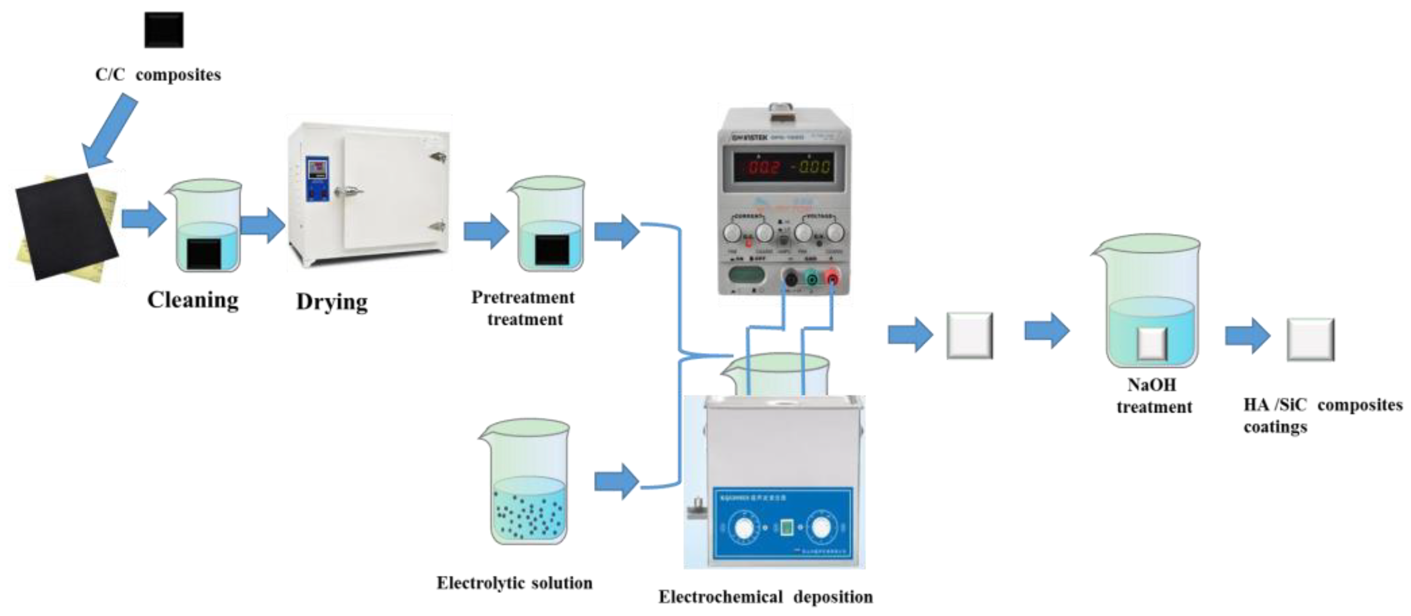
2. Materials and Methods

Several pieces (10 mm × 10 mm × 1.5 mm) were cut from a large C/C composite sheet (obtained using chemical vapor deposition) with a 1.2–1.3 g/cm3 density, polished using 600 grit SiC paper, cleaned with distilled water, dried at 100 °C for 2 h, and then soaked in H2O2 for 24 h, followed by distilled water rinsing and drying at 150 °C for 2h.
To prepare HA coatings, we used Ca(NO3)2 and 3 mmol/L NH4H2PO4 solutions in amounts taken to maintain the Ca/P mole ratio, equal to 1.67. To prepare SiC-containing hydroxyapatite, we used commercially purchased SiC (analytically pure), which was ultrasonically cleaned with ethanol for 30 min and dried at 70 °C for 24 h. SiC samples with 50, 100, and 200 nm particles were used. The contents of SiC added to hydroxyapatite synthesis were equal to 0.01, 0.1 and 1 g/L.
SiC-hydroxyapatite coatings were deposed onto the substrates electrochemically at 60 °C for 1 h under 7 V. The resulting samples were rinsed with distilled water, dried at 150 °C and then treated with 0.01 mol/L NaOH solution for 2 h, followed by rinsing and drying. All preparation steps are shown in Figure 1.
The sample crystallinity, morphology, and composition were obtained using X-ray diffraction (XRD, performed using Bruker D8ADVANCE instrument, Bilerica, Massachusetts, USA) and scanning electron microscopy (SEM, performed using Hitachi S-4800, Tokyo, Japan; Zeiss Sigma 300 setups, Jena, Germany) coupled with the energy dispersive spectroscopy. For SEM, all samples were coated with a thin layer of Au. Fourier transform infrared (FTIR, performed using IRAffinity-1s, shimadzu corporation, Kyoto, Japan) spectroscopy performed using a Shimadzu instrument was used to identify the surface functionality of the coatings.
The binding strength was tested with a scratch tester. The micron grade scratch tests were tested on micron grade scratch tester (MCT, Swiss CSM Instrument Co., Ltd., Peseux, Switzerland). For this purpose, four samples were prepared for each combination of SiC contents/particle size. The final value was reported as an average of four measurements.
The dissolution rates of the SiC-hydroxyapatite coating samples were measured using physiological saline solution (0.9% NaCl) at 36.5 °C. For this purpose, the hydroxyapatite coating pieces with a known weight were soaked for 1, 3, 5, 7, and 14 days, followed by a measuring of their Ca ion concentration and weight loss examination. For each coating composition and duration, four parallel tests were performed. After sampling 5 mL of the mother solution after 14 days of immersion, an appropriate amount of dilute nitric acid solution was added to the remaining solution. Additionally, 5 mL was of the mother solution was taken out again after the precipitates were completely dissolved. The resulting sample was named “14R”. After the dissolution tests, the Ca2+ content in the saline solution was detected by the inductively coupled plasma-mass spectrometry (ICP-MS, Thermo Tisher Scientific, Waltham, MA, USA).

3. Results

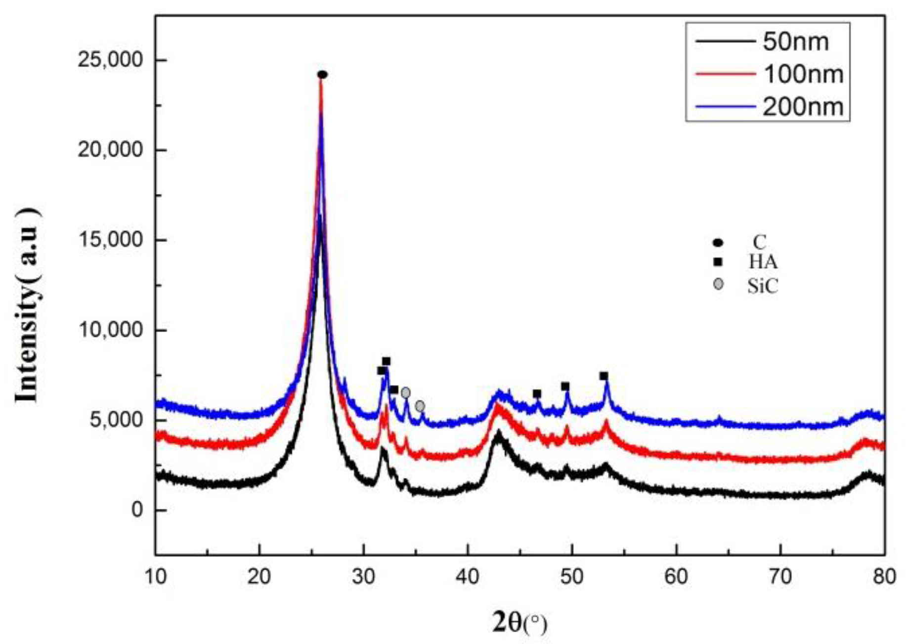
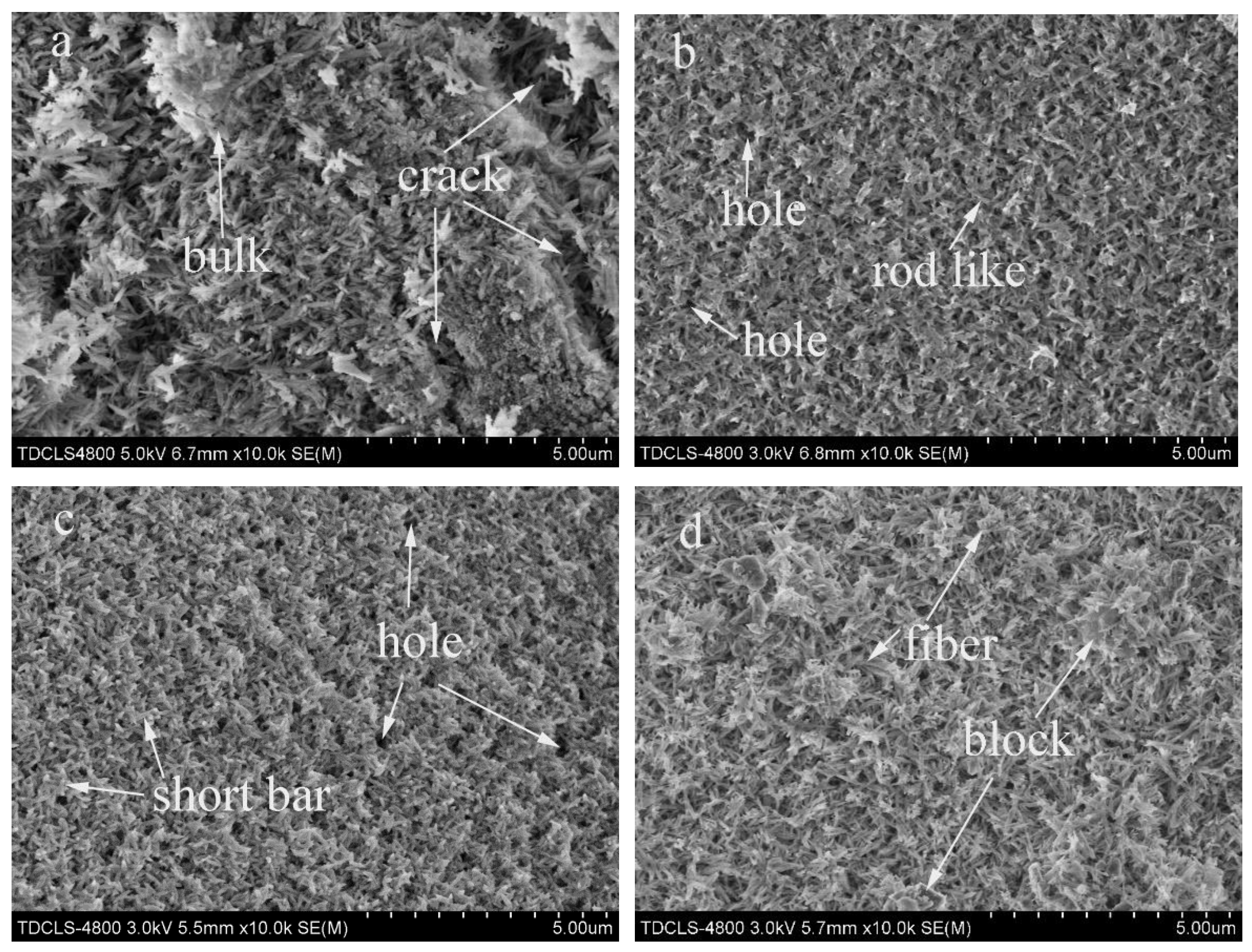
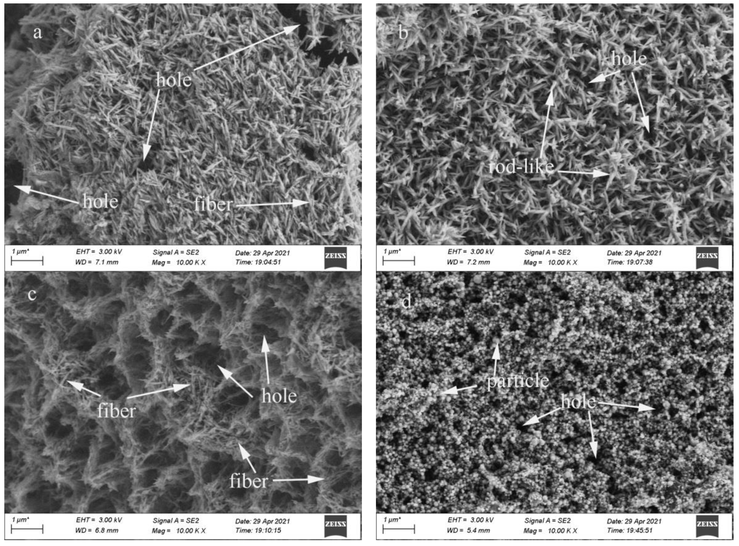
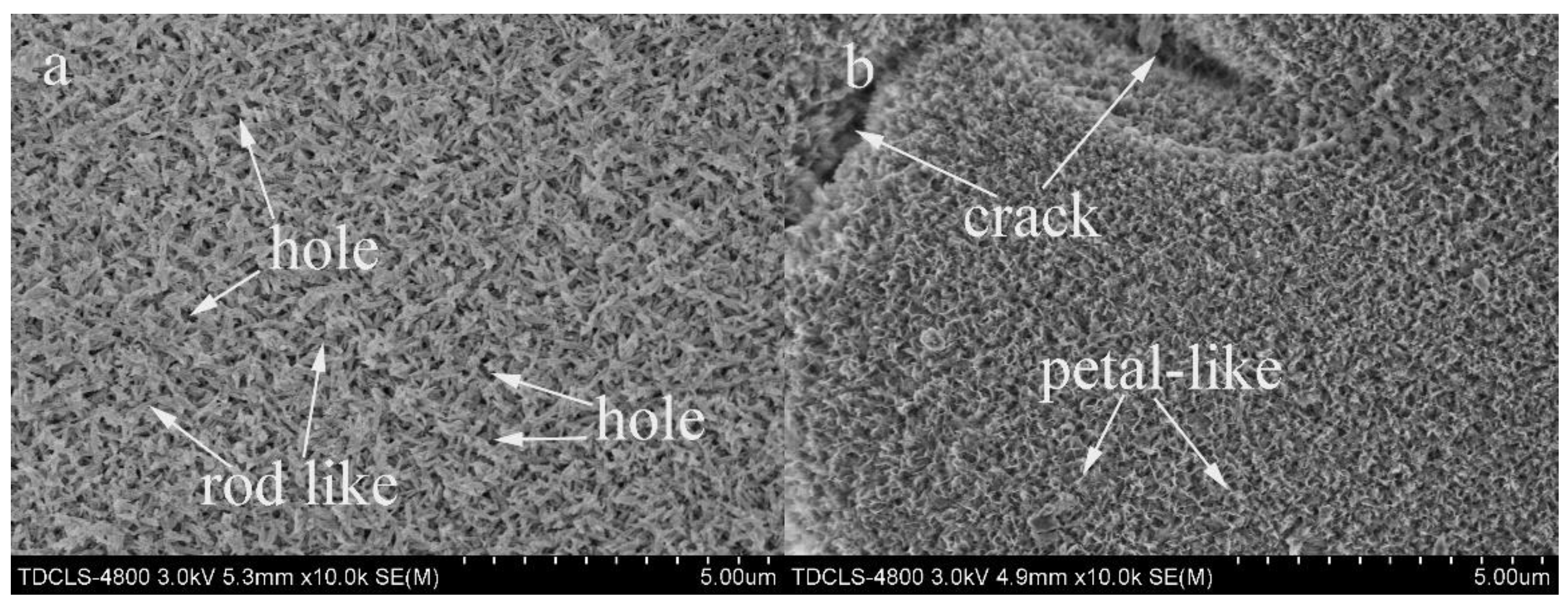
3.1. Effect of SiC Particle Sizes on the Hydroxyapatite Coating Properties

To analyze the effect of the SiC particles on the properties of the resulting hydroxyapatite coatings, we added 1 g/L SiC particles with average sizes equal to 50, 100, and 200 nm during the hydroxyapatite coating preparation. The XRD of the resulting samples showed the presence of the peaks attributed to the carbon matrix, hydroxyapatite coating and SiC particles (see Figure 2). The XRD peak intensity for the SiC phase gradually increased as the SiC particle size increased and was strongest for the hydroxyapatite coating containing 200 nm SiC particles. Additionally, as the SiC particle size increased, the intensity of the hydroxyapatite peaks increased and was the highest for the hydroxyapatite coating containing 200 nm SiC particles. No other peaks were observed in the XRD spectra of our SiC–hydroxyapatite samples. Thus, SiC and hydroxyapatite did not react strongly enough to form additional phases (see Figure 2).
The SEM of the SiC-hydroxyapatite coatings revealed some flaws (see Figure 3a). The SiC-hydroxyapatite coating containing 50 nm SiC particles contained short rod-like features and particles with numerous small holes (see Figure 3b). The coatings which contained larger SiC particles possessed a more fibrous morphology. The SiC-hydroxyapatite coatings containing 200 nm SiC particles possessed an interwoven fibrous morphology (see Figure 3d) with some lumpy tissues above the fibers.
The FTIR spectra of the SiC-hydroxyapatite coatings showed peaks at 948 and 1032 cm−1 (see Figure 4), which belonged to PO43− vibrations [31]. Additional bands at 556 and 605 cm−1 were attributed to the m4 vibrational mode of the P-O bond in the PO43− groups [32]. The band at 940–960 cm−1 was ascribed to the Si-C stretching bond [33]. The peaks in the 1460–1530 cm−1 range corresponded to the CO32− formed due to the atmospheric CO2 dissolution in the solutions during the sample preparation [34]. The bands at 1570–1640 cm−1 and 3440–3450 cm−1 were ascribed to the absorbed water [35,36] while the peak at 3570 cm−1 was ascribed to the OH vibrations.
The bonding strengths between the SiC-hydroxyapatite coatings and the C/C composite’s matrix were determined by scratch tests. The analysis of the chemical bonding strengths between the coatings and the C/C composites matrix showed that the bonding strength of the hydroxyapatite coatings significantly increased when SiC was incorporated. Additionally, the overall bonding strength increased as the SiC particle increased (see Figure 5). Thus, SiC-hydroxyapatite coatings containing 200 nm SiC particles exhibited the strongest bond between the hydroxyapatite coatings and the matrix. The bonding strengths for the SiC-hydroxyapatite coatings containing 50 and 100 nm SiC particles were almost the same. The pristine hydroxyapatite coating was loose and cracked (see Figure 3a) which explained its poor mechanical and binding strength. However, the SiC-hydroxyapatite coatings containing 200 nm SiC particles were uniform and dense (see Figure 3d), which explained why their binding strength was the highest of all the coatings.

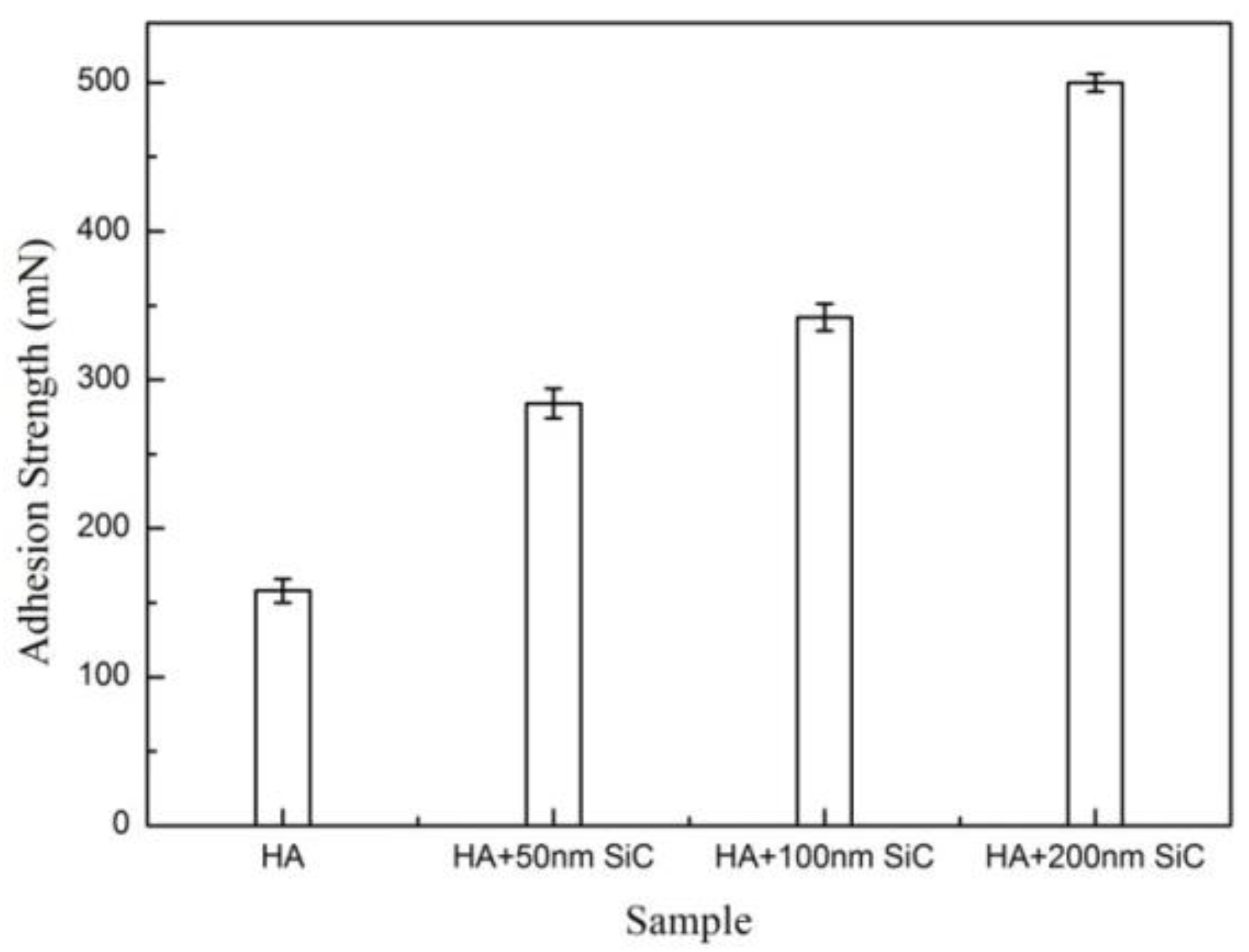
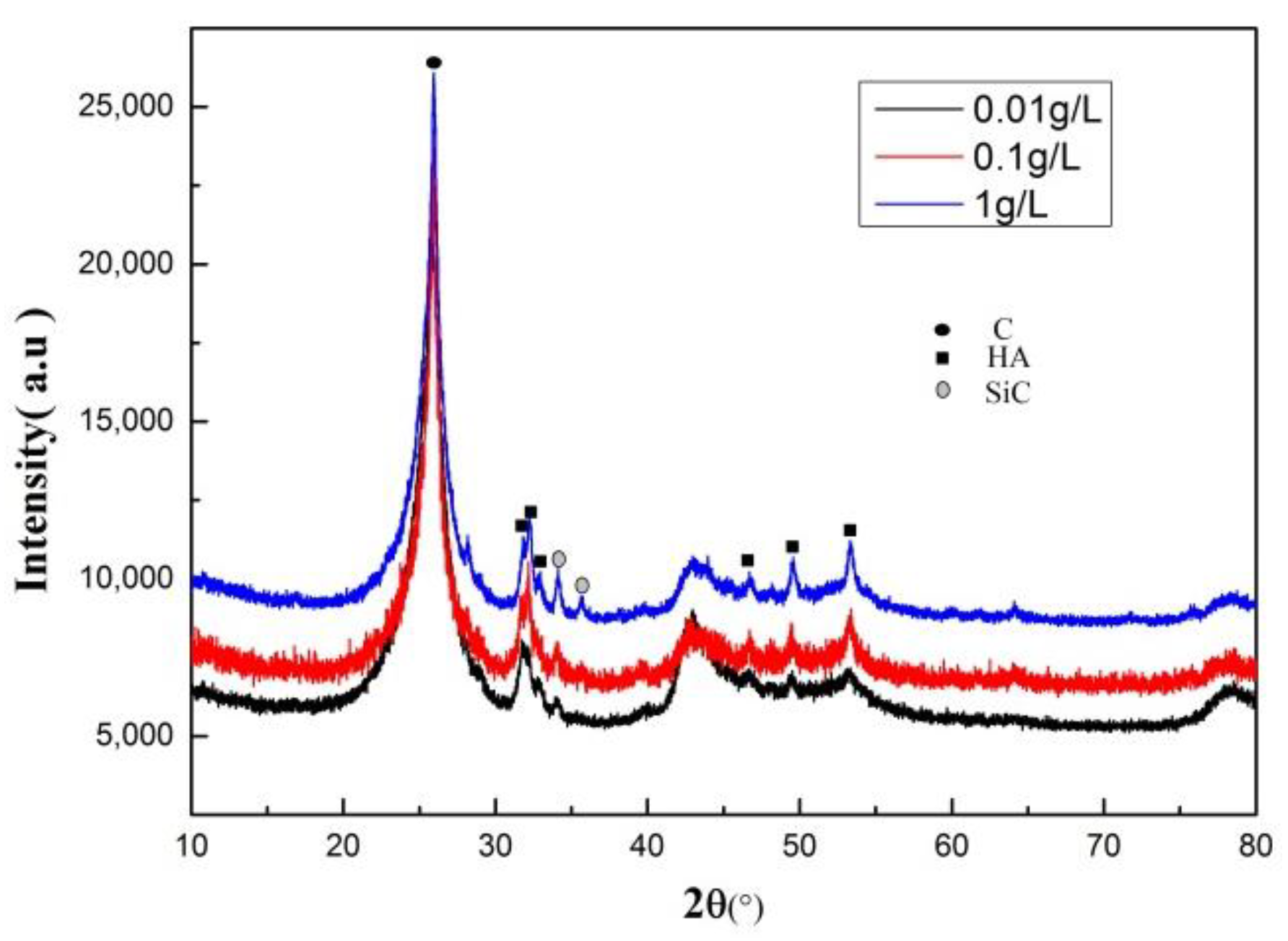
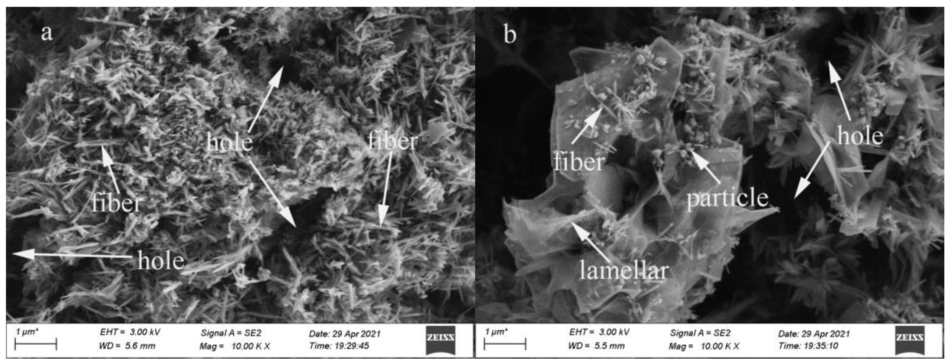
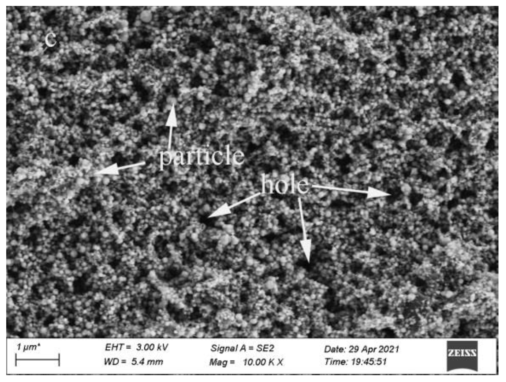
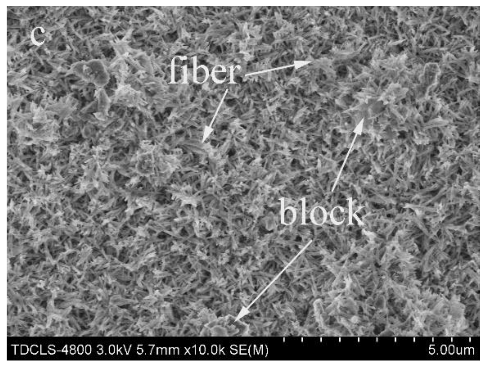
3.2. Effect of SiC Content on HA Coating Properties

To assess how SiC contents affected the properties of the hydroxyapatite coating deposited on the C/C substrate, we added 200 nm SiC particles at the 0.01, 0.1 and 1 g/L levels. The XRD of these coatings revealed peaks belonging to the C/C composites substrate, hydroxyapatite coatings and SiC particles (see Figure 6). As the SiC content increased, the intensities of these peaks did not change, indicating that the increased SiC particle content did not affect the structure and phase formation of the SiC-hydroxyapatite coatings on the C/C composites substrate.
The SEM of the SiC-hydroxyapatite coatings prepared using 0.01 g/L of 200 nm SiC particles showed rod-like features with small holes (see Figure 7a). The SEM of the SiC-hydroxyapatite coating prepared using 0.1 g/L of 200 nm SiC particles revealed flake-like tissues with large voids between them (see Figure 7b). The SiC-hydroxyapatite coatings prepared using 1 g/L of 200 nm SiC exhibited interwoven fibrous features (see Figure 7c).
The cross-section sample was polished using 600 grit SiC paper and 3000 grit SiC paper, cleaned with distilled water, then dried at 100 °C for 2 h. The EDS of the cross-section of the SiC-hydroxyapatite coatings deposited on the C/C substrate showed that the coating was 20–30 µm thick (see Figure 8). The SiC-hydroxyapatite coatings were clearly observed on the surface of the C/C composite with SiC particles embedded in hydroxyapatite (see Figure 8). The binding strength diagram of the C/C composites substrate and SiC-hydroxyapatite coating showed a tight interface without cracks, with the SiC-hydroxyapatite coatings penetrating the pores of the C/C composites substrate. Thus, the binding force between the coating and the matrix could be further improved.
Figure 9 shows the binding strength between the C/C composites matrix and SiC-hydroxyapatite coatings with the 200 nm SiC particles embedded at different levels. The highest binding force was observed when 1 g/L of 200 nm SiC particles was used during the preparation of the SiC-hydroxyapatite coating. The lowest binding force was observed at the 0.1 g/L level, which confirmed the results described above (in Figure 7). The structure of the coatings could explain such a weak binding force; the microstructure of hydroxyapatite coating prepared using 0.1 g/L of 200 nm SiC particles was petal-like and demonstrated numerous large cracks. A coating with such morphology was intrinsically fragile, its cracks propagated more easily, and its active material fell off. All these factors contributed to the weak adhesion between SiC-hydroxyapatite coatings and C/C composites.

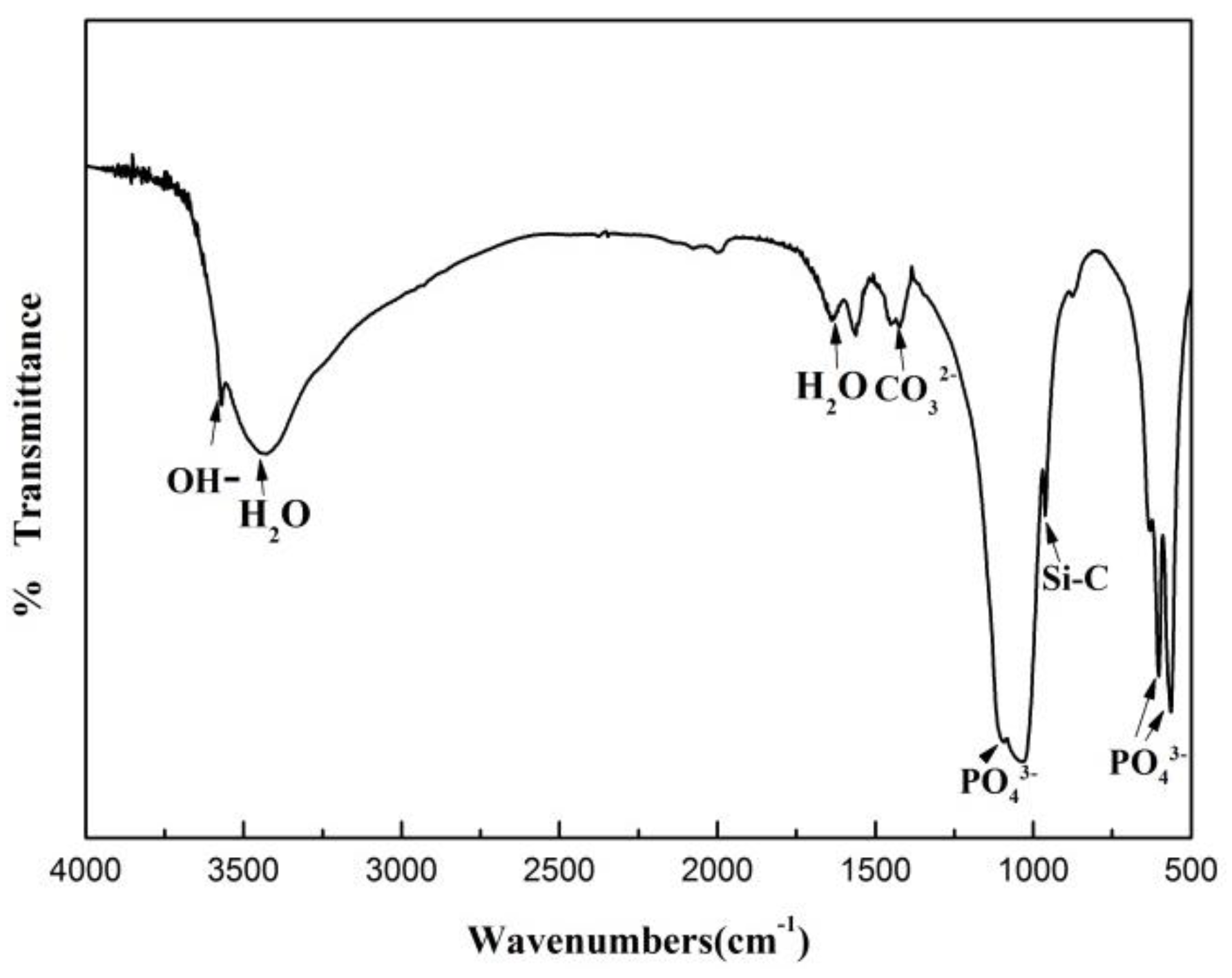
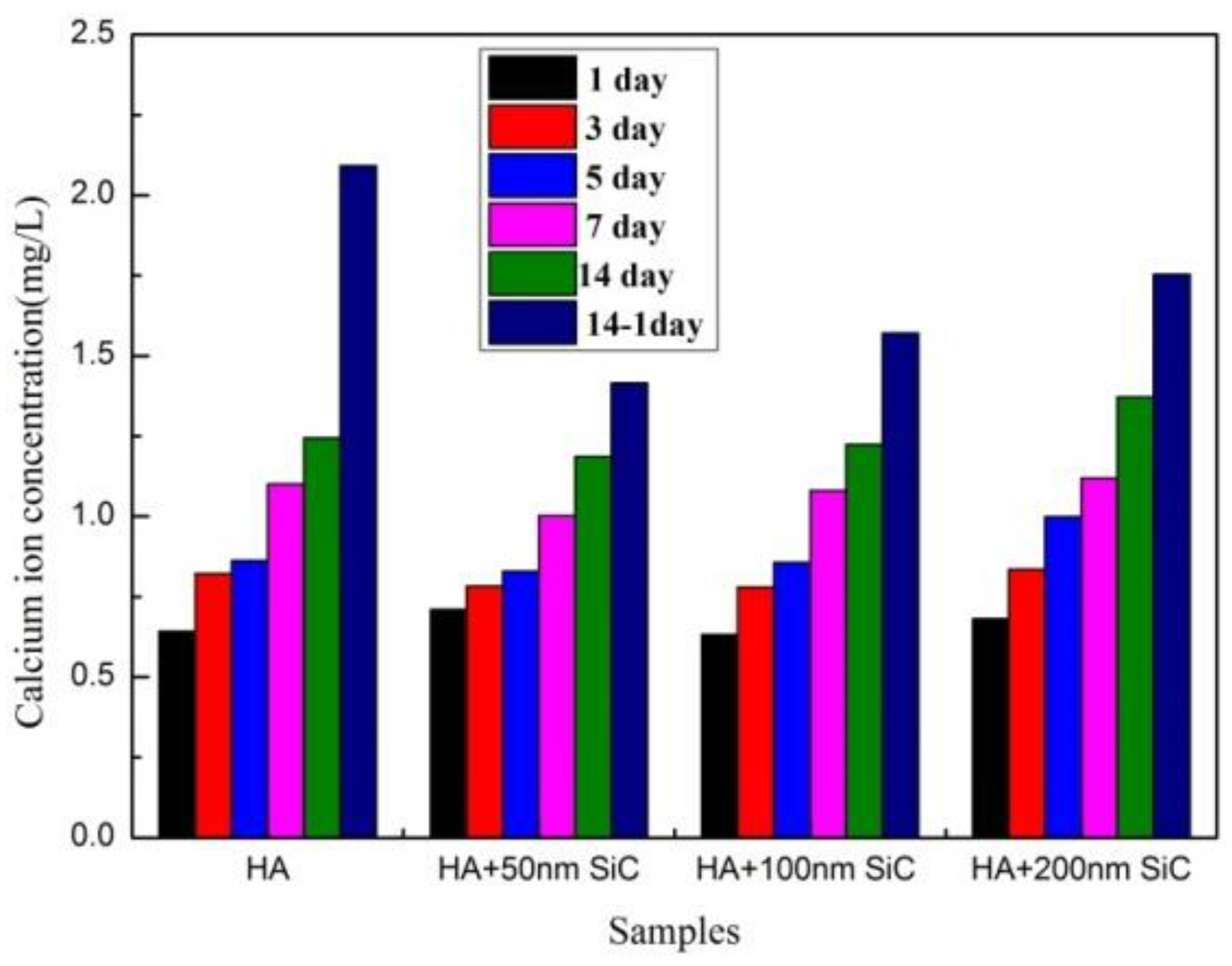
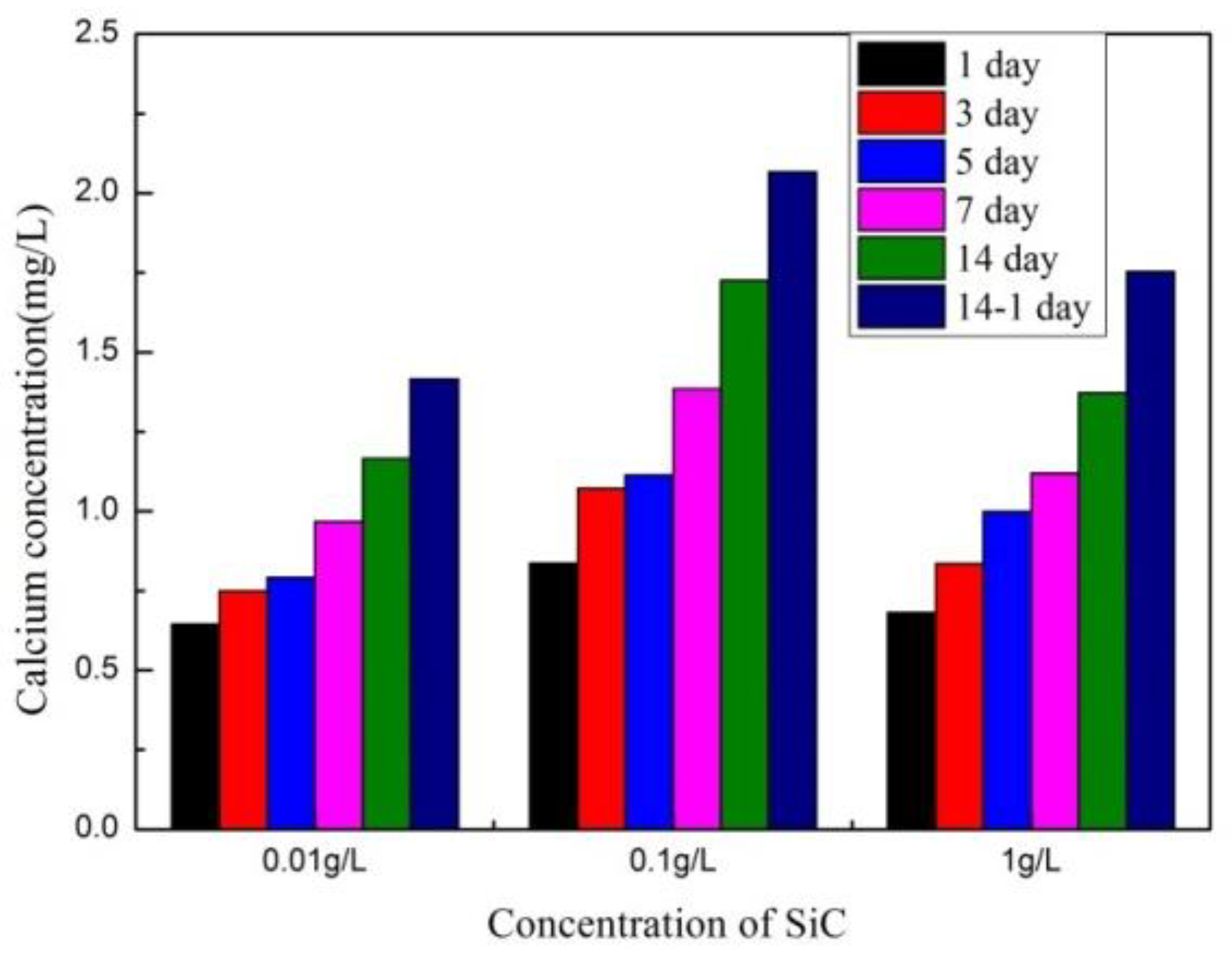
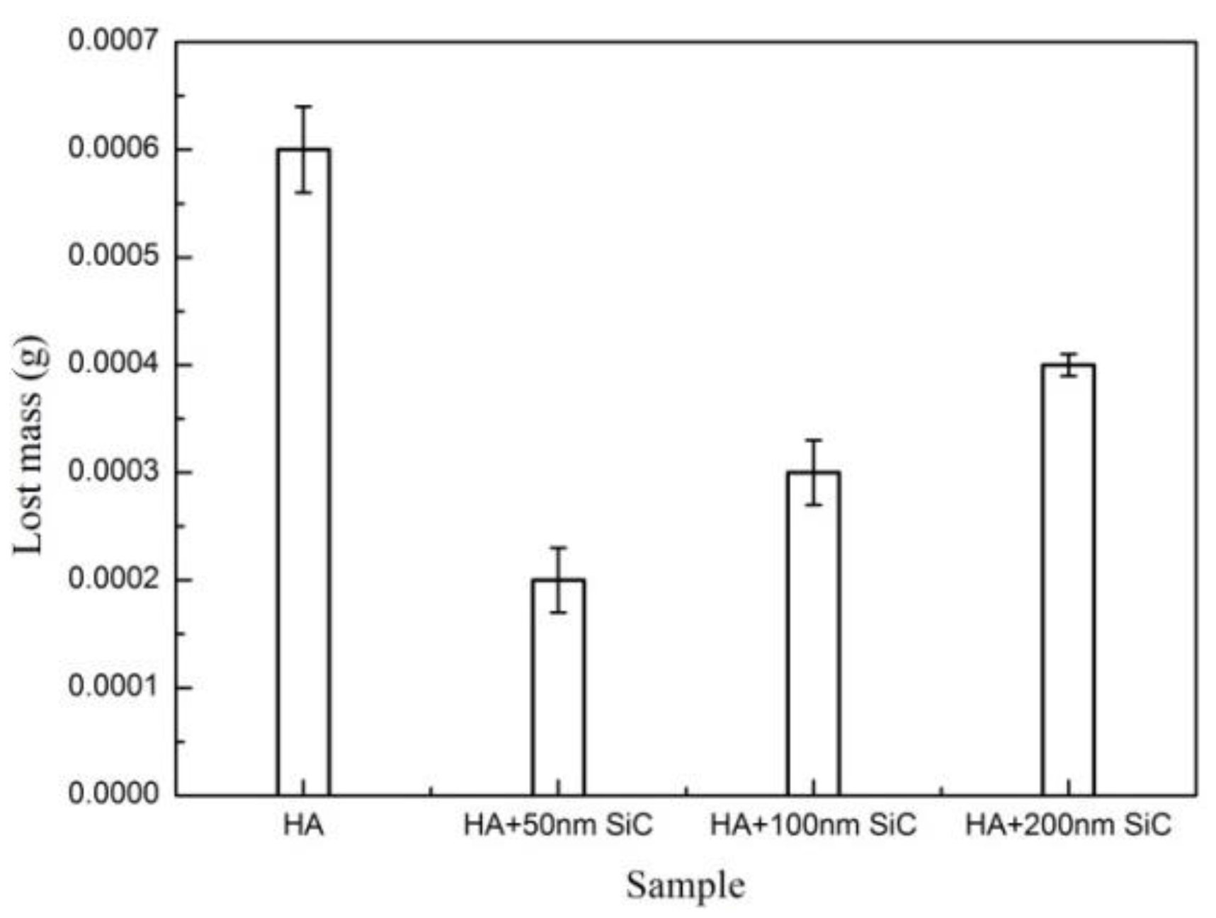
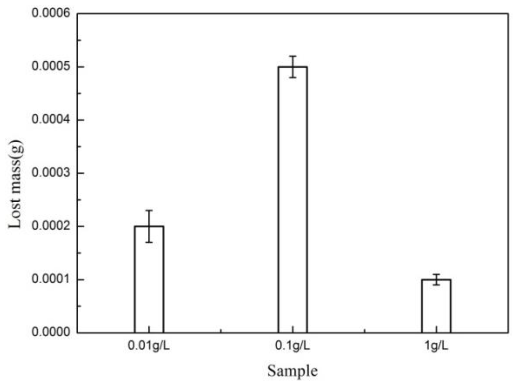
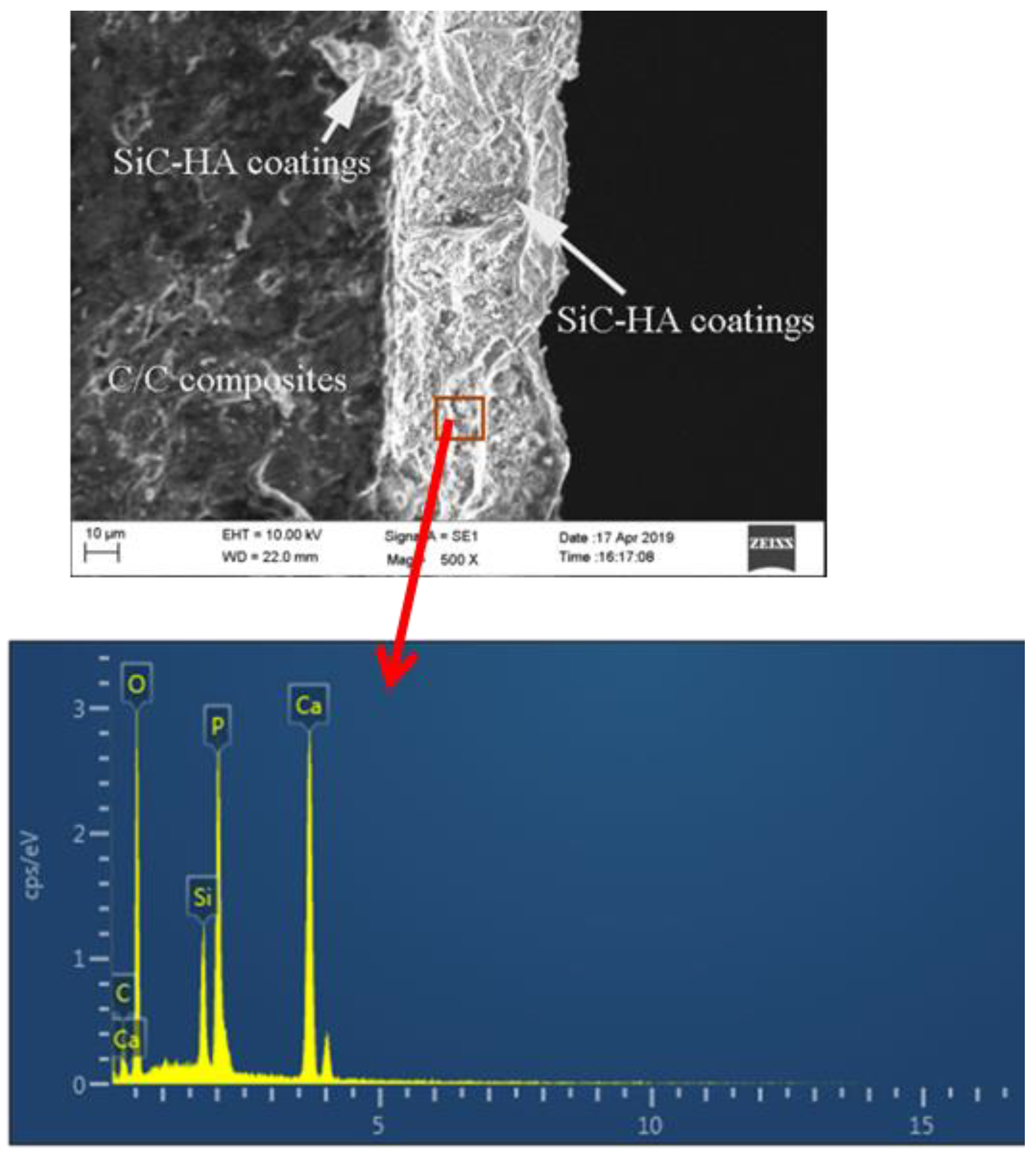
3.3. Dissolution of SiC-Hydroxyapatite Coatings

Dissolution is controlled by the processes occurring on the crystal surface, including the ion transport from the surface into the solution [37]. Figure 10 and Figure 11 show the Ca2+ concentration contents in the leachate after 1–14 days of soaking SiC-hydroxyapatite coatings in physiological saline solution. As expected, the Ca2+ concentration in the leachate increased with the immersion time. However, all hydroxyapatite coatings, independently of their SiC particle sizes and contents, showed similar Ca2+ concentration contents on days 1–14 (see Figure 10).
However, the data obtained for our coatings for the 1–14 days of soaking were significantly different. The Ca2+ concentration was the highest when the coating contained no SiC particles. The Ca2+ contents on days 14 and 14R were also very different because loosening the hydroxyapatite caused large coating pieces to detach. However, they were still too large to dissolve completely. The concentration of dissolved Ca2+ also did not differ significantly from day 1 to day 14 for all coatings (see Figure 11). The Ca2+ concentration in the leachate of the hydroxyapatite coating prepared using 0.1 g/L of 200 nm SiC particles was the highest (see Figure 11) because the corresponding SiC-hydroxyapatite coatings were loose with many cracks, which weakened their bonding strength.
The weight losses of the SiC-hydroxyapatite coatings agreed with the results discussed above on the Ca2+ content in the leachate (see Figure 12 and Figure 13). The pristine hydroxyapatite coating lost the most weight (see Figure 12). The Ca2+ concentration content in the leaching solution was largest after 14 days of soaking in dilute HNO3, which indicated the presence of large amounts of undisclosed chunks of hydroxyapatite coatings due to the coating peeling off during the soaking. Our data indicated that SiC presence in the hydroxyapatite inhibited its dissolution rate. The highest weight loss was observed for the SiC-hydroxyapatite coatings prepared using 0.1 g/L of 200 nm SiC particles (see Figure 13), which agreed with the data discussed above on the coating structure and morphology (see Figure 7b).
Figure 14 and Figure 15 show the morphologies of our SiC-hydroxyapatite coatings after dissolution tests. Large pieces of hydroxyapatite coating were detached, leaving large holes (see Figure 14a) and cracks. These processes contributed to the coating weight losses and the Ca2+ leaching out. The prolonged dissolution tests resulted in the formation of the larger holes and shorter rods tapered at both ends (see Figure 14b) as well as honeycomb-like features (see Figure 14c). As a result, the content of the short bar-like coating features (shown in Figure 3c) decreased, which implied their dissolution and the formation of honeycombs from the less soluble coating components. The fiber-like features on the coating surface also dissolved, leaving the granular tissues (see Figure 14d), and leading to Ca2+ and weight losses.
Figure 15a also shows numerous holes in the coating as well as an indication that the rod-like tissue became fibrous as the coatings were soaked in the diluted HNO3. Most of the petal-like tissues also dissolved, forming more holes (see Figure 15b). Thus, we concluded that the petal-like structures dissolved very quickly. Figure 15c also shows that the coatings’ fibrous tissues dissolved, leaving granular tissues. These data confirm that the solubility of the SiC-hydroxyapatite coatings strongly depended on their microstructure and morphology, both of which, in turn, were defined by the content and size of the embedded SiC particles.

4. Conclusions

Nano-SiC particles were introduced into hydroxyapatite coatings to improve their binding strengths to the C/C substrates. The structures, morphologies and binding strengths of the resulting SiC-hydroxyapatite coatings were studied as functions of the SiC particle contents and sizes. As the sizes of the embedded SiC particles increased, the hydroxyapatite diffraction peaks of the corresponding SiC-hydroxyapatite coatings also increased. However, the SiC content did not affect the diffraction peak strength. The SiC particle presence improved the adhesion of the hydroxyapatite coating to the C/C substrate. The best bonding strength was obtained for the coating prepared using 1 g/L of 200 nm SiC particles. The addition fo the SiC nanoparticles to the hydroxyapatite coatings changed their morphologies and microstructures, which, in turn, directly correlated with the hydroxyapatite coating dissolution.

Author Contributions

Conceptualization, L.Y. and Z.M.; formal analysis and data curation, Z.M.; writing—original draft preparation, L.Y. All authors have read and agreed to the published version of the manuscript.

Funding

This research was funded by the Hengyang Science and Technology Development Project (No.2016KG67), Hunan Provincial Department of Education(No.19C0534), Hengyang Science and Technology Department (2015KS21) and Hunan Institute of Technology(No.KFB20019).

Institutional Review Board Statement

Not applicable.

Informed Consent Statement

Not applicable.

Data Availability Statement

The data presented in this study are available on request from the corresponding author.

Conflicts of Interest

The authors declare no conflict of interest.

References

  1. Zhang, L.L.; Li, H.J.; Li, K.Z.; Zhang, Y.L.; Liu, S.J.; Guo, Q.; Li, S.X. Micro-oxidation treatment to improve bonding strength of Sr and Na co-substituted hydroxyapatite coatings for carbon/carbon composites. Appl. Surf. Sci. 2016, 378, 136–141. [Google Scholar] [CrossRef]
  2. Cao, N.; Dong, J.W.; Wang, Q.X.; Ma, Q.S.; Wang, F.; Chen, H.Y.; Xue, C.Q.; Li, M.S. Plasma sprayed hydroxyapatite coating on carbon/carbon composite scaffolds for bone tissue engineering and related tests in vivo. J. Biomed. Mater. Res. Part A 2010, 3, 1019–1027. [Google Scholar] [CrossRef]
  3. Zhai, Y.Q.; Li, K.Z.; Li, H.J.; Wang, C.; Liu, H. Influence of NaF concentration on fluorine-containing hydroxyapatite coating on carbon/carbon composites. Mater. Chem. Phys. 2007, 106, 22–26. [Google Scholar] [CrossRef]
  4. Zhang, L.L.; Li, S.X.; Li, H.J.; Pei, L.N. Bioactive surface modification of carbon/carbon composites with multilayer SiC-SiC nanowire-Si doped hydroxyapatite coating. J. Alloys Compd. 2018, 740, 109–117. [Google Scholar] [CrossRef]
  5. Ni, X.Y.; Tang, X.B.; Lin, T.; Zhao, Q.D.; Geng, C.R.; Cai, L.M.; Gua, W.D.; Miao, Y.L.; Chen, D. Properties of plasma-spray coated hydroxyapatite on carbon/carbon composites pretreated by an argon plasma. Carbon 2013, 63, 594–595. [Google Scholar] [CrossRef]
  6. Vilardella, A.M.; Cincaa, N.; Garcia-Giraltb, N.; Dostaa, S.; Canoa, I.G.; Noguésb, X.; Guilemanya, J.M. In-vitro comparison of hydroxyapatite coatings obtained by cold spray and conventional thermal spray technologies. Mater. Sci. Eng. C 2020, 107, 110306. [Google Scholar] [CrossRef] [PubMed]
  7. Jiang, J.W.; Han, G.; Zheng, X.S.; Chen, G.; Zhu, P.Z. Characterization and biocompatibility study of hydroxyapatite coating on the surface of titanium alloy. Surf. Coat. Technol. 2019, 375, 645–651. [Google Scholar] [CrossRef]
  8. Chambard, M.; Marsan, O.; Charvillat, C.; Grossin, D.; Fort, P.; Rey, C.; Gitzhofer, F.; Bertrand, G. Effect of the deposition route on the microstructure of plasma-sprayed hydroxyapatite coatings. Surf. Coat. Technol. 2019, 371, 68–77. [Google Scholar] [CrossRef]
  9. Wen, S.F.; Liu, X.L.; Ding, J.H.; Liu, Y.L.; Lan, Z.T.; Zhang, Z.M.; Chen, G.M. Hydrothermal synthesis of hydroxyapatite coating on the surface of medical magnesium alloy and its corrosion resistance. Prog. Nat. Sci. Mater. Int. 2021, 31, 324–333. [Google Scholar] [CrossRef]
  10. Lo, Y.S.; Chang, C.C.; Lin, P.-C.; Lin, S.P.; Wang, C.L. Direct growth of structurally controllable hydroxyapatite coating on Ti-6Al-4V through a rapid hydrothermal synthesis. Appl. Surf. Sci. 2021, 556, 149672. [Google Scholar] [CrossRef]
  11. Cao, N.; Yang, Z.G.; Yang, B.; Wang, W.B.; Boukherroubc, R.; Li, M. Construction of a bone-like surface layer on hydroxyl-modified carbon/carbon composite implants via biomimetic mineralization and in vivo tests. RSC Adv. 2016, 6, 9370–9378. [Google Scholar] [CrossRef]
  12. Cao, N.; Dong, J.W.; Wang, Q.X.; Ma, Q.S.; Xue, C.Q.; Li, M.S. An experimental bone defect healing with hydroxyapatite coating plasma sprayed on carbon/carbon composite implants. Surf. Coat. Technol. 2010, 205, 1150–1156. [Google Scholar] [CrossRef]
  13. Xiong, X.B.; Liu, L.; Ma, J.; Ni, X.Y.; Li, Y.Y.; Zeng, X.R. A simplified process for preparing adhesive hydroxyapatite coatings on carbon/carbon composites. Surf. Coat. Technol. 2019, 377, 124925. [Google Scholar] [CrossRef]
  14. Liu, L.; Ni, X.Y.; Xiong, X.B.; Ma, J.; Zeng, X.R. Low temperature preparation of SiO2 reinforced hydroxyapatite coating on carbon/carbon composites. J. Alloys Compd. 2019, 788, 768–778. [Google Scholar] [CrossRef]
  15. Guan, K.J.; Zhang, L.L.; Zhu, F.Y.; Li, H.J.; Sheng, H.C.; Guo, Y. Multi-layer SiC-graphene oxide-hydroxyapatite bioactive coating for carbon/carbon composites. J. Alloys Compd. 2020, 821, 153543. [Google Scholar] [CrossRef]
  16. Zhang, L.L.; Li, H.J.; Li, K.Z.; Zhang, S.Y.; Fu, Q.G.; Zhang, Y.L.; Lu, J.H.; Li, W. Preparation and characterization of carbon/SiC nanowire/Na-dopedcarbonated hydroxyapatite multilayer coating for carbon/carbon composites. Appl. Surf. Sci. 2014, 313, 85–92. [Google Scholar] [CrossRef]
  17. Li, H.; Khor, K.A.; Cheang, P. Titanium dioxide reinforced hydroxyapatite coatings deposited by high velocity oxy-fuel (HVOF) spray. Biomaterials 2002, 23, 85–91. [Google Scholar] [CrossRef]
  18. Natalia, M.; Stan, G.E.; Duta, L.; Mariana, C.C.; Coralia, B.; Sopronyi, M.; Luculescu, C.; Oktar, F.N.; Mihailescu, I.N. Structural, compositional, mechanical characterization and biological assessment of bovine-derived hydroxyapatite coatings reinforced with MgF2 or MgO for implants functionalization. Mater. Sci. Eng. C 2016, 59, 863–874. [Google Scholar] [CrossRef]
  19. Liu, Y.Y.; Zhang, L.L.; Guan, K.J.; Wang, Z.K.; Wang, Y.T.; Li, H.P.; Lei, X.Y.; Zhang, W.X.; Wang, K.; Hu, Y.Y. Boosting bonding strength of hydroxyapatite coating for carbon/carbon composites via applying tree-planting interface structure. Ceram. Int. 2021, 47, 11922–11928. [Google Scholar] [CrossRef]
  20. Fu, Q.G.; Gu, C.G.; Li, H.J.; Chu, Y.H.; Lu, J.H.; Zhang, L.L. Microstructure and mechanical properties of SiC nanowires reinforced hydroxyapatite coating on carbon/carbon composites. Mater. Sci. Eng. A 2013, 563, 133–137. [Google Scholar] [CrossRef]
  21. Zhao, F.; Zhang, L.L.; Guo, Y.; Sheng, H.C.; Zhang, P.X.; Zhang, Y.X.; Li, Q.; Yang, H.Y.; Mikhalovsky, S.V. Mechanically strong and bioactive carbon fiber-SiC nanowire-hydroxyapatite-pyrolytic carbon composites for bone implant application. Ceram. Int. 2021, 47, 3389–3400. [Google Scholar] [CrossRef]
  22. Hosseini, M.R.; Ahangari, M.; Johar, M.H.; Allahkaram, S.R. Optimization of nano HA-SiC coating on AISI 316L medical grade stainless steel via electrophoretic deposition. Mater. Lett. 2021, 285, 129097. [Google Scholar] [CrossRef]
  23. Tao, L.; Li, X.L.; Hu, S.X.; Wu, J. Enhanced osteoporotic effect of silicon carbide nanoparticles combine with nano-hydroxyapatite coated anodized titanium implant on healthy bone regeneration in femoral fracture. J. Photochem. Photobiol. B Biol. 2019, 197, 111515. [Google Scholar] [CrossRef]
  24. Hnatkoa, M.; Hičáka, M.; Labudováb, M.; Galuskovác, D.; Sedláčeka, J.; Lenčéša, Z.; Šajgalíka, P. Bioactive silicon nitride by surface thermal treatment. J. Eur. Ceram. Soc. 2020, 40, 1848–1858. [Google Scholar] [CrossRef]
  25. Dodevski, V.; Pagnacco, M.C.; Radović, I.; Rosić, M.; Janković, B.; Stojmenović, M.; Mitić, V.V. Characterization of silicon carbide ceramics obtained from porous carbon structure achieved by plant carbonization. Mater. Chem. Phys. 2020, 245, 122768. [Google Scholar] [CrossRef]
  26. Xu, L.-L.; Shi, X.-L.; Qian, Q.; Bai, X.-H.; Xu, L.; Wang, Q.-L. Hydrothermal sterilization in silver nitrate solution endows plasma sprayed hydroxyapatite coating with antibacterial property. Mater. Lett. 2020, 263, 127258. [Google Scholar] [CrossRef]
  27. Xu, M.; Girish, Y.R.; Rakesh, K.P.; Wu, P.; Manukumar, H.M.; Byrappa, S.M.; Udayabhanu; Byrappa, K. Recent advances and challenges in silicon carbide (SiC) ceramic nanoarchitectures and their applications. Mater. Today Commun. 2021, 28, 102533. [Google Scholar] [CrossRef]
  28. Vladescua, A.; Birlikb, I.; Braica, V.; Toparlib, M.; Celikb, E.; AkAzem, F. Enhancement of the mechanical properties of hydroxyapatite by SiC addition. J. Mech. Behav. Biomed. Mater. 2014, 40, 362–368. [Google Scholar] [CrossRef]
  29. Leila, C.-K.; Jafar, K.-A.; Amir, M.; Mohamadreza, E. The effect of hydroxyapatite nanoparticles on electrochemical and mechanical performance of TiC/N coating fabricated by plasma electrolytic saturation method. Surf. Coat. Technol. 2020, 394, 125817. [Google Scholar] [CrossRef]
  30. Mohammad, U.; Colin, H.; Vincent, S. Fabrication, characterisation and corrosion of HA coated AZ31B Mg implant material: Effect of electrodeposition current density. Surf. Coat. Technol. 2020, 385, 125363. [Google Scholar] [CrossRef]
  31. Li, K.-Z.; Guo, Q.; Zhang, L.-L.; Zhang, Y.-L.; Liu, S.-J.; Guo, K.-B.; Li, S.-X. Synthesis and characterization of Si-substituted hydroxyapatite bioactive coating for SiC-coated carbon/carbon composites. Ceram. Int. 2017, 43, 1410–1414. [Google Scholar] [CrossRef]
  32. McEntirea, B.-J.; Bala, B.-S.; Rahamanc, M.-N.; Chevalier, J.; Pezzotti, G. Ceramics and ceramic coatings in orthopaedics. J. Eur. Ceram. Soc. 2015, 35, 4327–4369. [Google Scholar] [CrossRef]
  33. Zhang, L.-L.; Pei, L.-N.; Li, H.-J.; Zhu, F.-Y. Design and fabrication of pyrolytic carbon-SiC fluoridated hydroxyapatite-hydroxyapatite multilayered coating on carbon fibers. Appl. Surf. Sci. 2019, 473, 571–577. [Google Scholar] [CrossRef]
  34. Batebi, K.; Abbasi Khazaei, B.A.; Afshar, A. Characterization of sol-gel derived silver/ fluor-hydroxyapatite composite coatings on titanium substrate. Surf. Coat. Technol. 2018, 352, 522–528. [Google Scholar] [CrossRef]
  35. Yuan, Q.H.; Zhang, Z.Q.; Yang, Y.; Jian, Y.L.; Li, R.L.; Dai, X.Y.; Wu, W.S.; Zhong, J.X.; Chen, C. Synthesis, characterization and biological performance study of Sr-doped hydroxyapatite/chitosan composite coatings. Mater. Chem. Phys. 2021, 270, 124752. [Google Scholar] [CrossRef]
  36. Yao, H.-L.; Hu, X.-Z.; Bai, X.-B.; Wang, H.-T.; Chen, Q.-Y.; Ji, G.-C. Comparative study of HA/TiO2 and HA/ZrO2 composite coatings deposited by high-velocity suspension flame spray (HVSFS). Surf. Coat. Technol. 2018, 351, 177–187. [Google Scholar] [CrossRef]
  37. Mao, Z.-L.; Yang, X.-J.; Zhu, S.-L.; Cui, Z.-D.; Li, Z.-Y. Effect of Na+ and NaOH concentrations on the surface morphology and dissolution behavior of hydroxyapatite. Ceram. Int. 2015, 41, 3461–3468. [Google Scholar] [CrossRef]
Figure 1. Experimental flow graph.
Coatings 11 01166 g001
Figure 2. X-ray diffraction of SiC-hydroxyapatite coatings under different SiC particle sizes.
Coatings 11 01166 g002
Figure 3. Surface morphology and EDS of SiC-hydroxyapatite coatings with different SiC particle sizes: (a) hydroxyapatite; (b) hydroxyapatite + 50 nm SiC; (c) hydroxyapatite + 100 nm SiC; (d) hydroxyapatite + 200 nm SiC.
Coatings 11 01166 g003
Figure 4. The FTIR spectra of the SiC-hydroxyapatite (200 nm, 1 g/L) coatings.
Coatings 11 01166 g004
Figure 5. Adhesion strength of SiC-hydroxyapatite coatings at different SiC particle sizes.
Coatings 11 01166 g005
Figure 6. X-ray diffraction of SiC-hydroxyapatite coatings at different SiC particle (200 nm) concentrations.
Coatings 11 01166 g006
Figure 7. Surface morphology and EDS of SiC-hydroxyapatite coatings at different concentrations of SiC particles: (a) 0.01 g/L; (b) 0.1 g/L; (c) 1 g/L.
Coatings 11 01166 g007aCoatings 11 01166 g007b
Figure 8. The cross section of SiC-hydroxyapatite coatings with 200 nm SiC particles.
Coatings 11 01166 g008
Figure 9. Adhesion strength of SiC-hydroxyapatite coatings at different concentrations of 200 nm SiC particles.
Coatings 11 01166 g009
Figure 10. Calcium concentration of different SiC particle sizes.
Coatings 11 01166 g010
Figure 11. Calcium concentration of different SiC particle (200 nm) concentrations.
Coatings 11 01166 g011
Figure 12. Lost mass of SiC-hydroxyapatite coatings with different SiC particle sizes.
Coatings 11 01166 g012
Figure 13. Lost mass of SiC-hydroxyapatite different SiC particle (200 nm) concentrations.
Coatings 11 01166 g013
Figure 14. Surface morphology of SiC-hydroxyapatite coatings with different SiC particle sizes after dissolution: (a) original sample; (b) 50 nm; (c) 100 nm; (d) 200 nm.
Coatings 11 01166 g014
Figure 15. Surface morphology of SiC-hydroxyapatite coatings with different SiC particle (200 nm) concentration after dissolution: (a) 0.01 g/L; (b) 0.1 g/L; (c) 1 g/L.
Coatings 11 01166 g015aCoatings 11 01166 g015b
Publisher’s Note: MDPI stays neutral with regard to jurisdictional claims in published maps and institutional affiliations.

