Use of an Ophthalmic Viscosurgical Device for Experimental Retinal Detachment in Rabbit Eyes
Abstract
:1. Introduction
2. Results and Discussion
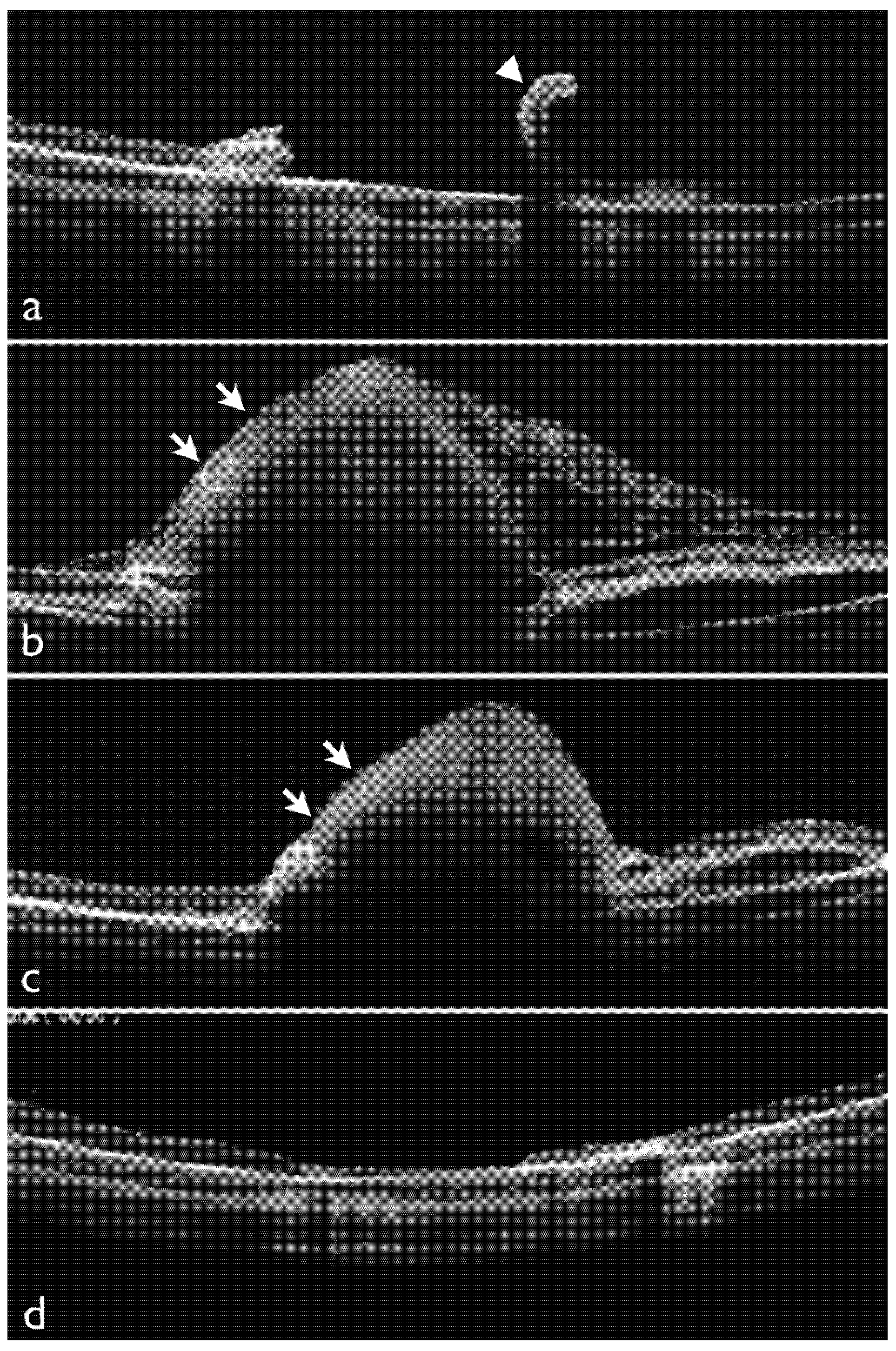
2.1. Effects of Dispersive OVD on Experimental Retinal Detachment Examined by OCT

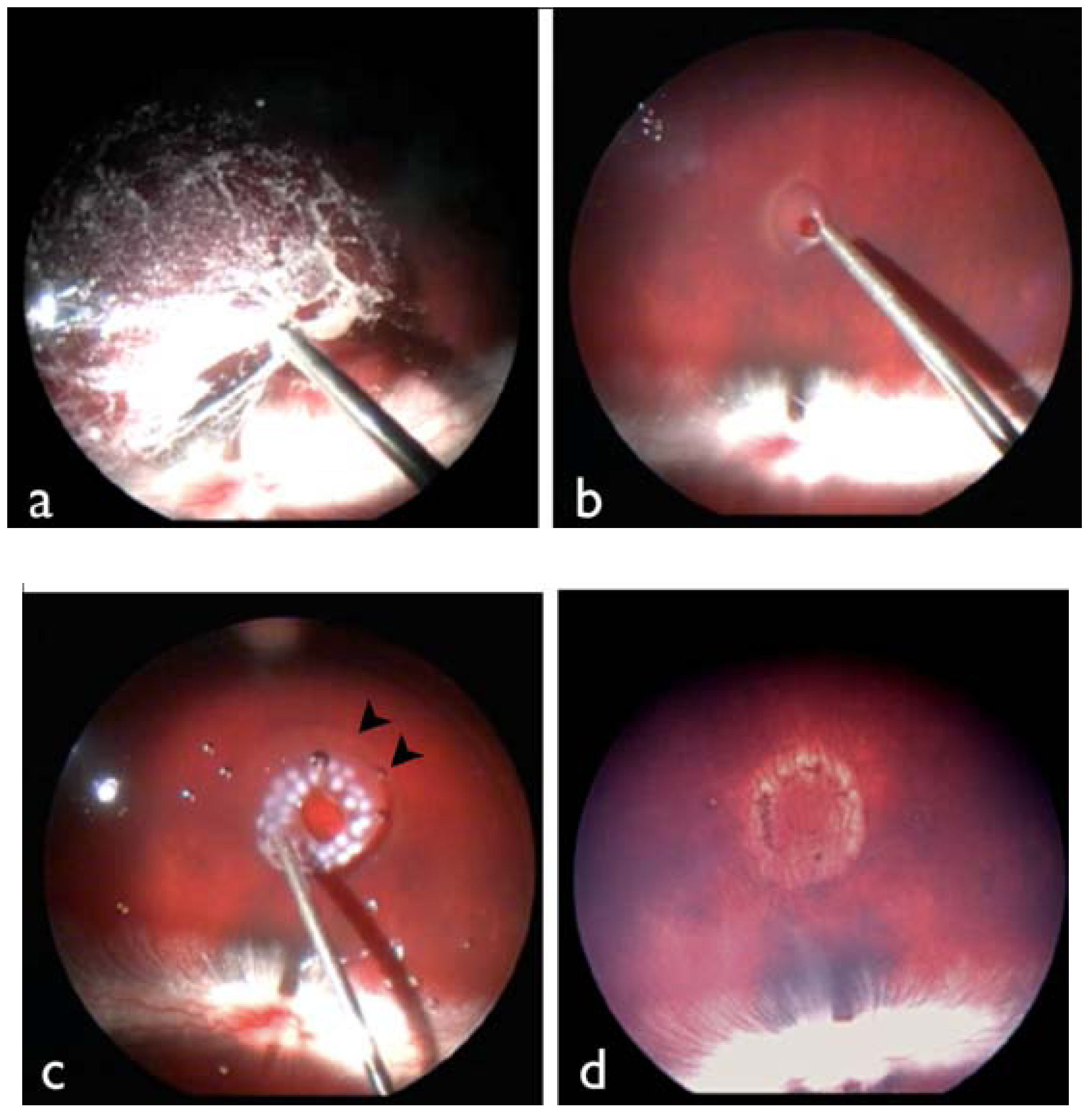
2.2. Observation of the Retinal Surface by SEM

2.3. Discussion
3. Experimental Section
3.1. Animals
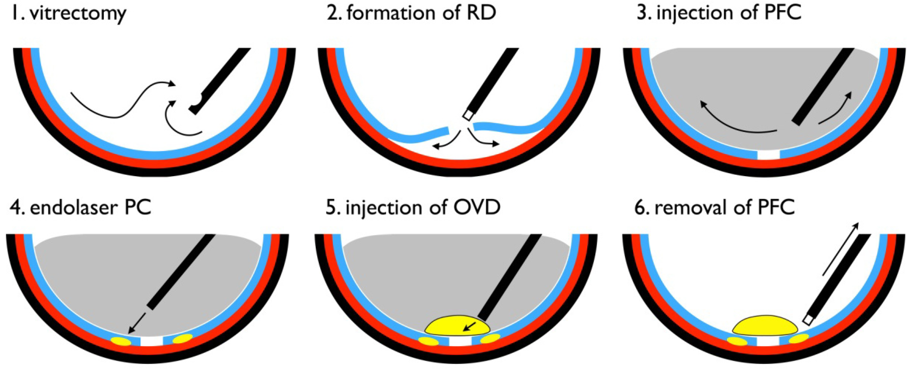
3.2. Vitrectomy with Experimental Retinal Detachment


3.3. Scanning Electron Microscopy
4. Conclusions
Conflict of Interest
References
- Minihan, M.; Tanner, V.; Williamson, T.H. Primary rhegmatogenous retinal detachment: 20 years of change. Br. J. Ophthalmol. 2001, 85, 546–548. [Google Scholar] [CrossRef]
- Heimann, H.; Bartz-Schmidt, K.U.; Bornfeld, N.; Weiss, C.; Hilgers, R.D.; Foerster, M.H. Scleral buckling versus primary vitrectomy in rhegmatogenous retinal detachment: A prospective randomized multicenter clinical study. Ophthalmology 2007, 114, 2142–2154. [Google Scholar] [CrossRef]
- Leaver, P. Expanding the role of vitrectomy in retinal reattachment surgery. Br. J. Ophthalmol. 1993, 77, 197. [Google Scholar] [CrossRef]
- Barrie, T. Debate overview. Repair of a primary rhegmatogenous retinal detachment. Br. J. Ophthalmol. 2003, 87, 790. [Google Scholar] [CrossRef]
- McLeod, D. Is it time to call time on the scleral buckle? Br. J. Ophthalmol. 2004, 88, 1357–1359. [Google Scholar] [CrossRef]
- Coleman, D.J.; Lucas, B.C.; Fleischman, J.A.; Dennis, P.H., Jr.; Chang, S.; Iwamoto, T.; Nalbandian, R.M. A biologic tissue adhesive for vitreoretinal surgery. Retina 1988, 8, 250–256. [Google Scholar] [CrossRef]
- Hida, T.; Sheta, S.M.; Proia, A.D.; McCuen, B.W. Retinal toxicity of cyanoacrylate tissue adhesive in the rabbit. Retina 1988, 8, 148–153. [Google Scholar] [CrossRef]
- Sueda, J.; Fukuchi, T.; Usumoto, N.; Okuno, T.; Arai, M.; Hirose, T. Intraocular use of hydrogel tissue adhesive in rabbit eyes. Jpn. J. Ophthalmol. 2007, 51, 89–95. [Google Scholar] [CrossRef]
- Liggett, P.E.; Cano, M.; Robin, J.B.; Green, R.L.; Lean, J.S. Intravitreal biocompatibility of mussel adhesive protein. A preliminary study. Retina 1990, 10, 144–147. [Google Scholar] [CrossRef]
- Smiddy, W.E.; Glaser, B.M.; Green, W.R.; Connor, T.B., Jr.; Roberts, A.B.; Lucas, R.; Sporn, M.B. Transforming growth factor beta. A biologic chorioretinal glue. Arch. Ophthalmol. 1989, 107, 577–580. [Google Scholar] [CrossRef]
- Teruya, K.; Sueda, J.; Arai, M.; Tsurumaru, N.; Yamakawa, R.; Hirata, A.; Hirose, T. Patching retinal breaks with Seprafilm in experimental rhegmatogenous retinal detachment of rabbit eyes. Eye 2009, 23, 2256–2259. [Google Scholar] [CrossRef]
- Liesegang, T.J. Viscoelastic substances in ophthalmology. Surv. Ophthalmol. 1990, 34, 268–293. [Google Scholar] [CrossRef]
- Glasser, D.B.; Katz, H.R.; Boyd, J.E.; Langdon, J.D.; Shobe, S.L.; Peiffer, R.L. Protective effects of viscous solutions in phacoemulsification and traumatic lens implantation. Arch. Ophthalmol. 1989, 107, 1047–1051. [Google Scholar] [CrossRef]
- Koch, D.D.; Liu, J.F.; Glasser, D.B.; Merin, L.M.; Haft, E. A comparison of corneal endothelial changes after use of Healon or Viscoat during phacoemulsification. Am. J. Ophthalmol. 1993, 115, 188–201. [Google Scholar]
- Baino, F. Towards an ideal biomaterial for vitreous replacement: Historical overview and future trends. Acta Biomater. 2011, 7, 921–935. [Google Scholar] [CrossRef]
- McDermott, M.L.; Hazlett, L.D.; Barrett, R.P.; Lambert, R.J. Viscoelastic adherence to corneal endothelium following phacoemulsification. J. Cataract. Refract. Surg. 1998, 24, 678–683. [Google Scholar]
- Hu, M.; Sabelman, E.E.; Tsai, C.; Tan, J.; Hentz, V.R. Improvement of Schwann cell attachment and proliferation on modified hyaluronic acid strands by polylysine. Tissue Eng. 2000, 6, 585–593. [Google Scholar] [CrossRef]
- Lane, D.; Motolko, M.; Yan, D.B.; Ethier, C.R. Effect of healon and viscoat on outflow facility in human cadaver eyes. J. Cataract. Refract. Surg. 2000, 26, 277–281. [Google Scholar] [CrossRef]
- Yamada, T.; Sawada, R.; Tsuchiya, T. The effect of sulfated hyaluronan on the morphological transformation and activity of cultured human astrocytes. Biomaterials 2008, 29, 3503–3513. [Google Scholar] [CrossRef]
- Hirata, A.; Okinami, S. Viability of topical endoscopic imaging system for vitreous surgery in rabbit eyes. Ophthalmic Surg. Lasers Imaging 2012, 43, 64–67. [Google Scholar]
© 2013 by the authors; licensee MDPI, Basel, Switzerland. This article is an open access article distributed under the terms and conditions of the Creative Commons Attribution license (http://creativecommons.org/licenses/by/3.0/).
Share and Cite
Hirata, A.; Yamamoto, S.; Okinami, S. Use of an Ophthalmic Viscosurgical Device for Experimental Retinal Detachment in Rabbit Eyes. J. Funct. Biomater. 2013, 4, 6-13. https://doi.org/10.3390/jfb4010006
Hirata A, Yamamoto S, Okinami S. Use of an Ophthalmic Viscosurgical Device for Experimental Retinal Detachment in Rabbit Eyes. Journal of Functional Biomaterials. 2013; 4(1):6-13. https://doi.org/10.3390/jfb4010006
Chicago/Turabian StyleHirata, Akira, Soichiro Yamamoto, and Satoshi Okinami. 2013. "Use of an Ophthalmic Viscosurgical Device for Experimental Retinal Detachment in Rabbit Eyes" Journal of Functional Biomaterials 4, no. 1: 6-13. https://doi.org/10.3390/jfb4010006
APA StyleHirata, A., Yamamoto, S., & Okinami, S. (2013). Use of an Ophthalmic Viscosurgical Device for Experimental Retinal Detachment in Rabbit Eyes. Journal of Functional Biomaterials, 4(1), 6-13. https://doi.org/10.3390/jfb4010006


