Article

Behavioral Changes in Caenorhabditis elegans After Exposure to Radial Extracorporeal Shock Waves

1
Department of Anatomy II, Ludwig-Maximilians-University of Munich, 81366 Munich, Germany
2
Department of Orthopedic Surgery, Kitasato University School of Medicine, Sagamihara 252-0329, Japan
*
Author to whom correspondence should be addressed.
J. Clin. Med. 2025, 14(20), 7206; https://doi.org/10.3390/jcm14207206 (registering DOI)
Submission received: 20 May 2025 / Revised: 3 October 2025 / Accepted: 10 October 2025 / Published: 13 October 2025
(This article belongs to the Special Issue Cerebral Palsy: Clinical Rehabilitation and Treatment)

Abstract

Background/Objectives: Cerebral palsy (CP) is a leading cause of motor disability in children and is commonly associated with spasticity. Treatment with radial extracorporeal shock waves (rESWs) is an established non-invasive therapy for spasticity, although its underlying mechanisms remain poorly understood. Caenorhabditis elegans (C. elegans) represents a powerful model for neuromuscular research due to its fully mapped nervous system, conserved cholinergic pathways and suitability for high-throughput behavioral analysis. This study aimed to test whether rESWs modulate cholinergic signaling at the neuromuscular junction (NMJ) in C. elegans. Methods: Wild-type and acr-16 mutant C. elegans were exposed in liquid to varying doses of rESWs, nicotine and carbachol in different combinations. Locomotor behavior was recorded using high-resolution video tracking, and parameters including peristaltic speed, body wavelength, reversals and omega bends were quantified. Results: Exposure to rESWs transiently altered locomotion, most notably by reducing forward speed and increasing the frequency of reversals. However, rESWs did not consistently modify behavioral responses to nicotine or carbachol, and these effects were not clearly dependent on NMJ-associated nicotinic receptors. Conclusions: Exploring C. elegans as a model for rESW effects on spasticity proved informative but also revealed important limitations. Results indicate that rESWs act on the nervous system more broadly, extending beyond neuromuscular structures. This contrasts with the clinical situation, where rESWs primarily target muscles and connective tissues. While this precludes C. elegans as a direct model for CP-related spasticity, the observation that rESWs influence nervous-system function at a systemic level points to potential therapeutic avenues for neurological diseases.

1. Introduction

Cerebral palsy (CP) is a leading cause of motor disability in children and is commonly associated with spasticity, a condition characterized by increased muscle tone and exaggerated reflexes [1]. Managing spasticity is critical to improving mobility and quality of life in individuals with CP [1].
Among the available treatment options, radial extracorporeal shock wave therapy (rESWT) has emerged as an established non-invasive modality for reducing spasticity due to CP [2,3,4]. Radial extracorporeal shock waves (rESWs) are applied transcutaneously and penetrate tissues to a depth of up to 2 cm [5]. Clinical studies have demonstrated that rESWT can reduce spasticity and improve motor function both immediately and for up to three months after repeated treatments [6]. Despite its therapeutic potential, the molecular and cellular mechanisms underlying these beneficial effects remain largely unknown.
To explore the mechanisms of rESWT, we have previously shown that in intact rat muscles, rESWs induce a reduction and degeneration of acetylcholine receptors (AChRs), along with a selective breakdown of the neuromuscular junction (NMJ). This results in a transient disruption of neuromuscular transmission and a measurable decrease in compound muscle action potentials [7,8,9].
Acetylcholine receptors (AChRs) are activated by the neurotransmitter acetylcholine (ACh) and are expressed in both the central nervous system (CNS) and the peripheral nervous system (PNS) [10]. They are subdivided into two main classes: muscarinic and nicotinic receptors [11]. Muscarinic AChRs (mAChRs) are G protein-coupled receptors found in cardiac and smooth muscle, secretory glands, and neural tissue [12,13,14]. Nicotinic AChRs (nAChRs), by contrast, are ligand-gated ion channels responsive to both ACh and nicotine. They modulate excitability in skeletal muscle (N1 subtype) and neurons (N2 subtype), with neuronal nAChRs located on postganglionic neuron cell bodies and muscular nAChRs situated at the NMJ on muscle cell surfaces, where they are essential for voluntary motor control [11,15]. Carbachol, a structural analog of ACh, functions as an agonist for both receptor types [16,17,18].
These cholinergic mechanisms are conserved in invertebrates. In the nematode Caenorhabditis elegans (C. elegans), NMJs are also cholinergic, and many of the proteins involved, including AChRs, are highly conserved across species [19,20,21]. In C. elegans, nAChRs at the NMJ are pharmacologically divided into levamisole-sensitive and nicotine-sensitive subtypes [22,23,24], both of which regulate locomotor behavior [22]. One crucial subunit of the nicotine-sensitive class is ACR-16, which closely resembles the vertebrate α7 subunit group [25,26,27]. Mutants lacking acr-16 have been employed in studies examining nicotine dependence [28].
The C. elegans nervous system is composed of only 302 neurons with a completely mapped connectome [29]. Its simplicity, along with low maintenance costs, short generation times, and ethical advantages, makes C. elegans an appealing model for high-throughput behavioral studies [30,31]. Locomotor behavior in C. elegans is straightforward to quantify and serves as a rapid readout of nervous system function [32,33,34]. Nicotine is known to affect several behaviors in C. elegans, such as head thrashing, pharyngeal pumping, egg-laying, and locomotion [22,31,35,36,37,38].
We have previously demonstrated that rESW exposure leads to reversible behavioral changes in C. elegans, including reduced locomotion speed and increased paralysis in a dose-dependent manner [39]. These findings support the use of C. elegans as a powerful model organism for studying how rESWs interact with AChRs at the NMJ. Moreover, it offers an ethical and economical alternative to vertebrate models, which are limited by higher costs, reduced fecundity, longer generation times, and stricter ethical regulations.
In the present study, we hypothesize that (i) exposure of C. elegans to rESWs reduces the effects of nicotine and carbachol on locomotor behavior; and (ii) these effects are mediated via nAChRs at the NMJ in C. elegans.
In line with our previous work investigating the behavioral effects of rESWs in C. elegans [39,40], worms in this study were maintained in liquid culture.
By elucidating how rESWs modulate neuromuscular transmission via nAChRs in C. elegans, this study aims to contribute mechanistic insights relevant to the therapeutic use of rESWT for spasticity in individuals with CP.

2. Materials and Methods

2.1. Nematodes

N2 wild-type and RB918 acr-16(ok789) mutant C. elegans, as well as OP50 Escherichia coli (E. coli), were obtained from the Caenorhabditis Genetics Center (Minneapolis, MN, USA). Note: the RB918 strain was provided by the C. elegans Gene Knockout Project at the Oklahoma Medical Research Foundation (Oklahoma City, OK, USA), which was part of the International C. elegans Gene Knockout Consortium [41].

2.2. Liquid Cultures

Stock C. elegans liquid cultures were maintained in 250 mL S Medium supplemented with 0.1% Tween 20 in 500 mL baffled flasks at 20 °C in a shaking incubator (250 rpm; New Brunswick Innova 40, Eppendorf, Hamburg, Germany) [42]. Worms were fed with pelleted E. coli OP50.
To prepare synchronized, young adult C. elegans for behavioral assays, cultures containing a high proportion of gravid hermaphrodites were selected and transferred into a 500 mL separatory funnel (Thermo Fisher Scientific, Waltham, MA, USA). After approximately 30 min of settling, the first 15 mL were collected into a 15 mL tube and centrifuged at 300× g at room temperature for 1 min. Worms were resuspended in 1.5 mL S Medium and subsequently treated with hypochlorite solution for 7–8 min to isolate eggs. After washing and pelleting, the remaining eggs were resuspended in 10 mL of S Medium containing 0.1% Tween 20 and incubated overnight at 20 °C on a rocker platform (30 rpm; PMR-30 Grant-Bio, Grant Instruments, Royston, UK) to reach the L1 larval stage.
On the following day, L1-stage larvae were transferred into 500 mL baffled flasks and maintained in 250 mL S Medium with 0.1% Tween 20 at 24 °C in a shaking incubator (250 rpm). Upon feeding with a pellet of E. coli OP50, larvae were allowed to grow for an additional 46 h to reach the young adult stage.
Synchronized young adult worms (>650 µm in length) were harvested by centrifugation (3000× g, 4 °C for 5 min). Worms were resuspended in cold NaCl solution and purified using the sucrose floatation method [43]. They were subsequently transferred into S Medium at a concentration of approximately 2 worms/µL. A volume of 10 µL of the worm suspension was dispensed into a single well of a U-bottom 96-well plate (VWR, Radnor, PA, USA), and the well was filled to 300 µL total volume with S Medium for subsequent assaying.

2.3. Assays

For each assay, each condition was tested with three technical replicates per experiment, and the entire set of experiments was independently repeated at least three times.
We applied rESW numbers and recovery periods as established in our previous study [39]. Nicotine and carbachol concentrations were selected within previously reported ranges [18,38], and dose–response experiments were conducted to substantiate the choice. Therefore, N2 wild-type worms were placed into a well containing S Medium with or without nicotine (0, 0.5, 2, 10, 20 mM) or carbachol (0, 1, 5, 10 mM). After one hour, the samples were rapidly transferred to food-free nematode growth medium (NGM) plates for video analysis. The results are provided in the Supplementary Materials (Figures S1 and S2).
Based on these conditions, the following assays were performed (Table 1):
Assays A1-2, B1-2, C1,4,7,10,13,16, D1-6 and E1-6: C. elegans N2 wild-type or acr-16 mutant worms were placed into a well containing S Medium with or without nicotine or carbachol, and were immediately exposed to a defined number of rESWs (0, 100 or 500 rESWs, respectively). One hour after the beginning of rESW exposure, samples were rapidly transferred to food-free NGM plates for video analysis.
Assays C2, 3, 5, 6, 8, 9, 11, 12, 14, 15, 17, 18: N2 wild-type worms were exposed to a defined number of rESWs (0, 100 or 500 rESWs, respectively). Following rESW exposure, worms were allowed to recover in the well for a designated time (30 or 180 min, respectively). Subsequently, nicotine was added for one hour, after which worms were rapidly transferred to food-free NGM plates for video analysis.

2.4. Application of rESWs

For the application of rESWs, the handpiece of a rESWT device (Swiss DolorClast; Electro Medical Systems, Nyon, Switzerland), equipped with a 6 mm applicator and inner 6 × 2 O-rings (Vi 975, G/FKM 75, C. Otto Gehrckens, Pinneberg, Germany), was mounted on a vertical drill stand (Wolfcraft, Kempenich, Germany). A 5.5 × 2 O-ring (Vi 670/FKM 80, C. Otto Gehrckens) was positioned around the applicator tip, and the handpiece was lowered into a single well of a 96-well plate until the well plate was held firmly in place [39]. A specified number of rESWs was delivered at 5 Hz and 2 Bar, corresponding to an energy flux density of approximately 0.016 mJ/mm2 at a 5 mm distance from the applicator.

2.5. Transfer and Video Capture

Following the designated waiting period, C. elegans liquid samples underwent a rapid transfer procedure for video analysis on NGM plates [39]. To prevent adherence of worms to pipette tips, 30 µL of S Medium containing 1% Tween 20 was added to the well. The well contents were then gently pipetted and released onto a membrane (Polyethersulfone Millipore Express PLUS Membrane, 47 mm diameter, 0.22 µm pore size; Millipore, Billerica, MA, USA) positioned on a vacuum filtration apparatus. The membrane was subsequently inverted onto an NGM plate (without an E. coli lawn) and removed just prior to video analysis.

2.6. Locomotion Data Collection, Processing and Analysis

For each included worm, the following behavioral parameters were analyzed (Table 2): absolute peristaltic speed, wavelength, number of reversals and number of omega bends. For peristaltic speed and wavelength, average values across all tracked frames were calculated. Reversal and omega bend frequencies were quantified as the percentage of total frames in which these behaviors occurred.
Behavioral video recording of worms on agar plates after rapid transfer was performed using the WormLab Imaging System (MBF Bioscience, Williston, VT, USA). Plates were positioned to ensure that worms were within the objective’s field of view (AF 60 mm 1:2.8D Micro; Nikon, Tokyo, Japan). Video capture was performed using a 20-megapixel monochrome digital camera (acA5472-17um; Basler, Ahrensburg, Germany) at 7.5 frames per second for a duration of 60 s (i.e., 450 frames per video), using WormLab software (Version 2022.1.1, MBF Bioscience). The resolution of each video was 5484 × 3660 pixels. For each sample, tracking was performed across frames 1 to 450.
After tracking, data were exported from WormLab into Microsoft Excel 2010 (Microsoft, Redmond, WA, USA) for further analysis. Only worms with body lengths greater than 650 µm that were continuously tracked for at least 30 s (i.e., ≥225 of 450 frames) were included in the final analysis. Tracks that did not meet these criteria—due to incomplete tracking, segmentation errors or insufficient duration—were excluded. Outliers were identified through visual inspection and removed only when they clearly resulted from tracking artifacts. No data points were excluded solely based on their deviation from the group distribution.

2.7. Statistical Analysis

Statistical analysis of all raw data was conducted using GraphPad Prism (version 10.4.1 for Windows; GraphPad Software, San Diego, CA, USA). One-way analysis of variance (ANOVA) was employed to assess statistical differences among groups. For post hoc pairwise comparisons, Bonferroni multiple comparison tests were applied. Details of the statistical analysis for all assays are provided in the Supplementary Materials (Tables S2, S4 and S7). All data were reported as the percentage (%) of the control group corresponding to each respective assay (Table 1). In addition, the Supplementary Materials (Tables S1, S3, S5 and S6) include a summary of absolute mean values, percent of control, corresponding 95% confidence intervals and the total number of worms analyzed per condition. Results are expressed as the arithmetic mean ± standard error of the mean (SEM), based on a minimum of three independent experiments. A p-value of <0.05 was considered statistically significant.

3. Results

N2 wild-type worms exposed to 2 mM nicotine (Assay A2; hereafter: AA2; Figure 1a) did not show significant differences in mean absolute peristaltic speed compared to control worms (AA1) (Figure 1b; Tables S1 and S2).
Similarly, the combination of 2 mM nicotine and 100 rESWs (AB2) did not significantly affect peristaltic speed relative to control worms (AA1) or exposure to 100 rESWs alone (AB1) (Figure 1b; Tables S1 and S2). Minor differences in mean wavelength and the number of omega bends were observed between nicotine-treated worms and controls (AA2 vs. AA1), but not between nicotine-treated worms with or without rESW exposure (AB2 vs. AA2) (Figure 1c,d; Tables S1 and S2). The mean number of reversals was unaffected by nicotine alone (AA2 vs. AA1; Figure 1e). However, combined exposure to 2 mM nicotine and 100 rESWs (AB2) significantly increased the number of reversals compared to AA1, AA2, and AB1 (Figure 1e), indicating disoriented locomotor behavior. Together, these findings suggest that the combination of 2 mM nicotine and 100 rESWs caused only modest behavioral changes in C. elegans.
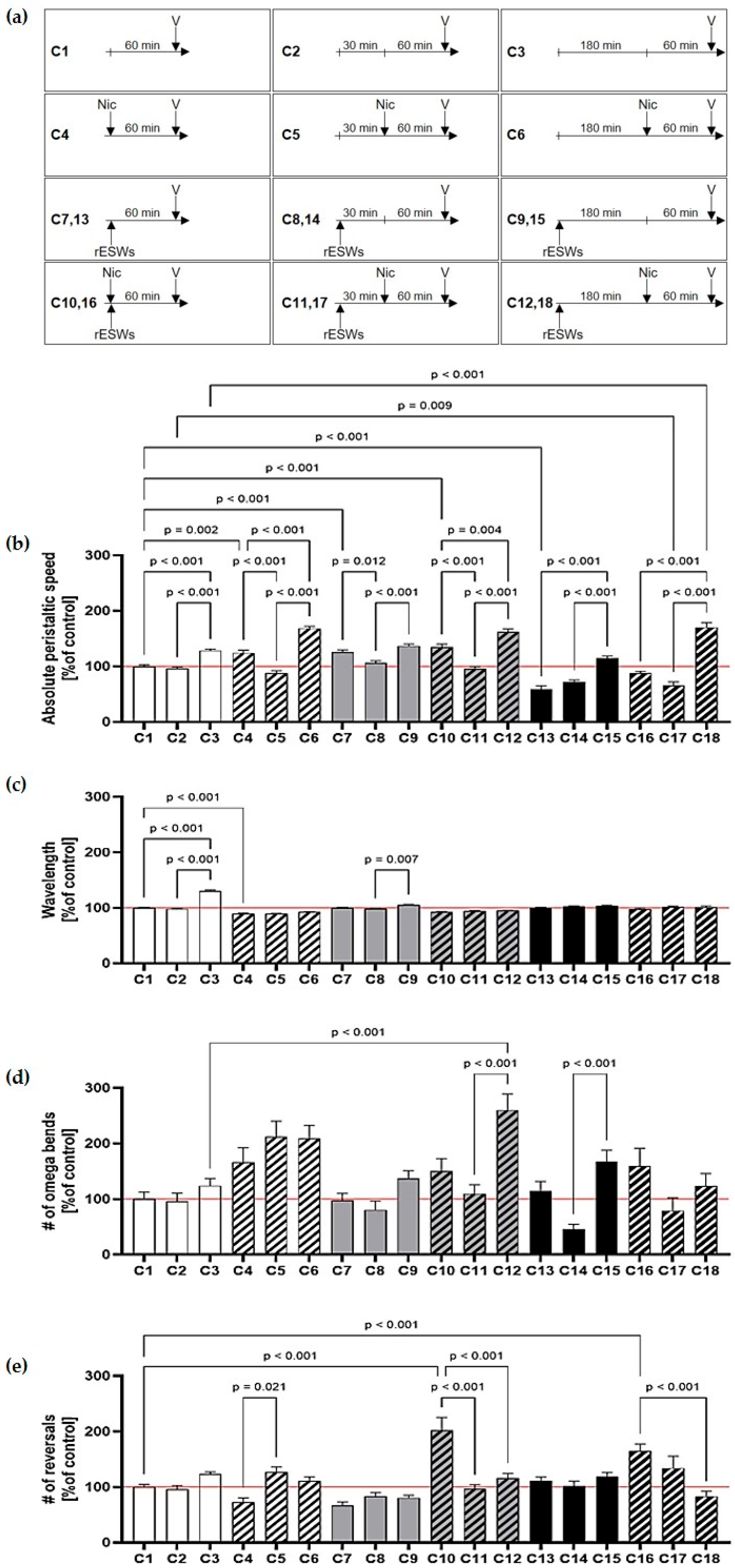
We next examined whether increasing recovery periods after rESW exposure influenced behavioral outcomes in N2 wild-type worms, and whether these effects were modulated by subsequent nicotine treatment (Figure 2a). Without a recovery period, nicotine alone led to a slight increase in mean peristaltic speed, while rESW exposure produced dose-dependent effects (Figure 2b; Tables S3 and S4).
Specifically, 100 rESWs increased peristaltic speed (AC7 vs. AC1), whereas 500 rESWs decreased it (AC13 vs. AC1). Notably, the addition of 2 mM nicotine abolished both of these rESW-induced effects (AC10 vs. AC4; AC16 vs. AC4; Figure 2b; Tables S3 and S4).
Recovery time following rESW exposure revealed a paradoxical pattern: after 30 min, worms treated with rESWs, with or without nicotine, exhibited significantly reduced peristaltic speeds compared to those without recovery (AC5 vs. AC4; AC8 vs. AC7; AC11 vs. AC10; Figure 2b). In contrast, after 180 min, rESW-treated worms without nicotine showed significantly increased peristaltic speeds compared to those without recovery (AC3 vs. AC1; AC15 vs. AC13; Figure 2b). This effect was further enhanced by nicotine (AC6 vs. AC4; AC12 vs. AC10; AC18 vs. AC16; Figure 2b; Tables S3 and S4), suggesting complex time-dependent interactions between nicotine and rESWs.
No systematic differences in wavelength or omega bend frequency were observed across assays, indicating generally coordinated movement (Figure 2c,d; Tables S3 and S4). Consistent with Assays A and B, neither rESWs nor nicotine alone significantly affected the number of reversals when no recovery period was provided (Figure 2e; Tables S3 and S4). However, worms exposed to both rESWs and nicotine without recovery showed significantly more reversals than those with a 180 min recovery (AC10 vs. AC12; AC18 vs. AC16; Figure 2e; Tables S3 and S4), further supporting transient disorientation that normalized over time.
To evaluate the effects of carbachol and rESWs on locomotion, N2 wild-type and acr-16 mutant worms were exposed to rESWs with or without carbachol (Figure 3a). In acr-16 mutants, 100 rESWs significantly increased mean peristaltic speed (AE2 vs. AE1; Figure 3b; Tables S6 and S7), while no such effect was observed in N2 wild-type worms (AD2 vs. AD1; Figure 3b; Tables S5 and S7).
Carbachol alone (1 mM) had no significant impact on peristaltic speed in either strain (AD3 vs. AD1; AE3 vs. AE1; Figure 3b; Table S7). However, the combination of 1 mM carbachol and 100 rESWs significantly reduced peristaltic speed in N2 wild-type worms (AD4 vs. AD1; Figure 3b; Table S7), but not in acr-16 mutants (AE4 vs. AE1; Figure 3b; Table S7), indicating that the observed effects were not solely mediated via NMJs.
Neither strain showed notable changes in wavelength or omega bend frequency after treatment with carbachol, rESWs, or their combination (Figure 3c,d; Table S7). As for reversals, both wild-type and acr-16 worms displayed a significant increase following combined exposure to 1 mM carbachol and 100 rESWs (AD4 vs. AD1; AE4 vs. AE1; Figure 3e; Table S7). Wild-type worms also showed increased reversal frequency after 500 rESWs (AD5 vs. AD1; Figure 3e; Table S7; not tested in acr-16 mutants), while acr-16 mutants exhibited a similar increase after exposure to 10 mM carbachol (AE5 vs. AE1; Figure 3e; Table S7; not tested in wild-type worms). As in Assays A–C, these results support the conclusion that high rESW intensity or high carbachol concentration induced disoriented locomotor behavior in C. elegans.

4. Discussion

Radial extracorporeal shock wave therapy (rESWT) is a clinically validated, non-invasive intervention used to manage spasticity in individuals with CP [2,3,4]. Previous work from our group demonstrated (i) the clinical efficacy of rESWT for CP-related spasticity, (ii) rESW-induced reduction and degeneration of AChRs at the NMJ in a rat model [8], and (iii) dose-dependent behavioral effects of rESWs in C. elegans [39].
In the present study, we investigated whether rESWs influence the effects of nAChR agonists nicotine and carbachol on C. elegans locomotion, using the nematode as a model system. Contrary to our hypothesis, rESWs did not produce significant modulatory effects on nicotine- or carbachol-induced changes in locomotion, nor did they show a specific dependence on nAChRs at the NMJ. Nicotine and carbachol are known to affect worm behavior in a concentration- and time-dependent manner [44]. Nicotine, for example, exerts biphasic effects, enhancing movement at low concentrations and inducing paralysis at high concentrations (>10 mM) [45]. In our study, 2 mM nicotine did not reduce peristaltic speed, suggesting this concentration may have been sub-threshold under the experimental conditions employed—specifically, liquid culture and the method of compound administration. Nevertheless, nicotine altered movement parameters such as wavelength and omega bends, reflecting orientation and foraging behaviors [46,47] and highlighting the influence of environmental context on locomotor assays [48].
Combined exposure to nicotine and rESWs produced comparable locomotor alterations. Our previous work demonstrated that rESWs can induce transient conduction deficits in motor nerves, reduce NMJ AChR density and decrease compound muscle action potentials in rats without causing overt paralysis [8]. In C. elegans, although NMJ morphology was not directly assessed, moderate rESW exposure did not disrupt nAChR function sufficiently to significantly alter nicotine-induced locomotion. High numbers of rESWs, however, reduced worm speed in wild-type N2 worms, an effect that attenuated after recovery and further diminished with one hour of nicotine exposure. These observations align with previously reported transient, biphasic and dose-dependent effects of rESWs [9,39,45,49].
Exposure to 1 mM carbachol with rESWs modified locomotion, although carbachol alone had no significant effect, suggesting that rESWs may influence cholinergic signaling beyond the NMJ, possibly influencing additional regions of the nervous system. Importantly, in our model, rESWs were applied in a liquid-based setup that exposes the entire organism uniformly [39]. Given that C. elegans is only ~1 mm in length, no shock wave applicators can selectively target peripheral structures, making it very likely that other tissues, including sensory head neurons, are affected. In contrast, clinical and rodent protocols use gel coupling to deliver rESWs locally to specific muscles, tendons or peripheral nerves [4,8,9]. These differences in application likely contribute to divergent mechanistic responses, with whole-body exposure in C. elegans reflecting systemic effects rather than isolated PNS modulation.
These results highlight broader implications of rESW action beyond peripheral tissues. While rESWs have not yet been applied to the CNS of vertebrates, our findings in C. elegans suggest they may influence CNS-related structures, potentially modulating neural circuits, plasticity or repair processes. Investigating these effects in vertebrate models may clarify underlying mechanisms—such as activity-dependent synaptic modulation, neurogenesis or glial responses—and inform potential therapeutic strategies for neurological disorders.
High rESW doses abolished carbachol-induced behavioral effects in N2 worms, consistent with substantial loss of nAChRs and impaired neurotransmission [8]. Of note, acr-16 mutants, which lack nicotine-sensitive nAChRs, exhibited locomotion similar to wild-type worms, showing no major baseline deficits while retaining substantial cholinergic postsynaptic currents [27]. In these mutants, 1 mM carbachol alone or combined with rESWs did not alter peristaltic speed, whereas 10 mM carbachol induced locomotor changes amplified by rESWs. These findings suggest potential inhibitory effects of rESWs on nicotine-independent nAChRs and/or mAChRs, with differing effects on wavelength and omega bend frequency further pointing to subtype-specific mechanisms.
Additionally, several off-target pathways may contribute to transient behavioral responses following rESW exposure. Interneurons in the ventral nerve cord and head ganglia, which modulate locomotion and posture without direct synaptic output to muscles, could be affected. Similarly, non-cholinergic systems such as GABAergic motor neurons and glutamatergic circuits may respond to mechanical stress or downstream signaling, altering coordination and linking sensory input to locomotor behavior [50,51]. This underlines the complexity of rESW effects, involving multiple neural subsystems, receptor-specific interactions and both targeted and off-target mechanisms.
In conclusion, consistent with our previous findings [39], rESWs induce transient behavioral changes in C. elegans, with responses modulated in complex ways by nAChR agonists. These effects seem to involve neural components analogous to those present in both the CNS and PNS of vertebrates. Since rESWs in C. elegans act on the whole organism rather than discrete neuromuscular targets as in vertebrates, the underlying mechanisms may differ from those mediating the therapeutic effects of rESWT in muscle spasticity associated with CP.
While this prevents using C. elegans as a model for isolated PNS effects, it also highlights a novel action of rESWs. Importantly, these findings suggest that rESW effects on CNS-related structures in vertebrates might offer new opportunities for developing therapeutic strategies for neurological diseases, highlighting a broader potential that extends beyond their established peripheral targets.

Supplementary Materials

The following supporting information can be downloaded at https://www.mdpi.com/article/10.3390/jcm14207206/s1: Figure S1: Dose–response analysis of nicotine on C. elegans locomotor behavior; Figure S2: Dose–response analysis of carbachol on C. elegans locomotor behavior; Table S1: Summary of absolute mean values of Assays A and B; Table S2: Details of the statistical analysis for Assays A and B; Table S3: Summary of absolute mean values of Assay C; Table S4: Details of the statistical analysis for Assay C; Table S5: Summary of absolute mean values of Assay D; Table S6: Summary of absolute mean values of Assay E; Table S7: Details of the statistical analysis for Assays D and E.

Author Contributions

Conceptualization, T.H., N.B.A. and C.S.; methodology and investigation, T.H., L.K. and L.M.; validation and formal analysis, T.H. and C.S.; interpretation, T.H., C.S., C.N.-K. and T.K.; writing—original draft preparation, T.H.; writing—review and editing, C.S., C.N.-K. and T.K.; supervision, C.S. All authors have read and agreed to the published version of the manuscript.

Funding

This research received no external funding.

Institutional Review Board Statement

All experiments were conducted using the nematode Caenorhabditis elegans (C. elegans). In accordance with German regulations, research involving C. elegans is not subject to institutional review board (IRB) approval, as the species is not classified as an animal under relevant animal welfare laws. The handling and maintenance of C. elegans were performed in accordance with published laboratory protocols at the Department of Anatomy II, LMU Munich (Munich, Germany).

Informed Consent Statement

Not applicable.

Data Availability Statement

The data that support the findings of this study are available on request from the corresponding author.

Acknowledgments

We would like to thank Sabine Tost, Frauke Paschoud, Beate Aschauer and Andrea Haderer for excellent technical assistance. C. elegans strains were provided by the CGC, which is funded by NIH Office of Research Infrastructure Programs (P40 OD010440).

Conflicts of Interest

C.S. served until December 2017 and serves since August 2024 as consultant for Electro Medical Systems (Nyon, Switzerland) and received funding from Electro Medical Systems for conducting basic research on radial extracorporal shock wave therapy. Regarding the present study, Electro Medical Systems had no role in the study design, data collection, analysis, interpretation, the writing of the manuscript, or the decision to publish the results. The remaining authors have no conflicts of interest.

Abbreviations

The following abbreviations are used in this manuscript:
AChacetylcholine
AChRacetylcholine receptor
CChcarbachol
C. elegansCaenorhabditis elegans
CNScentral nervous system
CPcerebral palsy
E. coliEscherichia coli
mAChRmuscarinic acetylcholine receptor
nAChRnicotinic acetylcholine receptor
Nicnicotine
NGMnematode growth medium
NMJneuromuscular junction
PNSperipheral nervous system
rESWradial extracorporeal shock wave
rESWTradial extracorporeal shock wave therapy
SEMstandard error of the mean

References

  1. Patel, D.R.; Bovid, K.M.; Rausch, R.; Ergun-Longmire, B.; Goetting, M.; Merrick, J. Cerebral palsy in children: A clinical practice review. Curr. Probl. Pediatr. Adolesc. Health Care 2024, 54, 101673. [Google Scholar] [CrossRef]
  2. Vidal, X.; Martí-Fàbregas, J.; Canet, O.; Roqué, M.; Morral, A.; Tur, M.; Schmitz, C.; Sitjà-Rabert, M. Efficacy of radial extracorporeal shock wave therapy compared with botulinum toxin type a injection in treatment of lower extremity spasticity in subjects with cerebral palsy: A randomized, controlled, cross-over study. J. Rehabil. Med. 2020, 52, jrm00076. [Google Scholar] [CrossRef] [PubMed]
  3. Vidal, X.; Morral, A.; Costa, L.; Tur, M. Radial extracorporeal shock wave therapy (rESWT) in the treatment of spasticity in cerebral palsy: A randomized, placebo-controlled clinical trial. Neuro. Rehabil. 2011, 29, 413–419. [Google Scholar] [CrossRef] [PubMed]
  4. Wang, T.; Du, L.; Shan, L.; Dong, H.; Feng, J.; Kiessling, M.C.; Angstman, N.B.; Schmitz, C.; Jia, F. A prospective case-control study of radial extracorporeal shock wave therapy for spastic plantar flexor muscles in very young children with cerebral palsy. Medicine 2016, 95, e3649. [Google Scholar] [CrossRef] [PubMed]
  5. Császár, N.B.; Angstman, N.B.; Milz, S.; Sprecher, C.M.; Kobel, P.; Farhat, M.; Furia, J.P.; Schmitz, C. Radial shock wave devices generate cavitation. PLoS ONE 2015, 10, e0140541. [Google Scholar] [CrossRef]
  6. Guo, X.; Li, L.; Yan, Z.; Li, Y.; Peng, Z.; Yang, Y.; Zhang, Y.; Schmitz, C.; Feng, Z. Efficacy and safety of treating chronic nonspecific low back pain with radial extracorporeal shock wave therapy (rESWT), reswt combined with celecoxib and eperisone (C + E) or C + E alone: A prospective, randomized trial. J. Orthop. Surg. Res. 2021, 16, 705. [Google Scholar] [CrossRef]
  7. Kenmoku, T.; Iwakura, N.; Ochiai, N.; Saisu, T.; Ohtori, S.; Takahashi, K.; Nakazawa, T.; Fukuda, M.; Takaso, M. Influence of different energy patterns on efficacy of radial shock wave therapy. J. Orthop. Sci. 2021, 26, 698–703. [Google Scholar] [CrossRef]
  8. Kenmoku, T.; Nemoto, N.; Iwakura, N.; Ochiai, N.; Uchida, K.; Saisu, T.; Ohtori, S.; Nakagawa, K.; Sasho, T.; Takaso, M. Extracorporeal shock wave treatment can selectively destroy end plates in neuromuscular junctions. Muscle Nerve 2018, 57, 466–472. [Google Scholar] [CrossRef]
  9. Kenmoku, T.; Ochiai, N.; Ohtori, S.; Saisu, T.; Sasho, T.; Nakagawa, K.; Iwakura, N.; Miyagi, M.; Ishikawa, T.; Tatsuoka, H.; et al. Degeneration and recovery of the neuromuscular junction after application of extracorporeal shock wave therapy. J. Orthop. Res. 2012, 30, 1660–1665. [Google Scholar] [CrossRef]
  10. Changeux, J.P. The nicotinic acetylcholine receptor: The founding father of the pentameric ligand-gated ion channel superfamily. J. Biol. Chem. 2012, 287, 40207–40215. [Google Scholar] [CrossRef]
  11. Papke, R.L. Merging old and new perspectives on nicotinic acetylcholine receptors. Biochem. Pharmacol. 2014, 89, 1–11. [Google Scholar] [CrossRef] [PubMed]
  12. Shapiro, R.A.; Tietje, K.M.; Subers, E.M.; Scherer, N.M.; Habecker, B.A.; Nathanson, N.M. Regulation of muscarinic acetylcholine receptor function in cardiac cells and in cells expressing cloned receptor genes. Trends Pharmacol. Sci. 1989. Suppl. 43–46. [Google Scholar]
  13. Leiber, D.; Marc, S.; Harbon, S. Pharmacological evidence for distinct muscarinic receptor subtypes coupled to the inhibition of adenylate cyclase and to the increased generation of inositol phosphates in the guinea pig myometrium. J. Pharmacol. Exp. Ther. 1990, 252, 800–809. [Google Scholar] [CrossRef] [PubMed]
  14. Levey, A.I.; Kitt, C.A.; Simonds, W.F.; Price, D.L.; Brann, M.R. Identification and localization of muscarinic acetylcholine receptor proteins in brain with subtype-specific antibodies. J. Neurosci. 1991, 11, 3218–3226. [Google Scholar] [CrossRef]
  15. Hogg, R.C.; Raggenbass, M.; Bertrand, D. Nicotinic acetylcholine receptors: From structure to brain function. Rev. Physiol. Biochem. Pharmacol. 2003, 147, 1–46. [Google Scholar] [CrossRef]
  16. Häggblad, J.; Eriksson, H.; Hedlund, B.; Heilbronn, E. Forskolin blocks carbachol-mediated ion-permeability of chick myotube nicotinic receptors and inhibits binding of 3h-phencyclidine to torpedo microsac nicotinic receptors. Naunyn Schmiedebergs Arch. Pharmacol. 1987, 336, 381–386. [Google Scholar] [CrossRef]
  17. Jones, C.K.; Byun, N.; Bubser, M. Muscarinic and nicotinic acetylcholine receptor agonists and allosteric modulators for the treatment of schizophrenia. Neuropsychopharmacology 2012, 37, 16–42. [Google Scholar] [CrossRef]
  18. Yanuar, R.; Semba, S.; Nezu, A.; Tanimura, A. Muscarinic acetylcholine receptor-mediated phosphorylation of extracellular signal-regulated kinase in HSY salivary ductal cells involves distinct signaling pathways. J. Oral Biosci. 2024, 66, 447–455. [Google Scholar] [CrossRef]
  19. Towers, P.R.; Edwards, B.; Richmond, J.E.; Sattelle, D.B. The Caenorhabditis elegans lev-8 gene encodes a novel type of nicotinic acetylcholine receptor alpha subunit. J. Neurochem. 2005, 93, 1–9. [Google Scholar] [CrossRef]
  20. Karlin, A. Emerging structure of the nicotinic acetylcholine receptors. Nat. Rev. Neurosci. 2002, 3, 102–114. [Google Scholar] [CrossRef]
  21. Mongan, N.P.; Baylis, H.A.; Adcock, C.; Smith, G.R.; Sansom, M.S.; Sattelle, D.B. An extensive and diverse gene family of nicotinic acetylcholine receptor alpha subunits in Caenorhabditis elegans. Recept. Channels 1998, 6, 213–228. [Google Scholar] [PubMed]
  22. Lewis, J.A.; Wu, C.H.; Berg, H.; Levine, J.H. The genetics of levamisole resistance in the nematode Caenorhabditis elegans. Genetics 1980, 95, 905–928. [Google Scholar] [CrossRef] [PubMed]
  23. Fleming, J.T.; Squire, M.D.; Barnes, T.M.; Tornoe, C.; Matsuda, K.; Ahnn, J.; Fire, A.; Sulston, J.E.; Barnard, E.A.; Sattelle, D.B.; et al. Caenorhabditis elegans levamisole resistance genes lev-1, unc-29, and unc-38 encode functional nicotinic acetylcholine receptor subunits. J. Neurosci. 1997, 17, 5843–5857. [Google Scholar] [CrossRef] [PubMed]
  24. Richmond, J.E.; Jorgensen, E.M. One GABA and two acetylcholine receptors function at the C. elegans neuromuscular junction. Nat. Neurosci. 1999, 2, 791–797. [Google Scholar] [CrossRef]
  25. Ballivet, M.; Alliod, C.; Bertrand, S.; Bertrand, D. Nicotinic acetylcholine receptors in the nematode Caenorhabditis elegans. J. Mol. Biol. 1996, 258, 261–269. [Google Scholar] [CrossRef]
  26. Francis, M.M.; Evans, S.P.; Jensen, M.; Madsen, D.M.; Mancuso, J.; Norman, K.R.; Maricq, A.V. The ROR receptor tyrosine kinase cam-1 is required for acr-16-mediated synaptic transmission at the C. elegans neuromuscular junction. Neuron 2005, 46, 581–594. [Google Scholar] [CrossRef]
  27. Touroutine, D.; Fox, R.M.; Von Stetina, S.E.; Burdina, A.; Miller, D.M., 3rd; Richmond, J.E. Acr-16 encodes an essential subunit of the levamisole-resistant nicotinic receptor at the Caenorhabditis elegans neuromuscular junction. J. Biol. Chem. 2005, 280, 27013–27021. [Google Scholar] [CrossRef]
  28. White, J.G.; Southgate, E.; Thomson, J.N.; Brenner, S. The structure of the nervous system of the nematode Caenorhabditis elegans. Philos. Trans. R. Soc. Lond. B Biol. Sci. 1986, 314, 1–340. [Google Scholar] [CrossRef]
  29. Emmons, S.W.; Yemini, E.; Zimmer, M. Methods for analyzing neuronal structure and activity in Caenorhabditis elegans. Genetics 2021, 218, iyab072. [Google Scholar] [CrossRef]
  30. Schafer, W.R. Deciphering the neural and molecular mechanisms of C. elegans behavior. Curr. Biol. 2005, 15, R723–R729. [Google Scholar] [CrossRef]
  31. Feng, Z.; Li, W.; Ward, A.; Piggott, B.J.; Larkspur, E.R.; Sternberg, P.W.; Xu, X.Z. A C. elegans model of nicotine-dependent behavior: Regulation by TRP-family channels. Cell 2006, 127, 621–633. [Google Scholar] [CrossRef]
  32. Husson, S.J.; Costa, W.S.; Schmitt, C.; Gottschalk, A. Keeping track of worm trackers. WormBook 2013, 1–17. [Google Scholar] [CrossRef]
  33. Roussel, N.; Sprenger, J.; Tappan, S.J.; Glaser, J.R. Robust tracking and quantification of C. elegans body shape and locomotion through coiling, entanglement, and omega bends. Worm 2014, 3, e982437. [Google Scholar] [CrossRef] [PubMed]
  34. Chung, T.; Chang, I.; Kim, S. Development of equation of motion deciphering locomotion including omega turns of Caenorhabditis elegans. eLife 2024, 12, RP92562. [Google Scholar] [CrossRef] [PubMed]
  35. Avery, L.; Horvitz, H.R. Effects of starvation and neuroactive drugs on feeding in Caenorhabditis elegans. J. Exp. Zool. 1990, 253, 263–270. [Google Scholar] [CrossRef]
  36. Raizen, D.M.; Avery, L. Electrical activity and behavior in the pharynx of Caenorhabditis elegans. Neuron 1994, 12, 483–495. [Google Scholar] [CrossRef] [PubMed]
  37. Polli, J.R.; Dobbins, D.L.; Kobet, R.A.; Farwell, M.A.; Zhang, B.; Lee, M.H.; Pan, X. Drug-dependent behaviors and nicotinic acetylcholine receptor expressions in Caenorhabditis elegans following chronic nicotine exposure. Neurotoxicology 2015, 47, 27–36. [Google Scholar] [CrossRef]
  38. Taki, F.A.; Pan, X.; Zhang, B. Nicotine exposure caused significant transgenerational heritable behavioral changes in Caenorhabditis elegans. EXCLI J. 2013, 12, 793–806. [Google Scholar]
  39. Angstman, N.B.; Kiessling, M.C.; Frank, H.G.; Schmitz, C. High interindividual variability in dose-dependent reduction in speed of movement after exposing C. elegans to shock waves. Front. Behav. Neurosci. 2015, 9, 12. [Google Scholar] [CrossRef]
  40. Angstman, N.B.; Frank, H.G.; Schmitz, C. Hypothermia ameliorates blast-related lifespan reduction of C. elegans. Sci. Rep. 2018, 8, 10549. [Google Scholar] [CrossRef]
  41. Consortium, C.E.D.M. Large-scale screening for targeted knockouts in the Caenorhabditis elegans genome. G3 2012, 2, 1415–1425. [Google Scholar] [CrossRef]
  42. Sulston, J.E.; Brenner, S. The DNA of Caenorhabditis elegans. Genetics 1974, 77, 95–104. [Google Scholar] [CrossRef]
  43. Portman, D.S. Profiling, C. elegans gene expression with DNA microarrays. WormBook 2006, 1–11. [Google Scholar] [CrossRef]
  44. Schafer, W.R. Genetic analysis of nicotinic signaling in worms and flies. J. Neurobiol. 2002, 53, 535–541. [Google Scholar] [CrossRef] [PubMed]
  45. Sobkowiak, R.; Kaczmarek, P.; Kowalski, M.; Kabaciński, R.; Lesicki, A. Behavior of Caenorhabditis elegans in a nicotine gradient modulated by food. Drug Chem. Toxicol. 2019, 42, 451–462. [Google Scholar] [CrossRef] [PubMed]
  46. Angstman, N.B.; Frank, H.G.; Schmitz, C. Advanced behavioral analyses show that the presence of food causes subtle changes in C. elegans movement. Front. Behav. Neurosci. 2016, 10, 60. [Google Scholar] [CrossRef] [PubMed]
  47. Sawin, E.R.; Ranganathan, R.; Horvitz, H.R. C. elegans locomotory rate is modulated by the environment through a dopaminergic pathway and by experience through a serotonergic pathway. Neuron 2000, 26, 619–631. [Google Scholar] [CrossRef]
  48. Gray, J.M.; Hill, J.J.; Bargmann, C.I. A circuit for navigation in Caenorhabditis elegans. Proc. Natl. Acad. Sci. USA 2005, 102, 3184–3191. [Google Scholar] [CrossRef]
  49. Matta, S.G.; Balfour, D.J.; Benowitz, N.L.; Boyd, R.T.; Buccafusco, J.J.; Caggiula, A.R.; Craig, C.R.; Collins, A.C.; Damaj, M.I.; Donny, E.C.; et al. Guidelines on nicotine dose selection for in vivo research. Psychopharmacology 2007, 190, 269–319. [Google Scholar] [CrossRef]
  50. Deng, L.; Denham, J.E.; Arya, C.; Yuval, O.; Cohen, N.; Haspel, G. Inhibition underlies fast undulatory locomotion in Caenorhabditis elegans. eNeuro 2021, 8, ENEURO.0241-20.2020. [Google Scholar] [CrossRef]
  51. Maricq, A.V.; Peckol, E.; Driscoll, M.; Bargmann, C.I. Mechanosensory signalling in C. elegans mediated by the GLR-1 glutamate receptor. Nature 1995, 378, 78–81. [Google Scholar] [CrossRef]
Figure 1. Effects of nicotine and radial extracorporeal shock wave (rESW) exposure on C. elegans locomotor behavior. (a) Timelines of the different assays as outlined in detail in Table 1 (Nic, nicotine; V, video analysis; rESWs, radial extracorporeal shock waves). (b) Mean absolute peristaltic speed was not significantly affected by exposure to 2 mM nicotine alone or in combination with 100 rESWs compared to control worms or those exposed to rESWs alone. (c,d) Differences in mean wavelength and number of omega bends were observed between nicotine-treated worms and controls, but no significant differences were found between nicotine-treated worms with or without rESW exposure. (e) The mean number of reversals remained unchanged following nicotine exposure alone; however, combined treatment with 2 mM nicotine and 100 rESWs significantly increased the number of reversals compared to control worms or those exposed to rESWs alone, suggesting altered locomotor orientation. The data are presented as mean ± SEM; statistical significance was determined as indicated in Section 2. Abbreviations: #, number.
Jcm 14 07206 g001
Figure 2. Effects of nicotine, radial extracorporeal shock waves (rESWs), and recovery time on C. elegans locomotor behavior. (a) Timelines of the different assays as outlined in detail in Table 1 (Nic, nicotine; V, video analysis; rESWs, radial extracorporeal shock waves). (b) rESW effects on the mean absolute peristaltic speed were dose- and recovery-dependent: 100 rESWs increased, 500 rESWs decreased speed, and nicotine abolished both effects. After 30 min recovery, speed was reduced, whereas after 180 min recovery, speed increased, particularly with nicotine. (c,d) Wavelength and omega bends remained largely unchanged. (e) Reversals increased without recovery but normalized after 180 min. The data are presented as mean ± SEM; statistical significance was determined as indicated in Section 2. Abbreviations: #, number.
Jcm 14 07206 g002
Figure 3. Effects of carbachol and radial extracorporeal shock waves (rESWs) on locomotor behavior of wild-type and acr-16 mutant C. elegans. (a) Timelines of the different assays as outlined in detail in Table 1 (CCh, carbachol; V, video analysis; rESWs, radial extracorporeal shock waves). (b) 100 rESWs increased speed in acr-16 mutants, but not in wild-type worms. Carbachol alone had no effect, while its combination with rESWs reduced speed in wild-type worms. (c,d) No consistent changes were observed in wavelength and omega bends. (e) Combined treatment altered reversals. Additional increases were seen with 500 rESWs in wild-type and with 10 mM carbachol in acr-16 mutants. The data are presented as mean ± SEM; statistical significance was determined as indicated in Section 2. Abbreviations: #, number.
Jcm 14 07206 g003
Table 1. Assay overview.
AssayStrainChemicalNumber of WormsConcentration [mM]Number of rESWsRecovery Period [min]
A1 (control)N2nicotine1309–131100-
A2N2nicotine65820-
B1 (control)N2nicotine2650100-
B2N2nicotine2452100-
C1 (control)N2nicotine343–351000
C2N2nicotine174–2060030
C3N2nicotine307–34800180
C4N2nicotine97200
C5N2nicotine1082030
C6N2nicotine4420180
C7N2nicotine175–18201000
C8N2nicotine216–219010030
C9N2nicotine250–2530100180
C10N2nicotine10721000
C11N2nicotine125210030
C12N2nicotine1082100180
C13N2nicotine273–27505000
C14N2nicotine174–178050030
C15N2nicotine208–2150500180
C16N2nicotine9025000
C17N2nicotine52250030
C18N2nicotine772500180
D1 (control)N2carbachol245–25700-
D2N2carbachol1680100-
D3N2carbachol139–14510-
D4N2carbachol2491100-
D5N2carbachol406–4130500-
D6N2carbachol398–4021500-
E1 (control)RB918carbachol13100-
E2RB918carbachol900100-
E3RB918carbachol17910-
E4RB918carbachol651100-
E5RB918carbachol99100-
E6RB918carbachol13610100-
Abbreviations: rESWs, radial extracorporeal shock waves.
Table 2. Summary of endpoint definitions as analyzed by the Wormlab software (MBF Bioscience).
Behavioral ParameterDefinition
Absolute peristaltic speedTotal peristaltic track length (i.e., the sum of forward and reverse movement distances) divided by time, expressed in micrometers per second (µm/s).
WavelengthTwice the distance between the negative and positive inflection points of the body waveform, measured in micrometers (µm).
Number of omega bendsAn omega bend is recorded when the bending angle drops below 90° and continues until the angle exceeds 90° again.
Number of reversalsA reversal is recorded when the worm transitions into backward movement.
Disclaimer/Publisher’s Note: The statements, opinions and data contained in all publications are solely those of the individual author(s) and contributor(s) and not of MDPI and/or the editor(s). MDPI and/or the editor(s) disclaim responsibility for any injury to people or property resulting from any ideas, methods, instructions or products referred to in the content.