Article

Improving the Solubility of Formononetin and Enabling Hydrogel-Based Wound-Oriented Applications Through the Effect of Hot-Melt Extrusion


by
Anna Gościniak
1,
Natalia Rosiak
1,
Miłosz Ignacyk
1,
Barbara Kaproń-Plech
2,
Piotr Trzaskoma
1,
Bozena Michniak-Kohn
3,4 and
Judyta Cielecka-Piontek
1,*
1
Department of Pharmacognosy and Biomaterials, Poznan University of Medical Sciences, Rokietnicka 3, 60-806 Poznan, Poland
2
Department of Clinical Genetics, Faculty of Medicine, Medical University of Lublin, 20-093 Lublin, Poland
3
Center for Dermal Research, Rutgers, The State University of New Jersey, 145 Bevier Road, Piscataway, NJ 08854, USA
4
Ernest Mario School of Pharmacy, Rutgers, The State University of New Jersey, 160 Frelinghuysen Rd, Piscataway, NJ 08854, USA
*
Author to whom correspondence should be addressed.
Appl. Sci. 2026, 16(4), 1975; https://doi.org/10.3390/app16041975
Submission received: 24 January 2026 / Revised: 11 February 2026 / Accepted: 14 February 2026 / Published: 17 February 2026
(This article belongs to the Special Issue Bioactive Natural Compounds: From Discovery to Applications)

Featured Application

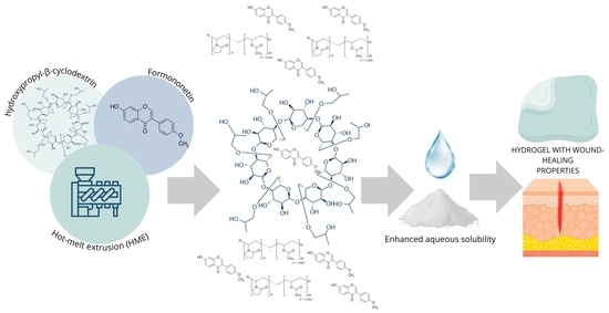
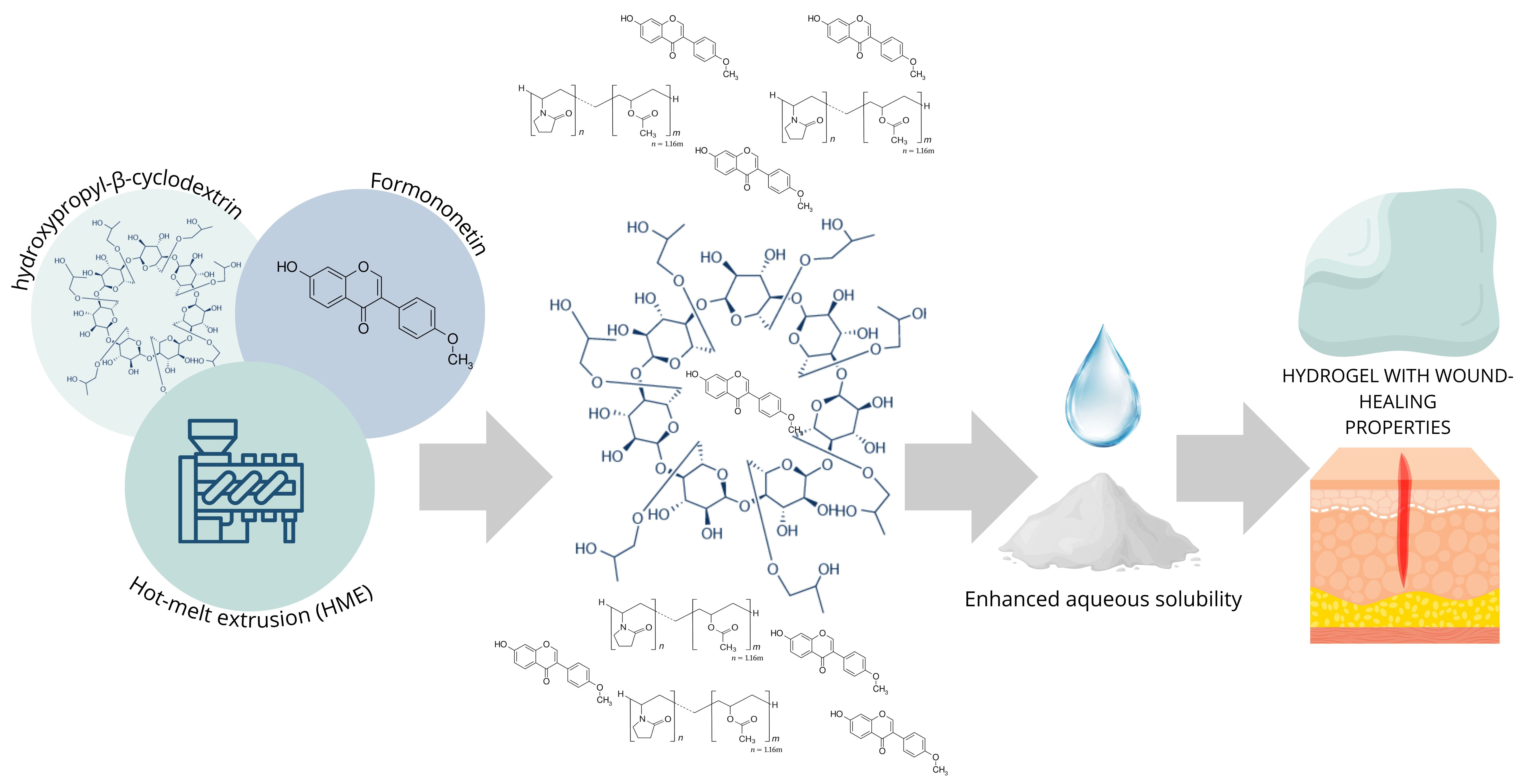
The developed solvent-free hot-melt extruded formononetin systems enable the formulation of hydrophilic hydrogels for topical delivery, offering a scalable approach for incorporating poorly water-soluble polyphenols into wound dressings.

Abstract

Isoflavones are plant-derived polyphenols with broad biological activity; however, their application in topical formulations is limited by poor aqueous solubility. The aim of this study was to enhance the aqueous solubility of formononetin using a solvent-free hot-melt extrusion (HME) approach and to enable its incorporation into a hydrogel formulation suitable for skin delivery. Amorphous formononetin-based systems were prepared by HME using polymeric carriers and hydroxypropyl-β-cyclodextrin, with and without prior inclusion complex formation. The resulting formulations were characterized using XRPD, DSC, and FT-IR/ATR to assess amorphization and intermolecular interactions. Aqueous solubility and skin permeability were evaluated using solubility testing, PAMPA, and Franz diffusion cells. The optimized amorphous system exhibited a substantial increase in apparent aqueous solubility compared to crystalline formononetin while maintaining comparable permeability. Cyclodextrin–formononetin interactions were effectively generated during the extrusion process, rendering pre-inclusion unnecessary. The selected system was successfully incorporated into a hydrogel matrix. This study demonstrates that solvent-free HME combined with cyclodextrins is an effective strategy for improving formononetin solubility and enabling its application in hydrogel-based topical delivery systems.

Graphical Abstract

1. Introduction

Isoflavones belong to the broad class of plant-derived polyphenols and have attracted considerable scientific interest due to their diverse biological activities [1]. Structurally related to endogenous estrogens, isoflavones are best known for their phytoestrogenic properties and have been extensively studied and applied in products intended for women during menopause [2,3]. In recent years, however, increasing attention has been directed toward the non-hormonal biological activities of isoflavones, revealing their potential in entirely different therapeutic areas. Recent studies demonstrate that selected isoflavones exhibit pronounced antioxidant, anti-inflammatory, antimicrobial, and pro-regenerative effects, which are highly relevant for skin and wound repair [4,5]. In vitro and in vivo studies have demonstrated that isoflavones may stimulate fibroblast proliferation and migration, enhance collagen synthesis, reduce excessive inflammatory responses, and modulate key signaling pathways involved in tissue regeneration, such as MAPK and PI3K/Akt [4,6,7,8,9].
Despite their favorable biological profile, the practical application of isoflavones is significantly limited by their poor aqueous solubility. Isoflavones possess rigid, planar aromatic structures that promote strong intermolecular interactions, such as π–π stacking, and a pronounced tendency toward crystallinity, which together contribute to their poor aqueous solubility and consequently low bioavailability [10]. Additionally, formononetin is particularly noteworthy, as it contains only a single hydroxyl group at the 7-position (Figure 1), further limiting its polarity and aqueous solubility [11].
This physicochemical drawback represents a major challenge for their incorporation into hydrophilic dosage forms, including topical and wound-care formulations. Numerous strategies have been proposed to overcome this limitation, such as particle size reduction, solid dispersions, complexation, and nanocarrier-based systems [12,13,14]. However, many of these approaches rely on the use of organic solvents or multistep processing, which may raise concerns regarding scalability, residual solvents, and environmental impact.
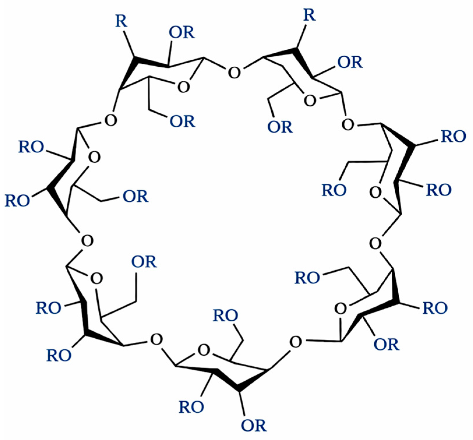
From a contemporary pharmaceutical and materials-engineering perspective, particular attention should be given to solvent-free technologies that comply with the principles of green chemistry and sustainable manufacturing. One such technique is hot-melt extrusion (HME), a continuous, solvent-free process widely employed to improve the apparent solubility of poorly water-soluble compounds by converting them into amorphous solid dispersions (ASDs) [15,16]. HME enables intimate mixing of the active compound with polymeric carriers under controlled thermal and mechanical conditions, leading to disruption of the crystalline lattice and enhancement of dissolution behavior. HME is readily scalable and well established in pharmaceutical manufacturing. Cyclodextrins constitute another well-recognized strategy for solubility enhancement, owing to their ability to form host–guest inclusion complexes with hydrophobic molecules [17]. Among them, hydroxypropyl-β-cyclodextrin is particularly attractive due to its high aqueous solubility and favorable safety profile (Figure 2) [18].
Traditionally, cyclodextrin inclusion complexes are prepared using solvent-based methods; however, recent reports indicate that cyclodextrin–drug interactions may also be generated directly during the hot-melt extrusion process, without the need for prior inclusion steps [19,20]. This emerging approach combines the advantages of cyclodextrin complexation with solvent-free processing, yet remains insufficiently explored.
Enhancing the aqueous solubility of formononetin, a representative isoflavone, creates new opportunities for its incorporation into hydrogel-based delivery systems. ASDs improve drug solubility and enable uniform incorporation into biocompatible hydrogels, offering an underexplored yet promising strategy for personalized wound care applications [21]. Hydrogels are widely used in wound management due to their ability to maintain a moist environment, absorb wound exudate, reduce pain, and protect the wound bed from external contamination [22]. Moreover, hydrogels can serve as effective carriers for bioactive compounds, enabling localized and controlled release at the site of application. By improving the solubility of formononetin, its biological potential, particularly its pro-regenerative and anti-inflammatory effects, can be more effectively exploited in a topical hydrogel formulation, allowing full utilization of its therapeutic properties in skin and wound-healing applications. The primary objective of this study was to investigate whether solvent-free hot-melt extrusion can be used to generate effective cyclodextrin–formononetin interactions, thereby eliminating the need for a prior solvent-based inclusion step while achieving substantial enhancement of aqueous solubility. A secondary aim was to evaluate whether the optimized amorphous system obtained by this approach can be successfully incorporated into a hydrogel matrix and retain biological activity relevant to wound-oriented topical applications.

2. Materials and Methods

2.1. HPLC-DAD Method Development and Validation

Formononetin was quantified using a reversed-phase HPLC-DAD system (Shimadzu LC-2050C, Kyoto, Japan). Separation was achieved on a Reprospher 100 C18 column 3 µm, 4.6 × 100 mm (Dr. Maisch GmbH, Ammerbuch-Entringen, Germany). The mobile phase consisted of 1% acetic acid in water (30%, v/v) and acetonitrile (70%, v/v) delivered isocratically at 1.0 mL/min. The column temperature was maintained at 40 °C and the injection volume was 10 µL. Detection was performed at 260 nm. The method was validated according to ICH Q2 guidelines.

2.2. Preparation of Formononetin-Based Systems

2.2.1. Preparation of Formononetin–Cyclodextrin Inclusion Complexes

Formononetin (200 mg) was dissolved in 150 mL of a methanol:water mixture (1:1, v/v). The appropriate amount of hydroxypropyl-β-cyclodextrin (HP-β-CD) was added under continuous stirring. The solvent was removed under reduced pressure at 50 °C using a rotary evaporator (BÜCHI Labortechnik AG, Flawil, Switzerland). The obtained solid residue was dried to constant weight and stored in a glass desiccator until further use.

2.2.2. Hot-Melt Extrusion Processing

Physical mixtures containing formononetin, HP-β-CD, and Kollidon® VA64 were prepared in defined mass ratios (Table 1). The powders were homogenized and fed into a twin-screw micro-extruder (HAAKE MiniCTW, Thermo Fisher Scientific, Karlsruhe, Germany). Extrusion was performed at a processing temperature of 150 °C and a screw speed of 90 rpm. The extrudates were cooled to room temperature and milled using a mill to obtain uniform powders (Tube Mill control, IKA-Werke GmbH & Co. KG, Staufen, Germany).

2.3. Solid-State Characterization

2.3.1. Powder X-Ray Diffraction (XRPD)

XRPD patterns were recorded using a Bruker D2 Phaser diffractometer with Cu Kα radiation (λ = 1.5406 Å) operating at 30 kV and 10 mA. Samples were scanned over the range 5–40° 2θ with a step size of 0.02° and a counting time of 2 s per step. Selected samples were analyzed with sample rotation. Diffractograms were processed using OriginPro software (OriginPro 2018 SR1 version 9.5.1.195, OriginLab Corporation, Northampton, MA, USA).

2.3.2. Differential Scanning Calorimetry (DSC)

Thermal analyses were performed using a DSC 214 Polyma (Netzsch, Selb, Germany). Approximately 10 mg of each sample was weighed into hermetically sealed aluminum pans. Samples were heated from 30 °C to 280 °C at a rate of 10 °C/min under a nitrogen purge. An empty pan was used as a reference.

2.3.3. FT-IR/ATR Spectroscopy

FT-IR spectra were recorded using a Shimadzu IRTracer-100 equipped with an ATR diamond crystal. Spectra were collected over 4000–400 cm−1 with a resolution of 2 cm−1, averaging 200 scans. Data were analyzed and processed in OriginPro.

2.4. Solubility Studies

Samples equivalent to 20 mg of formononetin were dispersed in 5 mL of distilled water and agitated for 24 h at 60 rpm at room temperature. Suspensions were filtered through 0.22 µm nylon filters, and the concentration of dissolved formononetin was determined by HPLC-DAD under the conditions described above.

2.5. Skin PAMPA Permeation Study

Permeation was assessed using the Skin PAMPA™ system (Pion Inc. Billerica, MA, USA). Membrane 96-well microfilter plates were hydrated overnight with 200 µL of hydration solution. Crystalline formononetin was first dissolved in dimethyl sulfoxide (DMSO) to obtain a working solution, which was subsequently diluted with phosphate-buffered saline (PBS, pH 7.4) to prepare the donor solution. Donor solutions containing the amorphous system were prepared in PBS (pH 7.4). The initial concentration of formononetin in the donor phase was identical for all tested samples. To minimize solvent-related effects, the final DMSO content in the donor phase was kept constant across all samples. Two hundred microliters of the donor solution was added to each donor well, and the plate was assembled and incubated for 5 h at 37 °C with shaking at 50 rpm. After incubation, samples from both donor and acceptor compartments were collected and analyzed by high-performance liquid chromatography with diode array detection (HPLC-DAD, Shimadzu Corp., Kyoto, Japan). Apparent permeability coefficients (Papp) were calculated using standard equations provided by the PAMPA model.

2.6. Scratch Wound Healing Assay

Human normal skin fibroblasts (Hs27 cell line) were purchased from the American Type Culture Collection (Manassas, VA, USA) and maintained in Dulbecco’s Modified Eagle’s Medium—high glucose (DMEM) supplemented with 10% Fetal Bovine Serum (FBS), penicillin (100 U/mL), and streptomycin (100 µg/mL). The cells were cultured in a humidified atmosphere at 5% CO2 and 37 °C. On the day of experiment, cells were collected from monolayers with trypsin/EDTA and seeded onto a 6-well plate at a concentration of 1 × 10⁵ cells/mL. When the confluency of cells reached ~ 90%, the vertical linear scratch was done with a sterile pipette tip. Cells were washed 3 times with PBS to remove cellular debris, and the fresh medium (control group) or medium with 10 µg/mL of FRM or FRM S was added to the respective wells. Images of the scratch were taken at 0 h, 24 h and 48 h using an Olympus CKX53 microscope coupled with an XM10 digital camera (Olympus, Tokyo, Japan). Scratch area at the beginning of the experiment (0 h) was considered 100%. The open wound area was measured with ImageJ software version 1.54g (NIH, Bethesda, MD, USA). Wound closure (in %) was calculated using the following formula:
C l o s e d   w o u n d   a r e a % = o p e n   w o u n d   a r e a   a t   0   h   o p e n   w o u n d   a r e a   a t   24   h   o r   48   h o p e n   w o u n d   a r e a   a t   0   h × 100 %
Results are expressed as the mean percentage of wound closure ± SD.

2.7. Hydrogel Preparation

To obtain a formononetin solution with enhanced solubility, the selected system was mixed with water while maintaining the same API-to-water ratio as used in the solubility study described in Section 2.4. The mixture was sealed with parafilm and stirred on a magnetic stirrer (IKA-Werke GmbH & Co. KG, Staufen, Germany) for 24 h at room temperature. The resulting mixture was then filtered through a 0.22 µm nylon syringe filter. Hydrogel formulations composed of sodium carboxymethylcellulose (CMC), gum arabic (GA), and gelatin (GEL) (with the previously prepared formononetin solution used as the solvent for all components) were prepared by weighing appropriate amounts of each component into a beaker to obtain solutions of 5% sodium carboxymethylcellulose, 3% gum arabic, and 20% gelatin. The solutions were then mixed in a 1:2:1 ratio (CMC:GA:GEL). The total mass of the mixture was recorded, and glycerol was added to achieve a final concentration of 2%. The formulation was stirred on a magnetic stirrer with heating until a clear mixture was obtained. Finally, the hydrogel was poured into a mold and stored in a refrigerator until further use.

2.8. In Vitro Release Studies

The in vitro release profile of formononetin from the hydrogel formulations was assessed using three independently prepared hydrogels containing the previously selected amorphous system. The release study was conducted in 100 mL vessels containing 50 mL of acceptor medium, phosphate buffer pH 7.4 with Tween 80 at 0.3% (v/v). The experiments were performed at 32 °C, matching the surface temperature of human skin. Before the experiment, the acceptor medium was pre-incubated at 32 °C. Each hydrogel sample was fully immersed in the acceptor solution using stainless steel mesh baskets (Agilent Technologies, Santa Clara, CA, USA), enabling diffusion from the entire hydrogel surface. The vessels were placed in a shaking incubator and agitated at 180 rpm throughout the study. At specified time points (5, 10, 15, 30, 45, 60, 120, 180, 240, 300, and 360 min), 1 mL samples were taken from the acceptor medium for HPLC analysis. Immediately after sampling, the removed volume was replaced with an equal volume of pre-incubated acceptor medium to maintain constant volume. The dilution effect from medium replacement was accounted for in the final calculations of cumulative release. The samples were filtered through 0.22 µm nylon syringe filters and then diluted two-fold in pure methanol prior to chromatographic analysis.

2.9. In Vitro Permeation Studies

In vitro permeation was evaluated using vertical Franz diffusion cells (Electrolab EDC-07, Navi Mumbai, India) with an effective diffusion area of 0.64 cm2 and an acceptor volume of 6 mL. Synthetic cellulose membranes were hydrated in phosphate-buffered saline (PBS, pH 7.4) for 1 h prior to use. The acceptor chamber was filled with a mixture of PBS and ethanol (60:40, v/v) maintained at 37 ± 1 °C. The medium was continuously stirred with a magnetic bar at 650 rpm. An accurately weighed amount of hydrogel formulation (1.3 g) containing the optimized amorphous formononetin system was placed on the membrane surface in the donor compartment. The donor was occluded to prevent evaporation. At predetermined time intervals, samples of the acceptor medium were withdrawn and immediately replaced with fresh medium to maintain a constant volume. The concentration of formononetin in each sample was determined using the validated HPLC-DAD method described previously. The cumulative amount of permeated formononetin per unit area (Q/A) was plotted as a function of time, and the steady-state flux (Jss) was calculated from the linear portion of the permeation profile using the equation:
J s s = d Q / d t A
where Q is the cumulative amount of permeated formononetin (µg), t is time (h), and A is the effective diffusion area (cm2).

3. Results and Discussion

3.1. Solid-State Properties of Formononetin Systems

3.1.1. Powder X-Ray Diffraction (XRPD)

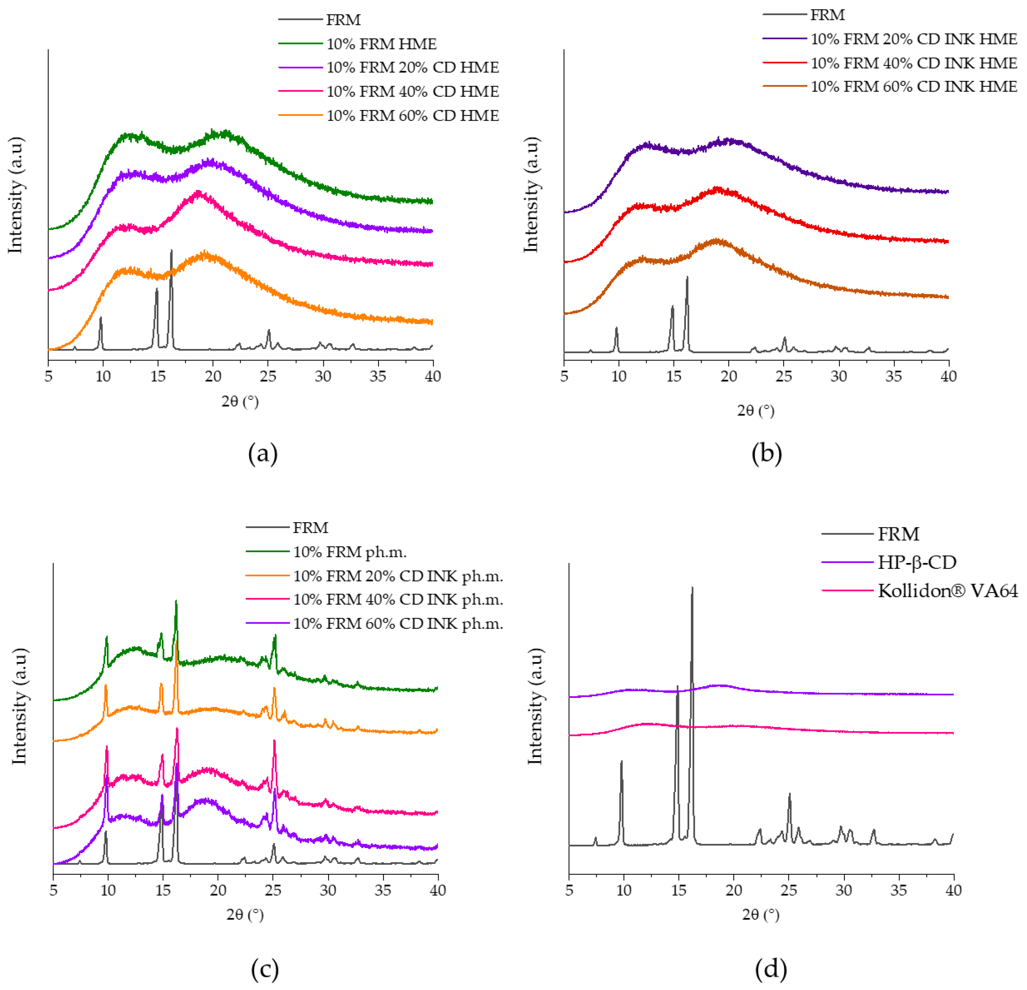
Figure 2 presents the XRPD patterns of pure formononetin (FRM), excipients, and all binary and ternary systems prepared using hot-melt extrusion (HME), pre-inclusion (INK), or physical mixing. In panel (a), crystalline FRM exhibited characteristic sharp reflections in the 5–40° 2θ range. In contrast, all HME formulations containing 10% formononetin showed broad halo patterns with complete disappearance of the drug’s diffraction peaks, regardless of the amount of HP-β-CD (20%, 40% or 60%). These results confirm that HME efficiently converted formononetin into an amorphous state.
Panel (b) shows the XRPD profiles of systems prepared using the pre-inclusion approach followed by extrusion (INK HME). All these samples also demonstrated fully amorphous patterns, indistinguishable from the directly extruded formulations. No residual crystalline reflections of formononetin were detected, indicating that prior inclusion complex formation did not influence the amorphization achieved during HME. Panel (c) presents the physical mixtures (ph.m.) of inclusion formononetin with HP-β-CD at different ratios with polymer. In all mixtures, intense and sharp peaks characteristic of crystalline formononetin were retained. Although increasing the proportion of HP-β-CD reduced peak intensity slightly, the mixtures remained predominantly crystalline. Panel (d) shows the diffraction patterns of the individual excipients. HP-β-CD and Kollidon® VA64 displayed amorphous halo profiles, in contrast to the crystalline formononetin reference. XRPD analysis demonstrated that, under the applied conditions, hot-melt extrusion was an effective approach for obtaining fully amorphous formononetin systems (Figure 3).
A comparable loss of crystalline reflections of formononetin was reported by Wang et al., where complexation with methyl-β-cyclodextrin into electrospun polyvinyl-alcohol nanofibers resulted in the disappearance of characteristic XRPD peaks, confirming disruption of the drug crystality [23]. In the nanosponge system described by Khan et al. [24], amorphization was attributed to molecular confinement of FMN in the porous β-CD network, whereas in the present study, complete loss of crystallinity was achieved through thermo-mechanical processing during HME. A study by Kim et al. [25] presents a distinct solid-state modification strategy, as formononetin was transformed into a new crystalline phase through cocrystal formation with imidazole, rather than being converted into an amorphous form. Powder X-ray diffraction confirmed the disappearance of the native formononetin reflections and the emergence of characteristic peaks corresponding to the newly formed cocrystal lattice. As a result, the solid-state properties of formononetin were altered by changing the crystal structure itself, providing an alternative pathway to improve its physicochemical performance.

3.1.2. Solubility Enhancement of Formononetin

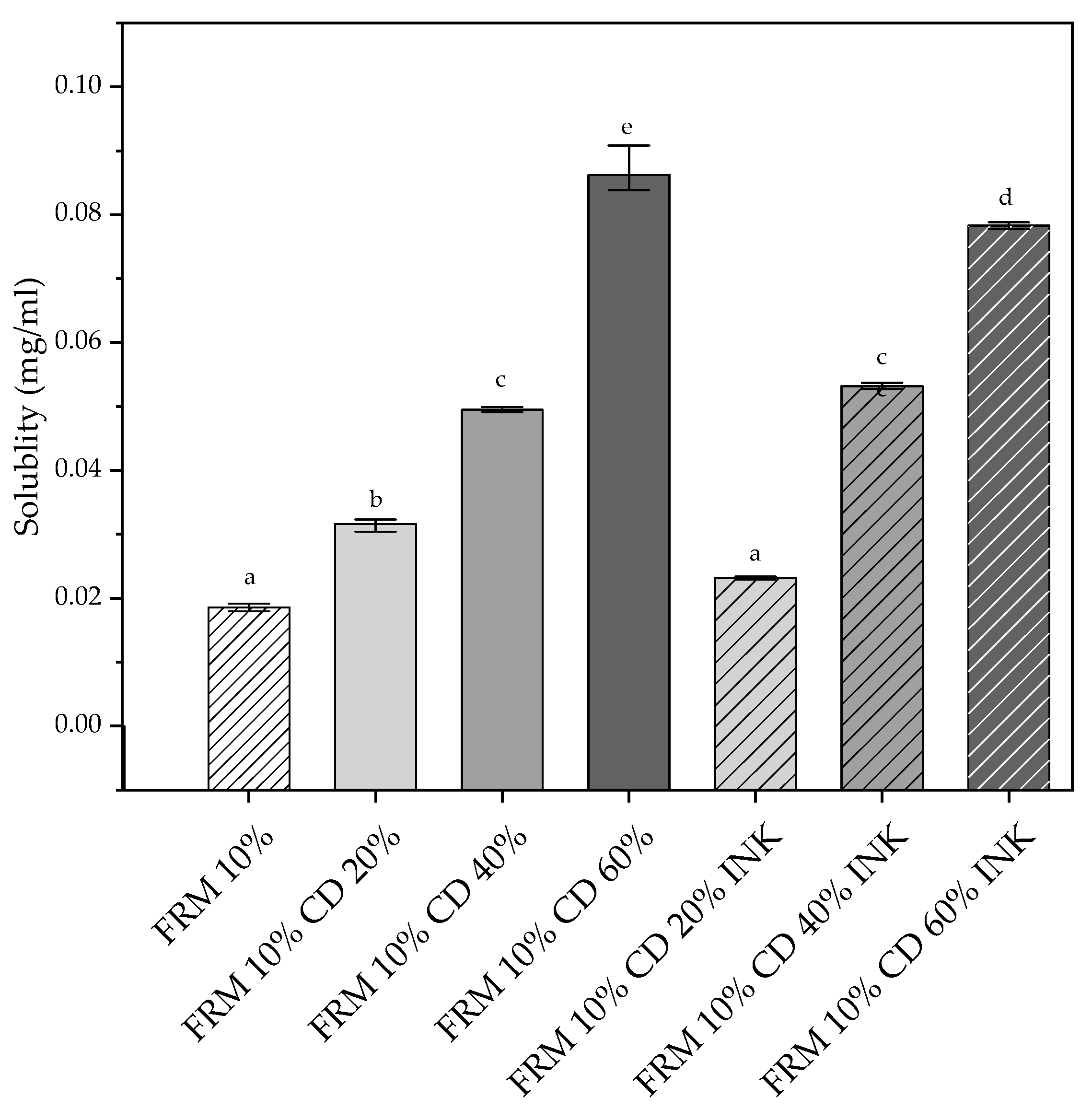
The aqueous solubility of formononetin was evaluated for selected amorphous systems obtained by HME. Under the applied experimental conditions, crystalline formononetin exhibited negligible solubility in water, consistent with literature reports describing its aqueous solubility as 0.56 µg/mL [23]. Figure 4 compares the aqueous solubility of formononetin in amorphous systems prepared by HME with and without a prior cyclodextrin pre-inclusion (INK) step. The formulation containing 10% formononetin without cyclodextrin (FRM 10%) showed a moderate increase in solubility compared to crystalline FRM, which can be attributed primarily to amorphization induced by the hot-melt extrusion process. However, the absence of cyclodextrin limited the extent of solubilization, indicating that polymer-based amorphization alone is insufficient to achieve maximal solubility enhancement. Incorporation of 20% HP-β-CD resulted in only a slight additional increase in formononetin solubility relative to the cyclodextrin-free system. This indicates that at this cyclodextrin level, the amount of HP-β-CD is too low to promote extensive host–guest complexation, and the solubility improvement remains largely governed by the amorphous state of the drug rather than by cyclodextrin-mediated inclusion. Similar to other isoflavone-based amorphous systems, the initial solubility enhancement at low cyclodextrin content can be primarily attributed to drug amorphization rather than to specific host–guest interactions [12]. A marked and statistically significant enhancement of solubility was observed for formulations containing 40% HP-β-CD. At this cyclodextrin content, formononetin solubility increased stepwise, indicating the formation of more effective drug–cyclodextrin interactions within the amorphous polymeric matrix. No statistically significant differences were detected between systems prepared by direct HME and those obtained via prior inclusion, demonstrating that the extrusion process alone is sufficient to induce efficient cyclodextrin–drug interactions at intermediate cyclodextrin loadings. The highest solubility values were obtained for formulations containing 60% HP-β-CD, reaching approximately 90 µg/mL, corresponding to a ~140-fold increase compared to crystalline formononetin. At this cyclodextrin level, the system prepared without prior inclusion exhibited significantly higher solubility than the corresponding INK formulation (p < 0.05), suggesting that direct extrusion promotes more effective molecular dispersion and interaction between formononetin and HP-β-CD when cyclodextrin is present in excess. The results demonstrate that effective solubility enhancement of formononetin requires a critical cyclodextrin threshold between 20% and 40% HP-β-CD. Below this level, solubility is dominated by amorphization, whereas above this threshold, cyclodextrin-mediated interactions become the primary determinant of solubility performance. Prior inclusion does not provide a systematic advantage and may even limit solubility enhancement at high cyclodextrin content, confirming hot-melt extrusion as a sufficient and efficient strategy for generating functional formononetin–cyclodextrin systems.
Comparable trends have been described for cyclodextrin-based systems. Wang et al. [23] demonstrated that the solubility of formononetin increased progressively with increasing methyl-β-cyclodextrin concentration, achieving approximately 20-fold and 50-fold enhancement at 8 and 20 mM Me-β-CD, respectively. Further incorporation of the inclusion complex into electrospun polyvinyl alcohol nanofibers enabled rapid dissolution, which was attributed to amorphization and the high surface area of the fibrous matrix. Agarwal et al. [26] reported that incorporation of formononetin into a phospholipid complex increased its aqueous solubility from 1.61 ± 0.06 µg/mL to 3.62 ± 1.49 µg/mL, and up to 31.46 ± 1.11 µg/mL when combined with a natural bioenhancer. Similarly, Guo et al. [27] showed a linear increase in formononetin solubility with increasing 2-hydroxypropyl-β-cyclodextrin concentration, following an AL-type phase solubility profile and indicating the formation of a stable 1:1 inclusion complex (Ks ≈ 2100 M−1).
Most HME-based solid dispersions reported for flavonoids rely primarily on polymer-induced amorphization, where improved dissolution is achieved by disruption of the crystalline lattice and molecular dispersion of the active compound within the polymer matrix [15,28,29]. In contrast, conventional cyclodextrin inclusion systems are typically prepared using solvent-based methods prior to further formulation, and their performance is governed by pre-formed host-guest complexes [30,31]. The present study demonstrates that, under hot-melt extrusion conditions, cyclodextrins enhance the aqueous solubility of formononetin by enabling effective drug–cyclodextrin interactions generated in situ during processing. Beyond process simplification, this approach offers functional advantages-at intermediate-to-high cyclodextrin loadings, direct extrusion resulted in solubility equal to or higher than that of pre-included systems, suggesting more efficient molecular contact and dynamic host–guest interactions under thermo-mechanical processing. This indicates that HME is not merely a shaping step but an active driver of cyclodextrin-mediated solubilization, providing a scalable and solvent-free alternative to conventional inclusion strategies.

3.1.3. DSC Analysis

Differential scanning calorimetry (DSC) is a fundamental tool for assessing a substance’s crystallinity and identifying its amorphous form. Amorphous materials are characterized by the disappearance of the sharp melting point (Tm), typical of crystalline structures, and the appearance of the glass transition (Tg), which reflects the transition of the material from a glassy to a rubbery state.
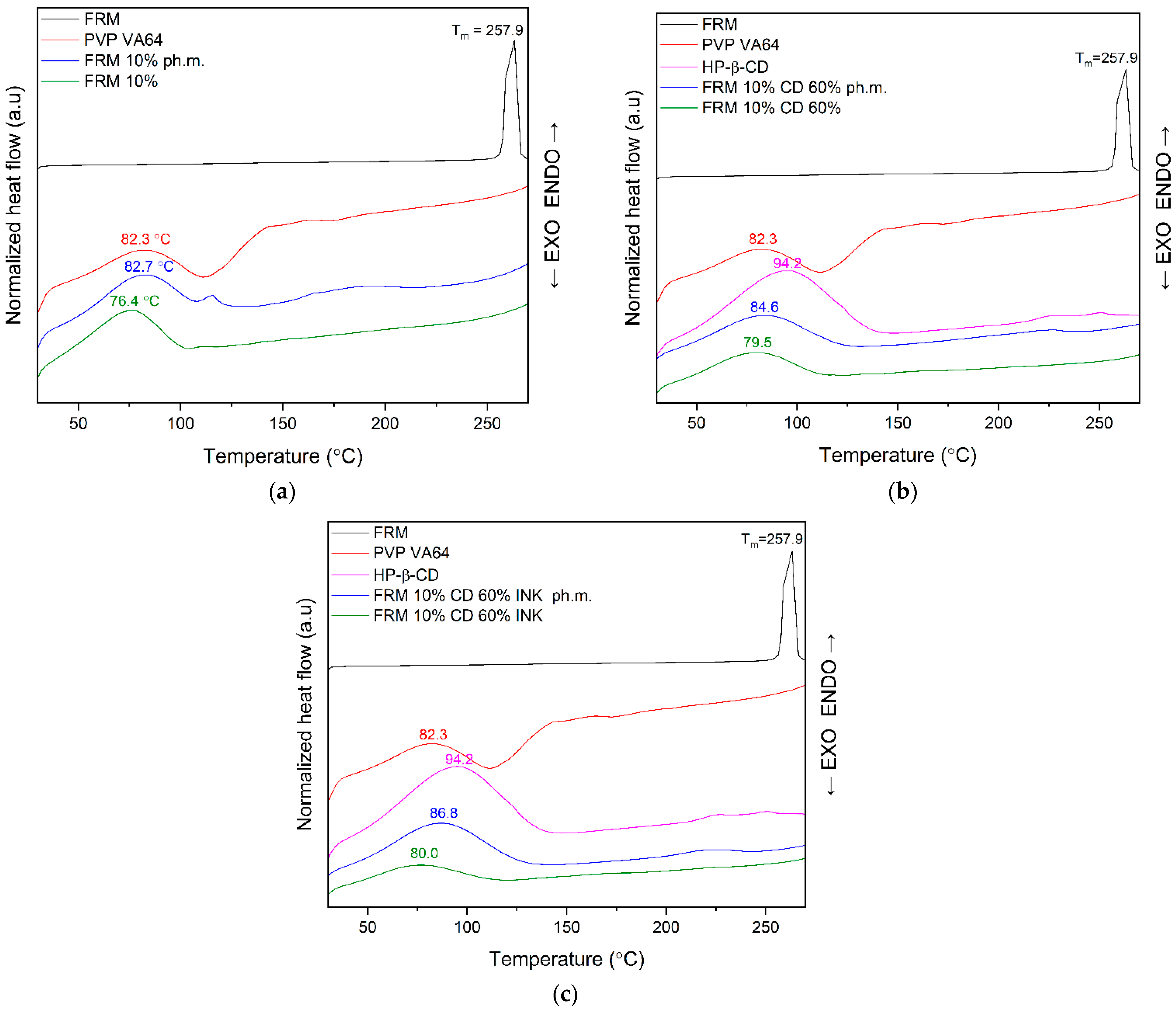
In this study, DSC was employed to evaluate the thermal behavior of crystalline FRM, PVP VA64, HP-β-CD, and amorphous formononetin-based systems prepared by different methods (physical mixture and hot-melt extrusion, HME) (Figure 5a–c).
Pure crystalline FRM exhibited a sharp melting endotherm at ~258 °C, confirming its crystalline nature and thermal stability (Figure 5a). PVP VA64 showed a broad endothermic event related to moisture loss. In the FRM 10% physical mixture, the polymer-related thermal event shifted to lower temperature, indicating plasticization of PVP VA64 by FRM, while the FRM melting peak was not clearly detectable due to low drug loading and dilution effects. In contrast, the FRM 10% HME system exhibited further shifting of the polymer thermal event and complete disappearance of the FRM melting peak, indicating amorphization of FRM during extrusion, consistent with XRPD results. HP-β-CD exhibited a broad dehydration-related endotherm (Figure 5b). In FRM 10% CD 60% systems, the polymer–cyclodextrin thermal event shifted toward lower temperatures after HME, accompanied by reduced intensity, indicating enhanced molecular mobility and stronger intermolecular interactions, while the absence of the FRM melting peak confirms loss of long-range crystalline order. Comparable behavior was observed for the corresponding INK systems, where extrusion induced further thermal-event shifts and confirmed the amorphous state of FRM (Figure 5c). In the HME-tested samples, the absence of the API melting peak may indicate amorphous API or partial amorphization in the matrix. For example, de Oliveira Eloy et al. [32] observed the melting point of ursolic acid (UA) only at 50% of the ph. m., while it was invisible in the thermogram at 20%, 10%, and 5% UA. Barghi et al. [33] attributed the disappearance of the endothermic glibenclamide peak in the PM to the gradual dissolution of the drug in the molten PEG during the DSC measurement. Also, Garbiec et al. [34], with 10% curcumin (CUR) content in ph.m., did not observe the melting point of CUR. The absence of the observed API melting peak may also be related to the dissolution of the API in the molten carrier during heating.

3.1.4. FT-IR Analysis

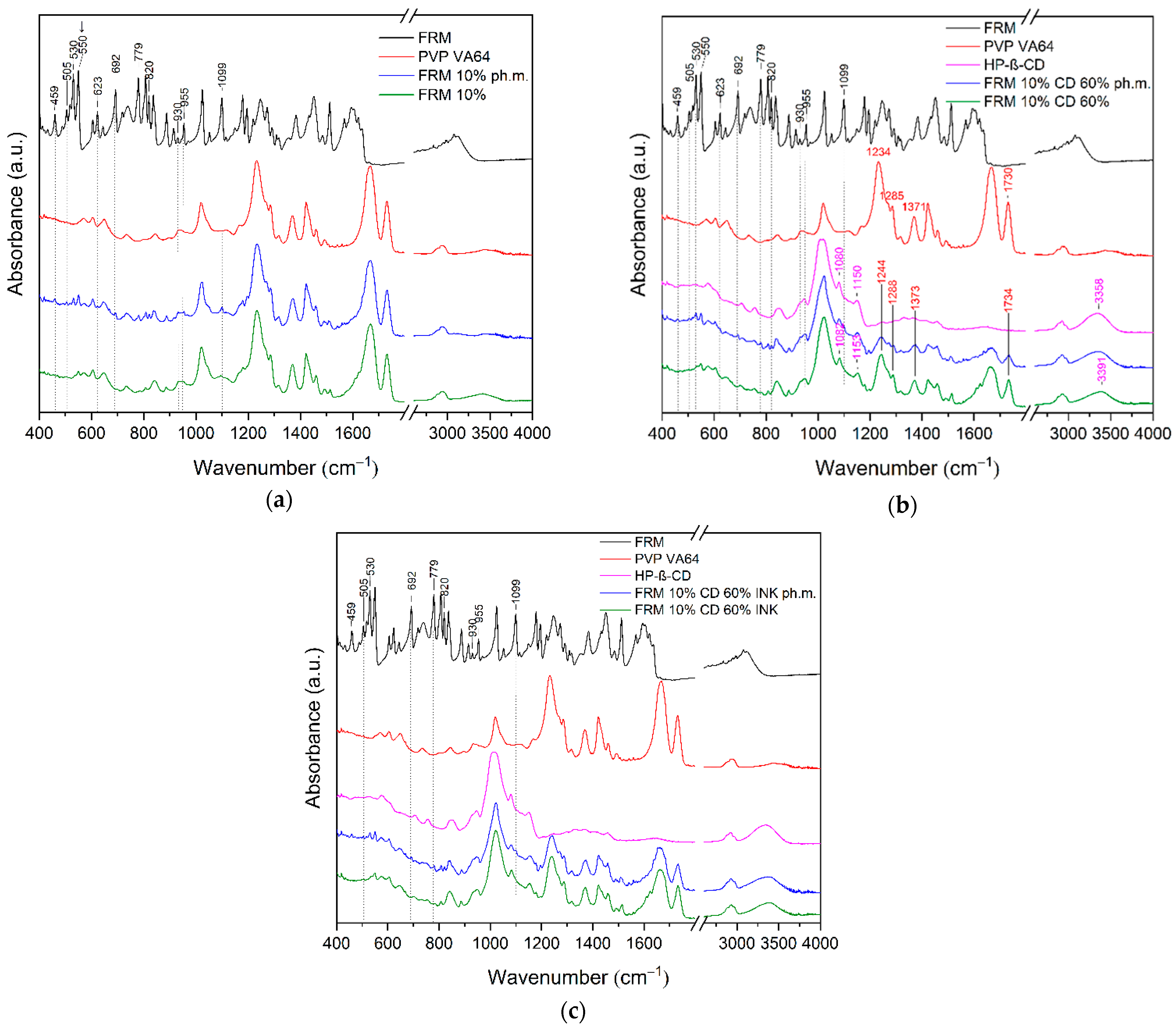
FT-IR spectroscopy was employed to investigate possible intermolecular interactions between FRM, PVP VA64, and HP-β-CD, as well as to evaluate changes in the solid-state organization of FRM resulting from inclusion complex formation and HME processing. The results are presented in Figure 6a–c.
The FT-IR spectrum of pure FRM exhibits characteristic absorption bands of the isoflavone structure, including aromatic C=C stretching (1608–1513 cm−1) and C=O stretching of the γ-pyrone moiety (1650–1620 cm−1), consistent with literature data. Additional bands related to C–O and C–O–C vibrations are observed in the fingerprint region, confirming the crystalline form of FRM [35,36].
In the FRM 10% ph.m. (Figure 6a), the characteristic FRM bands remain clearly detectable without significant shifts, indicating preserved crystalline structure and the absence of specific interactions with PVP VA64. In contrast, their strong attenuation or disappearance in the FRM 10% HME system indicates loss of long-range molecular order and amorphization of FRM, as confirmed by XRPD, while the FT-IR bands of PVP VA64 remain unchanged. In the FRM 10% ph.m. (Figure 6a), the characteristic absorption bands of FRM remain clearly detectable without significant shifts in wavenumber or changes in band shape. This indicates that FRM retains its molecular integrity and crystalline structure and that no specific chemical interactions occur between FRM and PVP VA64 in the physical mixture. The presence of these bands confirms that the components are separate phases and that their interaction is limited to simple physical blending. In contrast, the FT-IR spectrum of FRM 10% shows a pronounced disappearance or strong attenuation of the characteristic FRM bands (attenuation of intensity: 550 cm−1; disappearance: 459, 505, 530, 623, 692, 779, 820, 930, 955, 1099 cm−1). This spectral behavior suggests a loss of the long-range molecular order of FRM and indicates its transition from a crystalline to an amorphous state during the HME process (confirmed by XRPD). Nevertheless, no noticeable changes in the position or shape of the characteristic FT-IR bands of PVP VA64 are observed in the HME system. This may be due to the polymer being the dominant component and acting as a stable matrix for molecularly dispersed FRM molecules.
In the FT-IR spectra of FRM 10% CD 60% ph.m. (Figure 6b), the characteristic FRM bands remain visible, albeit with reduced intensity due to dilution by PVP VA64 and CD, indicating preserved crystalline structure and the absence of significant intermolecular interactions. In contrast, these bands are no longer clearly distinguishable in the FRM 10% CD 60% system, and their attenuation in the fingerprint region indicates loss of long-range molecular order and the presence of FRM in an amorphous or molecularly dispersed state. This effect is commonly interpreted as evidence of inclusion or strong host–guest interactions between the FRM molecule and cyclodextrin derivatives [37].
The FT-IR spectrum of FRM 10% CD 60% INK ph.m. (Figure 6c) is dominated by bands of PVP VA64 and HP-β-CD, while FRM-specific bands are no longer clearly distinguishable, indicating molecular dispersion or amorphization of the drug. The limited spectral changes compared to the FRM 10% CD 60% system suggest weaker intermolecular interactions. Consistent with XRPD data, FRM is likely partially included within the cyclodextrin cavity, with the remaining fraction dispersed in the PVP VA64 matrix, resulting in a fully amorphous final system.
The obtained results may explain the behavior of FRM during the solubility study, indicating the highest solubility for FRM 10% CD 60%, corresponding to the strongest interactions between system components. Slightly lower solubility is observed for FRM 10% CD 60% INK, while the lowest solubility is confirmed for FRM 10%. This correlation supports the conclusion that the extent of intermolecular interactions directly influences the solid-state organization and dissolution behavior of formononetin.
The observed FT-IR spectral changes are consistent with previously reported behavior of poorly soluble flavonoids and isoflavones incorporated into polymeric [38,39,40] and cyclodextrin-based carriers [37,41,42]. According to literature data, the disappearance or strong attenuation of API-specific bands in the fingerprint region is commonly associated with intermolecular interaction such as hydrogen bonding or molecular dispersion of the active compound within the carrier matrix, rather than with chemical degradation. Similar effects have been reported for structurally related isoflavones, such as genistein [12,43] and daidzein [44], dispersed in polymer solid dispersions or cyclodextrin inclusion complexes [31,45,46,47].
The combined XRPD, DSC, and FT-IR results demonstrate that formononetin processed by HME does not form a single, well-defined solid phase but exists as a mixed amorphous system. The complete disappearance of crystalline reflections in XRPD, together with the absence of the melting endotherm in DSC and attenuation of formononetin-specific bands in FT-IR without the emergence of new characteristic signals, confirms loss of long-range order without formation of a new crystalline phase. Accordingly, formononetin is proposed to be present in a heterogeneous amorphous state, in which a fraction of the molecules is molecularly dispersed and stabilized within the polymer matrix, while another fraction is transiently associated with the hydroxypropyl-β-cyclodextrin cavity through dynamic host–guest interactions. Such mixed polymer–cyclodextrin systems are well documented for solvent-free hot-melt extrusion, where cyclodextrin–drug interactions generated in situ coexist with polymer-mediated amorphization rather than forming a fully stoichiometric inclusion complex. Similar mixed amorphous polymer–cyclodextrin systems generated by solvent-free hot-melt extrusion have been reported by Munnangi et al. [20], who demonstrated that cyclodextrin–drug interactions can be formed in situ during HME and coexist with polymer-mediated amorphization rather than yielding fully stoichiometric inclusion complexes. Likewise, Marreto et al. [19] showed that in polymer–HP-β-cyclodextrin systems processed by HME, enhanced solubility arises from a combination of amorphization, dynamic host–guest interactions, and improved wettability, without the formation of a distinct crystalline cyclodextrin complex.

3.2. Permeability in PAMPA SKIN Model

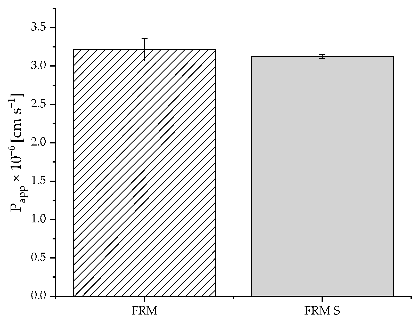
The permeability of formononetin was evaluated using the Skin PAMPA model to assess the impact of solubility enhancement on transmembrane transport. The results demonstrated that the apparent permeability coefficient (Papp) of formononetin from the amorphous system was comparable to that of crystalline formononetin (Figure 7). No statistically significant differences in Papp values were observed between the tested samples, indicating that the incorporation of formononetin into the polymer–cyclodextrin matrix did not adversely affect its passive diffusion across the artificial skin membrane. These findings confirm that the substantial increase in apparent aqueous solubility achieved by the HME process was not accompanied by a reduction in skin permeability, suggesting that formononetin remained available for transmembrane transport in the PAMPA Skin model. This outcome can be attributed to the intrinsic lipophilicity of formononetin and the passive diffusion–controlled nature of the model. As a lipophilic compound, formononetin exhibits favorable membrane affinity, and its permeation may approach a plateau, such that further solubility enhancement does not translate into increased transmembrane flux. Moreover, PAMPA Skin is a simplified, static model that reflects intrinsic passive permeability but does not account for dynamic biological processes or formulation-induced membrane interactions. Since cyclodextrin-assisted hot-melt extrusion improved solubility without altering the molecular structure or diffusion characteristics of formononetin, permeability remained unchanged under these conditions.
Previous work using artificial membrane models demonstrated high PAMPA permeability of formononetin across lipid-like barriers at physiological pH, indicating that its intrinsic permeation capacity is high. In a parallel artificial membrane permeability assay (PAMPA), formononetin demonstrated high permeability at both acidic and neutral pH values, indicating substantial intrinsic capacity for passive diffusion across lipid-like barriers [48]. Ex vivo skin permeation studies have shown that soy isoflavones such as genistein can permeate through excised human skin [49]. Technological modification of soy isoflavone-rich extracts has been shown to enhance in vitro permeability of constituent aglycones, suggesting formulation-dependent modulation of membrane transport. Daruházi et al. reported that inclusion of genistein and daidzein in cyclodextrin complexes significantly improved their aqueous solubility and enhanced their apparent permeability across Caco-2 cell monolayers compared to the pure aglycones, supporting a positive relationship between solubility enhancement and membrane transport for isoflavones [50]. Meng et al. [51] demonstrated that a novel daidzein–piperazine salt exhibited markedly increased aqueous solubility and a concomitant ~34% increase in Caco-2 apparent permeability relative to pure daidzein, illustrating that solubility enhancement can positively influence membrane transport of isoflavone derivatives. Nanomicelle-based genistein formulations have been shown to increase both solubility and Caco-2 cell permeability relative to free isoflavone, further supporting the notion that solubility enhancement strategies can enhance membrane transport of poorly soluble isoflavones [52]. In the present study, the intrinsic permeability of crystalline formononetin was already substantial, and incorporation into the solubility-enhancing formulation did not adversely affect its membrane permeability.

3.3. Wound Healing Potential

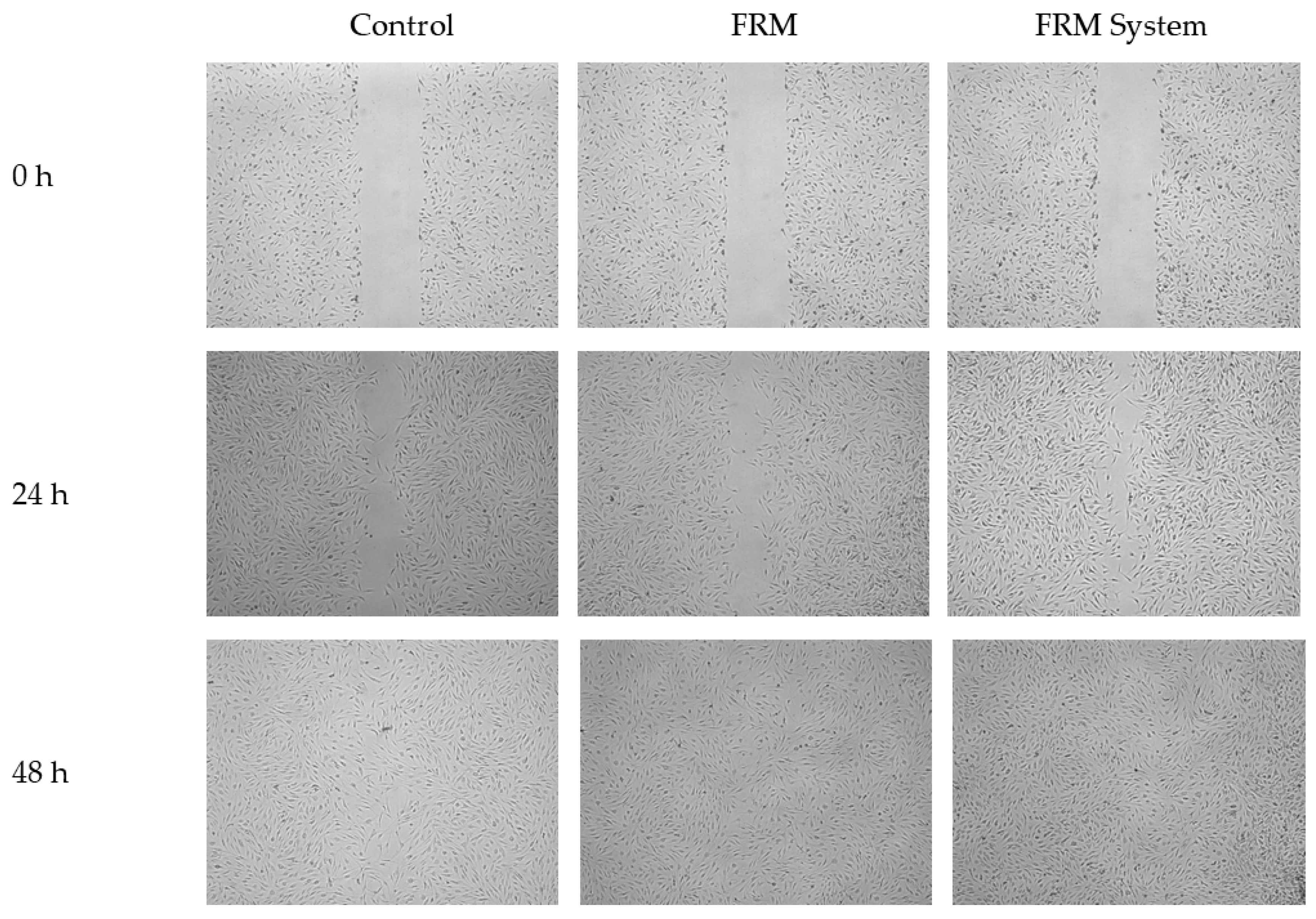
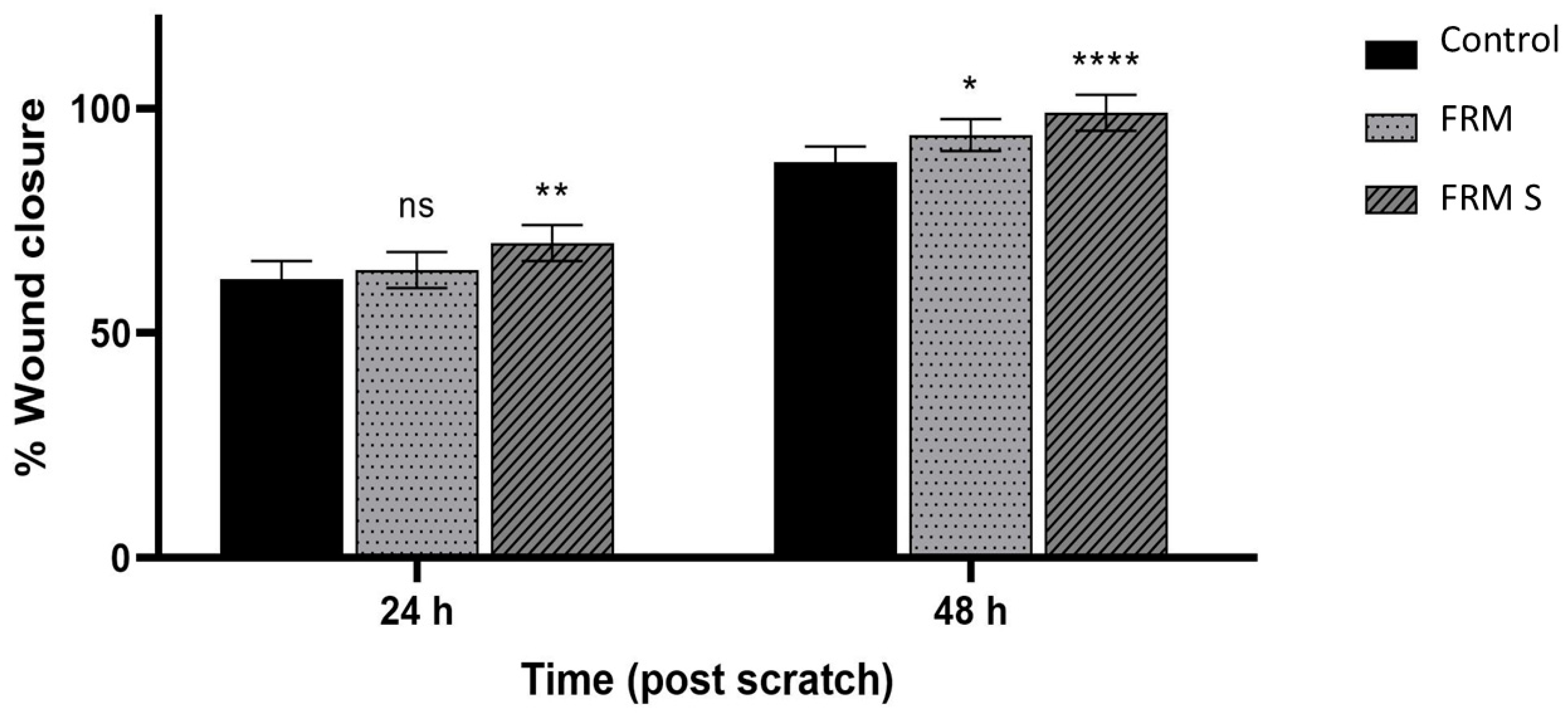
Quantitative analysis of the scratch assay revealed time-dependent wound closure in all experimental groups (Figure 8). After 24 h, untreated control cells achieved approximately 60% wound closure, whereas crystalline formononetin did not produce a statistically significant improvement at this time point. In contrast, cells treated with the amorphous system exhibited significantly higher wound closure at 24 h (** p < 0.01 vs. control), indicating more efficient early delivery of the active compound. After 48 h, wound closure increased in all groups, with both FRM and FRM S significantly enhancing fibroblast migration compared to the control (* p < 0.05 and **** p < 0.0001, respectively). Notably, FRM S achieved nearly complete wound closure (~100%), whereas FRM reached a slightly lower, although still significantly improved, level of closure (Figure 9). These findings demonstrate that the amorphous system remains functionally active in promoting wound closure. The scratch assay further confirms good biological compatibility of the formulated system with human skin fibroblasts.
These findings align with literature data indicating that formononetin supports wound closure primarily by promoting fibroblast migration and survival rather than by inducing nonspecific proliferative effects. Yang et al. [53] demonstrated that formononetin significantly promotes fibroblast proliferation and migration under thermal injury conditions while reducing apoptosis, oxidative stress, and inflammatory responses, effects that were linked to activation of the PI3K/AKT/mTOR pathway. Formononetin showed no significant effect on non-injured fibroblasts, indicating a context-dependent pro-regenerative action rather than nonspecific stimulation of cell growth. Other studies further support the wound-healing potential of formononetin when incorporated into biomaterial-based systems, such as chitosan–oxidized alginate scaffolds, which demonstrated accelerated fibroblast migration in vitro and near-complete wound closure within 48 h. These formononetin-loaded scaffolds also showed high biocompatibility, antibacterial activity, and enhanced wound regeneration in vivo, highlighting the suitability of formononetin as an active component of polymeric wound dressings [54].
The enhanced wound closure observed for the amorphous formononetin system is most likely related to its substantially improved apparent aqueous solubility, which enables higher and more consistent exposure of fibroblasts to biologically active concentrations of the compound. Although passive permeability remained unchanged in the PAMPA Skin model, this assay does not capture cell-dependent uptake mechanisms or membrane interactions that may contribute to biological responses. The limitations of the scratch assay as a simplified model were therefore acknowledged, and the experiment was used as a proof-of-concept to link physicochemical improvement with a measurable biological effect.

3.4. Hydrogel-Based Delivery

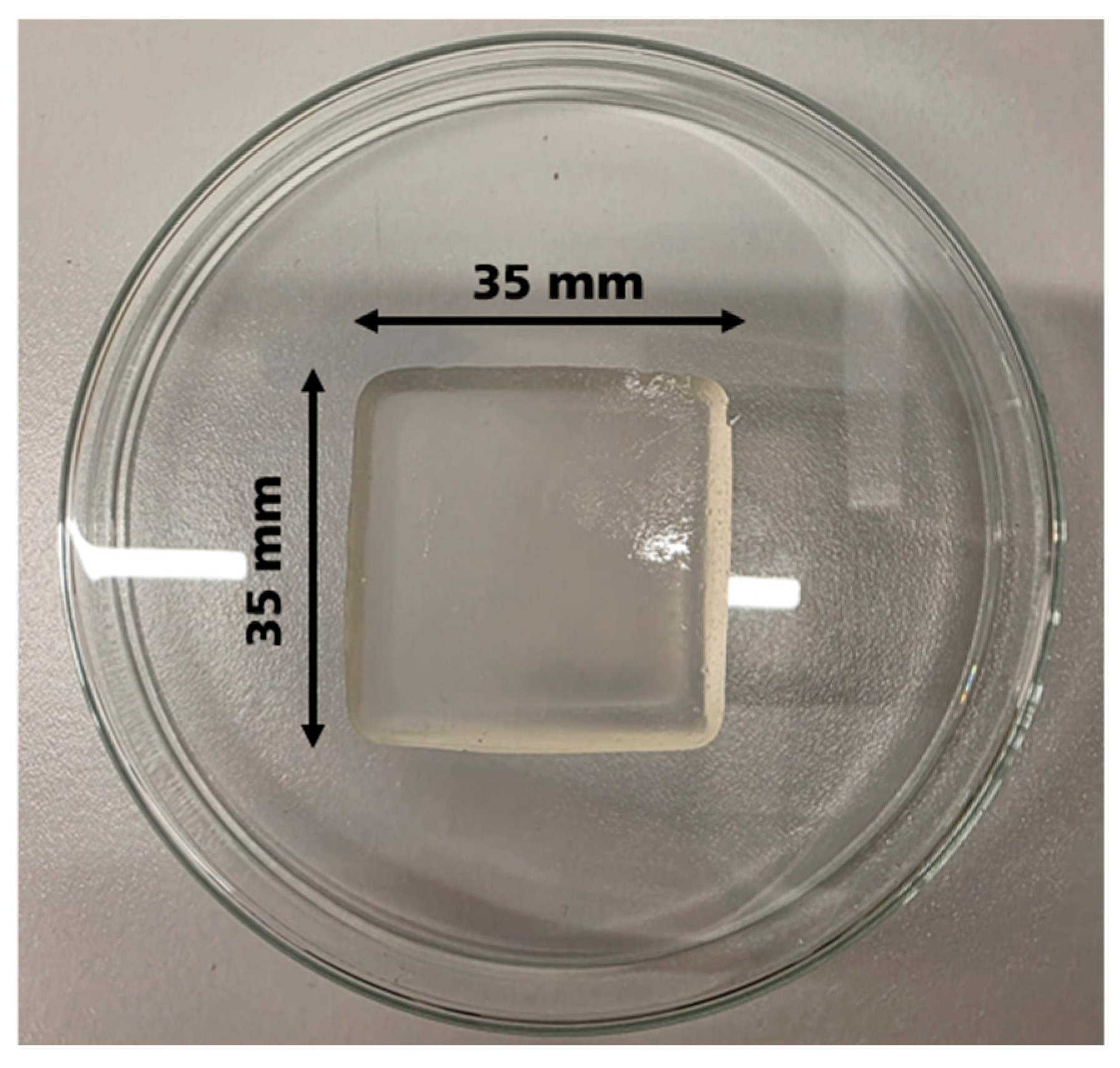
Figure 10 demonstrates the successful formation of a hydrogel patch obtained from the applied polymeric components. The system was prepared in the form of a self-supporting, square-shaped hydrogel (approximately 35 × 35 × 4 mm), which could be easily handled and transferred without structural damage. The material exhibited a uniform, translucent appearance and maintained its integrity upon placement in an aqueous environment. The obtained hydrogel showed sufficient mechanical coherence to retain its shape without additional crosslinking or external support, indicating that the applied formulation strategy enables the fabrication of stable hydrogel-based patches.
Similar self-supporting hydrogel patches have been widely reported as effective platforms for topical and wound-related applications. Both natural and hybrid polymeric hydrogels can be fabricated as mechanically coherent, free-standing materials that retain their shape under hydrated conditions, enabling easy handling and direct application to the wound site [55,56]. In such systems, structural stability is commonly achieved through physical or dual cross-linking within the polymer network, without the need for additional external support [57]. Recent studies further show that hydrogel patches are increasingly designed as active matrices that support the wound-healing process by providing a hydrated environment and enabling localized delivery of therapeutic agents [58,59].

3.4.1. In Vitro Release from Hydrogels

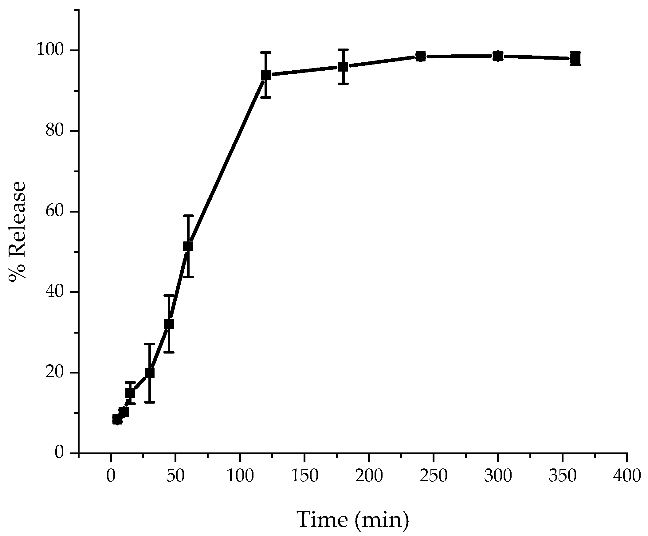
The release profile shows that the hydrogel releases more than 90% of the active compound within approximately 120 min (Figure 11). Kinetic modeling indicates that the release process does not follow zero-order or first-order kinetics, but is governed by a mixed mechanism typical of swelling hydrogel systems (Table 2). The best fit was obtained for the Korsmeyer–Peppas model (R2 = 0.953), with a diffusion exponent n = 0.775, indicating anomalous (non-Fickian) transport, where diffusion through the hydrated polymer network occurs simultaneously with polymer relaxation and swelling. The good agreement with the Higuchi model (R2 = 0.926) confirms the contribution of diffusion, while the high correlation with the Hixson–Crowell model (R2 = 0.950) suggests that matrix erosion and mass loss become significant at later stages of release. Such a sequential release mechanism is characteristic of hydrogel systems that rapidly swell and subsequently undergo gradual structural disintegration.
In vitro release studies allow direct assessment of how the hydrogel matrix controls the availability of the active compound before permeation occurs. Recent publications show that drug release from hydrogels depends mainly on the structure of the swollen polymer network, where both diffusion of the compound and relaxation of polymer chains determine the release rate [60].
Recent studies confirm that biopolymer-based hydrogels provide sustained and controllable release profiles. The extent of swelling and the density of the polymer network strongly affect release behaviour, often slowing drug liberation and limiting initial burst effects [60]. Similar observations have been reported for flavonoid-loaded hydrogels. In the case of kaempferol, release profiles were governed primarily by polymer type and environmental pH, indicating that the hydrogel matrix plays a decisive role in regulating polyphenol availability [61]. These findings are consistent with reports on polymeric hydrogel systems, where the internal structure of the gel acts as the main factor controlling diffusion of active compounds [62]. The release behaviour observed for formononetin in the present study follows the same pattern. The hydrogel matrix limited the rate of drug liberation, which in turn determined the amount of formononetin available for subsequent diffusion across the membrane. This controlled release profile supports the use of the developed hydrogel in applications where prolonged local availability of the active compound is required.

3.4.2. In Vitro Permeation

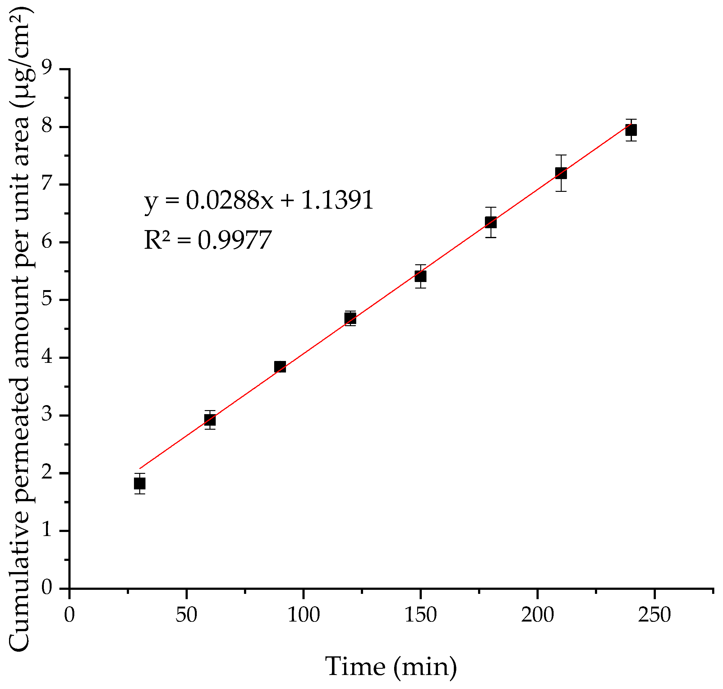
In vitro permeation studies demonstrated steady-state diffusion of formononetin across the membrane, enabling quantitative determination of membrane-controlled flux (Figure 10). Permeation of the active compound from the hydrogel formulation was evaluated using Franz diffusion cells. The cumulative amount of permeated compound was plotted as a function of time, and the permeation profile exhibited a clear linear region, indicating the establishment of steady-state transport across the membrane (Figure 12). The steady-state flux (J) was calculated from the slope of the linear portion of the cumulative permeation curve according to Fick’s first law. Linear regression of the selected time interval resulted in the equation y = 0.0288x + 1.1391 with a high coefficient of determination (R2 = 0.9977). The slope of the regression line corresponds to the permeation rate (dQ/dt), expressed in µg·min−1. The steady-state flux was calculated as 2.70 µg·cm−2·h−1.
The linear permeation profile and high R2 value indicate steady-state, diffusion-controlled transport of formononetin from the hydrogel formulation. This confirms that the hydrogel matrix enables controlled release while maintaining sufficient drug availability for membrane permeation, supporting its suitability for localized topical delivery.
Previous studies using skin-based models have shown that hydrogel systems can modulate drug availability by slowing release and promoting localized delivery rather than rapid systemic transport. Although a cellulose membrane does not replicate skin barrier properties, the present results demonstrate the diffusion feasibility of formononetin after release from the hydrogel and provide a formulation-oriented basis for further skin-relevant studies.
Previous studies have shown that isoflavone aglycone-rich nanoemulsions and their hyaluronic acid-based hydrogels effectively enhance dermal delivery by reducing release rates and increasing isoflavone retention within skin layers. In in vitro permeation studies using porcine skin, these systems promoted preferential accumulation of isoflavones in the dermis, supporting their suitability for localized topical applications rather than systemic delivery [63]. Dias et al. confirmed that formononetin showed high permeation when formulated individually, particularly after complexation with hydroxypropyl-β-cyclodextrin, while biochanin A exhibited a ~2.7-fold increase in epidermal and dermal permeation [64]. The literature indicates that hydrogel swelling and viscosity are critical parameters governing drug permeation, with highly swollen polymer networks often retarding diffusion and favoring controlled, localized delivery rather than rapid skin permeation [65]. In agreement with literature data on quercetin hydrogels, where cellulose-based matrices enhanced skin retention at the expense of rapid permeation, the moderate flux of formononetin observed in the study reflects controlled release governed by the hydrogel structure [66]. The Franz diffusion results confirm that the developed hydrogel efficiently releases formononetin and does not act as a diffusion barrier after drug liberation, supporting its use as a platform for controlled, localized delivery.

4. Conclusions

This study demonstrates that solvent-free hot-melt extrusion enables the efficient in situ formation of cyclodextrin–formononetin interactions, rendering a prior inclusion step unnecessary while providing substantial enhancement of apparent aqueous solubility. The hydrogel formulation and biological evaluation serve as application-oriented validation, confirming that the optimized amorphous system remains suitable for wound-related topical delivery.
Hot-melt extrusion combined with hydroxypropyl-β-cyclodextrin enabled the solvent-free production of fully amorphous formononetin-based systems, demonstrating a pronounced increase in apparent aqueous solubility compared to crystalline formononetin. The extent of solubility enhancement was composition-dependent, while no differences were observed between systems produced by direct extrusion and those prepared via a prior solvent-based inclusion step, indicating that cyclodextrin–formononetin interactions can be effectively generated during HME and rendering pre-inclusion unnecessary.
The optimized amorphous system maintained comparable passive permeability in the Skin PAMPA model, suggesting that solubility improvement did not compromise transmembrane transport. The selected formulation was successfully incorporated into a self-supporting hydrogel patch and exhibited rapid, non-Fickian release governed by combined diffusion and polymer relaxation, as described by the Korsmeyer–Peppas model. Franz diffusion studies further confirmed controlled membrane transport from the hydrogel, yielding a measurable steady-state flux consistent with localized topical delivery. These findings support solvent-free HME as a scalable and green strategy to enhance formononetin solubility and to enable its formulation into hydrogel-based skin delivery platforms for potential regenerative and wound-related applications. It should be noted that while the present study establishes a solid formulation and mechanistic basis for cyclodextrin-assisted solubility enhancement and controlled release of formononetin, additional parameters relevant to wound application, such as mechanical integrity, adhesion, and stability under wound-relevant conditions, were not addressed. These aspects should be evaluated in future studies to further support the translational potential of the proposed hydrogel system.

Author Contributions

Conceptualization, J.C.-P. and A.G.; methodology, A.G. and N.R.; software, A.G. and N.R.; validation, A.G., N.R. and M.I.; formal analysis, A.G. and N.R.; investigation, A.G., N.R., M.I., P.T. and B.K.-P.; resources, J.C.-P.; data curation, A.G.; writing—original draft preparation, A.G. and N.R.; writing—review and editing, J.C.-P., B.M.-K. and M.I.; visualization, A.G., N.R. and B.K.-P. supervision, J.C.-P. and B.M.-K.; project administration, M.I.; funding acquisition, J.C.-P. All authors have read and agreed to the published version of the manuscript.

Funding

This research was funded in whole by National Science Centre, Poland, the grant Preludium BIS nr 2023/50/O/NZ7/00445.

Institutional Review Board Statement

Not applicable.

Informed Consent Statement

Not applicable.

Data Availability Statement

The original data presented in this study are openly available in the Zenodo repository under the same title as the publication (DOI:10.5281/zenodo.18361488).

Conflicts of Interest

The authors declare no conflicts of interest.

Abbreviations

The following abbreviations are used in this manuscript:
HMEHot-melt Extrusion
XRPDX-ray Powder Diffraction
DSCDifferential Scanning Calorimetry
FRIT/ATRFourier Transformed Infrared Spectroscopy/Attenuated Total Reflectance
PAMPAParallel Artificial Membrane Permeability
MAPKMitogen-activated Protein Kinases
PI3K/AktPhosphatidylinositol 3-kinase/Protein Kinase B
HPLC-DADHigh-Performance Liquid Chromatography with Diode Array Detection
HP-β-CDHydroxypropyl-β-cyclodextrin
DMSODimethyl Sulfoxide
PBSPhosphate-Buffered Saline
DMEMDulbecco’s Modified Eagle’s Medium
FBSFetal Bovine Serum
EDTAEthylenediaminetetraacetic Acid
SDStandard Deviation
CMCCarboxymethylcellulose
GAGum Arabic
GELGelatin
INK HMEPre-inclusion Formulations Followed by Extrusion
Ph.m.Physical Mixture
Me-β-CDMethyl-β-cyclodextrin
TmMelting Temperature
FRMFormononetin
CDCyclodextrins
PappApparent Permeability Coefficient
mTORMechanistic Target of Rapamycin
FRM SFormononetin from Amorphous System
ANOVAAnalysis of Variance

References

  1. Zhang, Z.; Li, X.; Sang, S.; McClements, D.J.; Chen, L.; Long, J.; Jiao, A.; Jin, Z.; Qiu, C. Polyphenols as Plant-Based Nutraceuticals: Health Effects, Encapsulation, Nano-Delivery, and Application. Foods 2022, 11, 2189. [Google Scholar] [CrossRef] [Scilit] [PubMed]
  2. Desmawati, D.; Sulastri, D. Phytoestrogens and Their Health Effect. Open Access Maced. J. Med. Sci. 2019, 7, 495–499. [Google Scholar] [CrossRef] [Scilit]
  3. Patra, S.; Gorai, S.; Pal, S.; Ghosh, K.; Pradhan, S.; Chakrabarti, S. A Review on Phytoestrogens: Current Status and Future Direction. Phytother. Res. 2023, 37, 3097–3120. [Google Scholar] [CrossRef] [Scilit]
  4. Wójciak, M.; Drozdowski, P.; Skalska-Kamińska, A.; Zagórska-Dziok, M.; Ziemlewska, A.; Nizioł-Łukaszewska, Z.; Latalska, M. Protective, Anti-Inflammatory, and Anti-Aging Effects of Soy Isoflavones on Skin Cells: An Overview of In Vitro and In Vivo Studies. Molecules 2024, 29, 5790. [Google Scholar] [CrossRef] [Scilit]
  5. Mahajan, N.M.; Wanaskar, K.; Ali, N.; Mahapatra, D.K.; Iqbal, M.; Bhat, A.R.; Kaleem, M. Innovative Wound Healing Hydrogel Containing Chicken Feather Keratin and Soy Isoflavone Genistein: In Vivo Studies. Gels 2023, 9, 462. [Google Scholar] [CrossRef] [Scilit] [PubMed]
  6. Kim, Y.M.; Huh, J.S.; Lim, Y.; Cho, M. Soy Isoflavone Glycitin (4’-Hydroxy-6-Methoxyisoflavone-7-D-Glucoside) Promotes Human Dermal Fibroblast Cell Proliferation and Migration via TGF-β Signaling. Phytother. Res. 2015, 29, 757–769. [Google Scholar] [CrossRef] [Scilit]
  7. Pawlicka, M.A.; Zmorzyński, S.; Popek-Marciniec, S.; Filip, A.A. The Effects of Genistein at Different Concentrations on MCF-7 Breast Cancer Cells and BJ Dermal Fibroblasts. Int. J. Mol. Sci. 2022, 23, 12360. [Google Scholar] [CrossRef] [Scilit]
  8. Feng, Z.; Zhang, N.; Bai, J.; Lin, Q.; Xie, Y.; Xia, Y. Biochanin A Inhibits Cardiac Hypertrophy and Fibrosis in Vivo and in Vitro. Biomed. Pharmacother. 2024, 170, 116002. [Google Scholar] [CrossRef] [Scilit]
  9. Rizzo, J.; Min, M.; Adnan, S.; Afzal, N.; Maloh, J.; Chambers, C.J.; Fam, V.; Sivamani, R.K. Soy Protein Containing Isoflavones Improves Facial Signs of Photoaging and Skin Hydration in Postmenopausal Women: Results of a Prospective Randomized Double-Blind Controlled Trial. Nutrients 2023, 15, 4113. [Google Scholar] [CrossRef] [Scilit]
  10. Huang, S.; Xue, Q.; Xu, J.; Ruan, S.; Cai, T. Simultaneously Improving the Physicochemical Properties, Dissolution Performance, and Bioavailability of Apigenin and Daidzein by Co-Crystallization with Theophylline. J. Pharm. Sci. 2019, 108, 2982–2993. [Google Scholar] [CrossRef] [Scilit]
  11. Jin, M.; Wei, L.; Wang, J.; Shen, Y.; Gao, L.; Zhao, F.; Gao, Q.; Ma, Y.; Sun, Y.; Lin, Y.; et al. Formononetin: A Review of Its Source, Pharmacology, Drug Combination, Toxicity, Derivatives, and Drug Delivery Systems. Front. Pharmacol. 2025, 16, 1534798. [Google Scholar] [CrossRef] [Scilit]
  12. Garbiec, E.; Rosiak, N.; Zalewski, P.; Tajber, L.; Cielecka-Piontek, J. Genistein Co-Amorphous Systems with Amino Acids: An Investigation into Enhanced Solubility and Biological Activity. Pharmaceutics 2023, 15, 2653. [Google Scholar] [CrossRef] [Scilit]
  13. Liu, B.; Thum, C.; Wang, Q.; Feng, C.; Li, T.; Damiani Victorelli, F.; Li, X.; Chang, R.; Chen, S.; Gong, Y.; et al. The Fortification of Encapsulated Soy Isoflavones and Texture Modification of Soy Milk by α-Lactalbumin Nanotubes. Food Chem. 2023, 419, 135979. [Google Scholar] [CrossRef] [Scilit] [PubMed]
  14. Zverev, Y.F.; Rykunova, A.Y. Modern Nanocarriers as a Factor in Increasing the Bioavailability and Pharmacological Activity of Flavonoids. Appl. Biochem. Microbiol. 2022, 58, 1002–1020. [Google Scholar] [CrossRef] [Scilit]
  15. Azad, M.O.K.; Kim, W.W.; Jin, C.W.; Kang, W.S.; Park, C.H.; Cho, D.H. Development of a Polymer-Mediated Soybean Nanocomposite by Hot Melt Extrusion to Improve Its Functionality and Antioxidant Properties. Foods 2019, 8, 41. [Google Scholar] [CrossRef] [Scilit]
  16. Wdowiak, K.; Pietrzak, R.; Tykarska, E.; Cielecka-Piontek, J. Hot-Melt Extrusion as an Effective Technique for Obtaining an Amorphous System of Curcumin and Piperine with Improved Properties Essential for Their Better Biological Activities. Molecules 2023, 28, 3848. [Google Scholar] [CrossRef] [Scilit]
  17. Inam, M.; Sareh Sadat, M.-F.; Chen, W. Cyclodextrin Based Host-Guest Inclusion Complex, an Approach to Enhancing the Physicochemical and Biopharmaceutical Application of Poorly Water-Soluble Drugs. Chem. Res. Chin. Univ. 2023, 39, 857–861. [Google Scholar] [CrossRef] [Scilit]
  18. Musuc, A.M. Cyclodextrins: Advances in Chemistry, Toxicology, and Multifaceted Applications. Molecules 2024, 29, 5319. [Google Scholar] [CrossRef] [Scilit] [PubMed]
  19. Marreto, R.N.; Cardoso, G.; dos Santos Souza, B.; Martin-Pastor, M.; Cunha-Filho, M.; Taveira, S.F.; Concheiro, A.; Alvarez-Lorenzo, C. Hot Melt-Extrusion Improves the Properties of Cyclodextrin-Based Poly(Pseudo)Rotaxanes for Transdermal Formulation. Int. J. Pharm. 2020, 586, 119510. [Google Scholar] [CrossRef] [Scilit] [PubMed]
  20. Munnangi, S.R.; Youssef, A.A.A.; Narala, N.; Lakkala, P.; Vemula, S.K.; Alluri, R.; Zhang, F.; Repka, M.A. Continuous Manufacturing of Solvent-Free Cyclodextrin Inclusion Complexes for Enhanced Drug Solubility via Hot-Melt Extrusion: A Quality by Design Approach. Pharmaceutics 2023, 15, 2203. [Google Scholar] [CrossRef] [Scilit]
  21. Ignacyk, M.; Krasiński, Z.; Michniak-Kohn, B.; Cielecka-Piontek, J. Innovative Wound Healing Strategy Through Amorphization of Active Pharmaceutical Ingredients as an Effective Approach for Hydrogel Formulation. Pharmaceuticals 2025, 18, 1427. [Google Scholar] [CrossRef] [Scilit]
  22. Gounden, V.; Singh, M. Hydrogels and Wound Healing: Current and Future Prospects. Gels 2024, 10, 43. [Google Scholar] [CrossRef] [Scilit]
  23. Wang, Y.; Deng, Z.; Wang, X.; Shi, Y.; Lu, Y.; Fang, S.; Liang, X. Formononetin/Methyl-β-Cyclodextrin Inclusion Complex Incorporated into Electrospun Polyvinyl-Alcohol Nanofibers: Enhanced Water Solubility and Oral Fast-Dissolving Property. Int. J. Pharm. 2021, 603, 120696. [Google Scholar] [CrossRef] [Scilit] [PubMed]
  24. Khan, N.; Bhardwaj, N.; Saneja, A. Structural and Physicochemical Characterization of Formononetin Encapsulated β-Cyclodextrin Nanosponges for Improved Anticancer Activity. J. Mol. Struct. 2026, 1354, 144904. [Google Scholar] [CrossRef] [Scilit]
  25. Kim, J.; Lim, S.; Kim, M.; Ban, E.; Kim, Y.; Kim, A. Using the Cocrystal Approach as a Promising Drug Delivery System to Enhance the Dissolution and Bioavailability of Formononetin Using an Imidazole Coformer. Pharmaceuticals 2024, 17, 1444. [Google Scholar] [CrossRef] [Scilit]
  26. Agarwal, A.; Dadge, S.; Garg, R.; Sharma, R.K.; Chauhan, D.; Katekar, R.; Rathaur, S.; Mitra, K.; Gayen, J.R. Bioavailability Enhancement of Formononetin by Incorporation of Natural Bioenhancer in Phospholipid Complex. Pharm. Dev. Technol. 2024, 29, 1148–1161. [Google Scholar] [CrossRef] [Scilit] [PubMed]
  27. Guo, B.; Xu, D.; Liu, X.; Liao, C.; Li, S.; Huang, Z.; Li, X.; Yi, J. Characterization and Cytotoxicity of PLGA Nanoparticles Loaded with Formononetin Cyclodextrin Complex. J. Drug Deliv. Sci. Technol. 2017, 41, 375–383. [Google Scholar] [CrossRef] [Scilit]
  28. Ishimoto, K.; Shimada, Y.; Ohno, A.; Otani, S.; Ago, Y.; Maeda, S.; Lin, B.; Nunomura, K.; Hino, N.; Suzuki, M.; et al. Physicochemical and Biochemical Evaluation of Amorphous Solid Dispersion of Naringenin Prepared Using Hot-Melt Extrusion. Front. Nutr. 2022, 9, 850103. [Google Scholar] [CrossRef] [Scilit]
  29. Danilova, A.A.; Gusev, K.A.; Maimistov, D.N.; Flisyuk, E.V. Hot Melt Extrusion Technology as a Modern Strategy for Improving the Bioavailability of Flavonoids. Pharm. Chem. J. 2024, 58, 297–305. [Google Scholar] [CrossRef] [Scilit]
  30. You, G.-J.; Sun, L.-L.; Cao, X.-X.; Li, H.-H.; Wang, M.; Liu, Y.-N.; Ren, X.-L. Comprehensive Evaluation of Solubilization of Flavonoids by Various Cyclodextrins Using High Performance Liquid Chromatography and Chemometry. LWT 2018, 94, 172–177. [Google Scholar] [CrossRef] [Scilit]
  31. Fenyvesi, F.; Klusóczki, Á.; Rusznyák, Á.; Zsebik, B.; Bácskay, I.; Váradi, J. Cyclodextrin-Based Delivery Systems for Flavonoids: Mechanisms, Advances, Formulation, and Application Opportunities. Antioxidants 2025, 14, 998. [Google Scholar] [CrossRef] [Scilit]
  32. de Oliveira Eloy, J.; Saraiva, J.; de Albuquerque, S.; Marchetti, J.M. Solid Dispersion of Ursolic Acid in Gelucire 50/13: A Strategy to Enhance Drug Release and Trypanocidal Activity. AAPS PharmSciTech 2012, 13, 1436–1445. [Google Scholar] [CrossRef] [Scilit]
  33. Barghi, L.; Vekalati, A.; Jahangiri, A. Stability-Enhanced Ternary Solid Dispersions of Glyburide: Effect of Preparation Method on Physicochemical Properties. Adv. Pharmacol. Pharm. Sci. 2023, 2023, 2641153. [Google Scholar] [CrossRef] [Scilit]
  34. Garbiec, E.; Rosiak, N.; Sip, S.; Zalewski, P.; Cielecka-Piontek, J. Curcumin Solubility and Bioactivity Enhancement Through Amorphization with Tryptophan via Supercritical Fluid Technology. Int. J. Mol. Sci. 2025, 26, 855. [Google Scholar] [CrossRef] [Scilit]
  35. Srivastava, A.; Mishra, R.; Kumar, S.; Dev, K.; Tandon, P.; Maurya, R. Molecular Structure, Spectral Investigation (1H NMR, 13C NMR, UV–Visible, FT-IR, FT-Raman), NBO, Intramolecular Hydrogen Bonding, Chemical Reactivity and First Hyperpolarizability Analysis of Formononetin [7-Hydroxy-3(4-Methoxyphenyl)Chromone]: A Quantum Chemical Study. J. Mol. Struct. 2015, 1084, 55–73. [Google Scholar] [CrossRef] [Scilit]
  36. Guo, B.; Liao, C.; Liu, X.; Yi, J. Preliminary Study on Conjugation of Formononetin with Multiwalled Carbon Nanotubes for Inducing Apoptosis via ROS Production in HeLa Cells. Drug Des. Dev. Ther. 2018, 12, 2815–2826. [Google Scholar] [CrossRef] [Scilit] [PubMed]
  37. Wdowiak, K.; Rosiak, N.; Tykarska, E.; Żarowski, M.; Płazińska, A.; Płaziński, W.; Cielecka-Piontek, J. Amorphous Inclusion Complexes: Molecular Interactions of Hesperidin and Hesperetin with HP-Β-CD and Their Biological Effects. Int. J. Mol. Sci. 2022, 23, 4000. [Google Scholar] [CrossRef] [Scilit]
  38. Rosiak, N.; Tykarska, E.; Cielecka-Piontek, J. Myricetin Amorphous Solid Dispersions—Antineurodegenerative Potential. Molecules 2024, 29, 1287. [Google Scholar] [CrossRef] [Scilit] [PubMed]
  39. Rosiak, N.; Wdowiak, K.; Tykarska, E.; Cielecka-Piontek, J. Amorphous Solid Dispersion of Hesperidin with Polymer Excipients for Enhanced Apparent Solubility as a More Effective Approach to the Treatment of Civilization Diseases. Int. J. Mol. Sci. 2022, 23, 15198. [Google Scholar] [CrossRef] [Scilit]
  40. Rosiak, N.; Tykarska, E.; Cielecka-Piontek, J. Mechanochemical Approach to Obtaining a Multicomponent Fisetin Delivery System Improving Its Solubility and Biological Activity. Int. J. Mol. Sci. 2024, 25, 3648. [Google Scholar] [CrossRef] [Scilit]
  41. Stasiłowicz, A.; Tykarska, E.; Lewandowska, K.; Kozak, M.; Miklaszewski, A.; Kobus-Cisowska, J.; Szymanowska, D.; Plech, T.; Jenczyk, J.; Cielecka-Piontek, J. Hydroxypropyl-β-Cyclodextrin as an Effective Carrier of Curcumin—Piperine Nutraceutical System with Improved Enzyme Inhibition Properties. J. Enzym. Inhib. Med. Chem. 2020, 35, 1811–1821. [Google Scholar] [CrossRef] [Scilit]
  42. Stasiłowicz-Krzemień, A.; Rosiak, N.; Płazińska, A.; Płaziński, W.; Miklaszewski, A.; Tykarska, E.; Cielecka-Piontek, J. Cyclodextrin Derivatives as Promising Solubilizers to Enhance the Biological Activity of Rosmarinic Acid. Pharmaceutics 2022, 14, 2098. [Google Scholar] [CrossRef] [Scilit]
  43. Serebrenik, A.A.; Verduyn, C.W.; Kaytor, M.D. Safety, Pharmacokinetics, and Biomarkers of an Amorphous Solid Dispersion of Genistein, a Radioprotectant, in Healthy Volunteers. Clin. Pharmacol. Drug Dev. 2023, 12, 190–201. [Google Scholar] [CrossRef] [Scilit]
  44. Zhang, Z.; Wang, S.; Wang, Q.; Ye, Q.; Wang, W. Experimental and Theoretical Investigation of the Formation of Daidzein Ion-Pair Compounds: Solubility, Hydrogen Bonds, Stability. AAPS PharmSciTech 2025, 26, 60. [Google Scholar] [CrossRef] [Scilit] [PubMed]
  45. Danciu, C.; Soica, C.; Oltean, M.; Avram, S.; Borcan, F.; Csanyi, E.; Ambrus, R.; Zupko, I.; Muntean, D.; Dehelean, C.A.; et al. Genistein in 1:1 Inclusion Complexes with Ramified Cyclodextrins: Theoretical, Physicochemical and Biological Evaluation. Int. J. Mol. Sci. 2014, 15, 1962–1982. [Google Scholar] [CrossRef] [Scilit]
  46. Zafar, A.; Alruwaili, N.K.; Imam, S.S.; Alsaidan, O.A.; Alharbi, K.S.; Mostafa, E.M.; Musa, A.; Gilani, S.J.; Ghoneim, M.M.; Alshehri, S.; et al. Formulation of Ternary Genistein β-Cyclodextrin Inclusion Complex: In Vitro Characterization and Cytotoxicity Assessment Using Breast Cancer Cell Line. J. Drug Deliv. Sci. Technol. 2022, 67, 102932. [Google Scholar] [CrossRef] [Scilit]
  47. Zhang, M.; Liu, J.; Yu, Y.; Ren, W.; Yuan, R.; Liu, X.; Shang, X.; Du, Z.; Xu, M.-L.; Zhang, T. Alleviates Effects of γ-Cyclodextrin/Genistein Inclusion Complex on Oxidative Stress Injury Induced by Organic Hydrogen Peroxide in PC12 Cells via Nrf2/GPx4 Signaling Pathway. Food Biosci. 2025, 63, 105631. [Google Scholar] [CrossRef] [Scilit]
  48. Singh, S.P.; Wahajuddin; Tewari, D.; Pradhan, T.; Jain, G.K. PAMPA Permeability, Plasma Protein Binding, Blood Partition, Pharmacokinetics and Metabolism of Formononetin, a Methoxylated Isoflavone. Food Chem. Toxicol. 2011, 49, 1056–1062. [Google Scholar] [CrossRef] [Scilit] [PubMed]
  49. Minghetti, P.; Cilurzo, F.; Casiraghi, A.; Montanari, L. Evaluation of Ex Vivo Human Skin Permeation of Genistein and Daidzein. Drug Deliv. 2006, 13, 411–415. [Google Scholar] [CrossRef] [Scilit]
  50. Daruházi, Á.E.; Kiss, T.; Vecsernyés, M.; Szente, L.; Szőke, É.; Lemberkovics, É. Investigation of Transport of Genistein, Daidzein and Their Inclusion Complexes Prepared with Different Cyclodextrins on Caco-2 Cell Line. J. Pharm. Biomed. Anal. 2013, 84, 112–116. [Google Scholar] [CrossRef] [Scilit]
  51. Meng, J.; Qiu, C.; Lu, C.; He, X.; Zhao, X. A New Crystalline Daidzein-Piperazine Salt with Enhanced Solubility, Permeability, and Bioavailability. Front. Pharmacol. 2024, 15, 1385637. [Google Scholar] [CrossRef] [Scilit]
  52. Shen, H.; He, D.; Wang, S.; Ding, P.; Wang, J.; Ju, J. Preparation, Characterization, and Pharmacokinetics Study of a Novel Genistein-Loaded Mixed Micelles System. Drug Dev. Ind. Pharm. 2018, 44, 1536–1542. [Google Scholar] [CrossRef] [Scilit]
  53. Yang, M.; Yang, Z.; Huang, X.; Li, X.; Chou, F.; Zeng, S. Formononetin Alleviates Thermal Injury-Induced Skin Fibroblast Apoptosis and Promotes Cell Proliferation and Migration. Burns 2025, 51, 107256. [Google Scholar] [CrossRef] [Scilit]
  54. Stephen, S.B.; Francis, A.P.; Sharma, R.K.; Baskaran, V.; Arulsamy, E.P.; Sulthan, N.; Venkatachalam, S.; Subbaraj, G.K. Fabrication, Characterization, and Biocompatibility Studies of Formononetin-Loaded Chitosan-Oxidized Sodium Alginate Macromolecular Scaffolds for Potential Biomedical Applications. Int. J. Biol. Macromol. 2025, 331, 148402. [Google Scholar] [CrossRef] [Scilit]
  55. George, B.; Bhatia, N.; Kumar, A.; Gnanamani, A.; Thilagam, R.; Shanuja, S.K.; Vadakkadath Meethal, K.; Shiji, T.M.; Suchithra, T.V. Bioinspired Gelatin Based Sticky Hydrogel for Diverse Surfaces in Burn Wound Care. Sci. Rep. 2022, 12, 13735. [Google Scholar] [CrossRef] [Scilit] [PubMed]
  56. Liu, J.; Qu, M.; Wang, C.; Xue, Y.; Huang, H.; Chen, Q.; Sun, W.; Zhou, X.; Xu, G.; Jiang, X. A Dual-Cross-Linked Hydrogel Patch for Promoting Diabetic Wound Healing. Small 2022, 18, 2106172. [Google Scholar] [CrossRef] [Scilit]
  57. Ndlovu, S.P.; Ngece, K.; Alven, S.; Aderibigbe, B.A. Gelatin-Based Hybrid Scaffolds: Promising Wound Dressings. Polymers 2021, 13, 2959. [Google Scholar] [CrossRef] [Scilit] [PubMed]
  58. Li, W.; Yu, Y.; Huang, R.; Wang, X.; Lai, P.; Chen, K.; Shang, L.; Zhao, Y. Multi-Bioinspired Functional Conductive Hydrogel Patches for Wound Healing Management. Adv. Sci. 2023, 10, 2301479. [Google Scholar] [CrossRef] [Scilit]
  59. Dai, S.; Mao, L.; Chen, X.; Zhang, J.; Li, X.; Zhang, M.; Jiang, N.; Yang, K.; Duan, S.; Gan, Z.; et al. A Heterogeneous Hydrogel Patch with Mechanical Activity and Bioactivity for Chronic Diabetic Wound Healing. Biomaterials 2026, 324, 123531. [Google Scholar] [CrossRef] [Scilit] [PubMed]
  60. Patel, D.K.; Jung, E.; Won, S.-Y.; Priya, S.; Han, S.S. Nanocellulose-Assisted Mechanically Tough Hydrogel Platforms for Sustained Drug Delivery. Int. J. Biol. Macromol. 2024, 271, 132374. [Google Scholar] [CrossRef] [Scilit]
  61. Zhang, Q.; Yang, X.; Wu, Y.; Liu, C.; Xia, H.; Cheng, X.; Cheng, Y.; Xia, Y.; Wang, Y. In Vitro Evaluation of Kaempferol-Loaded Hydrogel as pH-Sensitive Drug Delivery Systems. Polymers 2022, 14, 3205. [Google Scholar] [CrossRef] [Scilit] [PubMed]
  62. Chummun, I.; Bekah, D.; Goonoo, N.; Bhaw-Luximon, A. Assessing the Mechanisms of Action of Natural Molecules/Extracts for Phase-Directed Wound Healing in Hydrogel Scaffolds. RSC Med. Chem. 2021, 12, 1476–1490. [Google Scholar] [CrossRef] [Scilit]
  63. Nemitz, M.C.; von Poser, G.L.; Teixeira, H.F. In Vitro Skin Permeation/Retention of Daidzein, Genistein and Glycitein from a Soybean Isoflavone Rich Fraction-Loaded Nanoemulsions and Derived Hydrogels. J. Drug Deliv. Sci. Technol. 2019, 51, 63–69. [Google Scholar] [CrossRef] [Scilit]
  64. Dias, P.H.; Scopel, M.; Martiny, S.; Bianchi, S.E.; Bassani, V.L.; Zuanazzi, J.A.S. Hydroxypropyl-β-Cyclodextrin-Containing Hydrogel Enhances Skin Formononetin Permeation/Retention. J. Pharm. Pharmacol. 2018, 70, 865–873. [Google Scholar] [CrossRef] [Scilit] [PubMed]
  65. Biswas, G.R.; Majee, S.B.; Roy, A. Combination of Synthetic and Natural Polymers in Hydrogel: An Impact on Drug Permeation. J. Appl. Pharm. Sci. 2016, 6, 158–164. [Google Scholar] [CrossRef] [Scilit]
  66. Szulc-Musioł, B.; Siemiradzka, W.; Dolińska, B. Formulation and Evaluation of Hydrogels Based on Sodium Alginate and Cellulose Derivatives with Quercetin for Topical Application. Appl. Sci. 2023, 13, 7826. [Google Scholar] [CrossRef] [Scilit]
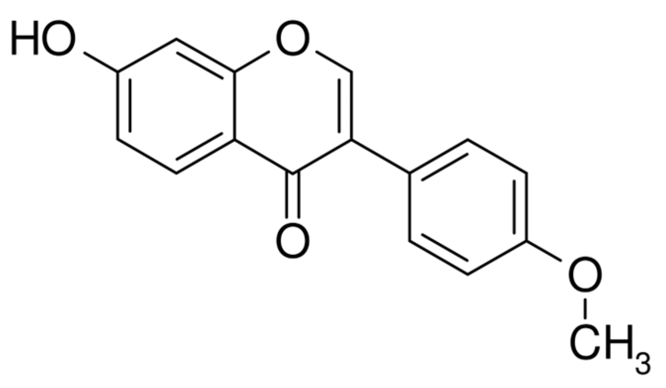
Figure 1. Chemical structure of formononetin.
Applsci 16 01975 g001
Figure 2. Structure of hydroxypropyl-β-cyclodextrin (HP-β-CD), where R = H or CH2–CHOH–CH3.
Applsci 16 01975 g002
Figure 3. XRPD patterns of formononetin, excipients, and prepared formulations. (a) Hot-melt extruded (HME) systems containing 10% formononetin with increasing amounts of HP-β-CD (20–60%). (b) Pre-inclusion formulations followed by extrusion (INK HME). (c) Physical mixtures (ph.m.) of formononetin with HP-β-CD at corresponding ratios. (d) Reference diffractograms of pure formononetin, HP-β-CD, and Kollidon® VA64.
Applsci 16 01975 g003
Figure 4. Aqueous solubility of formononetin (FRM) for selected systems. Different letters indicate statistically significant differences (p < 0.05).
Applsci 16 01975 g004
Figure 5. DSC analysis: (a) FRM, PVP VA64, FRM 10% ph.m., FRM 10% HME; (b) FRM, PVP VA64, HP-β-CD, FRM 10% CD 60% ph.m., FRM 10% CD 60% HME, (c) FRM, PVP VA64, HP-β-CD, FRM 10% CD 60% INK ph.m., FRM 10% CD 60% INK HME.
Applsci 16 01975 g005
Figure 6. FT-IR analysis: (a) FRM, PVP VA64, FRM 10% ph.m., FRM 10%; (b) FRM, PVP VA64, HP-β-CD, FRM 10% CD 60% ph.m., FRM 10% CD 60%; (c) FRM, PVP VA64, HP-β-CD, FRM 10% CD 60% INK ph.m., FRM 10% CD 60% INK.
Applsci 16 01975 g006
Figure 7. Apparent permeability coefficient (Papp) of crystalline formononetin (FRM) and formononetin system (FRM S) determined using the Skin PAMPA™ model (5 h, 37 °C). Data are presented as mean ± SD; n as stated in Methods.
Applsci 16 01975 g007
Figure 8. Wound closure effect of formononetin (FRN) and obtained system (FRN S) after 24 h and 48 h incubation.
Applsci 16 01975 g008
Figure 9. Wound-healing activity of formononetin (FRM) and formononetin system (FRM S) after 24 and 48 h at 10 µg/mL. Data were analyzed by one-way ANOVA with Tukey’s post hoc test. Statistical significance: ns p ≥ 0.05, * p < 0.05, ** p < 0.01, **** p < 0.0001 vs. control at the corresponding time points.
Applsci 16 01975 g009
Figure 10. Macroscopic appearance of the self-supporting hydrogel patch loaded with the optimized formononetin amorphous system (approx. 35 × 35 × 4 mm), demonstrating uniform structure and handling stability.
Applsci 16 01975 g010
Figure 11. In vitro release profile of formononetin from the hydrogel formulation (n = 3 independent hydrogels) in phosphate buffer pH 7.4 containing 0.3% Tween 80 at 32 °C. Data are presented as mean ± SD.
Applsci 16 01975 g011
Figure 12. Cumulative permeation of formononetin from the hydrogel formulation using Franz diffusion cells The inset/linear region shows the steady-state portion used to calculate flux (Jss).
Applsci 16 01975 g012
Table 1. Composition of formononetin-based systems prepared by hot-melt extrusion (HME) with hydroxypropyl-β-cyclodextrin (HP-β-CD) and Kollidon® VA64; “Pre-inclusion” indicates whether the formononetin–HP-β-CD inclusion complex was prepared prior to extrusion.
No.Formononetin (% M/M)HP-β-CD (% M/M)Kollidon® VA64 (% M/M)Pre-Inclusion
11090
2102070
3104050
4106030
5102070+
6104050+
7106030+
Table 2. Kinetic parameters (R2 and k) of formononetin release from the hydrogel fitted to common release models.
Zero-OrderFirst-OrderHiguchiHixson–CrowellKorsmeyer–Peppas
R20.7780.8200.9260.9500.953n = 0.775
k0.2810.0100.096−0.0230.278
Disclaimer/Publisher’s Note: The statements, opinions and data contained in all publications are solely those of the individual author(s) and contributor(s) and not of MDPI and/or the editor(s). MDPI and/or the editor(s) disclaim responsibility for any injury to people or property resulting from any ideas, methods, instructions or products referred to in the content.