Article

Preliminary Evaluation of a High-Class Treatment Dental Implant Surface: A TOF-SIMS Study

by
Vincenzo Ronsivalle
1,2,*,
Salvatore Bocchieri
1,2,
Antonino Licciardello
2,
Gabriele Cervino
3,
Cesare D’Amico
3,
Pierluigi Mariani
4 and
Marco Cicciù
1
1
Department of General Surgery and Surgical-Medical Specialties, School of Dentistry, University of Catania, 95124 Catania, Italy
2
Department of Chemical Sciences, University of Catania, 95124 Catania, Italy
3
Department of Biomedical and Dental Sciences, Morphological and Functional Images, University of Messina, G. Martino Polyclinic, 98125 Messina, Italy
4
Multidisciplinary Department of Medical-Surgical and Odontostomatological Specialties, University of Campania “Luigi Vanvitelli”, 80121 Naples, Italy
*
Author to whom correspondence should be addressed.
Appl. Sci. 2026, 16(4), 1936; https://doi.org/10.3390/app16041936
Submission received: 29 December 2025 / Revised: 3 February 2026 / Accepted: 13 February 2026 / Published: 14 February 2026
(This article belongs to the Special Issue Innovative Techniques and Materials in Implant Dentistry)

Abstract

Background: Surface chemistry and cleanliness are widely regarded as important factors influencing the host response to titanium dental implants. Despite advances in manufacturing and sterilization, trace residues may persist at the nanoscale even in commercially sterile devices. This study provides a preliminary evaluation of premium-grade titanium dental implants using time-of-flight secondary ion mass spectrometry (ToF-SIMS) to assess surface chemical uniformity and trace contaminant distribution. Method: Two commercially available titanium implants from Schütz Dental were analyzed under static and dynamic ToF-SIMS modes using Bi3+ and Cs+ ion beams. Both positive and negative ion spectra were collected to identify elemental and molecular species. Chemical mapping and depth profiling were performed to evaluate contaminant distribution and surface depth composition. Results: In the two implants analyzed, the surfaces were dominated by TiO+ and TiO2+ species, consistent with a native titanium oxide layer. In both analyzed implants, localized contaminants—including fluorine, chlorine, sulfur, CN groups, and organic residues—were detected within the outermost ~0.1 µm. These signals showed heterogeneous distribution along the thread-related regions within the analyzed ROIs, compatible with residues originating from machining, surface treatments, packaging, and/or sterilization steps. Conclusions: The present data support only the descriptive finding that trace contaminants were detected on the two analyzed implants. ToF-SIMS enabled nanoscale chemical mapping and depth profiling of these residues, supporting the feasibility of this approach for trace-level surface auditing and hypothesis generation. Any biological/clinical implications remain speculative and require dedicated in vitro/in vivo validation on larger sample sets.

1. Introduction

The meticulous control of surface purity on titanium dental implants is paramount for achieving successful osseointegration and long-term biocompatibility [1,2,3,4]. Pioneering studies by Ameen et al. highlighted the critical role of the implant–tissue interface, demonstrating how even subtle surface irregularities or residual manufacturing debris can significantly hinder biological response and tissue integration [5]. Indeed, the surface properties—including chemical composition, roughness, and morphology—directly influence cellular adhesion, proliferation, and differentiation, ultimately determining the clinical success of the implant [6,7].
The dynamic interaction between the implant surface and its surrounding biological environment dictates the formation of a stable osseous–implant interface, a process that can be compromised by the presence of surface contaminants [8]. Contaminants—such as fluorides, chlorides, hydrocarbons, or organic residues—may originate from machining, chemical etching, cleaning, or packaging and persist even in sterile products, altering the interfacial chemistry and delaying bone apposition [9].
The recent literature consistently demonstrates that the topographical and chemical characteristics of titanium implants are fundamental determinants of osseointegration, with variations in surface composition directly correlating to biological outcomes [10]. The physical and chemical nature of surface particles—including their morphology, grain size, and spatial distribution—affects mechanical load transfer and early cell anchorage during functional rehabilitation [11]. From an analytical standpoint, identifying and mapping trace residues at the outermost surface remains crucial, as these species may persist after manufacturing, sterilization, or packaging and may escape conventional inspection methods [12].
In this context, precise and non-destructive analytical techniques are essential to characterize surface composition and detect possible contaminants. Time-of-flight secondary ion mass spectrometry (ToF-SIMS) has emerged as a powerful tool for this purpose, providing nanometer-scale resolution and molecular-level chemical specificity. Through its ability to detect both inorganic and organic residues, ToF-SIMS enables a comprehensive evaluation of implant surface chemistry and cleanliness, complementing morphological observations obtained by means of SEM or AFM [13].
Importantly, ToF-SIMS also enables 2D chemical imaging, allowing localization of trace residues with sub-micrometric lateral resolution on complex implant morphologies [14,15]. When combined with sputter depth profiling, it can provide a semi-quantitative estimate of whether contaminants are confined to the outermost surface/oxide region or extend into deeper near-surface layers [16,17].
However, information on submicrometric trace residues on commercially sterile, premium-grade dental implants remains limited, particularly when analyzed under ‘as received’ conditions and on real threaded geometries [18,19,20]. This knowledge gap motivates the present work, which uses ToF-SIMS to provide a sensitive assessment of near-surface chemical heterogeneity at the micro–nano scale.
Since the present work is a purely analytical ToF-SIMS investigation, it does not include in vitro, in vivo, or clinical testing. Therefore, any biological considerations reported in the Discussion are literature-based hypotheses and are not outcomes demonstrated by the present dataset.
Therefore, the present study aims to provide a preliminary ToF-SIMS-based evaluation of high-class titanium dental implants, focusing on identifying trace surface contaminants, mapping their spatial distribution, and estimating their near-surface localization through depth profiling. Any potential associations with manufacturing or post-treatment steps are discussed as literature-based hypotheses.
Although ToF-SIMS and XPS have been previously applied to characterize dental implant surfaces, the novelty of the present preliminary study lies in its quality-control-oriented evaluation of commercially available premium-grade implants analyzed “as received” from sterile packaging. In particular, we employed ToF-SIMS chemical imaging on real threaded geometries to highlight localized nanoscale heterogeneity that may remain undetected by means of conventional surface inspection techniques. Moreover, by combining surface mapping with dual-beam depth profiling, we provide an estimate of the near-surface confinement of trace residues within the outermost region of the implant surface. Overall, this work aims to support ToF-SIMS as an advanced tool for nanoscale chemical auditing of high-standard implant devices, while acknowledging the semi-quantitative nature of ion intensities and the need for future biological validation.

2. Materials and Methods

2.1. Samples

Two commercially available titanium dental implants supplied by Schütz Dental GmbH (Rosbach vor der Höhe, Germany) were analyzed:
  • Implant Regular Ø 4.5 mm, L10 mm (referred to as sample 4.5);
  • Implant Mini Ø 3.5 mm, L13 mm (sample 3.5).
Both implants were obtained from the same manufacturing lot/batch (Lot: FKF0078), as reported on the sterile packaging. Therefore, the observed heterogeneity is more likely related to local surface features and threaded geometry rather than batch-to-batch variation; however, the limited sample size prevents definitive conclusions.
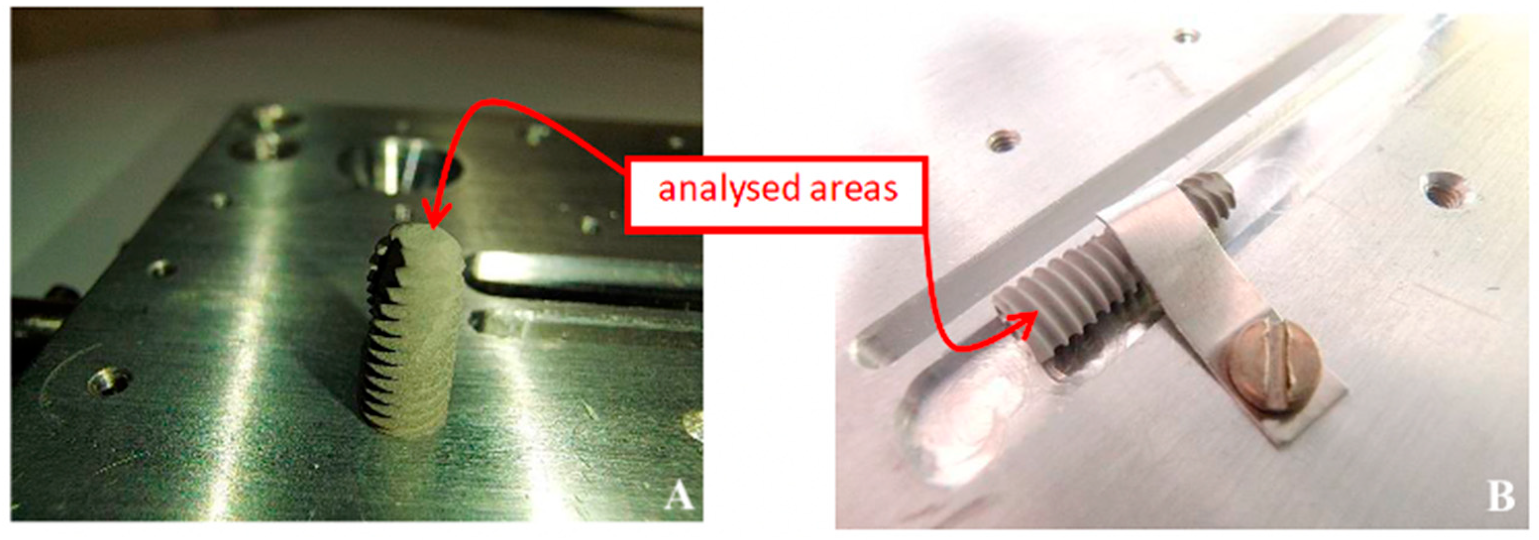
The implants were removed from their sterile containers under clean laboratory conditions and immediately mounted on a custom stainless-steel sample holder, ensuring exposure of either the top part (sample 4.5) or the threaded flank (sample 3.5) (Figure 1A,B).
Handling and mounting were performed with clean tools and minimal contact with the analysed ROIs, limiting ambient-air exposure to reduce external contamination.
Handling and mounting were performed using clean tools and a minimal-touch procedure (avoiding contact with the analysed ROIs) and minimizing ambient-air exposure to reduce the risk of external contamination.
This orientation was selected to evaluate possible contamination on both planar and complex threaded geometries, which are known to exhibit different residual accumulation patterns following machining and surface treatment [21,22].
Although handling was performed under clean laboratory conditions and exposure time to ambient air was minimized, adventitious contamination related to storage and mounting procedures cannot be fully excluded. Therefore, the ToF-SIMS data should be interpreted as representative of the ‘as received’ surface, including potential contributions from unavoidable experimental handling.

2.2. Mapped Regions and Depth-Profile Locations

For each implant, ToF-SIMS chemical maps were acquired from two distinct regions of interest (ROIs; n = 2 per implant) located on the exposed threaded surface accessible in the mounting configuration. Specifically, ROIs were selected a priori on predefined geometric zones of the mid-body threads: a thread-flank area and a thread valley/lower-flank area, both positioned away from the holder/clamp contact region to minimize handling-related artefacts. Depth profiling was performed at one location per implant (n = 1 per implant), positioned at the center of ROI-1 (thread flank). A brief low-dose survey scan was used only to verify focus/charging stability and accessibility of the predefined zones; ROIs were not chosen after post-hoc chemical screening for contaminant hotspots.

2.3. ToF-SIMS Analysis

Chemical surface analysis was performed using time-of-flight secondary ion mass spectrometry (ToF-SIMS), a technique that provides high mass resolution and spatially resolved chemical information from the outermost atomic layers (~1–2 nm) of solid surfaces [23].
In ToF-SIMS, the sample surface is bombarded with a focused primary ion beam, causing sputtering and emission of secondary ions from the outermost layers. These ions are accelerated into a time-of-flight mass spectrometer, where their mass-to-charge ratios are determined. ToF-SIMS allows simultaneous detection of elemental, molecular, and cluster species, making it suitable for identifying both inorganic and organic residues from manufacturing or surface treatments [13,24].
In the present work, a Bi3+ primary ion beam (0.1 pA, 25 keV) was employed for analysis, while a Cs+ sputter beam (60 nA, 10 keV) was used for depth profiling in dual-beam mode. Analyses were performed in both positive and negative ion polarities to obtain complementary elemental and molecular information.
  • Raster area: 200 × 200 µm2 for overview spectra; 51 × 51 µm2 for depth profiles.
  • Acquisition mode: static SIMS for surface spectra, dynamic SIMS for sputter depth profiling.
  • Vacuum level: <10−8 mbar during analysis.
Instrument calibration was carried out using standard metallic titanium and reference samples prior to each session to ensure mass accuracy within ±30 ppm.

2.4. Depth Profiling and Chemical Mapping

Depth profiling was performed in dual-beam mode, alternating analysis and sputtering cycles. Sputter time was converted to an approximate depth scale by comparing the applied sputtering conditions (Cs+ beam, energy/current and raster size) with literature-reported sputter rates for Ti-based substrates, and by monitoring the transition from contaminant-related signals to a stable predominance of Ti/TiO species. No direct crater-depth measurement was performed; therefore, the reported depth (~0.1 µm) should be considered an order-of-magnitude estimate. In addition, the threaded geometry and intrinsic roughness can lead to non-uniform erosion across peaks and valleys, introducing uncertainty and broadening of the apparent depth distribution [25].
Chemical maps were generated by scanning the Bi3+ beam across selected surface regions. For each implant, mapping was performed on representative areas of the exposed surface to account for local heterogeneity typical of threaded geometries. The maps were constructed from the integrated intensities of key species, including Ti+, TiO+, Cl, F, S, CN, Ca+, K+, and Si, to visualize spatial variations in contaminant distribution. Brighter regions in the resulting images indicate higher ion intensities, revealing heterogeneities in the distribution of surface residues.
Data processing and normalization were carried out using the ION-TOF SurfaceLab 7 software package (IONTOF GmbH, Münster, Germany). Signal intensities were normalized to total ion counts to correct for topographic and charging effects.
It should be noted that ToF-SIMS signal intensities represent relative ion yields and therefore provide semi-quantitative information rather than absolute concentrations. Ion yields may be influenced by matrix effects, surface topography, and ionization probability; thus, absolute quantification would require dedicated calibration standards.

2.5. Data Interpretation

Due to the high surface roughness and complex geometry of dental implants, the depth resolution was partially limited. Rough surfaces can cause shadowing effects during sputtering, leading to an apparent broadening of the depth profiles [26]. To minimize these artifacts, sputtering parameters were optimized, and comparative analyses were performed on both flat and curved regions.
Results were interpreted according to standard SIMS practices for dental biomaterials [9,10], focusing on the following:
  • The identification of contaminant species confined to the outermost layers.
  • Correlation of chemical mapping patterns with potential sources of contamination (e.g., machining lubricants, etching acids, or sterilization residues).
  • Estimation of contamination depth and lateral inhomogeneity.
Therefore, the depth profiles should be interpreted as an average chemical trend over rough topography, and sharp interfaces may appear broadened due to differential sputtering of ridges and valleys.

3. Results

3.1. Overview of ToF-SIMS Spectra

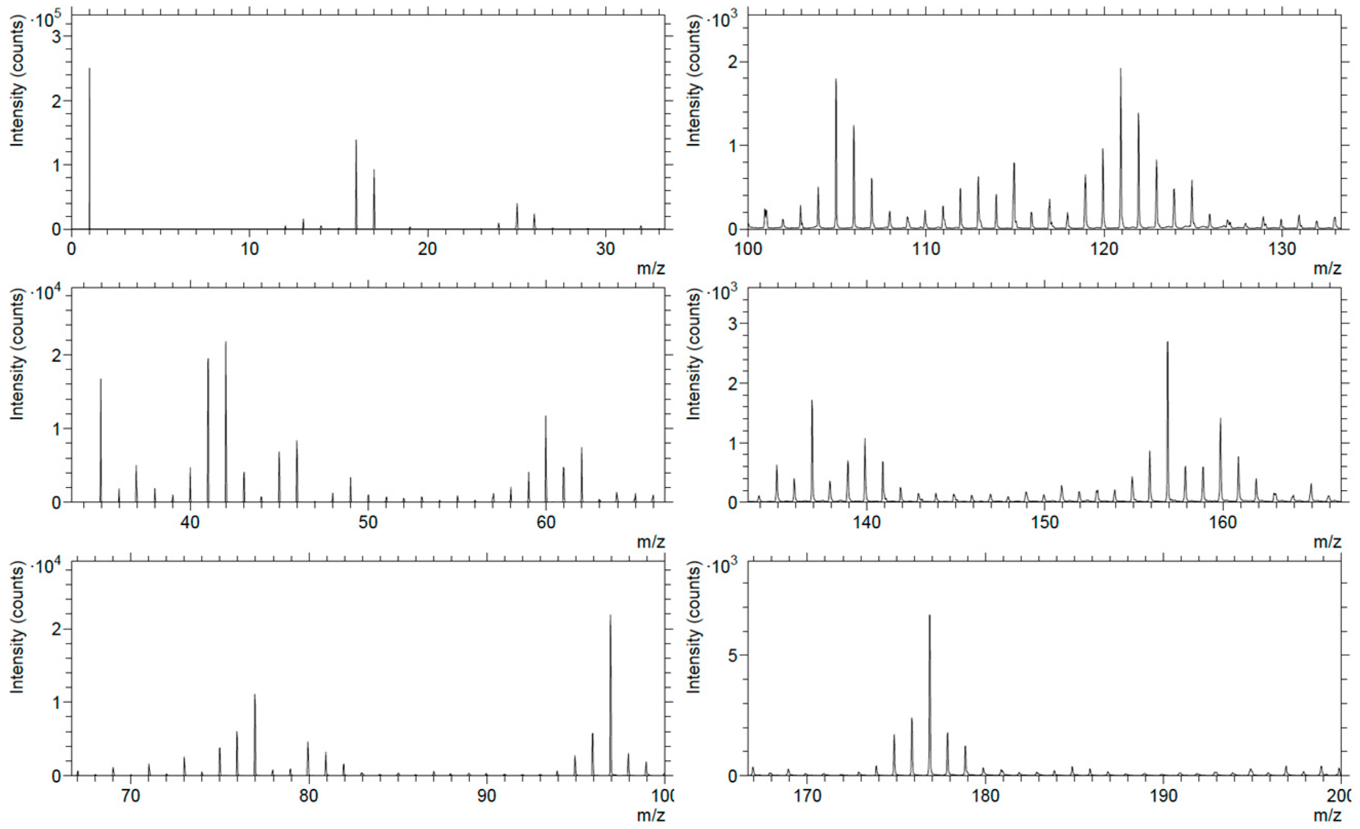
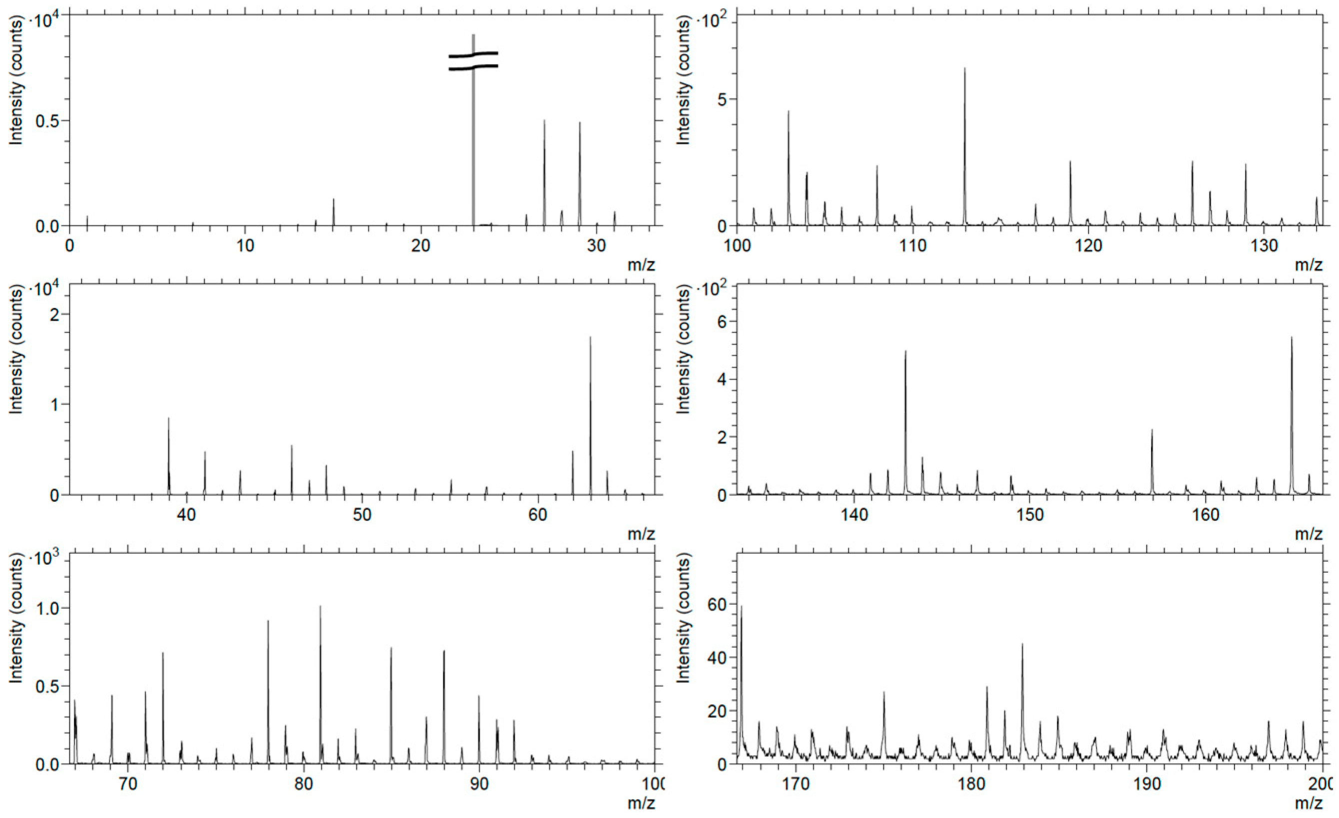
The positive and negative ion spectra obtained from sample 4.5 (Figure 2 and Figure 3) revealed chemical signatures typical of titanium-based materials, with dominant Ti+, TiO+, and TiO2+ peaks confirming the presence of a native titanium oxide layer on the implant surface. This oxide film is expected due to the spontaneous passivation of titanium in air and sterilization environments [27].
In the positive ion spectra (Figure 2), additional peaks corresponding to alkali and alkaline-earth metals (Na+, K+, Ca+, traces of Li+ and Mg+) were detected, suggesting the presence of adsorbed ionic species or residues from chemical treatments or packaging. Traces of Fe+, Ni+, and Si+ were also observed, which may reflect trace metallic/particulate residues associated with processing equipment or media [22]. Minor organic fragments containing CFx+ species were detected, indicating traces of fluorinated hydrocarbons; potential sources may include cleaning agents or lubricants, although the specific origin cannot be determined from the present data [28].
The negative ion spectra (Figure 3) confirmed the occurrence of fluorine (F), chlorine (Cl), sulfur (S), and carbon–nitrogen (CN) species. These findings are consistent with the presence of trace surface residues within the near-surface layer; possible sources may include surface treatments, sterilization steps, packaging-related contact, and/or adventitious adsorption [29]. The simultaneous detection of nitrogen- and oxygen-containing fragments (e.g., NO, CN, CNO) further supports the hypothesis of organic surface films likely adsorbed post-manufacturing [13].

3.2. Depth Profiling

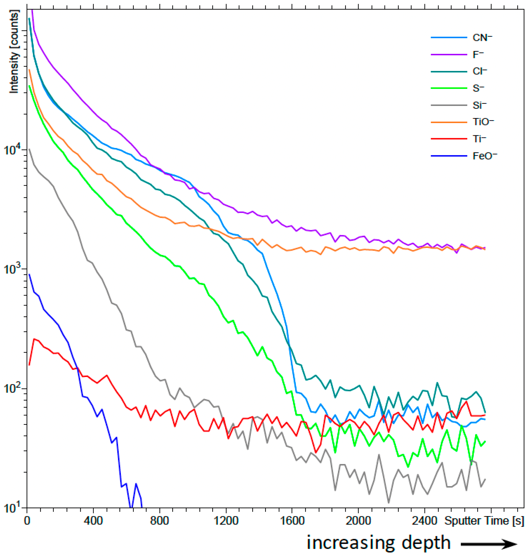
To determine whether the detected species were confined to the outermost surface or distributed deeper, depth profiling was performed using a dual-beam mode with a Bi3+ analysis beam and a Cs+ sputter beam (Figure 4).
The resulting depth profile shows that contaminant signals (Cl, F, CN, S) decrease rapidly with increasing sputter time, whereas titanium and titanium oxide species (Ti, TiO) become predominant beyond an estimated depth of approximately 0.1 µm. This indicates that most of the “exotic” species are confined to the outermost layer of the surface and do not penetrate into the bulk material, consistent with previous ToF-SIMS studies on dental titanium surfaces [6,10].
The profile of Si and FeO species remained detectable only within the first tens of nanometers, suggesting localized inclusions or residues likely from surface processing. It should be noted that the depth resolution is affected by the intrinsic surface roughness of the threaded implant, resulting in possible broadening of depth-related signals.

3.3. Chemical Mapping and Elemental Distribution

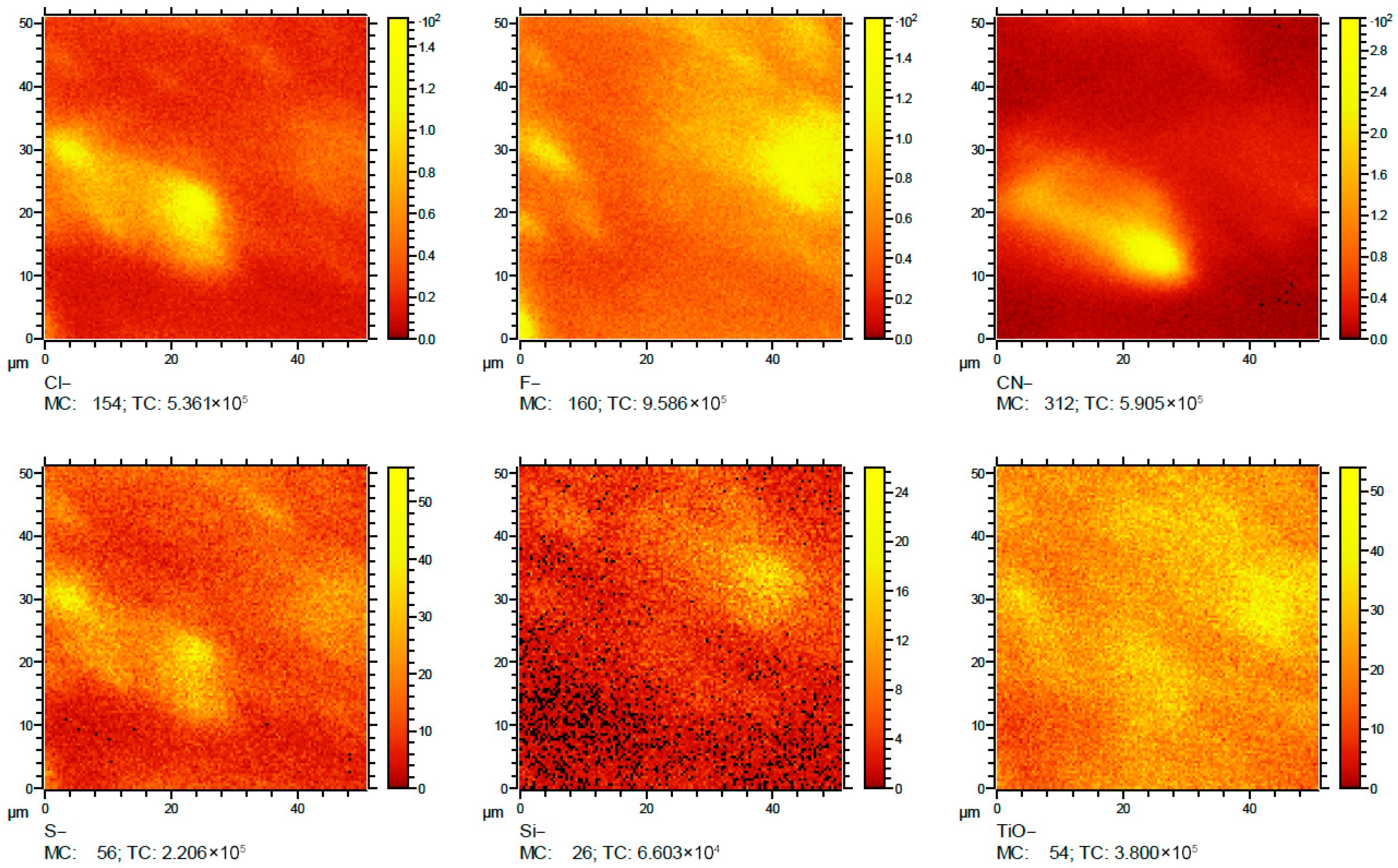
Chemical maps generated from negative ion spectra of sample 4.5 (Figure 5) revealed that the distribution of contaminants was highly heterogeneous. The fluorine (F) signal appeared intense in areas distinct from those rich in chlorine (Cl), while sulfur (S) and CN signals co-localized in regions of lower TiO intensity. This anti-correlation suggests that non-titanium phases or residual organic films partially cover the titanium substrate. Similar heterogeneous patterns have been reported in the literature for titanium implants following different surface-treatment protocols; however, the present study does not allow attribution of these patterns to a specific processing route [25,30].
In contrast, silicon (Si) was detected sporadically, indicating particulate residues rather than a uniform surface layer. The presence of halogens and CN groups indicates persistence of trace residues within the near-surface layer, which could be compatible with residues associated with surface treatments or sterilization/packaging steps. However, the present data do not allow unambiguous attribution to specific reagents or processing parameters [31].
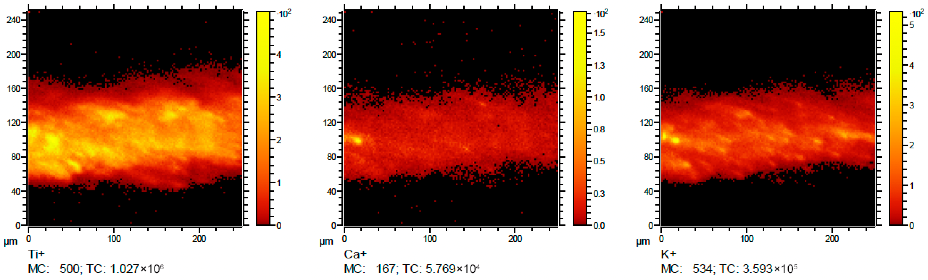
For sample 3.5, analyzed on the thread flank (Figure 6), a similar elemental composition was observed. However, due to the curvature of the threads and limited focus area, intensity distributions appeared confined along narrow linear zones corresponding to the ridge edges. The Ca+ and K+ signals exhibited localized enrichment along certain threads, possibly due to deposition of biological or environmental particles during handling or packaging, which is in line with the general concept reported in the literature that trace residues may be detected even on clinically used implants; however, the specific experimental conditions and quantitative levels are not directly comparable to the present ‘as received’ analytical assessment [9].

4. Discussion

4.1. Technological and Metrological Implications

In the two premium-grade titanium dental implants analyzed in this study (single batch), ToF-SIMS detected localized trace surface contamination despite commercial sterilization and industrial quality standards. This finding underscores the exceptional sensitivity of ToF-SIMS in detecting sub-micrometric residues that remain invisible to conventional quality assurance techniques such as optical or SEM inspection [22,28,31]. Both implants displayed surfaces dominated by titanium oxide species, as expected from passivated titanium alloys, but also contained trace levels of fluorine, chlorine, sulfur, CN fragments, and alkali/transition metals.
These “exotic” species were predominantly confined to the outermost ~0.1 µm, which is consistent with near-surface residues potentially associated with post-machining or finishing steps (e.g., surface treatments, rinsing, packaging, or sterilization). Importantly, ToF-SIMS alone cannot unambiguously assign the detected residues to a single manufacturing step; therefore, any process-related correlations should be regarded as hypotheses. Particularly, fluorine- and chlorine-related signals could be compatible with halogen-containing chemistries reported for certain micro-roughening protocols, which require thorough rinsing to minimize chemical persistence [27]. The co-localization of S and CN signals (Figure 5) indicates organic residues potentially related to industrial lubricants or post-treatment coatings, while sporadic Si signals likely arise from silica abrasives or environmental particulates [21,32]. The presence of such contaminants on premium implants—whose manufacturing protocols are typically optimized for surface purity—suggests that micro-contaminant retention may be intrinsic to complex implant geometries rather than solely a by-product of process quality. The heterogeneous spatial distribution of contaminants, especially on the threaded regions of the Ø 3.5 mm sample (Figure 6), highlights the metrological challenge of ensuring complete chemical homogeneity in implants with intricate morphologies. Although ToF-SIMS is intrinsically semi-quantitative, the present dataset enables internal comparisons through total-ion-normalized ion yields and depth-localization trends. In our measurements, contaminant-related signals were predominantly confined to the near-surface layer and chemical maps highlighted localized hotspots on threaded regions, supporting non-uniform spatial distribution within the same acquisition setting. However, the present ToF-SIMS data are intended to describe this heterogeneity from an analytical/metrological standpoint, and do not allow determination of its functional or biological impact, which will require dedicated in vitro/in vivo validation in future studies. Threaded designs increase the effective surface area and hinder uniform cleaning, drying, and packaging [29]. These observations reinforce the need for multi-technique surface auditing, where ToF-SIMS complements XPS, SEM, and EDS to monitor surface cleanliness at nanometric scales [10]. Importantly, these results show that even high-end, clinically certified implants may retain traces of manufacturing agents despite meeting all regulatory cleanliness thresholds. This aligns with recent findings by Rupp et al. [33], who emphasized that industrial cleanliness certifications may not fully reflect molecular-level purity. Consequently, ToF-SIMS may represent a valuable complementary approach for advanced nanoscale surface auditing in premium implant quality assessment, which should be supported by larger-scale validation studies [26,34].

4.2. Biological and Clinical Relevance

In this section, ToF-SIMS observations are discussed in relation to published biological evidence; however, these implications remain hypothetical in the absence of direct biological testing in the present study.
Given the very limited sample size (n = 2; single production lot), the present results do not allow any inference on the prevalence of these residues across the product line or on clinical relevance.
From a surface-science perspective, the detection of halogen- and sulfur-rich microdomains on the surface of high-standard implants may have potential implications for the biological interface. Although the implants analyzed belong to a premium class of devices, the ToF-SIMS results demonstrate that chemical heterogeneity can still persist at the nanoscale. Such trace residues may locally alter surface charge density and wettability, parameters that have been reported to influence protein adsorption and cell adhesion [7,12]. Specifically, residual fluorides and chlorides may form ionic species capable of perturbing the electrochemical equilibrium of the titanium oxide layer, whereas organic sulfur- or CN-containing fragments may create locally hydrophobic regions potentially affecting fibrin and protein binding [1]. These effects, although occurring at molecular dimensions, have been suggested in the literature as potential contributors to early interfacial events at the bone–implant interface [9]. It should be emphasized that these studies differ substantially in design and sample conditions, and therefore, they are cited here only as contextual evidence rather than as a rigorously quantitative comparison with the present ToF-SIMS dataset. Previous clinical and experimental reports have discussed possible associations between heterogeneous surface chemistry and peri-implant inflammatory conditions [1,9]. Shavit et al. [1] reported that implants exhibiting heterogeneous surface chemistry—even within top-tier products—may be associated with peri-implant inflammatory findings and early instability. Likewise, Duddeck & Albrektsson [9] discussed that micro-level contaminants may trigger macrophage activation and prolonged immune responses despite sterile packaging. Importantly, since no biological, cellular, or in vivo assessments were performed in the present work, these considerations should be interpreted as literature-based hypotheses rather than outcomes demonstrated here. Overall, these observations suggest that “premium” does not automatically equate to complete chemical uniformity at the nanoscale. Rather, ToF-SIMS reveals that trace residues may persist below traditional detection thresholds. For clinicians and manufacturers alike, this supports the value of trace-level chemical auditing as part of both pre-market and post-market quality assessments. The ToF-SIMS maps (Figure 5) showed distinct halogen-rich and TiO-depleted zones, suggesting that surface wettability and energy may not be fully homogeneous even within high-standard implants. Such heterogeneity may contribute to variability in early biological response reported in clinical studies comparing nominally identical implant models [33], although causality requires dedicated validation.

4.3. Perspectives for Future Research and Limitations

Since most detected contaminants are restricted to the uppermost 0.1 µm, future work should focus on controlled comparative experiments involving flat titanium coupons processed identically to threaded implants. These flat reference specimens would enable quantitative calibration of ToF-SIMS erosion rates and facilitate cross-technique validation with XPS or FTIR [10]. Moreover, given that the implants tested represent state-of-the-art commercial products, follow-up investigations could explore process-to-surface correlation, identifying the exact stage (machining, acid treatment, sterilization) where contamination originates. Combining these analytical insights with in vitro cell culture studies will help define thresholds of acceptable contamination levels for regulatory adoption, potentially leading to standardized certification frameworks for implant surface purity [35]. Ultimately, this pilot study suggests that even premium dental implants may exhibit submicroscopic chemical residues, supporting the potential value of ToF-SIMS as a complementary tool for nanoscale surface auditing, to be confirmed through larger-scale validation studies.
This study is preliminary in nature and limited by the analysis of only two implants from a single production lot; therefore, the present dataset supports only the descriptive finding that contaminants were detected on the two implants analyzed, and no inference on prevalence, source, or clinical relevance can be made. Moreover, it is not possible to determine whether the observed contamination pattern is typical of the investigated implant systems or reflects local variability limited to the specific analyzed units. Larger-scale studies including multiple specimens and different production lots are required to assess batch-to-batch variability and the reproducibility of the detected spatial patterns. However, the aim of this work was to provide an initial pilot feasibility assessment, and the acquisition of spectra/mapping on representative regions was used to strengthen the qualitative assessment. In addition, the complex geometry and surface roughness of the dental implants introduced some uncertainty in the depth resolution of the sputtering process, potentially broadening the apparent distribution of certain species. The ToF-SIMS technique, while extremely sensitive, provides only semi-quantitative information, meaning that absolute concentrations cannot be determined without appropriate calibration standards. Additionally, minor contributions from laboratory handling and mounting conditions may affect the outermost surface chemistry and should be considered as a potential source of experimental artefacts. Because a holder blank (holder and mounting procedure measured under identical conditions without an implant) was not acquired, background contributions from the holder and/or handling cannot be fully excluded. Moreover, biological implications were not directly assessed in this work, and the relationship between the detected contaminants and cellular responses remains to be confirmed through dedicated biological studies. These limitations require that the present findings are interpreted as an initial pilot feasibility assessment, rather than as statistically validated evidence. Larger-scale studies with multiple specimens per implant system are required to enable robust conclusions and appropriate statistical evaluation.

5. Conclusions

In the two premium-grade titanium dental implants analyzed, ToF-SIMS detected localized trace species (e.g., F, Cl, S, CN and organic-related fragments) mainly confined to the outermost ~0.1 µm and heterogeneously distributed across the threaded regions. Therefore, the present data support only the descriptive statement that contaminants were detected on these two implants, and no generalization to other implants or production lots can be made. ToF-SIMS enabled trace-level chemical mapping in this pilot setting; however, robust conclusions require larger studies including multiple specimens and production lots, and any biological implications remain hypothesis-generating pending dedicated validation.

Author Contributions

Conceptualization, M.C.; methodology, A.L.; data curation, P.M.; writing—original draft preparation, V.R.; writing—review and editing, M.C.; visualization, G.C. and C.D.; software, S.B. All authors have read and agreed to the published version of the manuscript.

Funding

This research received no external funding.

Institutional Review Board Statement

Not applicable.

Data Availability Statement

Data are available from the corresponding author upon request.

Conflicts of Interest

The authors declare no conflicts of interest.

References

  1. Shavit, A.S.; Rittel, D.; Shemtov-Yona, K. The Chemical and Microstructural Signature of Peri-Implantitis on Titanium Dental Implants’ Surface. Appl. Surf. Sci. Adv. 2024, 19, 100553. [Google Scholar] [CrossRef] [Scilit]
  2. Park, G.; Matsuura, T.; Komatsu, K.; Ogawa, T. Optimizing Implant Osseointegration, Soft Tissue Responses, and Bacterial Inhibition: A Comprehensive Narrative Review on the Multifaceted Approach of the UV Photofunctionalization of Titanium. J. Prosthodont. Res. 2025, 69, 136–152. [Google Scholar] [CrossRef] [Scilit]
  3. Kido, D.; Komatsu, K.; Suzumura, T.; Matsuura, T.; Cheng, J.; Kim, J.; Park, W.; Ogawa, T. Influence of Surface Contaminants and Hydrocarbon Pellicle on the Results of Wettability Measurements of Titanium. Int. J. Mol. Sci. 2023, 24, 14688. [Google Scholar] [CrossRef] [Scilit]
  4. Souza, E.G.; Nascimento, C.d.D.d.; Aguzzoli, C.; Santillán, E.S.B.; Cuevas-Suárez, C.E.; Nascente, P.d.S.; Piva, E.; Lund, R.G. Enhanced Antibacterial Properties of Titanium Surfaces through Diversified Ion Plating with Silver Atom Deposition. J. Funct. Biomater. 2024, 15, 164. [Google Scholar] [CrossRef] [Scilit]
  5. Ameen, A.P.; Short, R.D.; Johns, R.; Schwach, G. The Surface Analysis of Implant Materials. The Surface Composition of a Titanium Dental Implant Material. Clin. Oral Implant. Res. 1993, 4, 144–150. [Google Scholar] [CrossRef] [Scilit]
  6. Massaro, C.; Rotolo, P.; De Riccardis, F.; Milella, E.; Napoli, A.; Wieland, M.; Textor, M.; Spencer, N.D.; Brunette, D.M. Comparative Investigation of the Surface Properties of Commercial Titanium Dental Implants. Part I: Chemical Composition. J. Mater. Sci. Mater. Med. 2002, 13, 535–548. [Google Scholar] [CrossRef] [Scilit]
  7. Jayaraman, M.; Meyer, U.; Bühner, M.; Joos, U.; Wiesmann, H.P. Influence of Titanium Surfaces on Attachment of Osteoblast-like Cells In Vitro. Biomaterials 2004, 25, 625–631. [Google Scholar] [CrossRef] [Scilit] [PubMed]
  8. Sarvaiya, B.B.; Kumar, S.; Pathan, M.S.H.; Patel, S.; Gupta, V.; Haque, M. The Impact of Implant Surface Modifications on the Osseointegration Process: An Overview. Cureus 2025, 17, e81576. [Google Scholar] [CrossRef] [Scilit]
  9. Duddeck, D.U.; Albrektsson, T.; Wennerberg, A.; Larsson, C.; Mouhyi, J.; Beuer, F. Quality Assessment of Five Randomly Chosen Ceramic Oral Implant Systems: Cleanliness, Surface Topography, and Clinical Documentation. Int. J. Oral Maxillofac. Implant. 2021, 36, 863–874. [Google Scholar] [CrossRef] [Scilit] [PubMed]
  10. Palmquist, A.; Emanuelsson, L.; Sjövall, P. Chemical and Structural Analysis of the Bone-Implant Interface by TOF-SIMS, SEM, FIB and TEM: Experimental Study in Animal. Appl. Surf. Sci. 2012, 258, 6485–6494. [Google Scholar] [CrossRef] [Scilit]
  11. Balderrama, Í.d.F.; Stuani, V.d.T.; Cardoso, M.V.; Oliveira, R.C.; Lopes, M.M.R.; Greghi, S.L.A.; Adriana Campos Passanezi, S.A. The Influence of Implant Surface Roughness on Decontamination by Antimicrobial Photodynamic Therapy and Chemical Agents: A Preliminary Study In Vitro. Photodiagnosis Photodyn. Ther. 2021, 33, 102105. [Google Scholar] [CrossRef] [Scilit]
  12. Barberi, J.; Spriano, S. Titanium and Protein Adsorption: An Overview of Mechanisms and Effects of Surface Features. Materials 2021, 14, 1590. [Google Scholar] [CrossRef] [Scilit]
  13. Kravanja, K.A.; Finšgar, M. Analytical Techniques for the Characterization of Bioactive Coatings for Orthopaedic Implants. Biomedicines 2021, 9, 1936. [Google Scholar] [CrossRef] [Scilit]
  14. Fletcher, J.S.; Vickerman, J.C.; Winograd, N. Label Free Biochemical 2D and 3D Imaging Using Secondary Ion Mass Spectrometry. Curr. Opin. Chem. Biol. 2011, 15, 733–740. [Google Scholar] [CrossRef] [Scilit]
  15. Gilmore, I.S. SIMS of Organics—Advances in 2D and 3D Imaging and Future Outlook. J. Vac. Sci. Technol. A Vac. Surf. Film. 2013, 31, 050819. [Google Scholar] [CrossRef] [Scilit]
  16. Ekar, J.; Panjan, P.; Drev, S.; Kovač, J. ToF-SIMS Depth Profiling of Metal, Metal Oxide, and Alloy Multilayers in Atmospheres of H2, C2H2, CO, and O2. J. Am. Soc. Mass Spectrom. 2022, 33, 31–44. [Google Scholar] [CrossRef] [Scilit] [PubMed]
  17. Fletcher, J.S.; Vickerman, J.C. A New SIMS Paradigm for 2D and 3D Molecular Imaging of Bio-Systems. Anal. Bioanal. Chem. 2010, 396, 85–104. [Google Scholar] [CrossRef] [Scilit] [PubMed]
  18. Duddeck, D.U.; Albrektsson, T.; Wennerberg, A.; Larsson, C.; Beuer, F. On the Cleanliness of Different Oral Implant Systems: A Pilot Study. J. Clin. Med. 2019, 8, 1280. [Google Scholar] [CrossRef] [Scilit]
  19. Mtanis, T.; Biadsee, A.; Ormianer, Z. Assessing the Cleanliness of Dental Implants Using Scanning Electron Microscopy and Energy-Dispersive X-Ray Spectroscopy Analysis—A SEM and EDS In Vitro Study. J. Funct. Biomater. 2023, 14, 172. [Google Scholar] [CrossRef] [Scilit]
  20. Dhaliwal, J.S.; David, S.R.N.; Zulhilmi, N.R.; Sodhi Dhaliwal, S.K.; Knights, J.; de Albuquerque Junior, R.F. Contamination of Titanium Dental Implants: A Narrative Review. SN Appl. Sci. 2020, 2, 1011. [Google Scholar] [CrossRef] [Scilit]
  21. Pimenta, J.; Szmukler-Moncler, S.; Raigrodski, A.J. Physical Characterization of 3 Implant Systems Made of Distinct Materials with Distinct Surfaces. J. Prosthet. Dent. 2022, 128, 63–72. [Google Scholar] [CrossRef] [Scilit] [PubMed]
  22. Elias, C.N.; Lima, J.H.C.; Valiev, R.; Meyers, M.A. Biomedical Applications of Titanium and Its Alloys. J. Manag. 2008, 60, 46–49. [Google Scholar] [CrossRef] [Scilit]
  23. Sacher, E.; França, R. Surface Analysis Techniques for Dental Materials. In Dental Biomaterials; World Scientific: Singapore, 2018; pp. 1–31. [Google Scholar] [CrossRef] [Scilit]
  24. Viornery, C.; Chevolot, Y.; Léonard, D.; Aronsson, B.O.; Péchy, P.; Mathieu, H.J.; Descouts, P.; Grätzel, M. Surface Modification of Titanium with Phosphonic Acid To Improve Bone Bonding: Characterization by XPS and ToF-SIMS. Langmuir 2002, 18, 2582–2589. [Google Scholar] [CrossRef] [Scilit]
  25. Wang, Y.; Wen, C.; Hodgson, P.; Li, Y. Biocompatibility of TiO2 Nanotubes with Different Topographies. J. Biomed. Mater. Res. A 2014, 102, 743–751. [Google Scholar] [CrossRef] [Scilit]
  26. Rosa, M.B.; Albrektsson, T.; Francischone, C.E.; Schwartz Filho, H.O.; Wennerberg, A. The Influence of Surface Treatment on the Implant Roughness Pattern. J. Appl. Oral Sci. 2012, 20, 550–555. [Google Scholar] [CrossRef] [Scilit]
  27. Lausmaa, J. Surface Spectroscopic Characterization of Titanium Implant Materials. J. Electron Spectros. Relat. Phenom. 1996, 81, 343–361. [Google Scholar] [CrossRef] [Scilit]
  28. Inchingolo, A.M.; Malcangi, G.; Ferrante, L.; Del Vecchio, G.; Viapiano, F.; Inchingolo, A.D.; Mancini, A.; Annicchiarico, C.; Inchingolo, F.; Dipalma, G.; et al. Surface Coatings of Dental Implants: A Review. J. Funct. Biomater. 2023, 14, 287. [Google Scholar] [CrossRef] [Scilit] [PubMed]
  29. Park, J.H.; Olivares-Navarrete, R.; Baier, R.E.; Meyer, A.E.; Tannenbaum, R.; Boyan, B.D.; Schwartz, Z. Effect of Cleaning and Sterilization on Titanium Implant Surface Properties and Cellular Response. Acta Biomater. 2012, 8, 1966–1975. [Google Scholar] [CrossRef] [Scilit]
  30. Brabazon, D. Nanocharacterization Techniques for Dental Implant Development. In Emerging Nanotechnologies in Dentistry: Processes, Materials and Application; William Andrew Publishing: Norwich, NY, USA, 2011; pp. 307–331. [Google Scholar] [CrossRef] [Scilit]
  31. Peck, M.T.; Chrcanovic, B.R. Chemical and Topographic Analysis of Eight Commercially Available Dental Implants. J. Contemp. Dent. Pract. 2016, 17, 354–360. [Google Scholar] [CrossRef] [Scilit]
  32. Buser, D.; Schenk, R.K.; Steinemann, S.; Fiorellini, J.P.; Fox, C.H.; Stich, H. Influence of Surface Characteristics on Bone Integration of Titanium Implants. A Histomorphometric Study in Miniature Pigs. J. Biomed. Mater. Res. 1991, 25, 889–902. [Google Scholar] [CrossRef] [Scilit] [PubMed]
  33. Rupp, F.; Liang, L.; Geis-Gerstorfer, J.; Scheideler, L.; Hüttig, F. Surface Characteristics of Dental Implants: A Review. Dent. Mater. 2018, 34, 40–57. [Google Scholar] [CrossRef] [Scilit] [PubMed]
  34. Castilho, G.A.A.; Martins, M.D.; Macedo, W.A.A. Surface Characterization of Titanium Based Dental Implants. Braz. J. Phys. 2006, 36, 1004–1008. [Google Scholar] [CrossRef] [Scilit]
  35. Jordan, A.; Smojver, I.; Budimir, A.; Gabrić, D.; Vuletić, M. Evaluation of Different Procedures for Titanium Dental Implant Surface Decontamination-In Vitro Study. Bioengineering 2024, 11, 326. [Google Scholar] [CrossRef] [Scilit] [PubMed]
Figure 1. Implant sample: (A) Sample Ø 4.5 mm and (B) Sample Ø 3.5 mm.
Applsci 16 01936 g001
Figure 2. Positive ion spectra, sample Ø 4.5 mm.
Applsci 16 01936 g002
Figure 3. Negative ion spectra, sample Ø 4.5 mm.
Applsci 16 01936 g003
Figure 4. Dual-beam depth profile of sample Ø 4.5 mm, showing the intensity (counts) of the main negative secondary ions (CN, F, Cl, S, Si, TiO, Ti, and FeO) as a function of sputter time, converted into an approximate depth scale.
Applsci 16 01936 g004
Figure 5. Negative ion maps sample Ø 4.5 mm.
Applsci 16 01936 g005
Figure 6. Positive ion maps sample Ø 3.5 mm.
Applsci 16 01936 g006
Disclaimer/Publisher’s Note: The statements, opinions and data contained in all publications are solely those of the individual author(s) and contributor(s) and not of MDPI and/or the editor(s). MDPI and/or the editor(s) disclaim responsibility for any injury to people or property resulting from any ideas, methods, instructions or products referred to in the content.