Article

A Pilot Study on Whether Cycling with Different Levels of Electronic Assistance Changes Muscle Activity of the Lower Limb in People with Knee Osteoarthritis

Curtin School of Allied Health, Curtin University, Kent St., Bentley, Perth 6102, Australia
*
Author to whom correspondence should be addressed.
Appl. Sci. 2026, 16(4), 1713; https://doi.org/10.3390/app16041713
Submission received: 5 January 2026 / Revised: 5 February 2026 / Accepted: 7 February 2026 / Published: 9 February 2026
(This article belongs to the Special Issue Applied Biomechanics: Sports Performance and Rehabilitation)

Featured Application

Assisted e-cycling offers a promising therapeutic alternative for individuals with knee osteoarthritis (KOA) by enabling physical activity while reducing joint stress and pain. The adjustable assistance levels allow users to engage in beneficial exercise while minimising discomfort, potentially lowering kinesiophobia and increasing motivation. Integrating e-cycling into rehabilitation programs may support muscle strengthening, promote regular activity, and improve overall quality of life for those with KOA, particularly for individuals facing barriers to traditional exercise.

Abstract

Background: Knee osteoarthritis (KOA) often results in reduced physical activity. This exploratory study evaluated lower limb muscle activity across three e-cycling assistance levels and examined e-cycling’s influence on kinesiophobia, stress and exercise motivation in individuals with KOA. Methods: Ten participants cycled with no, low and high assistance on an e-bike. Muscle activity and knee kinematics were measured using surface electromyography and inertia measurement units. A subset of four participants completed questionnaires assessing kinesiophobia (TSK-17), perceived stress (PSS), and exercise motivation (BREQ-3). Muscle activity across the three levels of assistance was analysed using a linear mixed-effects model. Results: Peak and mean muscle activity of rectus femoris (p = 0.01; 0.039), vastus medialis oblique (p = 0.0002; 0.001) and biceps femoris (p = 0.002; 0.03) were lower during high-assistance compared to the no-assistance cycling. No significant differences were observed in mean muscle activity between no- and low-assistance cycling. Reported exertion and pain were low during e-cycling, with kinesiophobia (M = 35.8 ± 2.5 to 33.3 ± 3.5) decreasing following e-cycling, whereas perceived stress (M = 14 ± 5.7 to 14.5 ± 3.3) increased marginally in our small sample. In the small subgroup of participants, the Behaviour Regulation Exercise Questionnaire outcomes increased in four out of six subscores post-exercise. Conclusions: Considering the differences in muscle activity recorded, and given that this is a pilot study, we propose that e-cycling may be an ideal way of introducing graded exercise to KOA patients, potentially allowing them to maintain physical activity and self-management of their disease.

1. Introduction

Knee osteoarthritis (KOA) is a common degenerative joint condition that can affect adults from as early as 40 years of age [1]. In Australia, KOA is responsible for approximately 20% of the costs associated with musculoskeletal diseases [2]. It is characterised by an imbalance between articular damage and repair, low-grade inflammation driven by the secretion of catabolic factors, and an increased proportion of chrondrocytes undergoing apoptosis [3,4]. These processes ultimately lead to cartilage loss, subchondral bone sclerosis, and osteophyte formation, resulting in joint space narrowing and reduced ability to withstand mechanical loads [3,4]. Individuals with KOA frequently complain of pain, stiffness, and swelling, particularly after inactivity [1,4]. Furthermore, arthrogenic muscle inhibition is a common phenomenon in people with painful knee conditions where neural inhibition contributes to quadriceps weakness and impaired neuromuscular control. Elevated pain intensity is significantly associated with psychosocial variables such as kinesiophobia, anxiety and depression among individuals with KOA [5]. Consequently, individuals with KOA commonly experience fear of movement (FOM), reduced physical activity (PA), and diminished quality of life [4]. Reduced functional ability, increased treatment frequency and prolonged care contribute to the considerable economic burden of KOA [6]. Ultimately, there is a need for effective, rational and evidence-based treatments aimed at reducing both the economic and healthcare burden of the disease [4].
Exercise prescription for KOA has consistently been reported to be effective for improving function, weight management, and providing analgesic effects [7,8]. Aerobic and muscle strengthening exercises have been established to improve physical functioning and decrease pain and disability in people with KOA [9]. However, people with KOA often experience stress, anxiety and fear of pain, which can lead to kinesiophobia and a subsequent decline in physical function and capacity [10,11,12]. Lozano-Meca et al. [13] reported a significant association between FOM and the level of disability in people with KOA. A key challenge during conservative self-management of KOA is to implement management approaches that promote PA while concurrently reducing pain and other KOA symptoms [14].
Recommended treatment of KOA includes a strong focus on self-management of lifestyle factors with the aim of delaying surgical intervention [2]. Clinical guidelines for conservative management of KOA strongly recommend that general practitioners and physiotherapists prescribe muscle strengthening exercises and cardiovascular/aerobic exercise at a suitable intensity that increases overall physical activity levels, and address weight management [3,15]. Some passive strategies include short courses of manual therapy and pharmaceuticals, including nonsteroidal anti-inflammatory drugs, paracetamol, and corticosteroid injections [15]. It is recommended that the prescription of these be for short-term relief only, with monitoring of side effects, and not to replace lifestyle changes [16]. However, inclusion of pharmaceutical management in some cases may be necessary to facilitate individuals with KOA to begin participation in PA as the long-term management strategy [16]. Targeted exercise prescription with other pain relief strategies can also address key contributing factors to deconditioning, such as arthrogenic muscle inhibition of the quadriceps muscles [17].
As a low-impact exercise modality, cycling has been identified as effective for managing KOA by counteracting muscle loss, reducing pain and improving aerobic fitness [8,18]. Regular cycling is reported to reduce pain and improve function, quadriceps strength, VO2 max, gait speed and stiffness [14,18,19]. Moreover, cycling as an exercise modality has been shown to improve mood and create a more positive attitude towards PA [20]. For individuals with KOA, e-bike cycling (e-cycling) may increase PA and enhance knee muscle activation [14,21]. Analogous to pharmaceutical management as an established adjunctive intervention for facilitating PA participation, e-cycling integration into a graded exercise program may similarly enable individuals with KOA to begin PA participation while concurrently limiting pain and reduce disability [14].
There are currently no guidelines or research prescribing e-cycling as an exercise modality for people with KOA. In fact, the major international guidelines do not provide specific, detailed prescriptions for cycling as a standalone aerobic exercise modality, but rather include it within broader exercise recommendations [4,15,22,23]. Pain, stiffness, fear, anxiety and kinesiophobia can be barriers to exercise in people with KOA [10,11,12,13,24]. Understanding muscle activity during e-cycling and the psychological changes that occur before and after e-cycling can potentially support the integration of e-cycling into a multimodal exercise approach to conservative self-management of KOA.
We hypothesise that using e-cycling as an exercise modality will help to strengthen leg muscles, promote general PA, preserve mobility, lower stress levels and FOM, and increase motivation towards exercise. However, prior to any broader longitudinal application of this hypothesis, the acute effects of e-cycling needs to be systematically investigated. This pilot study’s primary purpose was to assess muscle activity of selected lower limb muscles during three different assistance levels of e-cycling. We hypothesised that e-cycling with low assistance would have no difference compared to no assistance, and that a high assistance level would elicit the lowest level of muscle activation. A secondary aim of the study was to investigate psychological outcomes in individuals with KOA before and after e-cycling. We hypothesised that e-cycling would lower FOM and stress while increasing motivation towards exercise in individuals with KOA.

2. Materials and Methods

2.1. Study Design

The study design was an experimental repeated-measures descriptive study with repeated measures of muscle activity across three different cycling assistance levels (no, low and high assistance). A subset of participants completed a pre- and post-assessment of psychosocial variables alongside an e-cycling task.

2.2. Participants

2.2.1. Recruitment

Participants (n = 10) were recruited over a 24-month period between June 2023 and August 2025, using convenience sampling and the snowball method from the local population, and were required to meet pre-defined inclusion and exclusion criteria. The recruited participants (n = 4) also completed an online questionnaire considering their psychosocial response to the exercise modality. Participants were provided with information sheets and a brief explanation of the study and procedures, followed by an opportunity to ask any relevant questions, prior to providing written informed consent and the commencement of data collection. Ethics approval was granted by the host institution human research ethics committee (HRE2024-0721).

2.2.2. Inclusion/Exclusion Criteria

Eligible adults were ≥45 years with mild-to-moderate KOA or a Kellgren–Lawrence grade of 2–3, confirmed with an X-ray within the last five years by their general practitioner [25], and had previously at a minimum recreationally cycled. A minimal exclusion criterion was applied, which included recent musculoskeletal injury within the past six months, neurological conditions, or comorbidities that affected e-bike usage. Medical treatment history participant data detailing whether they had undergone common KOA therapies, such as recent intra-articular corticosteroid, platelet-rich plasma, or hyaluronic acid injections, within the last 12 months was not recorded. Participants were free to withdraw from the study at any time.

2.3. Procedures, Materials and Measures

After providing written informed consent on confirmation of meeting the inclusion criteria, participants were sent an electronic questionnaire 12 h before data collection, which included a second consent confirmation. Participants were asked to complete the questionnaire to assess their psychological beliefs about their condition prior to the e-cycling session. The same questionnaire was sent immediately following the e-cycling session to gather the post-e-cycling session impression. Data collection was completed in a single session in an ecologically valid outdoor setting on the Curtin University Bentley Campus, with a randomised order of cycling assistance. Testing was conducted by members of the research team comprising of physiotherapists and a physiotherapy student.
Standardised methods to locate electrode placements and skin preparation were followed (Table 1), including shaving, rubbing (light abrasion) and disinfecting small portions of the skin in the anatomical location, according to published guidelines [26]. EMG activity signals from individual muscles were assessed to ensure proper placement of the electrodes. Knee kinematics of each pedal stroke were ascertained using synchronised inertial measurement units (IMU; Noraxon MyoMOTION, Noraxon, Scottsdale, AZ, USA) attached to the lower and upper leg, as per the manufacturer’s guidelines.
Maximal voluntary muscle activations were obtained from the rectus femoris (RF), vastus medialis oblique (VMO), biceps femoris (BF), gastrocnemius medialis (GM) and tibialis anterior (TA) during maximal voluntary isometric contractions (MVIC), following an adopted protocol from Hermens et al. [27]. Strong verbal encouragement was provided by the investigators during each position. RF and VMO were tested in a seated knee extension machine using the highest resistance available. BF was tested in the prone position, with manual resistance applied by the investigator, to the lower leg while participants tensed their knee flexors. GM was tested in the same position; participants were asked to push their forefoot into plantarflexion against manual resistance by the investigator. TA was tested with the participant sitting in an upright position and knees fully extended, with the investigator providing manual resistance on the dorsum of the foot and the participant pulling into dorsal extension. All MVIC tests were repeated three times with one minute of rest between each repetition.
Following the MVIC, participants were individually fitted to an e-bike (Smartmotion G3 ECity, Southport, QLD, Australia) and asked to cycle for 6 min at each assistance level on a 100 m loop at 4 m·s−1 in a counter-balanced randomised order. Participants were instructed to aim for a cadence of 70–80 rpm for each level of assistance, and each cycling bout was interspersed by 240 s of seated recovery. The 100 m loop was chosen, as it represented the limit of the wireless capability of the EMG system when the system was placed in the middle of the full distance. Each participant was given time to familiarise themselves with e-cycling at each assistance level, and data collection commenced after each participant expressed confidence in performing the trial. The final minute of each e-cycling bout was used for analysis to identify a consistent period of 10 revolutions at the desired speed (4 m·s−1). Immediately after dismounting, each participant provided a pain rating using a 100 mm Visual Analogue Scale (VAS) and a rating of perceived exertion (RPE) using the 6–20 Borg RPE scale.
After completing testing with six participants, the research team identified the need to better understand how the exercise modality may assist in reducing kinesiophobia in KOA sufferers. Thus, a subset of four participants completed three questionnaires: the Tampa Scale of Kinesiophobia (TSK-17), the Perceived Stress Scale (PSS) and the Behaviour Regulation Exercise Questionnaire (BREQ-3). The TSK-17, consisting of 17 items, as described by Miller et al. [28], is used to evaluate kinesiophobia in people with chronic musculoskeletal conditions, which participants ranked on a 4-point Likert scale with anchors of ‘strongly agree’ to ‘strongly disagree’. The PSS consists of 10 items and is similarly scored on a 5-point Likert scale, which is anchored using ‘never’ to ‘very often’ [29]. The PSS assesses the extent to which certain life circumstances are perceived as uncontrollable, unpredictable or overwhelming [30]. The BREQ-3 consists of 24 items, scored on a 5-point Likert scale anchored using ‘not true for me’ to ‘very true for me’ [31]. The BREQ-3 items are categorised into six subgroups: amotivation, external regulation (motivation by external factors), introjected regulation (motivation to increase self-esteem), identified regulation (motivation to achieve goals and self-satisfaction), integrated regulation (internalised external motivation), and intrinsic regulation (self-satisfaction) [31]. All three questionnaires have been reported to have good validity and reliability [32,33,34] in similar participant cohorts.

2.4. Data Analysis

All EMG data were recorded at 1500 Hz; data were full-wave-rectified, and root-mean-squared-smoothed using a 200 ms window [35]. The maximum value from the three MVIC attempts was selected for each muscle and used to normalise the EMG data obtained from each e-cycling trial [35].
To extract e-cycling kinematics for flexion and extension of the knee joint, maximum knee flexion was identified and used to determine ten full revolutions or cycles for each assistance level during the final minute of e-cycling. The average peak and mean activity from these 10 cycles were then obtained and exported to a corresponding spreadsheet programme for analysis, from which the peak and mean muscle activity were determined. All EMG data were then normalised to the MVIC value [35]. The normalised mean and peak muscle activity values of each muscle were analysed separately with a linear mixed-effects model to evaluate the effect of different assistance levels on muscle activity. To address individual differences in repeated measures, the assistance level was treated as a fixed effect, and the participant was included as a random effect. The difference between assistance levels was considered significant when α < 0.05, with the differences reported with 95% confidence intervals (CIs). If this occurred, Fisher’s Least Significant Difference test was used to conduct post hoc pairwise comparisons between assistance levels. The effect size of the results was determined by estimating Cohen’s d, with the effect then classified as small (≤0.1), medium (≤0.4) and large (≥0.8) [36].
Given the small sample size (n = 4), a secondary exploratory descriptive approach was employed to identify trends in psychological measures rather than applying a non-parametric statistical approach. Pre- and post-intervention results for TSK-17, PSS and BREQ-3 were evaluated according to standardised scoring guidelines, as described above. Mean values and standard deviation were calculated, and trends were explored.

3. Results

3.1. Baseline Characteristics

Ten participants were evaluated for the primary outcomes in this study. A subset of four completed the pre- and post-test psychological questionnaires, with one participant lost to follow-up because the post-intervention questionnaire was not completed. The EMG data for VMO and TA from two participants were omitted due to measurement error; therefore, nine participants’ EMG data were evaluated for the two muscles. A summary of relevant participant physical characteristics is shown in Table 2.

3.2. Primary Results

3.2.1. Mean Muscle Activity

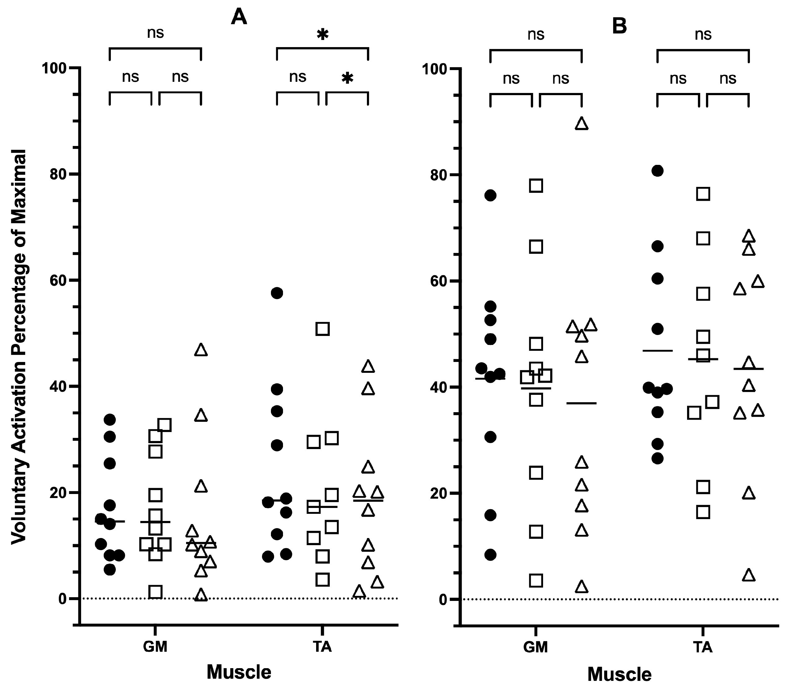
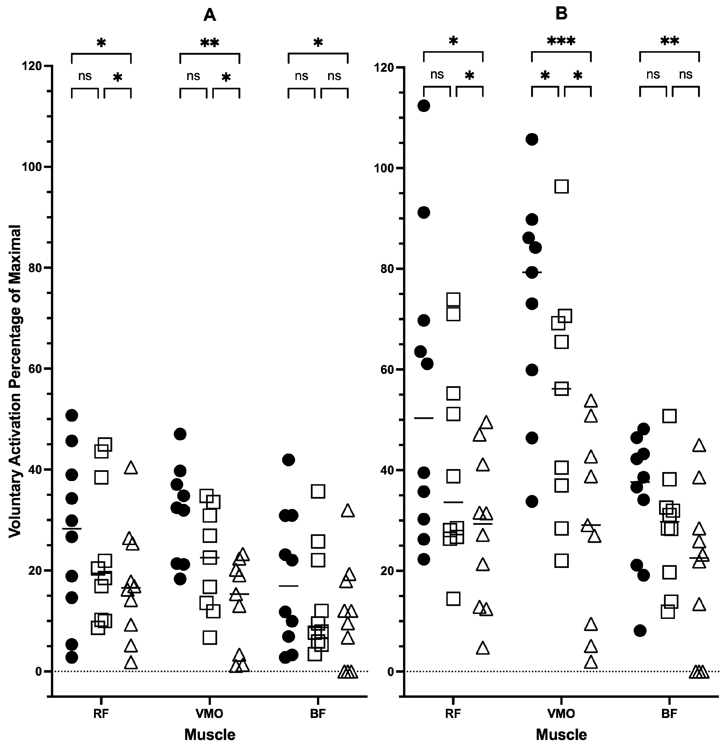
The mixed-effects analysis revealed significant differences in the interaction between muscle activity and assistance levels (p = 0.0006). A post hoc analysis found significant (p ≤ 0.05) changes with a large effect (d ≥ 0.8) between the assistance levels ‘no’ and ‘high’ for RF (p = 0.039; d = 0.8; 95% CI [2.26, 16.59]), VMO (p = 0.001; d = 1.6; 95% CI [11.40, 25.25]), BF (p = 0.03; d = 0.8; 95% CI [1.96, 12.86]) and TA (p = 0.03; d = 0.8; 95% CI [1.36, 9.73]), and between assistance levels ‘low’ and ‘high’ for RF (p = 0.02; d= 0.9; 95% CI [2.24, 9.77]), VMO (p = 0.04; d = 0.8; 95% CI [2.16, 15.33]) and TA (p = 0.02; d = 0.9; 95% CI [1.23, 6.78]) (Figure 1 and Table 3). No significant difference was detected between assistance levels in GM (Figure 1 and Table 3). Between the no and low assistance levels, no significant difference was shown for any muscle.

3.2.2. Peak Muscle Activity

The mixed-effects analysis for peak muscle activity revealed significant interaction between assistance levels and muscle activity (p = 0.0004). A post hoc analysis found significant (p ≤ 0.05) changes with a large effect (d ≥ 0.8) between the assistance levels ‘no’ and ‘high’ for RF (p = 0.01; d = 1.0; 95% CI [8.26, 46.27]), VMO (p = 0.0002; d = 2.1; 95% CI [28.27, 60.50]) and BF (p = 0.002; d = 1.3; 95% CI [6.46, 21.79]), and between low and high assistance levels for RF (p = 0.02; d = 0.9; 95% CI [2.91, 24.0]) and VMO (p = 0.02; d = 1.0; 95% CI [5.40, 45.04]) (Figure 2 and Table 4). No significant change between assistance levels was detected in the GM or TA (Figure 1B). Between no and low assistance, no significant difference was shown for any muscle, except VMO, which showed a significant difference with a large effect (p = 0.01; d = 1.1; 95% CI [5.42, 32.91]) (Figure 2 and Table 4).

3.2.3. RPE and Pain Scores

The median RPE was 10.5 for the no assistance level, 9.1 for the low assistance level and 8.2 for the high assistance level. All participants reported no pain during e-cycling at the no and low assistance levels. At the high assistance level, only one participant reported minimal pain, with a VAS score of 1/10.

3.3. Secondary Results

TSK-17 scores decreased from pre- (M = 35.8 ± 2.5) to post-intervention (M = 33.3 ± 3.5). Scores of the PSS increased from pre- (M = 14 ± 5.7) to post-intervention (M = 14.5 ± 3.3). Changes in the BREQ-3 subscores were minimal (Table 5). External, identified, integrated, and intrinsic regulation increased slightly, whereas amotivation remained unchanged, and introjected regulation decreased.

4. Discussion

The primary aim of this study was to evaluate the muscle activity of selected lower limb muscles during no-, low- and high-assistance e-cycling in people with KOA. Significant differences in mean muscle activity of the RF, VMO, BF and TA were detected when comparing no- to high-assistance cycling. For peak muscle activity, significant differences were observed in the RF, VMO and BF when comparing between no- to high-assistance cycling. In contrast, no differences between the no and low assistance levels were observed except for VMO peak activity. VMO activity significantly reduced, with a medium-to-large effect at the higher assistance levels. However, GM did not show any significant changes in peak or mean muscle activity, with only a low or medium effect between assistance levels. RPE progressively decreased from the no assistance, low assistance, to high assistance levels. Interestingly, mild knee pain was reported in one participant with high-assistance e-cycling.
Our study observed that muscle activity was similar between no-assistance and low-assistance e-cycling, except for VMO (peak and mean) activity, where assistance had a medium-to-large effect (Table 3 and Table 4). While these muscle activity observations are encouraging, we would be remiss not to place some context to the observations due to arthrogenic muscle inhibition. We did not record any measures related to pre- and post-joint effusion that our participants could have experienced, as even a small increased intra-articular fluid will stretch the joint capsule, thus activating mechanoreceptors and triggering inhibitory reflexes, which could account for some of our observed changes [37]. Other arthrogenic muscle inhibition mechanistic pathways (spinal reflex and supraspinal contributions) [38,39] are partially accounted for in our within-participant data normalisation, but we accept that transient inhibitory effects may also have influenced the results [40]. However, the reported pain scale and RPE responses do not support the supposition of an analgesic effect from e-cycling, whilst the supraspinal contribution could also have been altered by e-cycling. A clinical implication of our findings, which we cannot address with the pilot data, concerns whether e-cycling positively altered any delayed activation in the quadriceps that has been clinically observed elsewhere [40].
PA guidelines recommend at least 30 min of moderate-intensity PA for five days a week to achieve substantial health benefits in the general population [41]. Low levels of PA are common in individuals with KOA, often due to pain and stiffness in joints [42,43,44]. The outcomes reported here suggest that e-cycling, compared to no-assistance traditional cycling, may be more tolerable for inactive people due to lower perceived exertion, which may encourage people with KOA to engage in higher levels of PA via e-cycling [45]. This pilot study’s findings suggest that low-assistance e-cycling, which elicits muscle activity comparable to traditional cycling, may be a suitable alternative mode of exercise, potentially increasing motivation and adherence to exercise among individuals with KOA.
Muscle activity during high-assistance e-cycling was lower compared to no-assistance cycling. While high-assistance e-cycling may elicit relatively low muscle activity, it may also provide a low-intensity entry point to motivate individuals with KOA to start exercising. Moderate-intensity PA was induced when healthy individuals rode an e-bike across different landscapes, even when using high motor assistance [21,46]. In a laboratory-based study, it was also observed that no-, low- and high-assistance e-cycling produced sufficient intensity to align with the suggested levels of moderate-intensity exercise in healthy adults [47]. While there are differences in exercise tolerance and pain response in individuals with KOA, our results—and those from healthy individuals—provide initial supporting evidence to suggest that practitioners can use e-cycling as an exercise modality for people with KOA. Importantly, practitioners can encourage self-determination and have the individuals choose a self-selected assistance level during e-cycling according to their comfort levels, as our data show that individuals may potentially still achieve positive therapeutic effects of exercise, which could reduce the economic cost of KOA in the longer term [6]. If e-bikes are used for PA, additional resistance or aerobic exercise may be required to meet the minimum requirements for health benefits in otherwise healthy adults, and may further benefit sedentary individuals with higher baseline blood glucose, as greater reductions in blood glucose have been demonstrated after e-cycling [48]. High-assistance e-cycling could be used by clinicians as a low-barrier access strategy into PA for KOA individuals, helping to increase confidence and comfort before progressing to lower levels of assistance. While our sample size was limited, the observed exercise tolerance suggests that e-cycling is a feasible option for people with KOA, and that the self-selection of assistance levels may also increase participant motivation and long-term commitment to PA, but these suppositions need systematic confirmation.
In our exploratory study, VMO muscle activity was lower with higher assistance levels during e-cycling. This suggests that greater levels of electrical assistance lead to a lower VMO workload, which supports previous studies reporting that muscle activation increases with cycling intensity [49,50]. Furthermore, the use of high-assistance e-cycling in individuals with KOA could limit quadriceps strengthening effects; in contrast, it could also increase exercise tolerance and satisfaction. The application of these findings could support clinicians to personalise e-cycling prescription according to the patient’s specific goal, such as enhancing exercise compliance or muscle strengthening. Progression from initial exercise through high-assistance e-cycling to provide comfort and compliance, followed by a reduction in motor assistance, could be an effective approach to enhancing quadriceps activation and optimising clinical outcomes.
There was an overall pattern of decreasing RPE with higher levels of assistance, which was expected. In our study, e-cycling reduced RPE by up to two points during low-assistance and four points during high-assistance compared to no-assistance cycling. These findings are consistent with evidence showing that greater assistance by e-bike reduces RPE and increases pleasure compared with regular cycling [46,47]. The decrease in perceived exertion observed with e-cycling could reduce the exercise barrier and improve motivation; thus, e-cycling has the potential to be a practical and engaging form of PA for KOA individuals [47]. Providing lower-effort exercise options may improve PA levels and reduce KOA symptoms; therefore, low-assistance e-cycling may be an alternative to traditional cycling for people with KOA, as it may maintain muscle activity while reducing perceived exertion.
Individuals with KOA often express concern and anxiety about exercise-related pain [10]. All participants in this research reported no or mild pain when e-cycling at each assistance level. While increased PA levels have been associated with reduced pain and disability, excessive exercise may exacerbate knee pain and elevate FOM, reducing exercise adherence [43,51,52]. Our observation that e-cycling was mostly pain-free could help promote higher PA among individuals with KOA. However, this observation needs to be tempered by the small sample size. Additionally, we are unable to account for any participants who underwent intra-articular corticosteroid, platelet-rich plasma, or hyaluronic acid injection therapy within the last 12 months, as each of these treatments could influence the pain response.
Pre- and post-e-cycling assessments using the TSK-17, PSS and BREQ-3 showed minimal changes, likely due to the small sample size. The TSK-17 scores remained below 37, indicating a low to medium kinesiophobia risk [53], and PSS scores were within the low to moderate stress level, with no discernible trend [29]. Higher BREQ-3 subscores for autonomous motivation were observed post-exercise, suggesting that e-cycling may encourage self-motivated exercise in KOA individuals [54]. However, the small sample size means that these observations are preliminary and should be used to generate future research hypotheses.
The study’s main strengths are its pragmatic, field-based design, which reflects real-world e-cycling use, and its novel focus on psychosocial outcomes in individuals with KOA regardless of any recent common treatment program. Future studies with larger samples that consider the longitudinal responses are needed to further investigate the relationship between e-cycling and psychological outcomes.
The limitations of this study include potential expectation bias in self-reported outcomes, as participants may have anticipated some benefits from e-cycling. Also, a ceiling or floor effect may exist if participants were already motivated towards PA and low in FOM. The use of MVIC-normalised EMG data enables standardised comparisons [35], but may be affected by participants’ inexperience with maximal contractions or pain-related inhibition. While we delimited this study design to the analysis of only 100 m of e-cycling, it remains unclear if the differences in muscle activity would change over longer cycling distances or with repeated use. The small sample size and short-distance protocol with wireless constraints affect the generalisability and external validity of the results, reducing the ability to reflect real-world e-cycling exposure in the KOA population. Furthermore, cadence was difficult to maintain, and the power applied during e-cycling was not standardised, which could potentially introduce confounding factors. Future, larger and longitudinal studies are needed to clarify the psychological and functional effects and determine if observed changes are sustained. While exercise as a modality has clear benefits to sufferers of KOA [8], researchers seeking to design experiments to clarify the psychological and functional effects of e-cycling are strongly encouraged to ensure that confounding and known risk factors [1,4] are considered at the experimental design level and then included in the statistical process as deliberate covariates; pain or joint effusion changes could be applied as a random factor in an applied statistical model. These experimental considerations are paramount to improving the generalisability and clinical relevance of research seeking to improve exercise prescription guidelines for KOA.

5. Conclusions

The findings of this pilot study suggest that lower limb muscle activity during low-assistance e-cycling is not significantly different from that of no-assistance e-cycling in individuals with KOA. In contrast, high-assistance e-cycling generated significantly lower muscle activity compared to both the no and low assistance levels in this limited sample. While a low level of exertion was reported by the participants in both no-assistance and low-assistance trials, and pain levels were low across all levels of assistance. Overall, this pilot study suggests e-cycling is a low-exertion, pain-free activity that may effectively stimulate lower limb muscles in individuals with KOA. Low-assistance e-cycling could be explored as a convenient and viable alternative to traditional cycling for exercise in individuals with KOA. Further research is required to determine whether differences in muscle activity are significant during longer-duration e-cycling sessions, how activation levels present after prolonged periods of training at various assistance levels and whether there are any differences in lower limb strength following prolonged e-cycling use. Further studies are needed to explore changes in FOM, perceived stress and motivation with e-cycling in individuals with KOA.

Author Contributions

Conceptualisation, D.W.C. and K.J.N.; methodology, J.Y.C., T.M., T.F.Y., L.F.K., K.H.C., D.W.C. and K.J.N.; formal analysis, J.Y.C., T.M., T.F.Y., L.F.K., K.H.C., D.W.C. and K.J.N.; investigation, J.Y.C., T.M., T.F.Y., L.F.K. and K.H.C.; resources, D.W.C. and K.J.N.; writing—original draft preparation, J.Y.C., T.M., L.F.K. and K.H.C.; writing—review and editing, J.Y.C., T.M., T.F.Y., L.F.K., K.H.C., D.W.C. and K.J.N.; visualisation, J.Y.C., T.M., T.F.Y., L.F.K., K.H.C. and D.W.C.; supervision, D.W.C. and K.J.N.; project administration, D.W.C. and K.J.N. All authors have read and agreed to the published version of the manuscript.

Funding

This research received no external funding.

Institutional Review Board Statement

The study was conducted in accordance with the Declaration of Helsinki, and approved by the Human Research Ethics Committee of Curtin University (HRE2024-0721, 26 March 2024).

Informed Consent Statement

Informed consent was obtained from all subjects involved in the study.

Data Availability Statement

The original contributions presented in this study are included in the article. Further inquiries can be directed to the corresponding author.

Acknowledgments

We sincerely appreciate the time and effort of all participants who took part in the study.

Conflicts of Interest

The authors declare no conflicts of interest.

Abbreviations

The following abbreviations are used in this manuscript:
KOAKnee osteoarthritis
TSKTampa Scale of Kinesiophobia
PSSPerceived Stress Scale
BREQBehavioural Regulation in Exercise Questionnaire
FOMFear of movement
PAPhysical activity
E-bikeElectric bicycle
E-cyclingElectric bicycle cycling
EMGElectromyography
IMUInertial measurement units
RFRectus femoris
VMOVastus medialis oblique
BFBiceps femoris
TATibialis anterior
GMGastrocnemius medialis
VASVisual analogue scale
RPERate of perceived exertion
MVICMaximal voluntary isometric contractions
CIConfidence interval

References

  1. Dong, Y.; Yan, Y.; Zhou, J.; Zhou, Q.; Wei, H. Evidence on risk factors for knee osteoarthritis in middle-older aged: A systematic review and meta analysis. J. Orthop. Surg. Res. 2023, 18, 634. [Google Scholar] [CrossRef]
  2. Australian Institute of Health and Welfare. Osteoarthritis; AIHW: Canberra, Australia, 2024. [Google Scholar]
  3. Berteau, J.-P. Knee Pain from Osteoarthritis: Pathogenesis, Risk Factors, and Recent Evidence on Physical Therapy Interventions. J. Clin. Med. 2022, 11, 3252. [Google Scholar] [CrossRef] [PubMed]
  4. Geng, R.; Li, J.; Yu, C.; Zhang, C.; Chen, F.; Chen, J.; Ni, H.; Wang, J.; Kang, K.; Wei, Z.; et al. Knee osteoarthritis: Current status and research progress in treatment (Review). Exp. Ther. Med. 2023, 26, 481. [Google Scholar] [CrossRef]
  5. Bayrak, G.; Alkan, H. Factors influencing pain intensity in knee osteoarthritis: A cross-sectional biopsychosocial perspective. BMC Musculoskelet. Disord. 2025, 26, 196. [Google Scholar] [CrossRef]
  6. Bitton, R. The economic burden of osteoarthritis. Am. J. Manag. Care 2009, 15, S230–S235. [Google Scholar] [PubMed]
  7. Sharma, A.; Sharma, N.; Chahal, A. Home care program and exercise prescription for improving quality of life in geriatric population with knee osteoarthritis: A systematic review and meta-analysis. J. Bodyw. Mov. Ther. 2024, 40, 1645–1656. [Google Scholar] [CrossRef]
  8. Verhagen, A.P.; Ferreira, M.; Reijneveld-van de Vendel, E.A.E.; Teirlinck, C.H.; Runhaar, J.; van Middelkoop, M.; Hermsen, L.; de Groot, I.B.; Bierma-Zeinstra, S.M.A. Do we need another trial on exercise in patients with knee osteoarthritis?: No new trials on exercise in knee OA. Osteoarthr. Cartil. 2019, 27, 1266–1269. [Google Scholar] [CrossRef]
  9. Bosomworth, N.J. Exercise and knee osteoarthritis: Benefit or hazard? Can. Fam. Physician 2009, 55, 871–878. [Google Scholar]
  10. Gunn, A.H.; Schwartz, T.A.; Arbeeva, L.S.; Callahan, L.F.; Golightly, Y.; Goode, A.; Hill, C.H.; Huffman, K.; Iversen, M.D.; Pathak, A.; et al. Fear of Movement and Associated Factors Among Adults with Symptomatic Knee Osteoarthritis. Arthritis Care Res. 2017, 69, 1826–1833. [Google Scholar] [CrossRef]
  11. Kilinç, H.; Karahan, S.; Atilla, B.; Kinikli, G. Can Fear of Movement, Depression and Functional Performance be a Predictor of Physical Activity Level in Patients with Knee Osteoarthritis? Arch. Rheumatol. 2019, 34, 274–280. [Google Scholar] [CrossRef]
  12. Scopaz, K.A.; Piva, S.R.; Wisniewski, S.; Fitzgerald, G.K. Relationships of Fear, Anxiety, and Depression with Physical Function in Patients with Knee Osteoarthritis. Arch. Phys. Med. Rehabil. 2009, 90, 1866–1873. [Google Scholar] [CrossRef] [PubMed]
  13. Lozano-Meca, J.; Gacto-Sánchez, M.; Montilla-Herrador, J. Association of kinesiophobia with pain, disability and functional limitation in adults with knee osteoarthritis: A systematic review and meta-analysis. Geriatr. Nurs. 2024, 60, 481–490. [Google Scholar] [CrossRef] [PubMed]
  14. Salacinski, A.J.; Krohn, K.; Lewis, S.F.; Holland, M.L.; Ireland, K.; Marchetti, G. The Effects of Group Cycling on Gait and Pain-Related Disability in Individuals with Mild-to-Moderate Knee Osteoarthritis: A Randomized Controlled Trial. J. Orthop. Sports Phys. Ther. 2012, 42, 985–995. [Google Scholar] [CrossRef] [PubMed]
  15. Bannuru, R.R.; Osani, M.C.; Vaysbrot, E.E.; Arden, N.K.; Bennell, K.; Bierma-Zeinstra, S.M.A.; Kraus, V.B.; Lohmander, L.S.; Abbott, J.H.; Bhandari, M.; et al. OARSI guidelines for the non-surgical management of knee, hip, and polyarticular osteoarthritis. Osteoarthr. Cartil. 2019, 27, 1578–1589. [Google Scholar] [CrossRef]
  16. Australian Commission on Safety and Quality in Health. Osteoarthritis of the Knee Clinical Care Standard; Australian Commission on Safety and Quality in Health: Sydney, Australia, 2024. [Google Scholar]
  17. Sonnery-Cottet, B.; Hopper, G.P.; Gousopoulos, L.; Vieira, T.D.; Thaunat, M.; Fayard, J.-M.; Freychet, B.; Ouanezar, H.; Cavaignac, E.; Saithna, A. Arthrogenic Muscle Inhibition Following Knee Injury or Surgery: Pathophysiology, Classification, and Treatment. Video J. Sports Med. 2022, 2, 26350254221086295. [Google Scholar] [CrossRef]
  18. Luan, L.; Bousie, J.; Pranata, A.; Adams, R.; Han, J. Stationary cycling exercise for knee osteoarthritis: A systematic review and meta-analysis. Clin. Rehabil. 2021, 35, 522–533. [Google Scholar] [CrossRef]
  19. Øiestad, B.E.; Årøen, A.; Røtterud, J.H.; Østerås, N.; Jarstad, E.; Grotle, M.; Risberg, M.A. The efficacy of strength or aerobic exercise on quality of life and knee function in patients with knee osteoarthritis. A multi-arm randomized controlled trial with 1-year follow-up. BMC Musculoskelet. Disord. 2023, 24, 714. [Google Scholar] [CrossRef]
  20. Wild, K.; Woodward, A. Why are cyclists the happiest commuters? Health, pleasure and the e-bike. J. Transp. Health 2019, 14, 100569. [Google Scholar] [CrossRef]
  21. Bourne, J.E.; Sauchelli, S.; Perry, R.; Page, A.; Leary, S.; England, C.; Cooper, A.R. Health benefits of electrically-assisted cycling: A systematic review. Int. J. Behav. Nutr. Phys. Act. 2018, 15, 116. [Google Scholar] [CrossRef]
  22. Kolasinski, S.L.; Neogi, T.; Hochberg, M.C.; Oatis, C.; Guyatt, G.; Block, J.; Callahan, L.; Copenhaver, C.; Dodge, C.; Felson, D.; et al. 2019 American College of Rheumatology/Arthritis Foundation Guideline for the Management of Osteoarthritis of the Hand, Hip, and Knee. Arthritis Care Res. 2020, 72, 149–162. [Google Scholar] [CrossRef]
  23. Yan, L.; Li, D.; Xing, D.; Fan, Z.; Du, G.; Jiu, J.; Li, X.; Estill, J.; Wang, Q.; Belal, A.A.; et al. Comparative efficacy and safety of exercise modalities in knee osteoarthritis: Systematic review and network meta-analysis. BMJ 2025, 391, e085242. [Google Scholar] [CrossRef] [PubMed]
  24. Aykut Selçuk, M.; Karakoyun, A. Is There a Relationship Between Kinesiophobia and Physical Activity Level in Patients with Knee Osteoarthritis? Pain Med. 2020, 21, 3458–3469. [Google Scholar] [CrossRef]
  25. Kohn, M.D.; Sassoon, A.A.; Fernando, N.D. Classifications in Brief: Kellgren-Lawrence Classification of Osteoarthritis. Clin. Orthop. Relat. Res. 2016, 474, 1886–1893. [Google Scholar] [CrossRef] [PubMed]
  26. Hermens, H.J.; Freriks, B.; Disselhorst-Klug, C.; Rau, G. Development of recommendations for SEMG sensors and sensor placement procedures. J. Electromyogr. Kinesiol. 2000, 10, 361–374. [Google Scholar] [CrossRef] [PubMed]
  27. Hermens, H.J.; Freriks, B.; Merletti, R.; Stegeman, D.F.; Blok, J.H.; Rau, G.; Disselhorst-Klug, C.; Hägg, G. European Recommendations for Surface Electromyography: Results of the SENIAM Project; Roessingh Research and Development b.v.: Enschede, The Netherlands, 1999. [Google Scholar]
  28. Miller, R.P.; Kori, S.H.; Todd, D.D. The Tampa Scale: A Measure of Kinisophobia. Clin. J. Pain 1991, 7, 51. [Google Scholar] [CrossRef]
  29. Cohen, S.; Kamarck, T.; Mermelstein, R. A Global Measure of Perceived Stress. J. Health Soc. Behav. 1983, 24, 385–396. [Google Scholar] [CrossRef]
  30. Harris, K.M.; Gaffey, A.E.; Schwartz, J.E.; Krantz, D.S.; Burg, M.M. The Perceived Stress Scale as a Measure of Stress: Decomposing Score Variance in Longitudinal Behavioral Medicine Studies. Ann. Behav. Med. 2023, 57, 846–854. [Google Scholar] [CrossRef]
  31. Markland, D.; Tobin, V. A Modification to the Behavioural Regulation in Exercise Questionnaire to Include an Assessment of Amotivation. J. Sport Exerc. Psychol. 2004, 26, 191–196. [Google Scholar] [CrossRef]
  32. Dupuis, F.; Cherif, A.; Batcho, C.; Massé-Alarie, H.; Roy, J.-S. The Tampa Scale of Kinesiophobia: A Systematic Review of Its Psychometric Properties in People with Musculoskeletal Pain. Clin. J. Pain 2023, 39, 236–247. [Google Scholar] [CrossRef]
  33. Lee, E.-H. Review of the Psychometric Evidence of the Perceived Stress Scale. Asian Nurs. Res. 2012, 6, 121–127. [Google Scholar] [CrossRef]
  34. Palombi, T.; Lucidi, F.; Chirico, A.; Alessandri, G.; Filosa, L.; Tavolucci, S.; Borghi, A.M.; Fini, C.; Cavicchiolo, E.; Pistella, J.; et al. Is the Behavioral Regulation in Exercise Questionnaire a Valid Measure in Older People? Healthcare 2023, 11, 2707. [Google Scholar] [CrossRef]
  35. Konrad, P. The ABC of EMG: A Practical Introduction to Kinesiological Electromyography; Herman & Wallace Pelvic Rehabilitation Institute: Seattle, DC, USA, 2005; Volume 1, pp. 1–58. [Google Scholar]
  36. Zieliński, G. Effect Size Guidelines for Individual and Group Differences in Physiotherapy. Arch. Phys. Med. Rehabil. 2025, 106, 1844–1849. [Google Scholar] [CrossRef]
  37. Rice, D.; McNair, P.J.; Dalbeth, N. Effects of cryotherapy on arthrogenic muscle inhibition using an experimental model of knee swelling. Arthritis Care Res. 2009, 61, 78–83. [Google Scholar] [CrossRef]
  38. Hopkins, J.T.; Ingersoll, C.D. Arthrogenic Muscle Inhibition: A Limiting Factor in Joint Rehabilitation. J. Sport Rehabil. 2000, 9, 135. [Google Scholar] [CrossRef]
  39. Stokes, M.; Young, A. The Contribution of Reflex Inhibition to Arthrogenous Muscle Weakness. Clin. Sci. 1984, 67, 7–14. [Google Scholar] [CrossRef]
  40. Rice, D.A.; McNair, P.J.; Lewis, G.N.; Dalbeth, N. Quadriceps arthrogenic muscle inhibition: The effects of experimental knee joint effusion on motor cortex excitability. Arthritis Res. Ther. 2014, 16, 502. [Google Scholar] [CrossRef]
  41. Haskell, W.L.; Lee, I.-M.; Pate, R.R.; Powell, K.E.; Blair, S.N.; Franklin, B.A.; Macera, C.A.; Heath, G.W.; Thompson, P.D.; Bauman, A. Physical Activity and Public Health: Updated Recommendation for Adults from the American College of Sports Medicine and the American Heart Association. Med. Sci. Sports Exerc. 2007, 39, 1423–1434. [Google Scholar] [CrossRef]
  42. Fransen, M.; McConnell, S.; Harmer, A.R.; Van der Esch, M.; Simic, M.; Bennell, K.L. Exercise for osteoarthritis of the knee. Cochrane Database Syst. Rev. 2015, 1, CD004376. [Google Scholar] [CrossRef] [PubMed]
  43. Holla, J.F.M.; van der Leeden, M.; Knol, D.L.; Roorda, L.D.; Hilberdink, W.K.H.A.; Lems, W.F.; Steultjens, M.P.M.; Dekker, J. Predictors and Outcome of Pain-Related Avoidance of Activities in Persons with Early Symptomatic Knee Osteoarthritis: A Five-Year Followup Study. Arthritis Care Res. 2015, 67, 48–57. [Google Scholar] [CrossRef] [PubMed]
  44. Sliepen, M.; Mauricio, E.; Lipperts, M.; Grimm, B.; Rosenbaum, D. Objective assessment of physical activity and sedentary behaviour in knee osteoarthritis patients—Beyond daily steps and total sedentary time. BMC Musculoskelet. Disord. 2018, 19, 64. [Google Scholar] [CrossRef] [PubMed]
  45. Sundfør, H.B.; Fyhri, A. A push for public health: The effect of e-bikes on physical activity levels. BMC Public Health 2017, 17, 809. [Google Scholar] [CrossRef]
  46. Hughey, S.M.; Sella, J.; Adams, J.D.; Porto, S.C.; Bornstein, D.; Brown, K.; Amahrir, S.; Michalaka, D.; Watkins, K.; Davis, W.J. It’s electric! Measuring energy expenditure and perceptual differences between bicycles and electric-assist bicycles. J. Transp. Health 2022, 27, 101523. [Google Scholar] [CrossRef]
  47. Louis, J.; Brisswalter, J.; Morio, C.; Barla, C.; Temprado, J.-J. The Electrically Assisted Bicycle: An Alternative Way to Promote Physical Activity. Am. J. Phys. Med. Rehabil. 2012, 91, 931–940. [Google Scholar] [CrossRef]
  48. Bini, R.; Collins, B.; Hunter, J.; Hunter, N.; McVicar, J.; Gordon, B.; Kingsley, M. Associations between e-bike travel distance and changes in health. J. Transp. Health 2024, 39, 101925. [Google Scholar] [CrossRef]
  49. da Silva, J.C.L.; Ekblom, M.M.; Tarassova, O.; Andersson, E.; Rönquist, G.; Grundström, H.; Arndt, A. Effect of increasing workload on knee extensor and flexor muscular activity during cycling as measured with intramuscular electromyography. PLoS ONE 2018, 13, e0201014. [Google Scholar] [CrossRef]
  50. Holliday, W.; Theo, R.; Fisher, J.; Swart, J. Cycling: Joint kinematics and muscle activity during differing intensities. Sports Biomech. 2023, 22, 660–674. [Google Scholar] [CrossRef]
  51. Burrows, N.J.; Barry, B.K.; Sturnieks, D.L.; Booth, J.; Jones, M.D. The Relationship Between Daily Physical Activity and Pain in Individuals with Knee Osteoarthritis. Pain Med. 2020, 21, 2481–2495. [Google Scholar] [CrossRef]
  52. Focht, B.C.; Ewing, V.; Gauvin, L.; Rejeski, W.J. The unique and transient impact of acute exercise on pain perception in older, overweight, or obese adults with knee osteoarthritis. Ann. Behav. Med. 2002, 24, 201–210. [Google Scholar] [CrossRef]
  53. Liu, H.; Huang, L.; Yang, Z.; Li, H.; Wang, Z.; Peng, L. Fear of Movement/(Re)Injury: An Update to Descriptive Review of the Related Measures. Front. Psychol. 2021, 12, 696762. [Google Scholar] [CrossRef]
  54. Box, A.G.; Feito, Y.; Brown, C.; Petruzzello, S.J. Individual differences influence exercise behavior: How personality, motivation, and behavioral regulation vary among exercise mode preferences. Heliyon 2019, 5, e01459. [Google Scholar] [CrossRef]
Figure 1. Mean (A) and peak (B) voluntary muscle activity for lower leg muscles (GM: gastrocnemius; TA: tibialis anterior) during no assistance (⏺), low assistance (☐) and high assistance (∆). Note: ns denotes non significant p-value; * Denotes significant p-value (p < 0.05); Group mean shown with short solid black line.
Applsci 16 01713 g001
Figure 2. Mean (A) and peak (B) voluntary muscle activity for upper leg muscles (RF: rectus femoris; VMO: vastus medialis oblique; BF: biceps femoris) during no assistance (⏺), low assistance (☐) and high assistance (∆). Note: ns denotes non significant p-value; * Denotes significant p-value (p < 0.05); ** denotes significant p-value (p < 0.01); *** denotes significant p-value (p < 0.001); Group mean shown with short solid black line.
Applsci 16 01713 g002
Table 1. Electrode placement specifications in accordance with SENIAM guidelines [26].
Muscle SiteLocationOrientation
Rectus femoris50% distance from the ASIS to the superior portion of the patellaIn the direction of the line from the ASIS to the superior portion of the patella
Vastus medialis oblique80% distance from the ASIS to the medial knee joint spacePerpendicular to the line between the ASIS and the medial knee joint space
Biceps femoris50% distance from the ischial tuberosity and the lateral tibia epicondyleIn the direction of the line from the ischial tuberosity and the lateral tibial epicondyle
Tibialis anterior1/3 on the line between the fibula and the medial malleolusIn the direction of the line between the fibula and the medial malleolus
Gastrocnemius medialisOn the most pronounced bulge of the muscleIn the direction of the leg
Table 2. Participant characteristics (n = 10).
CharacteristicsMean or Count (±SD)
Age (yrs)68.1 ± 4.7
GenderFemale, n = 2
Male, n = 8
Height (cm)176 ± 11.2
Weight (kg)83.9 ± 17.6
Osteoarthritis GradeGrade 2, n = 4
Grade 3, n = 6
Table 3. Comparison of mean (%MVIC) muscle activity of lower leg muscles with the 95% confidence interval (95%CI) of the difference across the three levels of assistance.
MusclesAssistance Levels95% CIp-ValueCohen’s d
Rectus femoris
(n = 10)
No vs. Low−2.01 to 8.840.280.4
No vs. High2.26 to 16.590.04 *0.8
Low vs. High2.24 to 9.770.02 *0.9
Vastus medialis oblique
(n = 9)
No vs. Low1.44 to 17.730.060.7
No vs. High11.40 to 25.250.001 **1.6
Low vs. High2.16 to 15.330.04 *0.8
Biceps femoris
(n = 10)
No vs. Low−2.05 to 11.720.230.4
No vs. High1.96 to 12.860.03 *0.8
Low vs. High0.25 to 4.890.070.6
Tibialis anterior
(n = 9)
No vs. Low−4.13 to 4.460.940
No vs. High1.36 to 9.730.04 *0.8
Low vs. High1.23 to 6.780.03 *0.9
Gastrocnemius medialis
(n = 10)
No vs. Low−2.52 to 2.290.930
No vs. High−4.30 to 6.210.740.1
Low vs. High−4.42 to 6.560.730.1
* Denotes significant p-value (p < 0.05); ** denotes significant p-value (p < 0.01).
Table 4. Comparison of peak (%MVIC) muscle activity of lower leg muscles with the 95% confidence interval (95%CI) of the difference across the three levels of assistance.
MusclesAssistance Levels95% CIp-ValueCohen’s d
Rectus femoris
(n = 10)
No vs. Low−1.46 to 29.040.070.6
No vs. High8.26 to 46.270.01 *1.0
Low vs. High2.91 to 24.00.02 *0.9
Vastus medialis oblique
(n = 9)
No vs. Low5.42 to 32.910.01 *1.1
No vs. High28.27 to 60.500.0002 ***2.1
Low vs. High5.40 to 45.040.02 *1.0
Biceps femoris
(n = 10)
No vs. Low−2.13 to 12.350.140.5
No vs. High6.46 to 21.790.002 **1.3
Low vs. High−0.17 to 18.210.050.7
Tibialis anterior
(n = 9)
No vs. Low−13.67 to 9.250.67−0.1
No vs. High−7.48 to 14.330.500.2
Low vs. High−5.39 to 12.490.390.3
Gastrocnemius medialis
(n = 10)
No vs. Low−8.50 to 12.070.700.1
No vs. High−13.75 to 22.980.580.2
Low vs. High−9.10 to 14.750.600.2
* Denotes significant p-value (p < 0.05); ** denotes significant p-value (p < 0.01); *** denotes significant p-value (p < 0.001).
Table 5. Pre- and post-intervention BREQ-3 subscores (mean ± SD).
SubscoresPre-InterventionPost-Intervention
Motivation1 ± 01 ± 0
External regulation1.13 ± 0.251.5 ± 0.58
Introjected regulation3.13 ± 0.783 ± 0.98
Identified regulation4.38 ± 0.484.5 ± 0.54
Integrated regulation3.69 ± 0.903.88 ± 0.85
Intrinsic regulation3.5 ± 0.462.75 ± 0.52
Disclaimer/Publisher’s Note: The statements, opinions and data contained in all publications are solely those of the individual author(s) and contributor(s) and not of MDPI and/or the editor(s). MDPI and/or the editor(s) disclaim responsibility for any injury to people or property resulting from any ideas, methods, instructions or products referred to in the content.