Article

Valorization of Grape Pomace Extract Through Dextran–Grape Conjugates: A Sustainable Approach for Cosmetic and Dermatological Applications

by
Marisa Francesca Motta
1,†,
Anna Francesca Vattimo
2,†,
Fabio Amone
2,
Rocco Malivindi
1,2,
Ortensia Ilaria Parisi
1,2,* and
Francesco Puoci
1,2
1
Department of Pharmacy, Health and Nutritional Sciences, University of Calabria, 87036 Rende, CS, Italy
2
Macrofarm s.r.l., c/o Department of Pharmacy, Health and Nutritional Sciences, University of Calabria, 87036 Rende, CS, Italy
*
Author to whom correspondence should be addressed.
These authors contributed equally to this work.
Appl. Sci. 2025, 15(6), 3220; https://doi.org/10.3390/app15063220
Submission received: 11 February 2025 / Revised: 8 March 2025 / Accepted: 11 March 2025 / Published: 15 March 2025
(This article belongs to the Special Issue Cosmetics Ingredients Research - 2nd Edition)

Abstract

:
Industrial waste management is a growing concern, and the valorization of by-products through circular economy approaches represents a sustainable solution. In this context, dextran–grape conjugates (PLG–GRAPE) were obtained via a grafting reaction of grape pomace extract and dextran under aqueous conditions. To compare the properties of the polymeric graft with those of the free extract, total polyphenol content was assessed using the Folin–Ciocalteu assay, along with stability and diffusion studies. In addition, in vitro safety evaluations, including Neutral Red Uptake, h-CLAT, and skin irritation tests were performed to assess the biocompatibility. To evaluate the antioxidant, anti-inflammatory, and anti-aging properties of PLG–GRAPE, in vitro efficacy assays were performed on keratinocyte and fibroblast cell lines and full-thickness reconstructed human tissues exposed to damaging agents, such as UV radiation and pollutants. The results showed that the technology preserved the phenolic and antioxidant activity of the extract, while improving diffusion and stability properties. As demonstrated by the results of the in vitro studies, a favorable biocompatibility profile was observed, in addition to a significant capacity to reduce oxidative stress and inflammation in aged cells, thus, attenuating cellular aging and senescence. In conclusion, the study suggests that PLG–GRAPE has potential as a bioactive ingredient for cosmetic and dermatological applications, offering a sustainable and effective approach to utilizing industrial waste products.

1. Introduction

The circular economy is based on the principles of reducing, reusing, recycling, and recovering materials, as well as repurposing waste by interconnecting water, food, and energy resources [1]. Industrial by-products and biowaste have been increasingly recognized as sustainable sources of phytochemicals, which can be valorized into bioactive compounds [2]. Among these, grape pomace, a by-product of the winemaking process, has attracted particular attention. Traditionally used as animal feed or fertilizer, grape pomace has been acknowledged for nearly four decades as a potential source of bioactive compounds, offering an additional strategy to mitigate environmental pollution [3]. Comprising a mixture of grape skins, seeds, and stems, it accounts for approximately 20% of the weight of processed grapes. Since the winemaking process does not extract all the valuable compounds, grape pomace remains rich in polyphenols, although their distribution is inhomogeneous [4,5,6]. Consequently, it can be efficiently transformed into cost-effective raw materials for industries such as cosmetics, nutraceuticals, pharmaceuticals, and food, thus, reducing the environmental impact associated with waste disposal near wine production facilities [7]. The vinification process generates substantial amounts of waste; however, scientific and technological advancements have identified methods to recover these materials, sometimes yielding greater economic benefits than those derived from wine production itself [8]. The potential of these compounds in terms of application is essentially focused on their capacity for significant, widely demonstrated antioxidant and anti-inflammatory properties [9,10,11,12], which renders them suitable for the treatment of inflammatory skin diseases. Inflammatory skin diseases are distinguished by the release of pro-inflammatory cytokines (e.g., TNF-alpha, IL-6, IL-12) and persistent oxidative stress (ROS release), which contributes to structural skin damage [13].
Among different grape varieties, Magliocco Calabrese pomace has been identified as particularly rich in phenolic compounds with diverse physiological properties, including antiallergenic, anti-inflammatory, antimicrobial, antioxidant, antithrombotic, cardioprotective, and vasodilatory effects [14,15]. Furthermore, it has been largely demonstrated that anthocyanins, which are present in abundance in grape pomace, exhibit significant UV-shielding properties, thereby mitigating photoaging by reducing oxidative stress and collagen degradation [16,17]. However, the efficacy of these phenolic extracts largely depends on maintaining their stability, bioactivity, and bioavailability [18]. To enhance their preservation, various encapsulation strategies have been proposed, including the use of maltodextrin, primarily in the food industry [19].
Building upon these findings, the present study aims to expand on previously employed methods [20] by focusing on:
  • the reuse of waste raw materials, thereby promoting a circular economy through waste reduction and repurposing;
  • the application of rapid dynamic solid–liquid extraction (RSLDE, Naviglio®) to maximize the preservation of bioactive compounds [21];
  • the grafting of polymers in an aqueous environment, using dextran—a natural, biocompatible, and biodegradable polysaccharide—due to its moisturizing, film-forming, and carrier properties. Its biocompatibility and chemical stability make it particularly suitable for various applications, protecting active ingredients from degradation [22,23,24,25,26].
The novelty of this study lies in the choice of grape marc as the raw material of interest, exploiting its various components with multifunctional bioactive properties. Instead of being discarded, this material can be extracted and incorporated into technologies that have a controlled release system and that reduce its degradation by increasing its stability. Moreover, the objective of the study is to validate the biocompatibility and efficacy (anti-inflammatory, antioxidant and anti-aging properties) of dextran–grape seed conjugates (PLG–GRAPE) for utilization in cosmetic and dermatological applications. The present approach integrates the reuse of raw materials with long-release technology, thereby enhancing product performance and contributing to the principles of sustainability.

2. Materials and Methods

2.1. Reagents

Grape pomace was obtained from Biocal (Luzzi, CS, Italy). The grapes used for the production of the pomace were grown in Calabria (Italy) and the pomace was collected after the winemaking process.
The following reagents were purchased from Sigma-Aldrich s.r.l. (Milan, Italy): dextran from Leuconostoc spp. (DEX), hydrogen peroxide (H2O2), ascorbic acid (AA), 2,2-diphenyl-1-picrylhydrazyl radical (DPPH), Folin–Ciocalteu reagent, gallic acid monohydrate—98+%, A.C.S. reagent, sodium carbonate, 3-(4,5-dimethylthiazol-2-yl)-2,5-diphenyltetrazolium bromide (MTT), paraformaldehyde, Neutral Red (NR), acetic acid, nickel sulfate, propidium iodide, Triton X-100, and γ-globulin. FACS buffer is composed by Dulbecco’s Buffered Saline (PBS), 0.5–1% Bovine Albumin Serum (BSA), 0.1% sodium azide all purchased from Sigma-Aldrich s.r.l. (Milan, Italy).
Strat-M® membranes (25 mm discs) were purchased from Merck Millipore (Darmstadt, Germany).
4′,6-diamidino-2-phenylindole (DAPI), goat pAb IgG (TRITC), goat pAb IgG (FITC) were purchased from Abcam (Cambridge, UK).
Collagen III Monoclonal Antibody and Elastin Polyclonal Antibody, Human IL-12 ELISA Kit, and Human MMP9 ELISA were purchased from Invitrogen (Carlsbad, CA, USA).
The following kits were purchased from Abcam (Cambridge, UK): Human TNF-alpha ELISA Kit, Human IL-6 ELISA kit, Kit Beta Galactosidase Assay Kit, CPD ELISA Kit (cyclobutane pyrimidine dimers, DNA samples) and Mitochondrial DNA Isolation Kit.
Intracellular Total ROS Activity Assay Kit was purchased from Abnova (Heidelberg, Germany).
Human Collagen Type III ELISA Kit and Human Collagen Type I ELISA Kit were purchased from Cloude Clone (Wuhan, China).
8-hydroxy-2′-deoxyguanosine (8-OHdG) ELISA Kit, Human IL-1 alpha, and Human Hyaluronic Acid ELISA Kit were purchased from Abokine (Heidelberg, Germany).
Human Elastin ELISA Kit was purchased from Elabscience (Houston, TX, USA).
The DNeasy Blood and Tissue Kit was purchased from Qiagen (Venlo, Netherlands).
The MATTEK EPI-212-SIT and the full thickness skin tissues EFT-412 3D Human Epidermis Full Thickness Micro-Tissues were purchased from MatTek Life Sciences (Ashland, MA, USA).
The BJ (human fibroblast), Balb/3T3 Clone A31 (murine fibroblast), THP-1 (human monocyte), and HaCaT (human keratinocyte) cell lines were obtained from the American Type Culture Collection (ATCC) located in Manassas, VA, USA. The BJ and HaCat cells were cultured in MEM containing 10% FBS and 1% Penicillin/Streptomycin and 1% sodium pyruvate. The Balb/3T3 clone cells were cultured in DMEM containing 10% BCS and 1% Penicillin/Streptomycin. The THP-1 cells were maintained in RPMI 1640 containing 10% FBS, 1% Penicillin/Streptomycin, and 0.05% β-Mercapto-ethanol. All cell lines were maintained at a temperature of 37 °C in a humidified atmosphere of 5% CO2.

2.2. Extract Preparation Using a Naviglio Extractor®

Grape pomace, supplied by Biocal, was dried using a cold dryer for 12 h and transformed into small particles in a grinder to increase the surface area and improve extraction efficiency.
Subsequently, the pomace was subjected to extraction with a Naviglio extractor® model NEXNA0040 (Atlas Filtri, Italy), using water as a solvent. The extraction was performed according to the method reported in literature with some modifications [21].
A quantity of 500 g of the processed material was enclosed in microporous polyethylene pouches (50 μm). To maximize interaction between the components, a plant material-to-solvent ratio of 1:10 was maintained. The extraction vessel was then filled with water, and the pouch containing the plant material was introduced. After securing the system, the internal pressure was progressively raised to about 9 bar, enhancing solvent infiltration. The extraction was performed by subjecting the material to 30 extraction cycles, lasting a total of 2 h. During the dynamic phase, a total of 12 impacts were applied. Both the dynamic operation phase and the static operation phase were carried out for a duration of 2 min.
After extraction, the aqueous grape pomace extract, in its final form as an extractive solution, was filtered to remove any residual particulate matter before further processing and was stored at 4 °C until use.

2.3. Preparation and Characterization of the Polymeric Conjugates

2.3.1. Grafting Reaction

Dextran–grape pomace conjugates (PLG–GRAPE) were synthesized through a grafting reaction carried out under aqueous conditions at ambient temperature as previously described [27,28].
Different conjugates were prepared using dextran at concentrations of 2.5%, 5%, 10%, and 20% (w/w). The reaction utilized a redox pair, comprising hydrogen peroxide and ascorbic acid, as the initiating system, ensuring biocompatibility and avoiding the formation of toxic by-products.

2.3.2. Total Polyphenol Content Quantification

The Folin–Ciocalteu reagent procedure was employed to determine the total polyphenol content of the synthesized PLG–GRAPE conjugates, as described [27], with slight modifications. Specifically, before analysis, all samples were diluted to 23% (v/v) in distilled water. Each sample (1 mL) was mixed with 1 mL of distilled water, 1 mL of 0.2 N Folin–Ciocalteu reagent and 1 mL of 7.5% (w/v) sodium carbonate solution. The mixture was then incubated in the dark at room temperature for two hours, and the absorbance was subsequently measured at 760 nm by using a UV-Visible Evolution 201 spectrophotometer (Thermo Fisher Scientific, Waltham, MA, USA).
The same procedure was applied to both the initial grape pomace extract and a control sample (blank), consisting of a 20% (w/w) aqueous dextran solution, to evaluate any interference of the polymeric material in the Folin–Ciocalteu assay.
The total polyphenol content was expressed as milligrams equivalents of a reference polyphenol, such as catechin, per gram of tested sample (mg eq CA/g), based on the equation derived from the catechin calibration curve. This one was recorded using five different catechin standard solutions of known concentrations (ranging from 1 × 10⁻³ to 1 × 10⁻⁴ M). The slope and intercept of the resulting regression equation were calculated using the least squares method.
The assay was repeated in triplicate.

2.3.3. DPPH Assay

In order to evaluate the antioxidant activity of PLG–GRAPE conjugates and the grape pomace extract, their ability to scavenge a stable free radical, 2,2-diphenyl-1-picrylhydrazyl (DPPH), was analyzed based on the protocol as described [29,30].
For this aim, one milliliter of each sample was subsequently mixed with 4 mL of a 188 μM ethanolic DPPH solution and 5 mL of absolute ethanol. The resulting solutions were incubated in darkness, and after 15 min, the radical scavenging capacity was assessed by measuring the absorbance at 517 nm through a UV-Visible Evolution 201 spectrophotometer (Thermo Fisher Scientific, MA, USA).
A control sample (Blank), consisting of a 20% w/w aqueous dextran solution, was subjected to the same experimental conditions to investigate its potential interference in the DPPH assay.
The DPPH assay is a widely used method for evaluating antioxidant capacity, based on the ability of antioxidants to scavenge 2,2-diphenyl-1-picrylhydrazyl (DPPH) radicals.
In the adopted method, the antioxidant molecule converts the radical into a yellow-colored compound, diphenylpicrylhydrazine. The reaction extent depends on the hydrogen-donating capacity of the antioxidant.
The scavenging activity of the tested samples was assessed based on DPPH reduction and expressed as the percentage of inhibition, calculated using the following Equation (1):
I n h i b i t i o n % = A 0 A 1 A 0 × 100
where A0 is the absorbance of a control sample prepared under the same conditions without any test compound, and A1 is the absorbance of the tested sample.
The assay was repeated in triplicate.

2.3.4. In Vitro Diffusion Studies

The in vitro diffusion studies were performed at a temperature of 37 ± 0.5 °C using Franz diffusion cells. In vitro diffusion studies utilizing Franz diffusion cells (Disa, Milan, Italy) with a permeation area of 0.4614 cm2, are a highly effective approach, not only for the development of novel formulations [31,32] but also as a tool for quality control purposes [33,34].
In this study, a Strat-M® synthetic membrane was used as an alternative to human skin and was positioned between the donor and receptor compartments of the diffusion cell, with the two chambers securely assembled [35].
The receptor compartment was filled with 5.5 mL of phosphate buffer (pH 7.4, 10−3 M), and following a 20–30 min equilibration period at 37 °C, 0.5 mL of the PLG–GRAPE and grape pomace extract samples were introduced into the donor compartment.
At predetermined intervals (0, 2, 4, 6, 8 and 24 h), aliquots were withdrawn from the receptor compartment and analyzed to determine the total polyphenol content using the Folin–Ciocalteu assay. The spectrophotometric measurements were performed with a UV-Visible Evolution 201 spectrophotometer (Thermo Fisher Scientific, MA, USA). To maintain sink conditions, the withdrawn aliquots were replaced with fresh phosphate buffer.
The assay was repeated in triplicate.

2.3.5. Stability Studies

Stability studies were conducted on PLG–GRAPE conjugates and the grape pomace extract. The present protocol aims to evaluate the stability of the tested samples under oxidation conditions, such as UV/H2O2 treatment [36,37].
In this case, samples were placed in a photoreactor and exposed to irradiation for 12 h using a mercury lamp with an emission spectrum ranging from 200 to 600 nm, in the presence of hydrogen peroxide (H2O2), which undergoes photolysis, generating hydroxyl radicals responsible for the degradation process through homolytic cleavage, as shown in Equation (2):
H2O2 + hv → 2OH•
The stability of each sample was assessed at specific time points (0, 2, 3, 6, 12 and 24 h) evaluating the total polyphenol content using the Folin–Ciocalteu assay. The spectrophotometric measurements were performed with a UV-Visible Evolution 201 spectrophotometer (Thermo Fisher Scientific, MA, USA), and the results were reported as the percentage of degradation.
Stability experiments were conducted in a photoreactor fitted with a high-pressure mercury lamp (TQ 718, 250 W) from Heraeus Noblelight Company (Hanau, Germany).
The assay was repeated in triplicate.

2.4. In Vitro Safety Assessment

2.4.1. Neutral Red Uptake Assay

The Neutral Red Uptake (NRU) assay, based on ISO 10993-5:2009 guidelines [38], evaluates the cytotoxicity of medical devices.
Using Balb/3T3 Clone A31 cells, an extraction according to ISO 10993-12:2021 [39] was performed [40]. Cells (2.5 × 10⁴/well) were treated with PLG–GRAPE conjugates in DMEM at three concentrations (3%, 0.6% and 0.3% of sample diluted in water) for 24 h at 37 °C with 5% CO2. Following treatment, cells were incubated with neutral red (50 μg/mL) for 3 h. Absorbance at 540 nm was measured using a Synergy H1 microplate reader (Hybrid Reader, BioTek Instruments, Agilent, CA, USA), and viability (%) was calculated using the following Equation (3):
% V i a b i l i t y = A b s s a m p l e A b s b l a n k A b s c o n t r o l A b s b l a n k

2.4.2. Skin Sensitization Potential Test (h-CLAT)

The Human Cell Line Activation Test (h-CLAT) is a method of determining the skin sensitization potential of substances. It does this by evaluating the expression of immune-related markers CD54 and CD86 on THP-1 monocytic cells.
The cells are treated with test substances at eight crescent concentrations (381.01 µg/mL, 457.21 µg/mL, 548.65 µg/mL, 658.39 µg/mL, 790.07 µg/mL, 984.08 µg/mL, 1137.70 µg/mL, 1365.24 µg/mL), alongside positive (NiSO4 100 µg/mL) and negative (culture medium) controls. Following a period of incubation, the expression levels of CD54/CD86 and cell viability are measured by means of flow cytometry CytoFLEX BH38234 model purchased from Beckman Coulter s.r.l (Milan, Italy). The effective concentrations (EC150/EC200) are then calculated in order to identify allergenic responses, with increased marker expression being linked to immune activation. To ensure reliability, tests are repeated in triplicate according to OECD 442E guidelines [41] and as before performed [42].

2.4.3. Skin Irritation Test

The «In vitro Skin Irritation: Reconstructed Human Epidermis Test Method» has been developed for the prediction of chemical-induced skin irritation. The principle behind the method is the measurement of the ability of a chemical to produce a decrease in cell viability, as reflected in the MTT assay, on a reconstructed human epidermis model. The fundamental principle of the human skin model assay is predicated on the hypothesis that irritant chemicals are able to penetrate the stratum corneum by diffusion or erosion and are cytotoxic to the underlying cell layers.
The study utilized a reconstructed human epidermis (MatTek EPIDERM), which features 0.6 cm2 of keratinocytes cultured on polycarbonate filters in an air-liquid medium. The tissues underwent rigorous quality controls, including viability (MTT OD > 0.8), barrier integrity (ET50: 4–9 h), and sterility.
The study followed OECD 439 [43] and ISO 10993-10:2010 [44] guidelines for assessing skin irritation. The concentration of the tested sample employed in this study was 3% in water, which is in accordance with the highest use concentration of the developed polymeric conjugate that can be included in a cosmetic/dermatological formulation. An amount of 30 µL of the tested sample diluted at 3% was, thus, applied directly to tissues according to ISO 10993-12:2021 [39] for 60 min. After 60 min, tissues were rinsed with Dulbecco’s Buffered Saline (DPBS).
The assessment of viability was conducted using the MTT assay after 42 h, with classifications assigned based on tissue viability.
The percentage of relative viability for the test substance and positive control was calculated in relation to the negative control, and the mean relative viability of the three individual tissues and the corresponding relative standard deviation were determined.
The categorization of the sample tested was based on the following criteria:
  • Mean tissue viability < 50%: IRRITANT
  • Mean tissue viability > 50%: NON-IRRITATING.

2.5. In Vitro Efficacy Assessment

2.5.1. Experimental Conditions

In order to evaluate the antioxidant, anti-inflammatory, and anti-aging properties of the tested sample, a co-culture of cellular HaCat and BJ cells and tissue models (full-thickness skin tissues) were subjected to aging conditions induced by the following:
Urban dust exposure: a standardized urban dust mixture containing typical environmental pollutants was utilized, including polycyclic aromatic hydrocarbons (PAHs), nitro-substituted PAHs (nitro-PAHs), polychlorinated biphenyls (PCBs), pesticide residues, and inorganic airborne particles. The concentration of the mixture was determined to be 100 mg/mL for its effects on tissues and 0.1 mg/mL for its effects on cells, with the aim of simulating the environmental pollution-induced stress experienced in nature.
UV radiation stress: cells and tissues were irradiated with the SunTest solar simulator for a period of two hours in order to replicate the stress induced by UV radiation.
At the same time as the damage, cells and tissues were treated with the tested sample at three concentrations (3%, 0.6% and 0.3% diluted in water).
Following a 24-h exposure period, a comprehensive assessment was conducted to evaluate oxidation, inflammation, and senescence parameters. Finally, parameters related to skin well-aging and trophism were assessed.
The results were then compared to those obtained from tissues and cells that had not been exposed to damage or treated (CTR-) with any samples, as well as to tissues and cells that had only been exposed to damage but not treated with any samples (CTR+).

2.5.2. DCFDA Cellular ROS Assay

In order to assess the potential mechanisms of the technology in terms of antioxidant efficacy, the intracellular generation of ROS was measured using the DCFDA Cellular ROS Detection Assay Kit following the manufacturer’s instructions.
The cells were exposed to the sample for a period of 24 h, as indicated in Section 2.5.1. Fluorescence intensity was measured at 483-14 nm/530-30 nm, and the resulting data were used to calculate the fold changes. The fluorescence spectra were measured using a Synergy H1 microplate reader (Hybrid Reader, BioTek Instruments, Agilent, CA, USA).

2.5.3. Enzyme-Linked Immunosorbent Assay (ELISA)

Cells were treated as indicated in paragraph 2.5.1. After 24 h of exposition, ELISA evaluations were assessed.
All ELISA tests were performed in strict accordance with the manufacturer’s instructions, with all kits relying on the competitive binding of an antigen with its primary antibody. The immune complex (antigen-antibody) is then recognized by a secondary antibody conjugated with a peroxidase, resulting in the addition of the peroxidase substrate. This produces a colorimetric reaction with an intensity proportional to the amount of immune complexes present and, thus, to the amount of marker to be assayed. Quantitative determination involves the construction of a calibration curve using known and increasing concentrations of standards. All standards and samples were measured with a micro-plate reader (Synergy H1, Hybrid Reader, BioTek Instruments, Agilent, CA, USA) at a wavelength of 450 nm.
The samples used for the dosage of collagen type 1, elastin, hyaluronic acid, IL-1 alpha, TNF-ALFA, IL-6, and IL12 consisted of the supernatant from cells or tissues.
The measurement of cyclobutene pyrimidine dimers (CPDs) was conducted on DNA extracted through the DNeasy Blood and Tissue Kit in strict accordance with the supplier’s instructions.
The quantification of 8-OHdG levels was performed on both DNA and mitochondrial DNA (mtDNA) extracted through the DNeasy Blood and Tissue Kit and the Mitochondrial DNA Isolation Kit, respectively, adhering to the instructions of the respective suppliers.

2.5.4. Senescence-Associated β-Galactosidase Activity

Cells were treated as indicated in Section 2.5.1. After 24 h of exposition, senescence evaluation was performed.
The measurement of cellular senescence was conducted by utilizing a Beta Galactosidase Assay Kit following the manufacturer’s instructions. This kit employs a fluorogenic fluorescein galactosidase substrate, which, upon cleavage by β-galactosidase, generates a fluorescent product that can be measured with an ELISA plate reader. The kit guidelines were followed to prepare the samples, positive controls were provided by the kit and the standard curve.
The fluorescence intensity of each sample was subsequently quantified using a microplate reader (Synergy H1, Hybrid Reader, BioTek Instruments, Agilent, CA, USA), with excitation and emission wavelengths set at 475 nm and 500–550 nm, respectively. The quantification of β-gal levels in each sample was then determined by utilizing a β-galactosidase standard curve.

2.5.5. Immunofluorescence

BJ cells (1.5 × 10⁴) were cultured on immunofluorescence slides, treated with PLG–GRAPE, as indicated in Section 2.5.1, for 24 h, then fixed, permeabilized, and blocked. The detection of collagen was accomplished through the utilization of an anti-type 1 collagen antibody and elastin antibody, followed by an overnight incubation at 4 °C in conjunction with TRITC-conjugated secondary and FITC-conjugated secondary antibodies. DAPI was used for nuclear staining, while collagen fluorescence was visualized via phycocyanin. Examination of the slides was conducted using an OLYMPUS FV3000 confocal microscope (Tokyo, Japan) at 20x magnification, with untreated samples serving as negative controls.

2.6. Statistical Analysis

Each experiment was conducted in triplicate. All data were presented as mean and standard deviation (SD). Differences between the groups were assessed using a Student’s T-test by employing GraphPad Prism 8.3.0 (GraphPadSoftware, Inc., San Diego, CA, USA).

3. Results

3.1. Characterization of the PLG–GRAPE Conjugates

3.1.1. Evaluation of the Total Polyphenol Content: Folin–Ciocalteu Assay

In consideration of the obtained results (Table 1), it was observed that variations in the amount of dextran utilized during the preparation of the polymeric conjugates have no influence on the total polyphenol content of the final product.

3.1.2. Determination of DPPH Radical Scavenging Activity

Based on the adopted experimental protocol and the obtained results (Table 1), all the tested samples demonstrated significant antioxidant activity, particularly in terms of their DPPH scavenging ability. The DPPH assay, widely recognized for its effectiveness in measuring free radical scavenging capacity, revealed that each sample exhibited a marked reduction in DPPH radical concentration, indicating their potential to neutralize free radicals. This suggests that the samples possess substantial antioxidant properties, which could contribute to their effectiveness in various applications, such as in skincare and/or dermatological formulations.

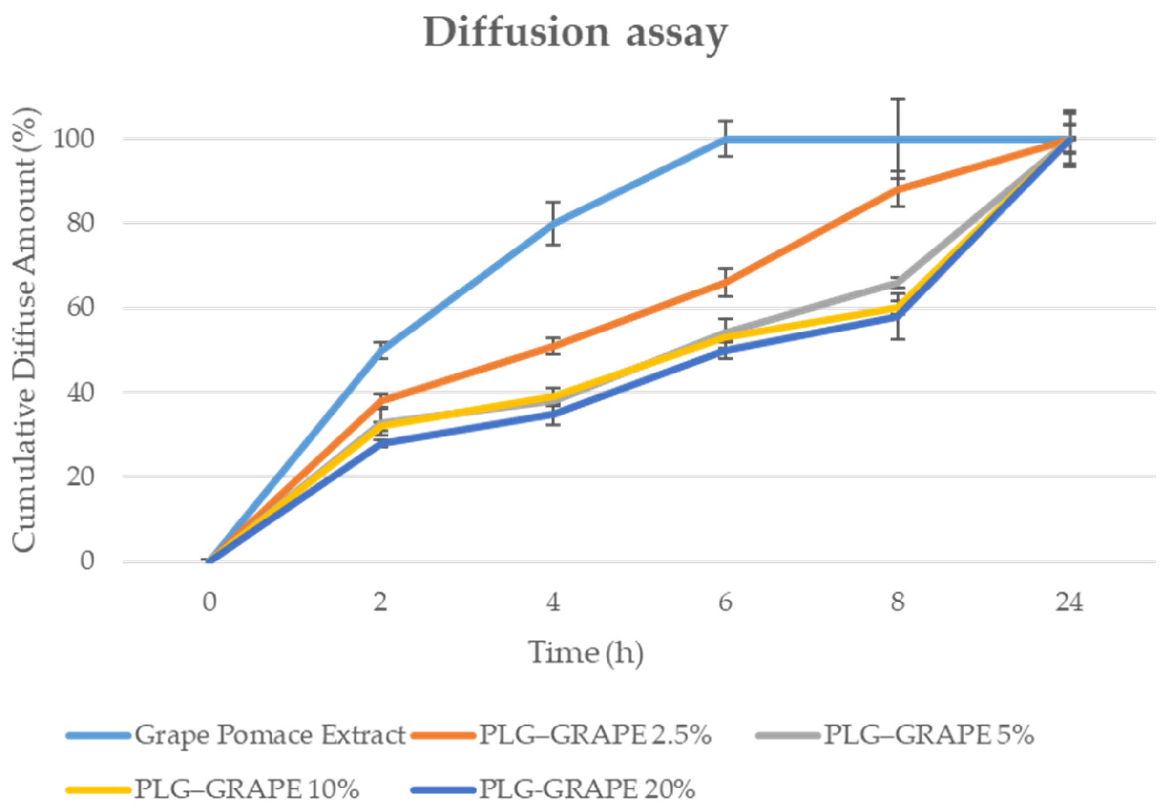
3.1.3. In Vitro Diffusion Studies by Vertical Franz Cells

The results were presented as the cumulative percentage of the diffused amount for the tested samples, with the corresponding diffusion profiles illustrated in Figure 1.
The results demonstrate that the grape pomace extract exhibits a rapid rate of diffusion, reaching 50% diffusion within 2 h, 80% at 4 h, and 100% at 6 h. In contrast, polymer conjugates demonstrate a slower and more controlled diffusion rate. PLG–GRAPE 2.5% reached 38% at 2 h, gradually increasing to 66% at 6 h, and finally reaching 100% after 24 h. PLG–GRAPE 5% exhibited a comparable trend, reaching 33% diffusion at 2 h, 54% at 6 h, and 100% at 24 h. Conversely, PLG–GRAPE 10% and 20% exhibited the slowest initial diffusion, reaching 32% and 28% diffusion at 8 h, respectively. At 8 h, PLG–GRAPE 10% reached 53%, while PLG–GRAPE 20% reached 50%. Both samples finally reached full diffusion at 24 h.
These data demonstrate that polymer conjugates facilitate sustained and gradual release, in contrast to the rapid diffusion observed for the extract.

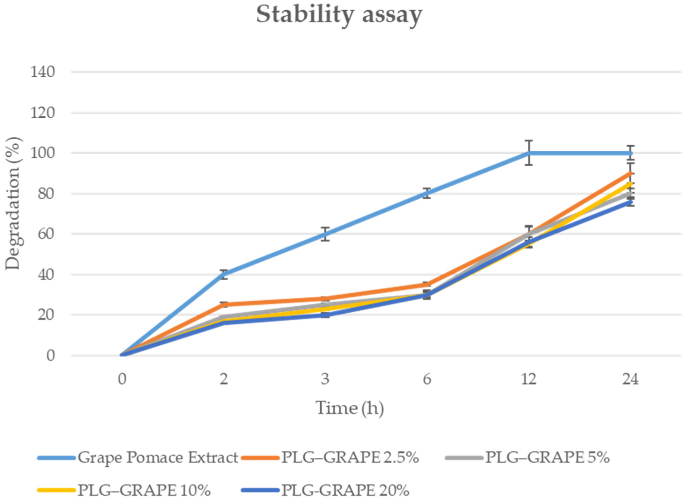
3.1.4. Stability Studies

The stability of each sample was assessed at each interval through absorbance measurement.
The analysis and data were expressed as a degradation percentage and are shown in Figure 2.
The stability data indicates a significant difference between the grape pomace extract and the PLG–GRAPE conjugates under oxidative conditions. The extract demonstrates rapid degradation, reaching 60% loss at 3 h and 100% degradation by 12 h. Conversely, the PLG–GRAPE conjugates exhibit enhanced stability, with slower degradation rates proportional to the DEX concentration. For instance, PLG–GRAPE 2.5% degrades by 60% at 12 h, while PLG–GRAPE 20% degrades by only 56% at the same time and reaches 76% degradation at 24 h.
These data demonstrate that polymer conjugates reduce the degradation of the active matrix. According to the results obtained from diffusion and stability assays, which indicate PLG–GRAPE 20% as the optimal combination, this was selected for the in vitro safety and efficacy test.

3.2. Safety Assessment

3.2.1. Cytotoxicity Studies

The study evaluated the cytotoxic potential of the PLG–GRAPE extract on BALB/3T3 murine fibroblast cells through the Neutral Red Uptake (NRU) assay, in accordance with the guidelines stipulated in ISO 10993-5:2009.
As demonstrated in Figure 3, cell viability was not significantly impacted by either treatment at decrescent doses (3%, 0,6%, 0,3%) in comparison to the negative control (untreated cells). These results suggest that the sample lacks cytotoxic activity.
Prior to performing the NRU assay, the condition of the cell culture was assessed through microscopic examination following 24 h of incubation. Biological reactivity, including any signs of abnormalities or cellular damage, was evaluated and scored on a scale from 0 to 4 in accordance with ISO 10993-5:2009 guidelines [17].
The BALB/3T3 fibroblast cells displayed normal morphology and no evidence of biological reactivity after exposure to the vehicle medium or the test substances. Conversely, cells treated with positive control (1% SDS) exhibited a maximum reactivity score of four. These results demonstrate that the tested samples are non-toxic and do not cause any cellular abnormalities.

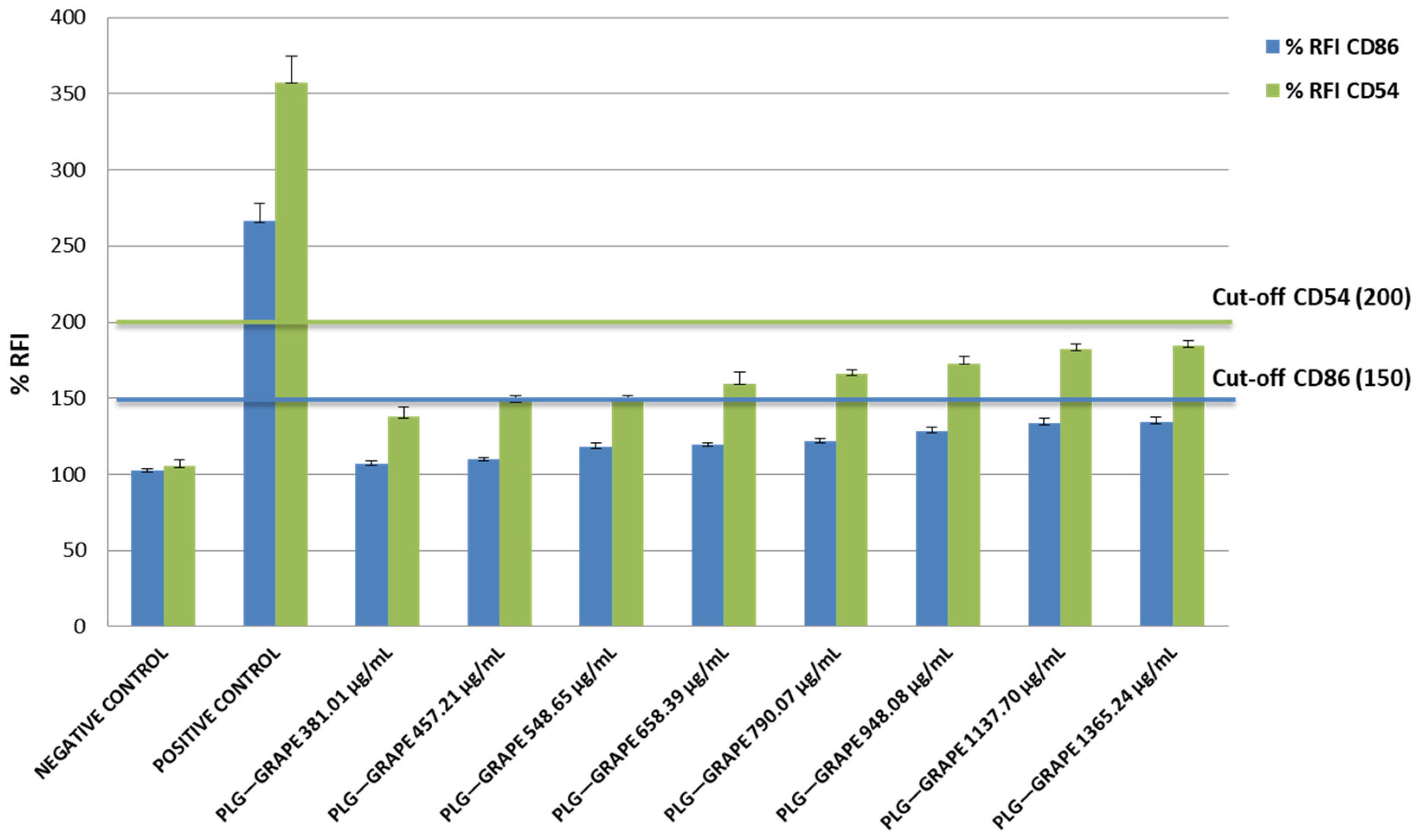
3.2.2. Skin Sensitization Potential Test (h-CLAT)

As shown in Figure 4, the data demonstrate that the sample, even at increasing doses, did not display any skin-sensitizing effects.
The relative fluorescence intensity (RFI%) values for CD86 and CD54 remained below the established threshold levels, thus, confirming the absence of immune activation or allergenic responses.

3.2.3. Skin Irritation Test

As indicated in Table 2, tissues treated with the tested sample exhibited a viability of 87.53%. Thus, under the employed experimental conditions, the sample is classified as non-irritating. Furthermore, the mean OD value measured in the negative control was 1.52 1.56 ± 2.11%, demonstrating compliance with the OECD acceptability criteria. The viability of the positive control of 7.23 also met the acceptability criteria.

3.3. Efficacy Assessment

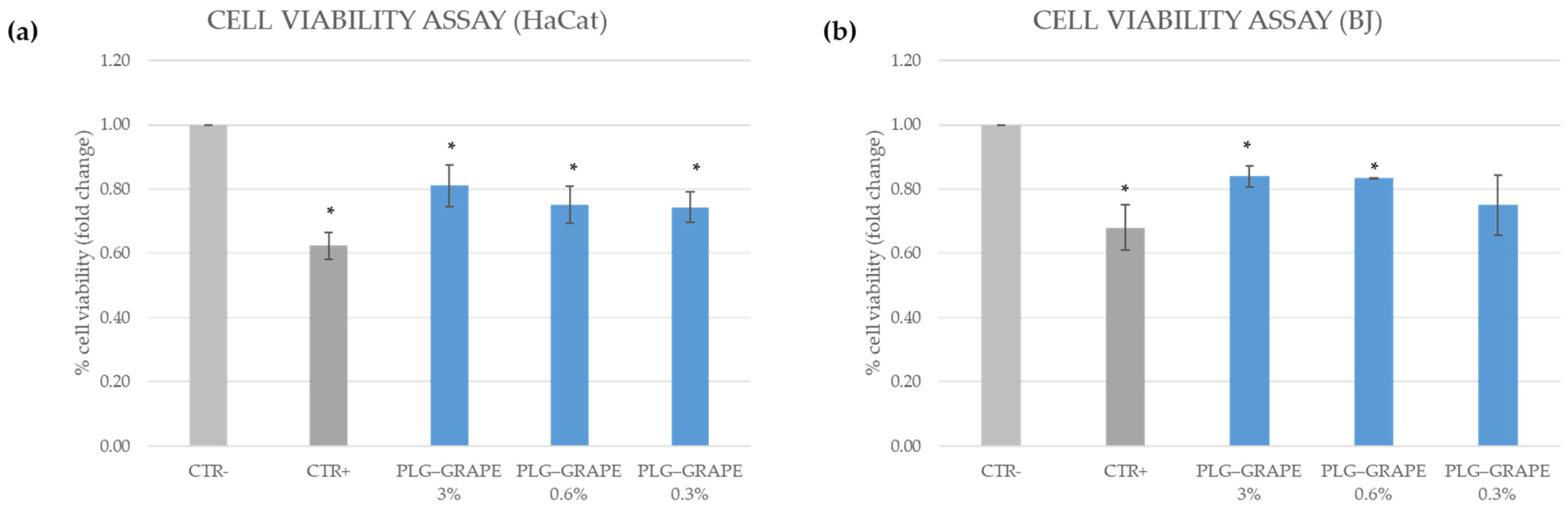
3.3.1. Cell Viability Assay

As demonstrated in Figure 5, the cell viability evaluation revealed that cells and tissues exposed to urban dust and UV irradiation exhibited a significant decline in viability, reaching approximately 40% reduction. The treatment of cells with the test sample resulted in a viability reduction of approximately 20%. These findings demonstrate that the test sample is able to preserve both HaCat cells (Figure 5a) and BJ cells (Figure 5b) from mortality induced by pollution and UV.

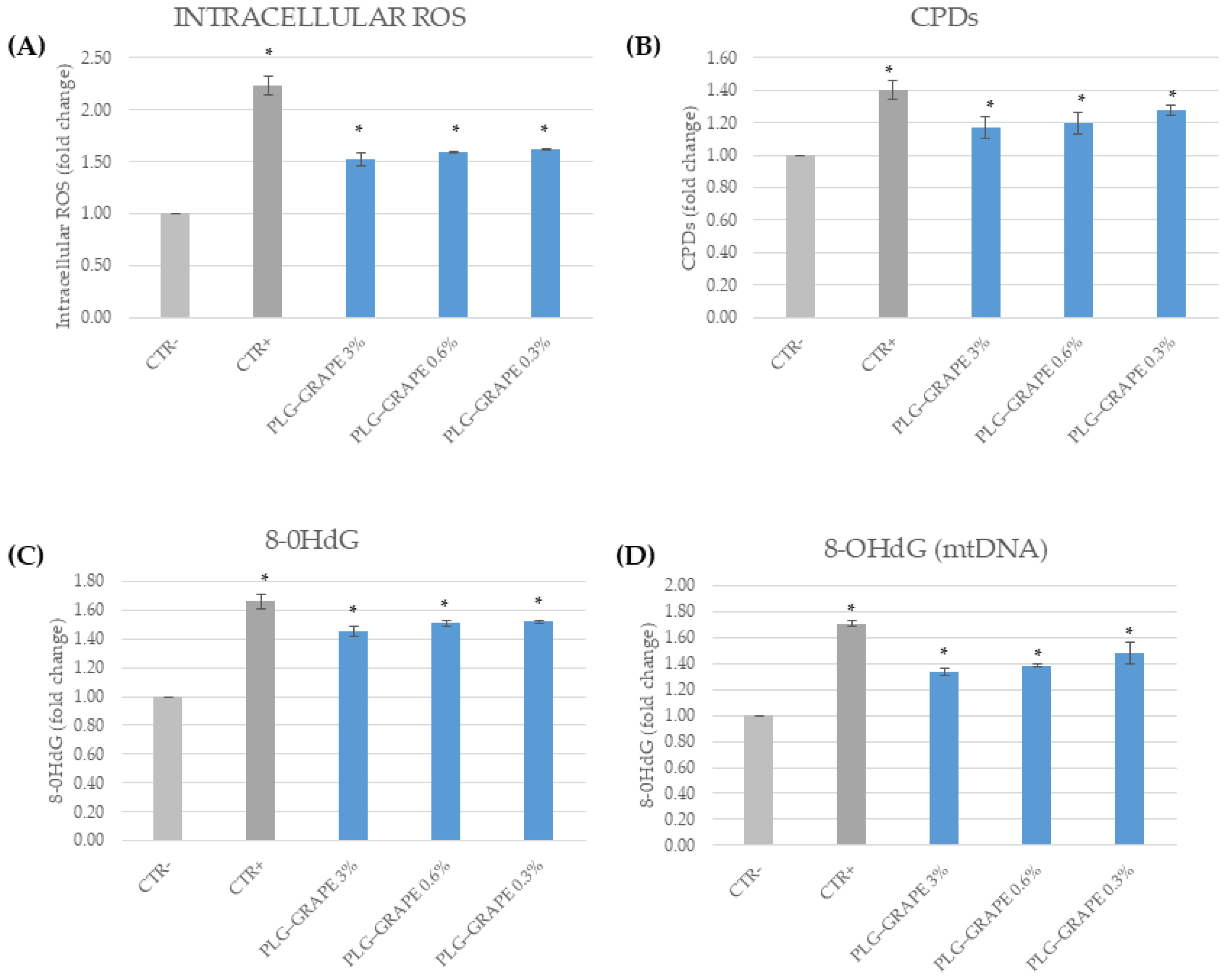
3.3.2. Antioxidant Effect

The antioxidant effect was evaluated through the quantification of intracellular ROS, 8-OHdG (in DNA and mtDNA) and CPDs. The latter two evaluations were conducted to assess the capacity of the sample to protect DNA from oxidative damage. Figure 6A demonstrates a significant increase in ROS levels for the positive control (CTR+) in comparison to the negative control (CTR−). Treatment with PLG–GRAPE resulted in a reduction of ROS levels, approaching those observed in the negative control group, thereby indicating a high degree of antioxidant activity.
Figure 6B shows higher levels of CPDs in the CTR+ group compared to the CTR− group. Treatment with PLG–GRAPE led to a reduction in CPD levels, thereby indicating partial protection of the DNA.
Figure 6C demonstrates elevated levels of 8-OHdG in the CTR+ group. Treatment with PLG–GRAPE resulted in a significant reduction of these levels, highlighting their capacity to mitigate oxidative damage to DNA.
Furthermore, Figure 6D reveals increased 8-OHdG levels in CTR+ and demonstrates that PLG–GRAPE effectively reduces these levels, thus, showing protection against mitochondrial DNA oxidative damage.

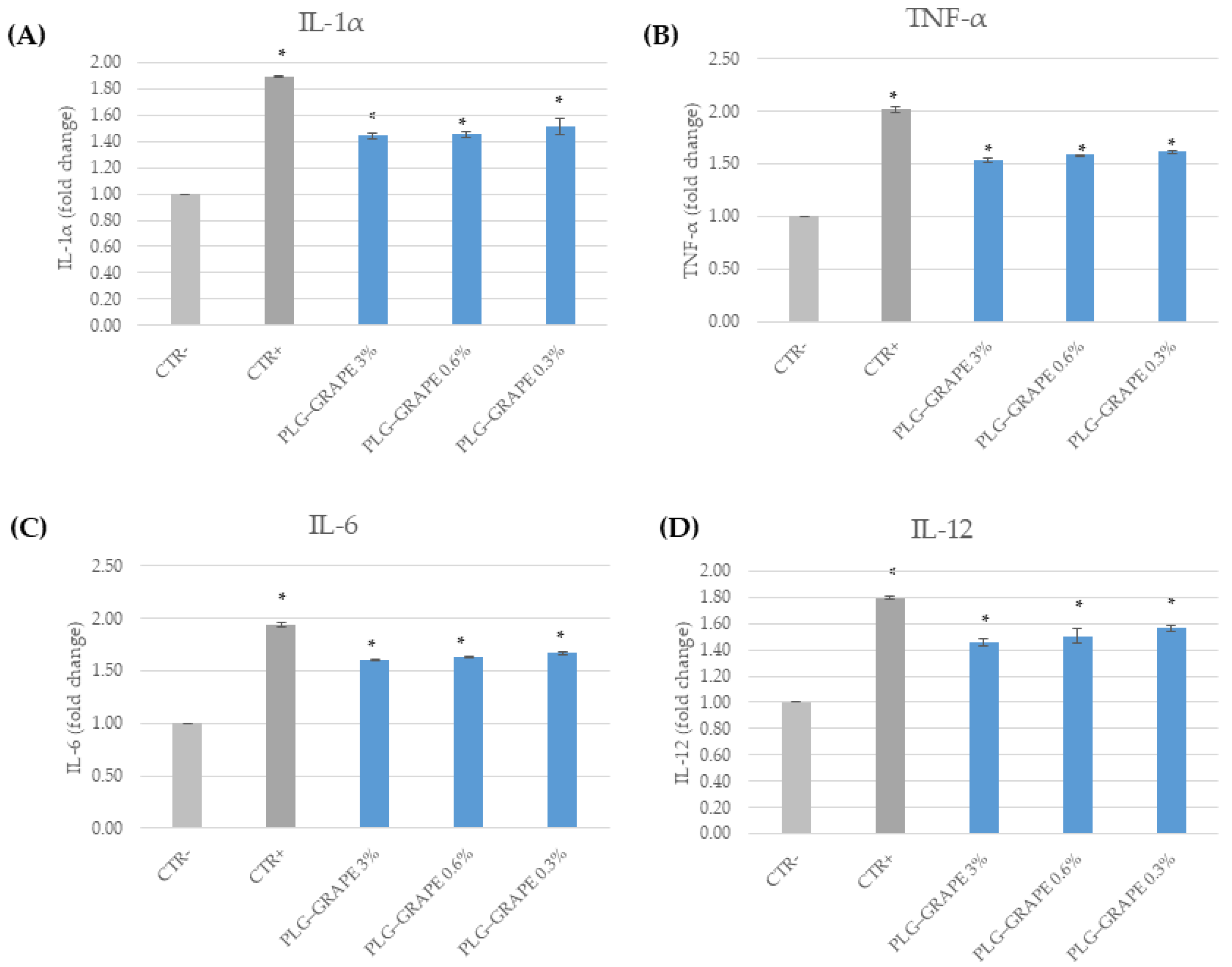
3.3.3. Anti-Inflammatory Efficacy

The anti-inflammatory effect was evaluated through the dosage of the following pro-inflammatory cytokines: IL-1α, TNF-α, IL-12, and IL-6 on HaCat cells.
The results presented in Figure 7A demonstrate that IL-1α levels are significantly elevated in the positive control (CTR+) compared to the negative control (CTR-). Treatment with PLG–GRAPE at all concentrations resulted in a reduction of IL-1α levels, indicating anti-inflammatory efficacy.
As demonstrated in Figure 7B, TNF-α levels were found to be increased in the CTR+ group. PLG–GRAPE treatments have been shown to reduce TNF-α levels in a concentration-independent manner, thus, demonstrating consistent anti-inflammatory effects.
Furthermore, Figure 7C shows a significant increase in IL-6 levels for CTR+ compared to CTR-. Conversely, PLG–GRAPE treatments have been observed to reduce IL-6 levels, thereby suggesting effective modulation of inflammatory responses.
Furthermore, Figure 7D shows elevated IL-12 levels in CTR+. PLG–GRAPE treatments have been demonstrated to reduce IL-12 levels, thereby confirming its ability to counteract pro-inflammatory cytokine release.

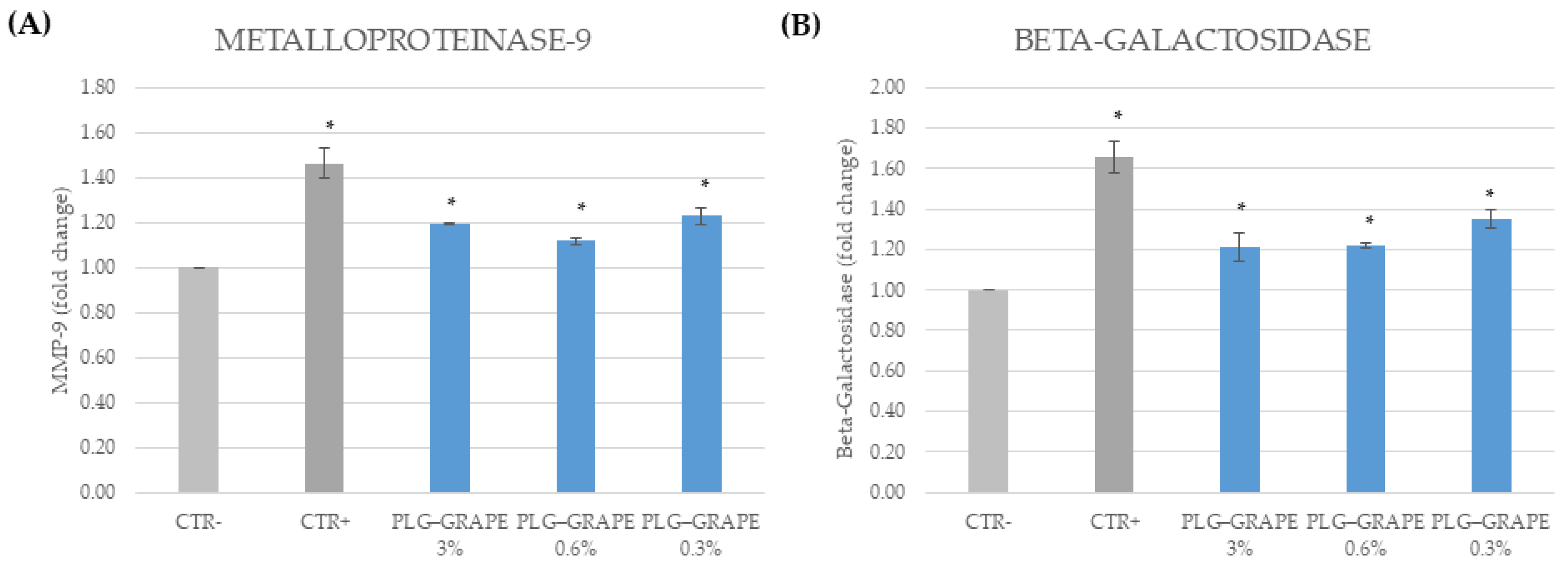
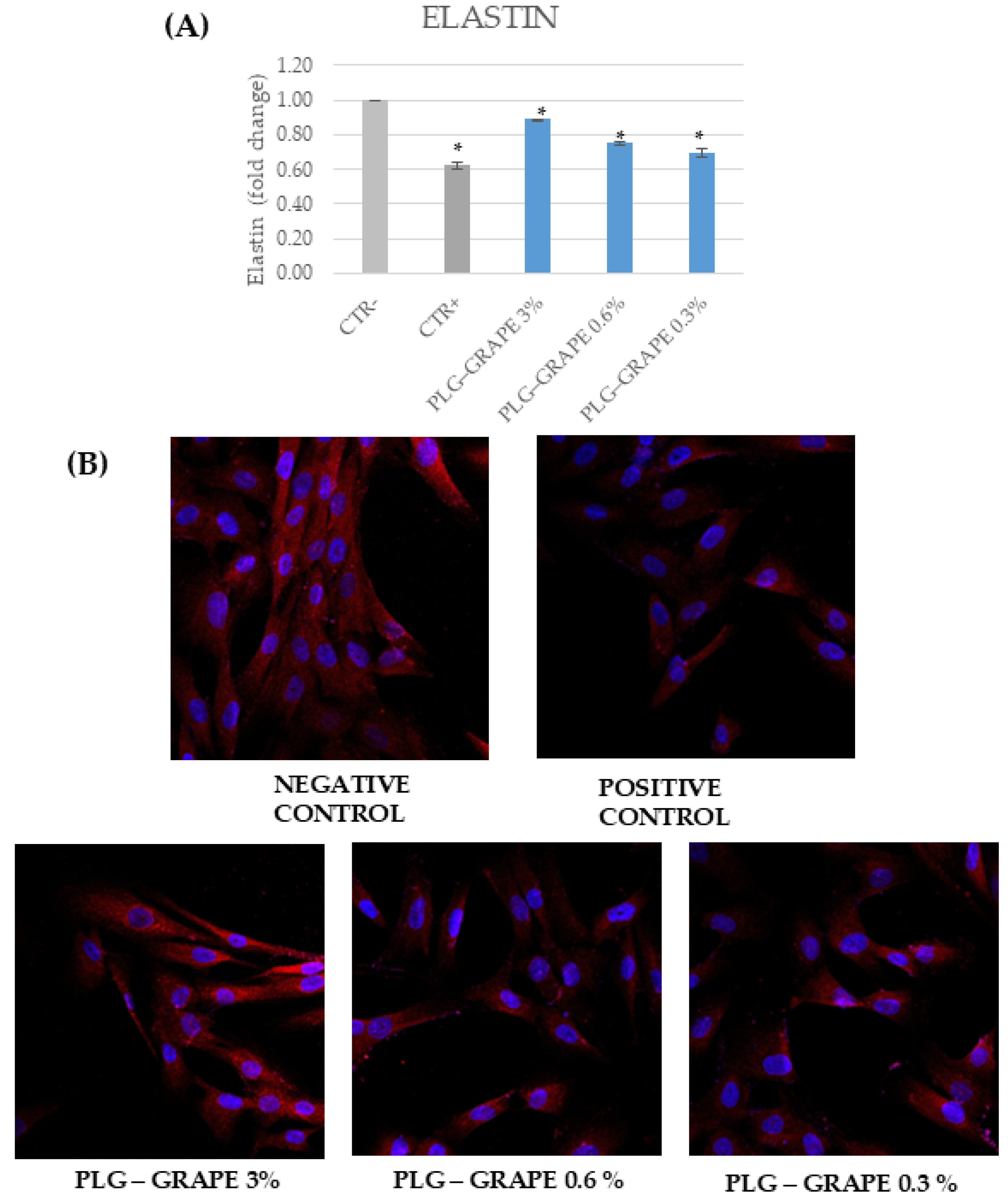
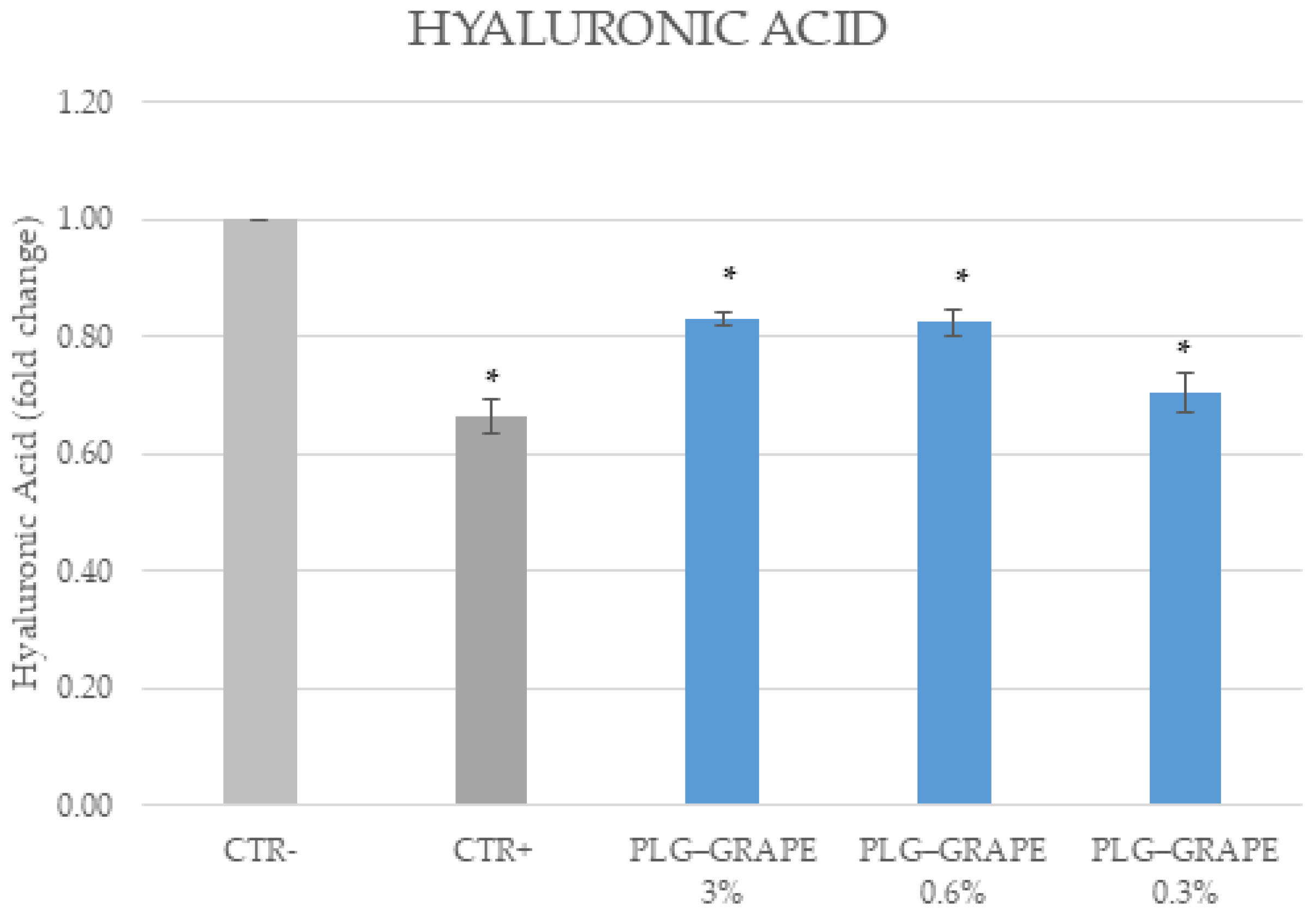
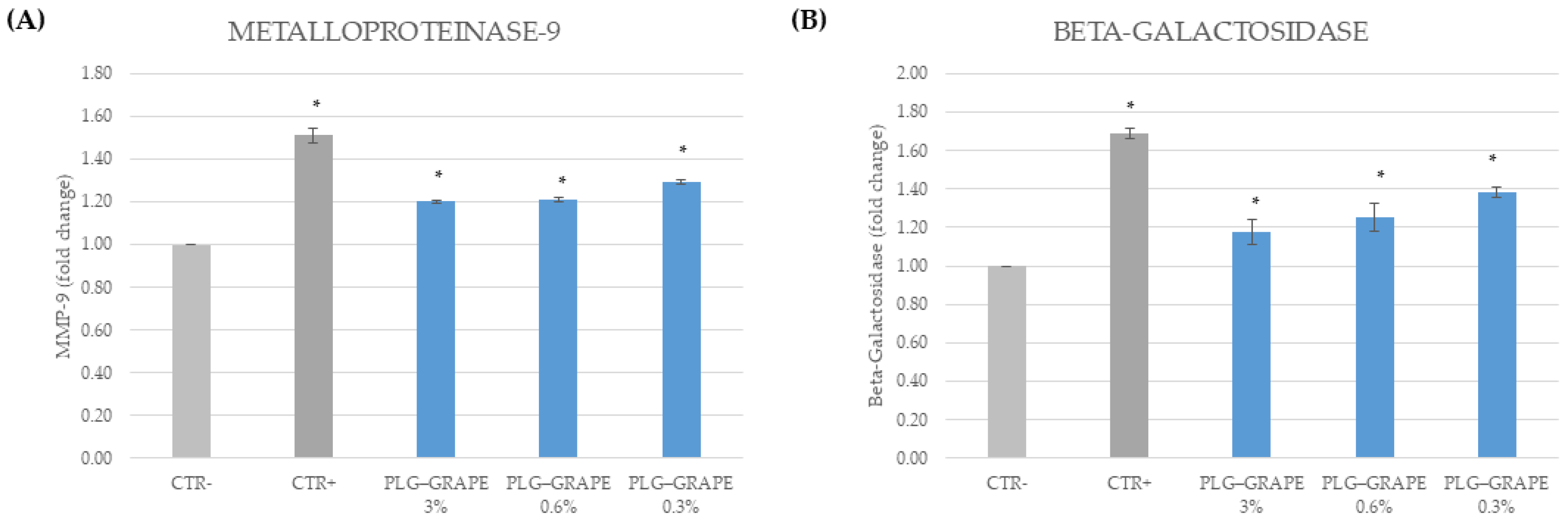
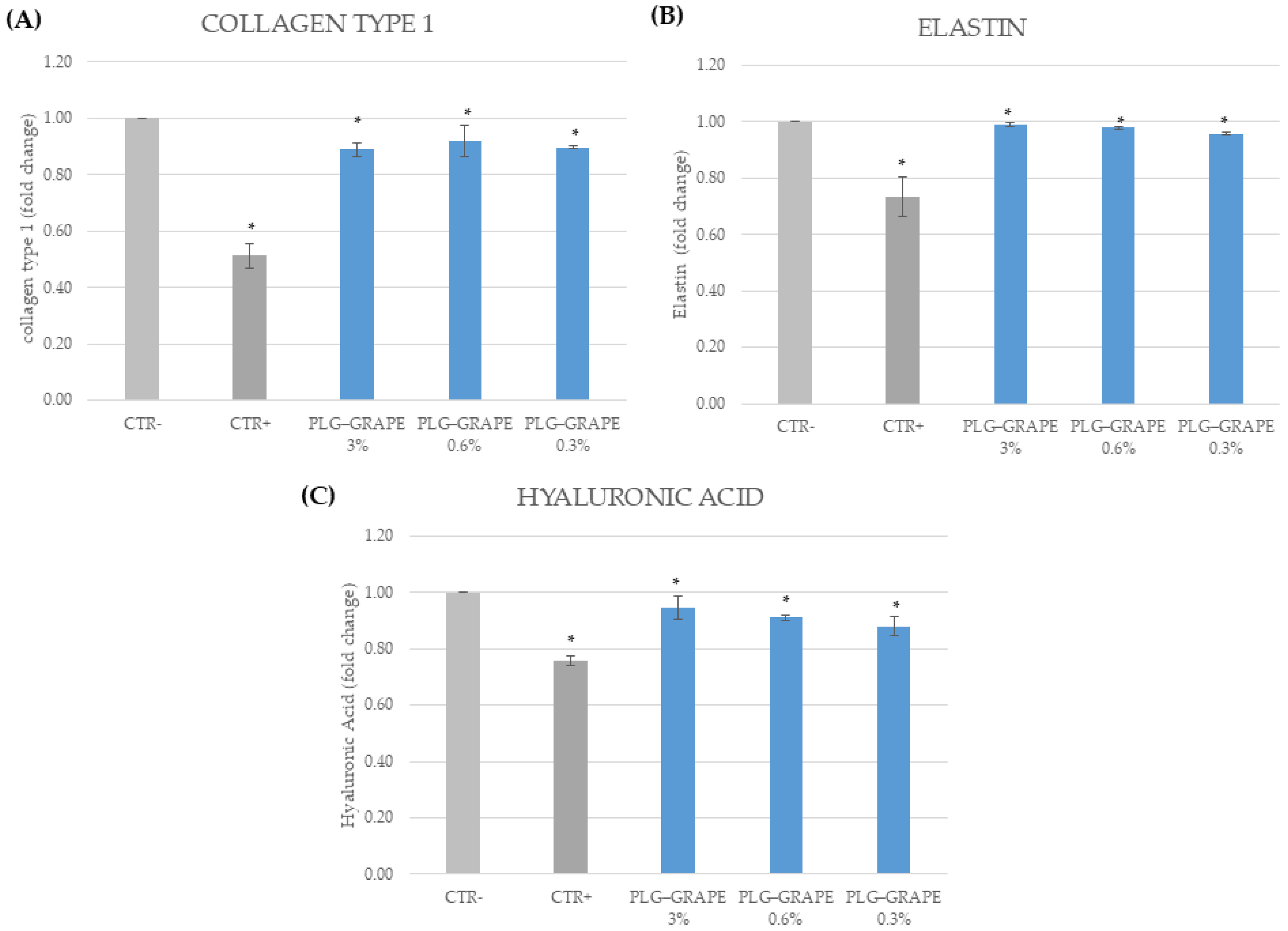
3.3.4. Anti-Age Effect

The anti-aging effect was evaluated in the first instance through the evaluation of both senescence-related (beta-galactosidase) and aging-related markers (MMP-9). Subsequently, the expression levels of well-aging and trophic-related markers (collagen type 1, hyaluronic acid and elastin) were analyzed. These evaluations were conducted on BJ-cells and full-thickness skin tissues.

Results on BJ-Cells

As demonstrated in Figure 8, exposure to UV and pollution (CTR+) resulted in elevated MMP-9 levels and beta-galactosidase activity in comparison to the negative control (CTR-). Treatment with PLG–GRAPE, at all concentrations, resulted in a reduction of MMP-9 levels and beta-galactosidase activity.
Furthermore, the expression of key components of the extracellular matrix was evaluated, with results from the assessment of collagen type I (Figure 9A,B), elastin (Figure 10A,B), and hyaluronic acid (Figure 11) revealing that cells exposed to UV radiation and pollution exhibited significantly lower levels of collagen type I, elastin, and hyaluronic acid to the negative control. Treatment with PLG–GRAPE led to a significant enhancement in the levels of all these key extracellular matrix components, thereby further validating the anti-aging efficacy of the product.

Results on Full Thickness Skin Tissues

The experiments were also conducted on full-thickness skin tissue. As demonstrated in Figure 12, also in this case, the expression of MMP-9 and the activity of beta-galactosidase increased in the CTR+ samples compared to the CTR− samples. However, this increase was significantly reduced in the tissues treated with PLG–GRAPE. These results confirm the anti-aging efficacy of the sample in a more complex model.
Furthermore, the key components of the extracellular matrix were evaluated in full-thickness skin tissues. The assessment of collagen type I (Figure 13A) elastin (Figure 13B), and hyaluronic acid (Figure 13C) revealed that tissues exposed to UV radiation and pollution exhibited significantly lower levels of collagen type I, elastin, and hyaluronic acid compared to the negative control. Treatment with PLG–GRAPE led to a significant enhancement in the levels of these critical extracellular matrix components, thus, validating the anti-aging efficacy of the product in full-thickness skin tissues.

4. Discussion

The management and elimination of agricultural by-products remain significant challenges. Addressing food waste sustainably necessitates innovative extraction technologies that maximize waste valorization while minimizing environmental [45]. Recent advancements in green extraction techniques have demonstrated enhanced efficiency in recovering polyphenols from plant by-products, supporting their application in functional formulations [46]. Among several raw materials, grape pomace, rich in bioactive compounds, presents a valuable resource for developing natural additives [47].
Within this context, the Naviglio® rapid solid-liquid dynamic extractor emerges as an advanced solid-liquid extraction technology, offering several advantages over existing extraction methods. Unlike many conventional techniques that rely on heating the extraction system to enhance extraction efficiency, the Naviglio extractor® operates at room or sub-room temperature, preventing thermal degradation of heat-sensitive compounds, and allowing the use of deionized water as a solvent. Moreover, it utilizes pressure increases in the extracting liquid applied to the solid matrix, which helps to reduce extraction times while simultaneously enhancing the efficiency of the extraction process [48].
In this study, Naviglio extractor® was employed to prepare a grape pomace extract, which was then utilized to produce a dextran–grape conjugate (PLG–GRAPE). The extraction of the grape pomace was performed using water as the solvent, making the entire process highly appealing as it avoids the use of organic solvents in the extraction stages, thus, ensuring an eco-friendly approach.
Therefore, the primary objective of this study was to undertake a comparative analysis of dextran–polyphenol conjugates (PLG–GRAPE) and free grape pomace extract with regard to stability, bioactivity, diffusion, and sustainability. From the data obtained, the total polyphenol content in our grape pomace extract (18.00 ± 0.6 mg eq CA/g) aligns with values reported in studies employing conventional extraction techniques [49]
Regarding antioxidant activity, the DPPH radical inhibition capacity (65.0 ± 0.8%) of our grape pomace extract is consistent with previous findings on Vitis vinifera extracts [50]. Polyphenols, known for their potent free radical scavenging ability, play a crucial role in oxidative stress reduction [51]. Notably, PLG–GRAPE conjugates exhibited antioxidant activity (63.7 ± 0.7% to 65.2 ± 0.4%) comparable to the free extract, indicating that dextran conjugation preserved bioactivity.
The protective effects of PLG–GRAPE against oxidative stress and inflammation were further confirmed through in vitro studies. Exposure to oxidative agents (e.g., UV radiation, pollutants) increased reactive oxygen species (ROS) levels, DNA damage markers (e.g., 8-OHdG, CPDs), and pro-inflammatory cytokines (IL-1α, TNF-α, IL-6, IL-12), leading to cellular senescence and extracellular matrix degradation [52]. Treatment with PLG–GRAPE significantly reduced ROS levels, preserved nuclear and mitochondrial DNA, and prevented CPD formation. Furthermore, PLG–GRAPE effectively reduced pro-inflammatory cytokines and cellular senescence markers (e.g., β-galactosidase, MMP-9), restoring extracellular matrix components, further supporting the therapeutic potential of polyphenol conjugates in dermatological applications [53,54].
In terms of safety, PLG–GRAPE was confirmed as biocompatible through the Neutral Red Uptake test (ISO 10993-5:2009 [38]). Additionally, it was classified as non-sensitizing and non-irritating according to h-CLAT (OECD Test Guideline 442E [41]) and skin irritation tests (OECD Test Guideline 439 [43]), supporting its suitability for cosmetic and topical formulations.
Overall, our findings demonstrate that grape pomace-derived polyphenols, particularly in the form of PLG–GRAPE conjugates, retain strong bioactivity, stability, and safety, highlighting their promising role in sustainable dermatological formulations.

5. Conclusions

In consideration of the findings, PLG–GRAPE is identified as a technology that shows considerable promise, offering a multifaceted approach to harnessing the potential of pomace extract. Its stability is maintained, ensuring a sustained release of beneficial compounds over an extended duration. The study’s outcomes demonstrate the significant antioxidant and anti-inflammatory properties of PLG–GRAPE on keratinocytes and fibroblast cell lines. These properties contribute to its anti-aging effects, even on reconstructed tissue. In addition, safety tests confirm the technology’s biocompatibility, making it suitable for dermatological applications. Consequently, the technology has the potential to serve as a promising bioactive ingredient in cosmetic formulations designed for anti-aging, anti-inflammatory, and antioxidant properties, particularly in dermatological products intended for the treatment of inflammatory skin conditions. The use of grape pomace, a by-product of the wine industry, underscores the environmental sustainability of this approach by repurposing industrial waste into high-value bioactive compounds. The future outlook for these conjugates is focused on their formulation into finished products, with the potential for in vivo studies to provide more substantial evidence of the efficacy and safety of their therapeutic applications in dermatological fields.

Author Contributions

Conceptualization, F.A., R.M., O.I.P. and F.P.; methodology, R.M. and M.F.M.; validation, M.F.M. and A.F.V.; formal analysis, M.F.M., A.F.V. and R.M.; investigation, M.F.M. and A.F.V.; writing—original draft preparation, M.F.M. and A.F.V.; writing—review and editing, O.I.P.; visualization, M.F.M. and A.F.V.; supervision, F.A. and O.I.P.; project administration, F.A., O.I.P. and F.P. All authors have read and agreed to the published version of the manuscript.

Funding

This research received no external funding.

Institutional Review Board Statement

Not applicable.

Informed Consent Statement

Not applicable.

Data Availability Statement

The original contributions presented in the study are included in the article, further inquiries can be directed to the corresponding author.

Conflicts of Interest

Some authors (F. Amone, R. Malivindi, O.I. Parisi and F. Puoci) are shareholders of the Spin-off Macrofarm s.r.l., the company that will commercialize the product discussed in this manuscript. Other authors (A.F. Vattimo) are employees of the same company but do not hold equity. Additionally, some authors (O.I. Parisi and F. Puoci) are listed as inventors on a related patent (EP2535087A2). All the other authors declare no competing interests.

References

  1. Omran, B.A.; Baek, K.-H. Valorization of Agro-Industrial Biowaste to Green Nanomaterials for Wastewater Treatment: Approaching Green Chemistry and Circular Economy Principles. J. Environ. Manag. 2022, 311, 114806. [Google Scholar] [CrossRef]
  2. Pham, T.-N.; Cazier, E.A.; Gormally, E.; Lawrence, P. Valorization of Biomass Polyphenols as Potential Tyrosinase Inhibitors. Drug Discov. Today 2024, 29, 103843. [Google Scholar] [CrossRef]
  3. López-Astorga, M.; Molina-Domínguez, C.C.; Ovando-Martínez, M.; Leon-Bejarano, M. Orujo de Uva: Más Que Un Residuo, Una Fuente de Compuestos Bioactivos. Epistemus 2023, 16. [Google Scholar] [CrossRef]
  4. Luchian, C.E.; Cotea, V.V.; Vlase, L.; Toiu, A.M.; Colibaba, L.C.; Răschip, I.E.; Nadăş, G.; Gheldiu, A.M.; Tuchiluş, C.; Rotaru, L. Antioxidant and Antimicrobial Effects of Grape Pomace Extracts. BIO Web Conf. 2019, 15, 04006. [Google Scholar] [CrossRef]
  5. Fontana, A.R.; Antoniolli, A.; Bottini, R. Grape Pomace as a Sustainable Source of Bioactive Compounds: Extraction, Characterization, and Biotechnological Applications of Phenolics. J. Agric. Food Chem. 2013, 61, 8987–9003. [Google Scholar] [CrossRef]
  6. Rockenbach, I.I.; Gonzaga, L.V.; Rizelio, V.M.; Gonçalves, A.E.d.S.S.; Genovese, M.I.; Fett, R. Phenolic Compounds and Antioxidant Activity of Seed and Skin Extracts of Red Grape (Vitis vinifera and Vitis labrusca) Pomace from Brazilian Winemaking. Food Res. Int. 2011, 44, 897–901. [Google Scholar] [CrossRef]
  7. Europe, P.M.C. Europepmc.org. Available online: https://europepmc.org/article/PMC/4200838 (accessed on 10 February 2025).
  8. Soceanu, A.; Dobrinas, S.; Sirbu, A.; Manea, N.; Popescu, V. Economic Aspects of Waste Recovery in the Wine Industry. A Multidisciplinary Approach. Sci. Total Environ. 2021, 759, 143543. [Google Scholar] [CrossRef]
  9. Agati, G.; Brunetti, C.; Di Ferdinando, M.; Ferrini, F.; Pollastri, S.; Tattini, M. Functional Roles of Flavonoids in Photoprotection: New Evidence, Lessons from the Past. Plant Physiol. Biochem. 2013, 72, 35–45. [Google Scholar] [CrossRef]
  10. Alexis, A.F.; Grimes, P.; Boyd, C.; Downie, J.; Drinkwater, A.; Garcia, J.K.; Gallagher, C.J. Racial and Ethnic Differences in Self-Assessed Facial Aging in Women. Dermatol. Surg. 2019, 45, 1635–1648. [Google Scholar] [CrossRef] [PubMed]
  11. Arct, J.; Pytkowska, K. Flavonoids as Components of Biologically Active Cosmeceuticals. Clin. Dermatol. 2008, 26, 347–357. [Google Scholar] [CrossRef] [PubMed]
  12. Liu, X.; Liu, C.; Li, Y.; Yuan, K.; Zhang, W.; Cai, D.; Peng, Z.; Hu, Y.; Sun, J.; Bai, W. Bioactivity and Application of Anthocyanins in Skin Protection and Cosmetics: An Extension as a Functional Pigment. Phytochem. Rev. 2023, 22, 1441–1467. [Google Scholar] [CrossRef]
  13. Khan, A.Q.; Agha, M.V.; Sheikhan, K.S.A.M.; Younis, S.M.; Tamimi, M.A.; Alam, M.; Ahmad, A.; Uddin, S.; Buddenkotte, J.; Steinhoff, M. Targeting Deregulated Oxidative Stress in Skin Inflammatory Diseases: An Update on Clinical Importance. Biomed. Pharmacother. 2022, 154, 113601. [Google Scholar] [CrossRef]
  14. Quan, T. Human Skin Aging and the Anti-Aging Properties of Retinol. Biomolecules 2023, 13, 1614. [Google Scholar] [CrossRef] [PubMed]
  15. Rojo, L.E.; Roopchand, D.; Graf, B.; Raskin, I. Role of Anthocyanins in Skin Aging and UV Induced Skin Damage. ResearchGate. Available online: https://www.researchgate.net/publication/281031602_Role_of_Anthocyanins_in_Skin_Aging_and_UV_Induced_Skin_Damage (accessed on 31 May 2023).
  16. Restuccia, D.; Sicari, V.; Pellicanò, T.M.; Spizzirri, U.G.; Loizzo, M.R. The impact of cultivar on polyphenol and biogenic amine profiles in Calabrian red grapes during winemaking. Food Res. Int. 2017, 102, 303–312. [Google Scholar] [CrossRef]
  17. Brezoiu, A.-M.; Matei, C.; Deaconu, M.; Stanciuc, A.-M.; Trifan, A.; Gaspar-Pintiliescu, A.; Berger, D. Polyphenols Extract from Grape Pomace. Characterization and Valorisation through Encapsulation into Mesoporous Silica-Type Matrices. Food Chem. Toxicol. 2019, 133, 110787. [Google Scholar] [CrossRef]
  18. Burin, V.M.; Ferreira-Lima, N.E.; Panceri, C.P.; Bordignon-Luiz, M.T. Bioactive Compounds and Antioxidant Activity of Vitis Vinifera and Vitis Labrusca Grapes: Evaluation of Different Extraction Methods. Microchem. J. 2014, 114, 155–163. [Google Scholar] [CrossRef]
  19. Millinia, B.L.; Mashithah, D.; Nawatila, R.; Kartini, K. Microencapsulation of Roselle (Hibiscus sabdariffa L.) Anthocyanins: Effects of Maltodextrin and Trehalose Matrix on Selected Physicochemical Properties and Antioxidant Activities of Spray-Dried Powder. Future Foods 2024, 9, 100300. [Google Scholar] [CrossRef]
  20. Parisi, O.; Malivindi, R.; Amone, F.; Ruffo, M.; Malanchin, R.; Carlomagno, F.; Piangiolino, C.; Nobile, V.; Pezzi, V.; Scrivano, L.; et al. Safety and Efficacy of Dextran-Rosmarinic Acid Conjugates as Innovative Polymeric Antioxidants in Skin Whitening: What Is the Evidence? Cosmetics 2017, 4, 28. [Google Scholar] [CrossRef]
  21. Gallo, M.; Formato, A.; Giacco, R.; Riccardi, G.; Luongo, D.; Formato, G.; Amoresano, A.; Naviglio, D. Corrigendum to “Mathematical Optimization of the Green Extraction of Polyphenols from Grape Peels through a Cyclic Pressurization Process”. Heliyon 2019, 5, e01593. [Google Scholar] [CrossRef]
  22. Ojogbo, E.; Ogunsona, E.O.; Mekonnen, T.H. Chemical and Physical Modifications of Starch for Renewable Polymeric Materials. Mater. Today Sustain. 2020, 7–8, 100028. [Google Scholar] [CrossRef]
  23. Dahiya, D.; Nigam, P.S. Dextran of Diverse Molecular-Configurations Used as a Blood-Plasma Substitute, Drug-Delivery Vehicle and Food Additive Biosynthesized by Leuconostoc, Lactobacillus and Weissella. Appl. Sci. 2023, 13, 12526. [Google Scholar] [CrossRef]
  24. Sleightholm, R.; Watley, D.; Wahlmeier, S.; Patel, A.; Foster, J.M. The Efficacy of Dextran-40 as a Venous Thromboembolism Prophylaxis Strategy in Cytoreductive Surgery and Hyperthermic Intraperitoneal Chemotherapy. Am. Surg. 2017, 83, 134–140. [Google Scholar] [CrossRef] [PubMed]
  25. Chen, F.; Huang, G.; Huang, H. Preparation and Application of Dextran and Its Derivatives as Carriers. Int. J. Biol. Macromol. 2019, 145, 827–834. [Google Scholar] [CrossRef]
  26. Moreno-Mendieta, S.; Guillén, D.; Hernández-Pando, R.; Sánchez, S.; Rodríguez-Sanoja, R. Potential of Glucans as Vaccine Adjuvants: A Review of the α-Glucans Case. Carbohydr. Polym. 2017, 165, 103–114. [Google Scholar] [CrossRef]
  27. Puoci, F.; Iemma, F.; Pezzi, V.; Picci, N.; Cirillo, G.; Curcio, M.; Parisi, O.I. Cosmetic, Pharmaceutical or Nutraceutical Formulations Containing Antioxidant Molecules and Polymeric Materials. Patent EP2535087A2, 19 December 2012. [Google Scholar]
  28. Pagliarulo, C.; De Vito, V.; Picariello, G.; Colicchio, R.; Pastore, G.; Salvatore, P.; Volpe, M.G. Inhibitory Effect of Pomegranate (Punica granatum L.) Polyphenol Extracts on the Bacterial Growth and Survival of Clinical Isolates of Pathogenic Staphylococcus aureus and Escherichia coli. Food Chem. 2016, 190, 824–831. [Google Scholar] [CrossRef]
  29. Puoci, F.; Morelli, C.; Cirillo, G.; Curcio, M.; Parisi, O.I.; Maris, P.; Sisci, D.; Picci, N. Anticancer Activity of a Quercetin-Based Polymer Towards HeLa Cancer Cells. Anticancer Research. Available online: https://www.semanticscholar.org/paper/Anticancer-activity-of-a-quercetin-based-polymer-Puoci-Morelli/a84e3b46520410aece13910b9e997c5dba91fb93 (accessed on 10 February 2025).
  30. Ng, S.-F.; Rouse, J.; Sanderson, D.; Eccleston, G. A Comparative Study of Transmembrane Diffusion and Permeation of Ibuprofen across Synthetic Membranes Using Franz Diffusion Cells. Pharmaceutics 2010, 2, 209–223. [Google Scholar] [CrossRef] [PubMed]
  31. Parisi, O.; Scrivano, L.; Amone, F.; Malivindi, R.; Ruffo, M.; Vattimo, A.F.M.; Pezzi, V.; Puoci, F. Interconnected PolymerS TeChnology (IPSTiC): An Effective Approach for the Modulation of 5α-Reductase Activity in Hair Loss Conditions. J. Funct. Biomater. 2018, 9, 44. [Google Scholar] [CrossRef]
  32. Siewert, M.; Dressman, J.; Brown, C.K.; Shah, V.P.; Aiache, J.-M.; Aoyagi, N.; Bashaw, D.; Brown, C.; Brown, W.; Burgess, D.; et al. FIP/AAPS Guidelines to Dissolution/in Vitro Release Testing of Novel/Special Dosage Forms. AAPS PharmSciTech 2003, 4, 43–52. [Google Scholar] [CrossRef]
  33. Shah, V.P.; Elkins, J.S.; Williams, R.L. Evaluation of the Test System Used for in Vitro Release of Drugs for Topical Dermatological Drug Products. Pharm. Dev. Technol. 1999, 4, 377–385. [Google Scholar] [CrossRef]
  34. Ueda, C.T.; Shah, V.P.; Derdzinski, K.; Ewing, G.; Flynn, G.; Maibach, H.; Marques, M.; Rytting, H.; Shaw, S.; Thakker, K.; et al. Topical and Transdermal Drug Products. Dissolution Technol. 2010, 17, 12–25. [Google Scholar] [CrossRef]
  35. Millipore, M. Substituting Strat-M® Membrane for Human Skin in Evaluating Effect of Encapsulation on Diffusion of Sunscreen Formulations; Application Note [Online]; EMD Millipore: Billerica, MA, USA, 2013. [Google Scholar]
  36. Zanta, C.L.P.S.; Martínez-Huitle, C.A. Degradation of 2-Hydroxybenzoic Acid by Advanced Oxidation Processes. Braz. J. Chem. Eng. 2009, 26, 503–513. [Google Scholar] [CrossRef]
  37. Legrini, O.; Oliveros, E.; Braun, A.M. Photochemical Processes for Water Treatment-Legrini. Chem. Rev. 1993, 93, 671–698. [Google Scholar] [CrossRef]
  38. ISO 10993-5:2009; Biological Evaluation of Medical Devices—Part 5: Tests for In vitro Cytotoxicity. ISO: Geneva, Switzerland, 2009.
  39. ISO 10993-12:2021; Biological Evaluation of Medical Devices—Part 12: Sample Preparation and Reference materials. ISO: Geneva, Switzerland, 2021.
  40. Parisi, O.I.; Aiello, D.; Casula, M.F.; Puoci, F.; Malivindi, R.; Scrivano, L.; Testa, F. Mesoporous Nanocrystalline TiO2 Loaded with Ferulic Acid for Sunscreen and Photo-Protection: Safety and Efficacy Assessment. RSC Adv. 2016, 6, 83767–83775. [Google Scholar] [CrossRef]
  41. Draft proposal for a new test guideline. In vitro Skin Sensitization: Human Cell Line Activation Test (h-CLAT). In Oecd Guideline for the Testing of Chemicals; OECD: Paris, France, 2024.
  42. Patitucci, F.; Motta, M.F.; Dattilo, M.; Malivindi, R.; Leonetti, A.E.; Pezzi, G.; Prete, S.; Mileti, O.; Gabriele, D.; Parisi, O.I.; et al. 3D-Printed Alginate/Pectin-Based Patches Loaded with Olive Leaf Extracts for Wound Healing Applications: Development, Characterization and in Vitro Evaluation of Biological Properties. Pharmaceutics 2024, 16, 99. [Google Scholar] [CrossRef] [PubMed]
  43. OECD 439: In vitro Skin Irritation: Reconstructed Human Epidermis Test Method. In OECD Guidelines for the Testing of Chemicals; OECD: Paris, France, 2013.
  44. ISO 10993-10:2010; Biological Evaluation of Medical Devices—Part 10: Tests for Irritation and Skin Sensitization. ISO: Geneva, Switzerland, 2010.
  45. Circular Economy in Europe—Developing the Knowledge Base. Europa.eu. Available online: https://www.eea.europa.eu/en/analysis/publications/circular-economy-in-europe (accessed on 18 January 2016).
  46. Di Prima, G.; Belfiore, E.; Migliore, M.; Scarpaci, A.G.; Angellotti, G.; Restivo, I.; Allegra, M.; Arizza, V.; De Caro, V. Green Extraction of Polyphenols from Waste Bentonite to Produce Functional Antioxidant Excipients for Cosmetic and Pharmaceutical Purposes: A Waste-to-Market Approach. Antioxidants 2022, 11, 2493. [Google Scholar] [CrossRef]
  47. Naviglio, D. Naviglio’s Principle and Presentation of an Innovative Solid–Liquid Extraction Technology: Extractor Navi-glio®. Anal. Lett. 2003, 36, 1647–1659. [Google Scholar] [CrossRef]
  48. Ferri, M.; Lima, V.; Zappi, A.; Fernando, A.L.; Melucci, D.; Tassoni, A. Phytochemicals Recovery from Grape Pomace: Extraction Improvement and Chemometric Study. Foods 2023, 12, 959. [Google Scholar] [CrossRef] [PubMed] [PubMed Central]
  49. Chedea, V.S.; Macovei, Ș.O.; Bocșan, I.C.; Măgureanu, D.C.; Levai, A.M.; Buzoianu, A.D.; Pop, R.M. Grape Pomace Polyphenols as a Source of Compounds for Management of Oxidative Stress and Inflammation—A Possible Alternative for Non-Steroidal Anti-Inflammatory Drugs? Mol./Mol. Online/Mol. Annu. 2022, 27, 6826. [Google Scholar] [CrossRef]
  50. Veskoukis, A.S.; Kyparos, A.; Nikolaidis, M.G.; Stagos, D.; Aligiannis, N.; Halabalaki, M.; Chronis, K.; Goutzourelas, N.; Skaltsounis, L.; Kouretas, D. The Antioxidant Effects of a Polyphenol-Rich Grape Pomace Extractin VitroDo Not Correspondin VivoUsing Exercise as an Oxidant Stimulus. Oxidative Med. Cell. Longev. 2012, 2012, 1–14. [Google Scholar] [CrossRef]
  51. Zhang, H.; Tsao, R. Dietary Polyphenols, Oxidative Stress and Antioxidant and Anti-Inflammatory Effects. Curr. Opin. Food Sci. 2016, 8, 33–42. [Google Scholar] [CrossRef]
  52. Gromkowska-Kępka, K.J.; Puścion-Jakubik, A.; Markiewicz-Żukowska, R.; Socha, K. The Impact of Ultraviolet Radiation on Skin Photoaging—Review of in Vitro Studies. J. Cosmet. Dermatol. 2021, 20, 3427–3431. [Google Scholar] [CrossRef] [PubMed]
  53. Calvo, M.J.; Navarro, C.; Durán, P.; Galan-Freyle, N.J.; Parra Hernández, L.A.; Pacheco-Londoño, L.C.; Castelanich, D.; Bermúdez, V.; Chacin, M. Antioxidants in Photoaging: From Molecular Insights to Clinical Applications. Int. J. Mol. Sci. 2024, 25, 2403. [Google Scholar] [CrossRef] [PubMed]
  54. Zillich, O.V.; Schweiggert-Weisz, U.; Eisner, P.; Kerscher, M. Polyphenols as active ingredients for cosmetic products. Int. J. Cosmet. Sci. 2015, 37, 455–464. [Google Scholar] [CrossRef] [PubMed]
Figure 1. In vitro diffusion profiles. Results are expressed as mean ± standard deviation, based on three replicates.
Applsci 15 03220 g001
Figure 2. In vitro stability studies: degradation percentages. Results are expressed as mean ± standard deviation, based on three replicates.
Applsci 15 03220 g002
Figure 3. Clone A31 Balb/3T3 fibroblast cell viability NRU test (%) in the absence or in the presence of increasing doses of sample. Each column represents the mean ± SD of three wells/group.
Applsci 15 03220 g003
Figure 4. Results of %RFI of CD86 and CD54 measured for each experimental group. Results are expressed as mean ± st. dev, based on three replicates.
Applsci 15 03220 g004
Figure 5. Evaluation of cell viability: (a) Results obtained from HaCat cells; (b) Results obtained from BJ cells. Values are shown as mean ± st. dev (* p < 0.05 t-test vs. negative control), based on three replicates.
Applsci 15 03220 g005
Figure 6. Evaluation of antioxidant activity on HaCat cells through: (A) Intracellular ROS quantification; (B) CPDs quantification; (C) 8-OHdGquantification in total DNA; (D) 8-OHdGquantification in mtDNA. Values are shown as mean ± st. dev, based on three replicates. (* p < 0.05 t-test vs. negative control).
Applsci 15 03220 g006
Figure 7. Evaluation of anti-inflammatory activity on HaCat cells through: (A) IL-1α quantification; (B) TNF-α quantification; (C) IL-6 quantification; (D) IL-12 quantification. Values are shown as mean ± st. dev, based on three replicates. (* p < 0.05 t-test vs. negative control).
Applsci 15 03220 g007
Figure 8. Evaluation of anti-age activity on BJ cells through: (A) MMP-9 quantification; (B) Beta-galactosidase activity evaluation. Values are shown as mean ± st. dev, based on three replicates (* p < 0.05 t-test vs. negative control).
Applsci 15 03220 g008
Figure 9. Evaluation of Collagen type three levels through ELISA method (A) and immunofluorescence technique (B). Values are shown as mean ± st. dev, based on three replicates (* p < 0.05 t-test vs. negative control).
Applsci 15 03220 g009
Figure 10. Evaluation of elastin levels using the ELISA method (A) and immunofluorescence technique (B). Values are shown as mean ± st. dev, based on three replicates (* p < 0.05 t-test vs. negative control).
Applsci 15 03220 g010
Figure 11. Evaluation of hyaluronic acid levels using the ELISA method. Values are shown as mean ± st. dev, based on three replicates (* p < 0.05 t-test vs. negative control).
Applsci 15 03220 g011
Figure 12. Evaluation of anti-age activity on full-thickness skin tissues through MMP-9 quantification (A) and Beta-galactosidase activity evaluation (B). Values are shown as mean ± st. dev based on three replicates (* p < 0.05 t-test vs. negative control).
Applsci 15 03220 g012
Figure 13. Evaluation of anti-age activity on full-thickness skin tissues through collagen type one quantification (A); elastin quantification (B) and hyaluronic acid dosage (C). Values are shown as mean ± st. dev based on three replicates (* p < 0.05 t-test vs. negative control).
Applsci 15 03220 g013
Table 1. Total polyphenol content expressed as mg eq CA/g of the liquid extract obtained from the Naviglio extraction procedure and DPPH inhibition expressed as a percentage (mean value ± standard deviation, based on three replicates).
SampleTotal Polyphenol ContentDpph Inhibition (%)
(mg eq CA/g)
Grape pomace extract18.00 ± 0.665.0 ± 0.8
Blank0.05 ± 0.40.2 ± 0.9
PLG–GRAPE 2.5%17.91 ± 1.064.1 ± 0.6
PLG–GRAPE 5%17.30 ± 0.764.9 ± 0.4
PLG–GRAPE 10%18.11 ± 0.563.7 ± 0.7
PLG–GRAPE 20%17.90 ± 1.165.2 ± 0.4
Table 2. Results of cell viability (%) measurement in the negative control, positive control, and tested product-treated tissues.
Sample% Cell Viability (Mean Value)
± ST.DEV
Results
Negative Control100%
Positive Control
(SDS 5%)
7.23% ± 3.24%Irritating
Tested Product
(PLG–GRAPE)
87.53% ± 4.33%Non-Irritating
Disclaimer/Publisher’s Note: The statements, opinions and data contained in all publications are solely those of the individual author(s) and contributor(s) and not of MDPI and/or the editor(s). MDPI and/or the editor(s) disclaim responsibility for any injury to people or property resulting from any ideas, methods, instructions or products referred to in the content.

