Article

Antioxidant and Anti-Inflammatory Effects of Agarum cribrosum Extract and Its Fractions in LPS-Induced RAW 264.7 Macrophages

1
Department of Food Science and Nutrition, Pukyong National University, Busan 48513, Republic of Korea
2
Department of Smart Green Technology Engineering, Pukyong National University, Busan 48513, Republic of Korea
3
Department of Dermatology, The Warren Alpert Medical School of Brown University, Providence, RI 02903, USA
*
Author to whom correspondence should be addressed.
Appl. Sci. 2023, 13(18), 10048; https://doi.org/10.3390/app131810048
Submission received: 20 July 2023 / Revised: 30 August 2023 / Accepted: 4 September 2023 / Published: 6 September 2023
(This article belongs to the Special Issue Antioxidants in Natural Products III)

Abstract

:
Excessive oxidative stress and chronic inflammation are implicated in the pathogenesis of chronic inflammation diseases. The purpose of this study was to determine whether the antioxidant and anti-inflammatory effects of Busan-grown Agarum cribrosum ethanol extract (ACE) and its organic solvent five fractions are exhibited in lipopolysaccharide (LPS)-stimulated RAW 264.7 macrophages. ACE and its five fractions from ACE evaluated the total antioxidant activity and the effect of pro-inflammatory cytokines and antioxidant genes expression in LPS-induced RAW 264.7 macrophages. ACE and its ethyl acetate (EtOAc) fraction showed a high total phenolic content and total antioxidant capacity by decreasing free radicals scavenging activity. ACE and its EtOAc fraction significantly repressed LPS-induced tumor necrosis factor α and interleukin-1β gene expression. Additionally, ACE and its EtOAc fraction significantly diminished the LPS-stimulated gene expression of inducible nitric oxide synthase and cyclooxygenase 2 genes with a concomitant decrease in their protein levels in the macrophages. The gene expression of NADPH oxidase 2 was significantly abolished by ACE and its EtOAc fraction in LPS-induced macrophages, while other antioxidant genes showed minimal effects. The results suggest that ACE and its EtOAc fraction exert inhibitory effects on LPS-stimulated inflammation and oxidative stress in macrophages accompanied by total antioxidant activity.

1. Introduction

Excessive oxidative stress and chronic inflammation are closely linked to the development of multiple chronic inflammation diseases such as cancer, rheumatoid arthritis, cardiovascular diseases, type 2 diabetes, and neurological disorders [1,2]. An important role of macrophages is to initiate inflammatory responses by producing pro-inflammatory cytokines such as interleukin-1β (IL-1β), IL-6, and tumor necrosis factor α (TNFα) [3,4]. Furthermore, excessive reactive oxygen species (ROS) activate the macrophages that stimulate pro-inflammatory transcription factors such as nuclear factor-κB (NFκB) and activating protein-1 (AP-1) [5]. Therefore, the reduction of intracellular ROS levels in macrophages leads to the inhibition of pro-inflammatory pathways in macrophages [3,6].
Natural seaweed is an emerging material in the field of biomedicine due to its high content of bioactive compounds with potential as anti-inflammatory and antioxidant agents [7,8]. Phenolic compounds in edible brown seaweeds play a crucial role in protecting algae from toxic ROS with their strong radical scavenging ability [9]. Phlorotannins are unique phenolic compounds with tridimensional polymerization of phloroglucinol, which can be found abundantly in edible brown seaweeds, but not in terrestrial plants [10]. Individual compounds in phlorotannins, including phlorofucofuroeckol A, dieckol, eckol, and 8,8′-bieckol showed excellent antioxidant properties [11]. These phlorotannins not only showed higher superoxide and 2,2-diphenyl-1-picrylhydrazyl (DPPH) radical scavenging capacities than ascorbic acid and α-tocopherol, but also inhibited phospholipid peroxidation in the liposomal system [11]. Furthermore, brown algal phlorotannins, dieckol, 6,6′-bieckol, phlorofucofuroeckol A (PFF-A), and PFF-B are the main anti-inflammatory components that inhibit inducible nitric oxide synthase (iNOS) and cyclooxygenase 2 (COX2) gene expression in lipopolysaccharide (LPS)-induced macrophages [12]. In addition, eckol, 6,6′-bieckol, 8,8′-bieckol, dieckol, and PFF-A inhibited inflammation-related enzymes and pro-inflammatory cytokines, which was attributed to the suppression of the activation of NFκB and mitogen-activated protein kinases (MAPKs) signaling pathways [13].
Agarum cribrosum (AC) is one of the brown seaweeds rich in phlorotannins. AC has been distributed along the eastern coast in the Republic of Korea, Russia, Japan, British Columbia, northern Washington, and Alaska [14]. Several studies have demonstrated that AC has antioxidant properties and anti-inflammatory effects. The ultrasound extract of Gangneung-grown AC phlorotannin showed the strongest free-radical scavenging activity against 2,2′-azino-bis (3-ethylbenzothiazoline-6-sulfonic acid) (ABTS), DPPH, and hydrogen peroxide [15]. AC fractions from Gangneung, which are rich in phlorotannins such as trifuhalol A, inhibited the mRNA expression of IL-1β, IL-6, and TNFα through the suppression of the NFκB and MAPKs signaling pathway in LPS-induced macrophages [16]. However, it has not been confirmed whether Busan-grown AC ethanol extract (ACE) and its fractions have inhibitory effects on inflammation and oxidative stress in LPS-stimulated macrophages. In this study, the total antioxidant activity of ACE and its five different organic solvent fractions such as Hex (n-hexane), CHCl3 (chloroform), EtOAc (ethyl acetate), BuOH (butanol), and H2O (water) from ACE was evaluated. Based on the total antioxidant activity of ACE and its five fractions, we selected one of the fractions of ACE with the best total antioxidant activity to determine whether the ACE and fraction of ACE can inhibit inflammation and oxidative stress in LPS-stimulated RAW 264.7 macrophages.

2. Materials and Methods

2.1. Chemical Reagents

Organic solvents including Hex, CHCl3, EtOAc, and BuOH were provided by Duksan Co., Ltd. (Siheung, Republic of Korea) for the purpose of fractionation. The total phenolic content and total antioxidant activity assay employed Folin–Ciocalteu’s phenol reagent, phloroglucinol, DPPH, and 2,2′-azobis (2-amidinopropane) dihydrochloride (AAPH), all obtained from Sigma-Aldrich (St. Louis, MO, USA). ABTS diammonium salt, 2,4,6-tris (2-pyridyl)-s-triazine (TPTZ), and ferric chloride hexahydrate was procured from Roche (Basel, Switzerland). Cell experiments utilized fetal bovine serum (FBS) and penicillin–streptomycin solution from Hyclone (Logan, UT, USA), while Dulbecco’s modified Eagle’s medium (DMEM, high glucose), 3-(4,5-dimethylthiazol-2-yl)-2,5-diphenyl tetrazolium bromide (MTT), and LPS (Escherichia coli O26:B6) were sourced from Sigma-Aldrich.

2.2. Preparation of the Extraction and Fractions

In May of 2016, AC was gathered from the Busan coast of the Republic of Korea. The dried AC (94.70 g) underwent two extractions with 80% ethanol (1000 mL each), employing sonication for 20 min at room temperature. The resulting AC extract was then passed through Whatman filter paper No. 1 and condensed using a rotary vacuum evaporator (Büchi Labortechnik AG, Flawil, Switzerland) at a temperature of 40 °C. The ACE (44.00 g) was dissolved in deionized water (DW, 1000 mL) and subjected to liquid–liquid partitioning using solvents of varying polarities. The aqueous crude extract underwent fractionation, wherein the process was repeated two or three times with equivalent volumes (1000 mL) of Hex, CHCl3, EtOAc, and BuOH. This resulted in the isolation of the following fractions: Hex (0.2 g), CHCl3 (0.6 g), EtOAc (0.9 g), BuOH (2.0 g), and H2O (17.8 g). To determine the yield of each fraction, they were concentrated utilizing the rotary vacuum evaporator (Büchi Labortechnik AG) and subsequently stored at a temperature of −20 °C until ready for analysis.

2.3. Total Phenolic Content

The quantification of total phenolic content in both ACE and its five fractions was conducted using the Folin–Ciocalteu method [17]. To outline the process, 200 µL of ACE or each fraction were combined with 200 µL of the Folin–Ciocalteu reagent and 2.6 mL of DW within a test tube. Following a 6 min reaction period, a subsequent addition of 2 mL of 7% sodium carbonate solution was added to the mixture, which was then allowed to incubate at room temperature in darkness for a duration of 90 min. Using a Shimadzu spectrophotometer (Shimadzu, Kyoto, Japan), the absorbance of the resulting blue-colored samples was measured at a wavelength of 750 nm. The total phenolic content (TPC) was quantified in terms of mg of phloroglucinol equivalents (PGE) per g of dry weight of either the ACE or its individual fractions. This determination was made by comparing the absorbance readings with a calibration curve generated using a standard phloroglucinol compound.

2.4. DPPH Radical Scavenging Activity

The assessment of DPPH radical scavenging activity was carried out as previously outlined in our methods [17]. In brief, a solution of DPPH radical was created by blending 7.89 mg of DPPH with 200 µL of 80% aqueous methanol, resulting in a concentration of 0.1 mM. This DPPH solution was then appropriately diluted with 80% aqueous methanol until the absorbance reached 0.700 ± 0.020 at 517 nm using a Shimadzu spectrophotometer (Shimadzu, Kyoto, Japan). Next, a total of 50 µL of ACE or each ACE fraction was combined with 2.95 mL of the DPPH solution, and the mixture was incubated at room temperature in darkness for a period of 30 min. Subsequently, the absorbance at 517 nm was gauged utilizing the Shimadzu spectrophotometer (Shimadzu). For reference, 80% aqueous methanol served as the blank, while vitamin C was utilized as the standard. The DPPH radical scavenging activity was expressed in terms of mg of vitamin C equivalents (VCE) per g of dry weight of either the ACE or its individual fractions.

2.5. ABTS Radical Scavenging Assay

The evaluation of ABTS scavenging activity was conducted in accordance with the previously outlined methods [17]. To provide a concise overview, an ABTS radical solution was created by blending 1.0 mM AAPH and 2.5 mM ABTS within 100 mL of phosphate-buffered solution (PBS). This mixture was then subjected to an incubation period at 80 °C for 40 min in darkness, generating the ABTS radical. Subsequently, the ABTS radical solution underwent filtration using a 0.45 µm PVDF filter and was diluted with PBS until the absorbance reached 0.700 ± 0.020 at 734 nm using a Shimadzu spectrophotometer (Shimadzu). In the experimental phase, 5 µL of both ACE and each ACE fraction were combined with 245 µL of the ABTS radical solution. The resulting mixture was incubated at 37 °C in darkness for a duration of 10 min, after which the absorbance at 734 nm was quantified utilizing a microplate reader (Fisher Scientific, Pittsburgh, PA, USA). The ABTS radical scavenging activity was quantified as mg of vitamin C equivalents (VCE) per g of dry weight of either the ACE or its individual fractions.

2.6. Ferric-Reducing Antioxidant Power Assay

The ferric-reducing antioxidant power (FRAP) of ACE and its corresponding fractions was evaluated following the procedures outlined in our previous methods [17]. The FRAP solution was prepared by combining a 20 mM solution of ferric chloride hexahydrate (FeCl3·6H2O), a 10 mM TPTZ solution (dissolved in 40 mM HCl), and a 300 mM acetate buffer (pH 3.6) in a volumetric ratio of 1:1:10, respectively. This FRAP solution was maintained at a temperature of 37 °C until it was ready for use. In the experimental phase, a total of 6 µL of ACE or each individual ACE fraction was blended with 200 µL of the prepared FRAP solution and then subjected to incubation at 37 °C for a duration of 4 min. Following this incubation, the absorbance at 593 nm was measured using a microplate reader (Fisher Scientific, Pittsburgh, PA, USA). For reference, DW was used as the blank, and ferrous sulfate (FeSO4, ranging from 0.1 mM to 1 mM) was employed as the standard. The FRAP values were expressed in terms of mM of FeSO4 equivalents (FSE) per g of dry weight of either the ACE or its individual fractions.

2.7. Cell Culture

The RAW 264.7 macrophages were sourced from the American Type Culture Collection (Manassas, VA, USA). These macrophages were cultivated in high-glucose DMEM, complemented with 10% FBS and antibiotics (100 units/mL penicillin A and 100 µg/mL streptomycin), and were maintained at a temperature of 37 °C in a humidified atmosphere containing 5% CO2.

2.8. Cell Viability

Cell viability was assessed using the colorimetric MTT assay. RAW 264.7 macrophages were seeded into a 96-well plate at a density of 6.25 × 10⁴ cells per well and exposed to various concentrations (ranging from 0 to 100 µg/mL) of both ACE and the EtOAc fraction. Following a 24 h incubation with the samples, a solution of MTT (500 μg/mL) was introduced to the cells and allowed to incubate at a temperature of 37 °C for 90 min. Subsequently, the MTT solution was removed, and any resulting insoluble formazan dye was dissolved using dimethyl sulfoxide. The absorbance at 570 nm was then quantified through spectrophotometric analysis employing a Thermo Fisher Scientific spectrophotometer (Waltham, MA, USA).

2.9. Quantitative Real-Time PCR (qRT-PCR)

RAW 264.7 macrophages designated for quantitative real-time PCR were exposed to ACE and the EtOAc fraction at concentrations of 0, 25, 50, and 100 µg/mL for a duration of 6 hr. Following this treatment, the cells were subsequently stimulated with 100 ng/mL of LPS for a period of 3 h, both in the presence and absence of ACE and the EtOAc fraction. Total RNA was isolated from RAW 264.7 macrophages using a homemade Trizol reagent. Subsequently, the RNA (1 µg) was reverse-transcribed into cDNA using the Compact cDNA Synthesis Kit (Smart Gene, Daejeon, Republic of Korea) within the GeneAmp PCR System 9700 (PerkinElmer, Inc., Waltham, MA, USA). Quantitative real-time PCR (qRT-PCR) was performed using the SYBR Green Q-PCR Master Mix (Smart Gene) on a QuantStudio™ 1 Real-Time PCR system (Thermo Fisher Scientific). The qRT-PCR process followed standard thermal cycling conditions, including polymerase activation at 95 °C for 2 min, followed by 40 cycles of denaturation at 95 °C for 5 s and annealing/extension at 60 °C for 30 s. The primers utilized were sourced from Macrogen Co., Ltd. (Seoul, Republic of Korea). To ensure consistency, all mRNA levels were normalized using glyceraldehyde 3-phosphate dehydrogenase (GAPDH) as an internal control.

2.10. Western Blot Analysis

RAW 264.7 macrophages selected for Western blot analysis were pre-exposed to ACE and the EtOAc fraction at concentrations of 0, 25, 50, and 100 µg/mL for a duration of 6 h. Following this pretreatment, the cells were subsequently stimulated with 100 ng/mL of LPS for a period of 24 h, both in the presence and absence of ACE and the EtOAc fraction. To prepare for analysis, macrophages were lysed using CETi lysis buffer (TransLab, Daejeon, Republic of Korea). The protein concentrations within the lysates were determined utilizing the Pierce™ BCA protein assay kit from Thermo Fisher Scientific. Equal quantities of protein (30 µg) were then subjected to separation through 8% SDS–polyacrylamide gel electrophoresis (SDS-PAGE) and subsequently transferred onto PVDF membranes. Following the transfer, the membranes were blocked using a blocking buffer at room temperature for a span of 1 h. Subsequently, they were incubated overnight at 4 °C with primary antibodies targeting iNOS, COX2, and β-Actin (diluted at 1:500–1000). After undergoing three washes with Tris-buffered saline containing 0.1% Tween 20, the membranes were exposed to goat anti-mouse and goat anti-rabbit IgG-conjugated secondary antibodies (diluted at 1:10,000) for a duration of 1 h at room temperature. Detection of proteins was accomplished through an ECL detection solution and visualization using the ImageQuant LAS 500 (GE Healthcare, Chicago, IL, USA). The relative intensity of each protein band was determined using the Image Studio Lite Western Blot Quantification software and subsequently normalized to the intensity of the corresponding loading control.

2.11. Statistical Analysis

A one-way analysis of variance (ANOVA) alongside Tukey’s multiple comparison test was performed to establish significant distinctions among the various treatments. This analysis was executed using GraphPad 9.0 (GraphPad Software, La Jolla, CA, USA). Results were deemed statistically significant when p values were less than 0.05. All provided values were expressed as the mean along with the corresponding standard deviation (SD).

3. Results

3.1. Extraction Yield and TPC

The phenolic hydroxyl structure of phenolic extracts can bind with free radicals to form a relatively stable semiquinone structure, which is responsible for the antioxidant and anti-inflammatory effects of phenolic extracts [18]. We determined the extraction yields and TPC of ACE and five organic solvent fractions of ACE (Table 1). ACE showed the extraction yield and TPC as 48.57% and 144.62 ± 10.33 mg PGE/g dry weight, respectively. The TPC of the five fractions from ACE were ranged from 17.04 to 320.97 mg PGE/g dry weight. The EtOAc fraction showed the highest TPC among the five fractions of ACE as 320.97 ± 3.41 mg PGE/g dry weight, followed by H2O (173.66 ± 2.69 mg PGE/g dry weight), BuOH (157.10 ± 2.23 mg PGE/g dry weight), CHCl3 (47.80 ± 3.10 mg PGE/g dry weight), and Hex (17.04 ± 0.37 mg PGE/g dry weight). The extraction yields of the five different fractions were ranged from 0.67 to 52.29%, and were Hex (0.67%), CHCl3 (2.00%), EtOAc (3.00%), BuOH (6.66%), and H2O (52.29%). The H2O fraction showed the highest extraction yield compared to the other fractions of ACE.

3.2. Total Antioxidant Capacity

The total antioxidant capacities of the ACE and its organic solvent fractions were determined using ABTS, DPPH, and FRAP assays (Table 1). The antioxidant capacity of ACE and the five different fractions of ACE observed a broad range. The highest total antioxidant capacity of the ABTS (highest 1144.48 ± 25.76 and lowest 9.38 ± 0.78 mg VCE/g dry weight), DPPH (highest 37.63 ± 0.93 and lowest 1.97 ± 0.19 mg VCE/g dry weight), and FRAP (highest 0.41 ± 0.05 and lowest 0.02 ± 0.00 mM FSE/g dry weight) assays was 122-, 19-, and 20-fold higher than the lowest total antioxidant capacity, respectively. ACE showed ABTS, DPPH, and FRAP radical scavenging capacities with 744.48 ± 9.62 mg VCE/g dry weight, 18.70 ± 0.24 mg VCE/g dry weight, and 0.26 ± 0.01 mM FSE/g dry weight, respectively. ABTS radical scavenging activity was the highest for EtOAc (1144.48 ± 25.76 mg VCE/g dry weight) among the five fractions of ACE, followed by H2O (822.86 ± 27.67 mg VCE/g dry weight), BuOH (763.33 ± 39.01 mg VCE/g dry weight), CHCl3 (110.48 ± 6.60 mg VCE/g dry weight), and Hex (9.38 ± 0.78 mg VCE/g dry weight). In the case of DPPH activity, EtOAc (37.63 ± 0.93 mg VCE/g dry weight), BuOH (19.27 ± 0.44 mg VCE/g dry weight), H2O (15.57 ± 0.80 mg VCE/g dry weight), CHCl3 (5.02 ± 0.08 mg VCE/g dry weight), and Hex (1.97 ± 0.19 mg VCE/g dry weight) had the lowest DPPH radical scavenging activity. For FRAP activity, the EtOAc fraction also showed the highest FRAP activity among the five fractions of ACE, being 0.41 ± 0.05 mM FSE/g dry weight, followed by H2O (0.29 ± 0.01 mM FSE/g dry weight), BuOH (0.25 ± 0.01 mM FSE/g dry weight), CHCl3 (0.09 ± 0.02 mM FSE/g dry weight), and Hex (0.02 ± 0.00 mM FSE/g dry weight). Therefore, the EtOAc fraction of ACE showed the highest antioxidant activity in all assays evaluating total antioxidant activity.
ACE and its fractions with high TPC showed superior results in total antioxidant activity, suggesting that TPC has a positive correlation with total antioxidant activity. Thus, we further performed Pearson correlation analysis to measure the relation between the TPC and three antioxidant assays (Table 2). The TPC assay was significantly positively correlated with the ABTS (r = 0.94, p < 0.01), DPPH (r = 0.98, p < 0.01), and FRAP (r = 0.96, p < 0.01) assays. In addition, the ABTS assay showed a significantly positive correlation between the DPPH (r = 0.97, p < 0.01) and FRAP (r = 0.99, p < 0.01) assays. This result indicated that polyphenol compounds in ACE and its fractions have strong total antioxidant activity.

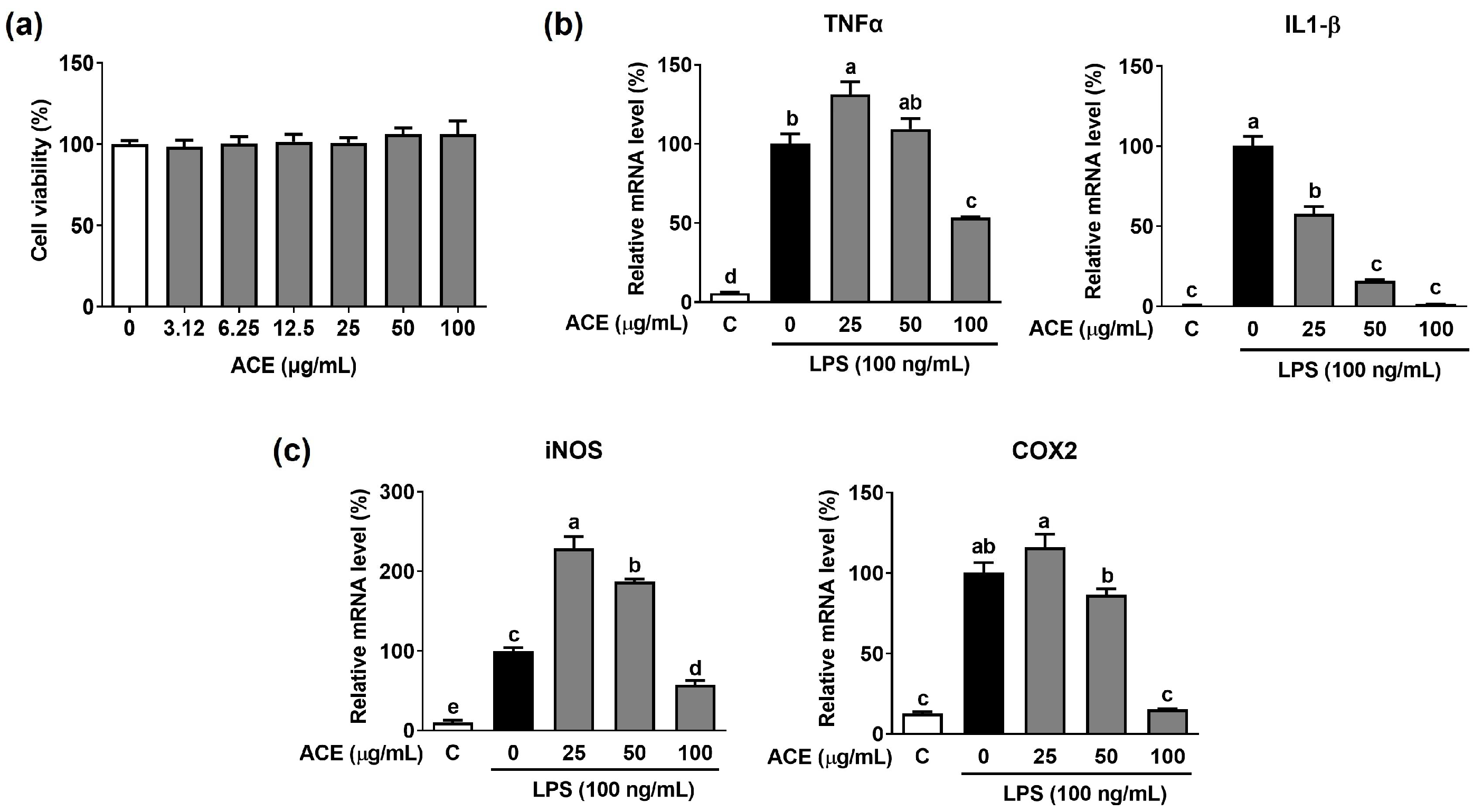
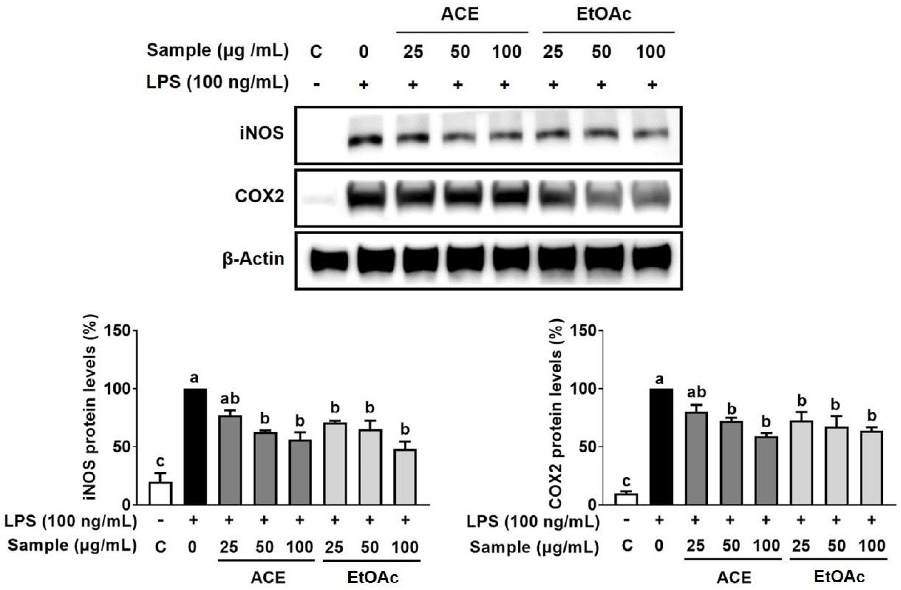
3.3. Inhibitory Effect of ACE on LPS-Stimulated Inflammation in Macrophages

The antioxidant effect by free radical scavenging activity is closely related to the suppression of pro-inflammatory gene expression [19]. Since ACE and its five fractions showed high TPC and total antioxidant activity, we investigated whether the ACE can inhibit LPS-induced inflammation in macrophages. When the cytotoxicity of ACE was measured at 0–100 µg/mL on RAW 264.7 macrophages, cell viability was ~98% at 100 µg/mL ACE (Figure 1a). Thus, the following experiments were performed at ACE concentrations under 100 µg/mL. The mRNA expression of TNFα and IL-1β was significantly elevated by LPS, whereas ACE significantly diminished the LPS-induced expression of TNFα and IL-1β in macrophages (Figure 1b). LPS significantly increased the expression of inflammation-inducible enzymes such as iNOS and COX2 in macrophages, while 100 µg/mL of ACE significantly decreased the mRNA expression of iNOS and COX-2 (Figure 1c). However, 25 and 50 µg/mL of ACE showed minimal effects on the mRNA expression of iNOS and COX2 in LPS-stimulated macrophages. The LPS-increased protein levels of iNOS and COX2 were also noticeably decreased by ACE in macrophages (Figure 2).

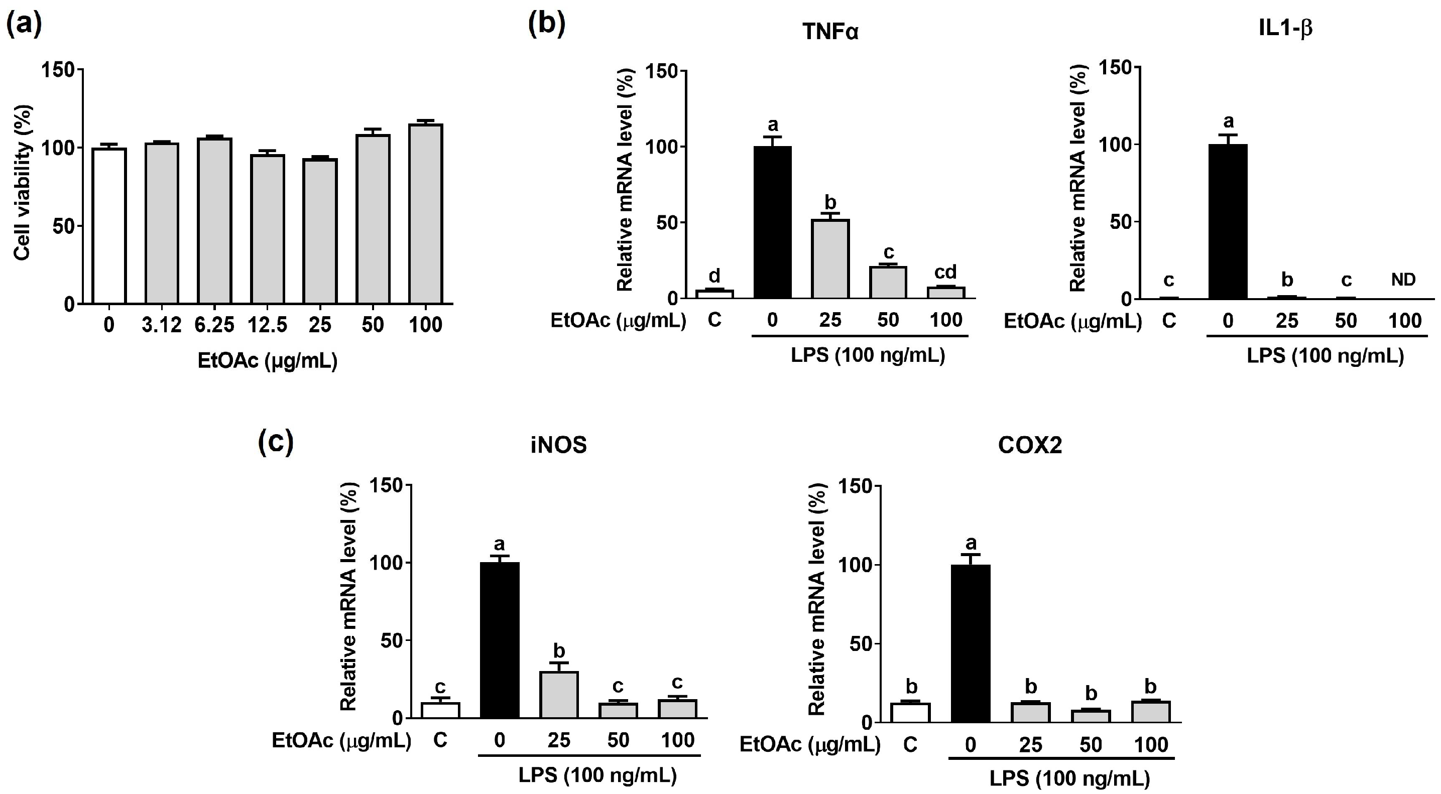
3.4. Effect of EtOAc Fraction from ACE on LPS-Induced Inflammation in Macrophages

As the EtOAc fraction showed the highest TPC and antioxidant activity among other ACE fractions, we additionally confirmed the inhibitory effect of the EtOAc fraction on inflammation in LPS-stimulated macrophages. We measured the cell viability of the EtOAc fraction in macrophages and found that ~90% of cells were viable when treated 0–100 µg/mL of the EtOAc fraction (Figure 3a). LPS significantly increased TNFα and IL-1β gene expression levels, which were completely abolished by the EtOAc fraction in macrophages (Figure 3b). Additionally, the EtOAc fraction significantly repressed the mRNA expression of iNOS and COX2 in LPS-stimulated macrophages (Figure 3c). Increases in the protein expression of iNOS and COX2 by LPS were markedly decreased by the EtOAc fraction in macrophages (Figure 2).

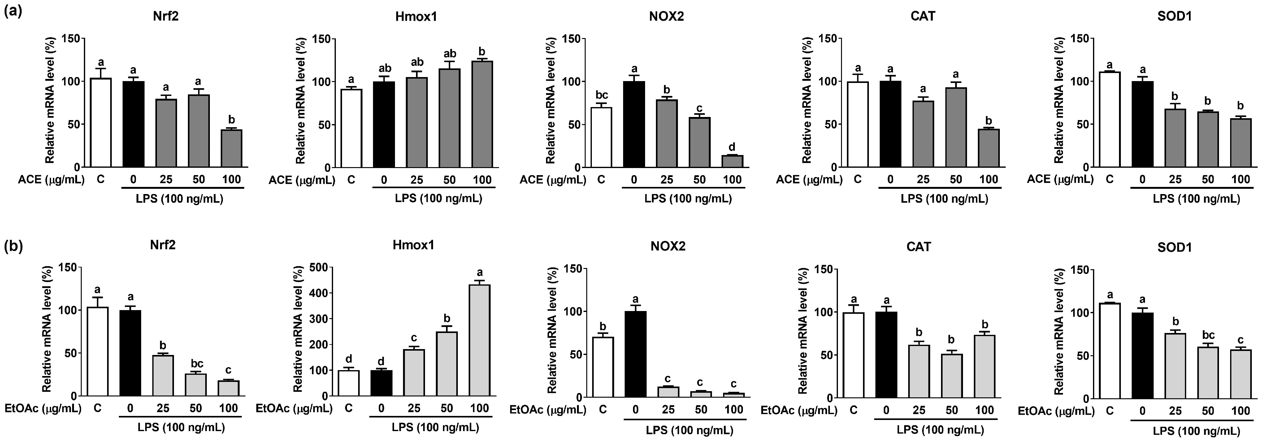
3.5. Effects of ACE and Its EtOAc Fraction on LPS-Induced Antioxidant Gene Expression in Macrophages

We observed that ACE and its EtOAc fraction have a potent antioxidant effect because of their high TPC and total antioxidant activity. Therefore, the effects of ACE and its EtOAc fraction on the expression of antioxidant genes in LPS-stimulated macrophages were evaluated. LPS significantly stimulated the mRNA expression of NADPH oxidase 2 (NOX2), an enzyme that generates ROS, which was significantly suppressed by ACE and its EtOAc fraction in macrophages (Figure 4a,b). Although the mRNA expression of the other antioxidant genes, such as nuclear factor E2-related factor 2 (Nrf2), catalase (CAT), and superoxide dismutase (SOD1), was not altered by LPS, ACE and its EtOAc fraction significantly suppressed the expression of Nrf2, CAT, and SOD1 in LPS-stimulated macrophages. However, the expression of heme oxygenase-1 (Hmox1) was significantly increased by the EtOAc fraction in LPS-induced macrophages.

4. Discussion

Seaweed phenolic compounds are considered to exert antioxidant and anti-inflammatory properties that mitigate oxidative stress and inflammation at the cellular levels [20,21]. Studies have shown that Gangneung-grown AC exerts antioxidant activity and inhibits the gene expression of pro-inflammatory cytokine such as IL-1β, IL-6, and TNFα in LPS-induced macrophages [15,16,22]. However, the effects of Busan-grown AC ethanol extract and its fractions on antioxidant and inflammation in LPS-induced macrophages were not confirmed. In this study, we found that Busan-grown ACE and its EtOAc fraction inhibited the mRNA expression of pro-inflammatory cytokines and antioxidant genes in LPS-stimulated RAW 264.7 macrophages accompanied by high TPC and total antioxidant activity.
Phlorotannins are known to be the most representative phenolic compounds in edible brown seaweed extracts [23]. In the present study, we found that the EtOAc fraction of ACE exhibited the highest TPC among five fractions of ACE, and thus had the highest content of phlorotannins formed through the polymerization of phloroglucinol. In addition, ACE and its EtOAc fractions showed high DPPH and ABTS radical scavenging activity and FRAP activity. The antioxidant properties of ACE and its EtOAc fraction are partly attributed to a high phlorotannin content, which has a unique structure. The strong antioxidant capacity of phlorotannin is associated with the degree of polymerization, the phenol ring, and the phenolic hydroxyl group on the benzene ring skeleton [24]. The presence of phlorotannins in the ACE and EtOAc fractions may confer the intrinsic stability of the polyphenol structure and potent free-radical scavenging activity. Furthermore, studies have well supported that phlorotannins exert excellent antioxidant properties through their free-radical scavenging activity, inhibition of ROS production, and antioxidant defense mechanism [25]. Phlorotannin compounds showed a high scavenging ability on DPPH and superoxide anion radicals two-to-ten-folds higher than ascorbic acid, α-tocopherol, and catechin, which are known as strong antioxidants [26]. Phlorotannin compound-isolated Ishige okamurae significantly reduced intercellular ROS production in RAW 264.7 macrophages [27]. Furthermore, in zebrafish exposed to ethanol, AAPH, high glucose condition, and UV-B radiation, the phlorotannin compounds from Eisenia cava inhibited intercellular ROS, DNA damage, and lipid peroxidation [28]. Thus, the study to find the phlorotannin compounds of the EtOAc fraction of ACE with potent total antioxidant activity by reducing free-radical scavenging activity is warranted to test this possibility.
LPS, a major pathogenic infectious agent of Gram-negative bacteria, directly activates macrophages, which promotes the secretion of inflammation-inducible enzymes and pro-inflammatory cytokines [29]. In this study, ACE and its EtOAc fraction significantly suppressed the gene expression of IL-1β, TNFα, iNOS, and COX2 in LPS-induced macrophages. In addition, we found that ACE and its EtOAc fraction showed high TPC and total antioxidant capacity by reducing DPPH and ABTS radical scavenging activity and FRAP activity. Oxidative stress by increasing ROS production can lead to the oxidation of amino acids, lipid peroxidation, and oxidative DNA damage, triggering an inflammatory response [30]. Thus, the antioxidant capacity of ACE and its EtOAc fraction may contribute to the repressed mRNA expression of IL-1β, TNFα, iNOS, and COX2 in LPS-induced macrophages. In particular, LPS significantly increased NOX2 gene expression, which was abolished by ACE and its EtOAc fraction in LPS-induced macrophages. NOX2 is known to generate ROS in macrophages which contributes to inflammation [31]. Although their effect on ROS production in macrophages was not evaluated in this study, ACE and its EtOAc fraction can reduce increased ROS production by inhibiting NOX2 expression in LPS-induced macrophages.
Large amounts of the pro-inflammatory cytokines are generated by iNOS and COX2 during inflammatory processes. Additionally, iNOS and COX2 expression are stimulated by growth factors and pro-inflammatory cytokines [32]. In this study, ACE and its EtOAc fraction significantly decreased the LPS-stimulated mRNA levels of iNOS and COX2 with a concomitant decrease in their protein levels in macrophages. Gangneung-grown AC fractions with a high content of phlorotannins such as trifuhalol A significantly reduced nitric oxide (NO) production and iNOS and COX2 gene expression in LPS-stimulated RAW 264.7 macrophages [16], which indicates that ACE has the inhibitory property of NO production. Thus, the inhibition of iNOS and COX2 gene expression by ACE and its EtOAc fraction may be attributed to the NO production inhibitory effect by ACE in macrophages. Since NO can form peroxynitrite, which increases nitrification and oxidative stress in macrophages in response to ROS [33], it is possible that the antioxidant activities of the ACE and EtOAc fraction contribute to the reduction of NO production. Finally, the effect of reducing iNOS and COX2 expression in LPS-stimulated macrophages by ACE and EtOAc fractions may be responsible for their antioxidant and anti-inflammatory properties.

5. Conclusions

In conclusions, this study shows that ACE and its EtOAc fraction has inhibitory effects on inflammation and oxidative stress in LPS-stimulated RAW 264.7 macrophages. ACE and its EtOAc fraction inhibited the LPS-stimulated mRNA expression of pro-inflammatory cytokines and inflammation-inducible enzymes. In addition, a high TPC and total antioxidant capacity by ACE and its EtOAc fraction can contribute to the inhibitory effects of ACE and its EtOAc fraction on inflammation and oxidative stress. However, further studies are required to investigate the phlorotannin compounds of the EtOAc fraction from ACE that has potent antioxidant and anti-inflammatory properties. Therefore, our findings suggest that ACE has excellent potential as a nutraceutical material for the prevention and treatment of chronic inflammation diseases.

Author Contributions

Conceptualization, S.G.L. and M.-B.K.; methodology, M.-B.K. and H.L.; software, M.-B.K. and H.L.; validation, M.-B.K. and H.L.; formal analysis, H.L.; investigation, M.-B.K. and H.L.; resources, S.G.L.; data curation, M.-B.K., H.L. and S.G.L.; writing—original draft preparation, M.-B.K., H.L., T.V. and S.G.L.; writing—review and editing, M.-B.K., H.L., T.V. and S.G.L.; visualization, M.-B.K. and H.L.; supervision, S.G.L.; project administration, S.G.L.; funding acquisition, M.-B.K. and S.G.L. All authors have read and agreed to the published version of the manuscript.

Funding

This research was funded by the Future Fisheries Food Research Center by the Ministry of Oceans and Fisheries, Korea, funding number 201803932 to S.G.L., and by funds from the Basic Science Research Program through the National Research Foundation (NRF) funded by the Ministry of Education, funding number 202300247455 to M.-B.K.

Institutional Review Board Statement

Not applicable.

Informed Consent Statement

Not applicable.

Data Availability Statement

Not applicable.

Conflicts of Interest

The authors declare no conflict of interest.

References

  1. Rius-Pérez, S.; Torres-Cuevas, I.; Millán, I.; Ortega, Á.L.; Pérez, S. PGC-1α, inflammation, and oxidative stress: An integrative view in metabolism. Oxid. Med. Cell. Longev. 2020, 2020, 1452696. [Google Scholar] [CrossRef] [PubMed]
  2. Yang, Y.; Qian, J.; Li, B.; Lu, M.; Le, G.; Xie, Y. Metabolomics based on 1H-NMR reveal the regulatory mechanisms of dietary methionine restriction on splenic metabolic dysfunction in obese mice. Foods 2021, 10, 2439. [Google Scholar] [CrossRef] [PubMed]
  3. Kim, M.-B.; Kang, H.; Li, Y.; Park, Y.-K.; Lee, J.-Y. Fucoxanthin inhibits lipopolysaccharide-induced inflammation and oxidative stress by activating nuclear factor E2-related factor 2 via the phosphatidylinositol 3-kinase/AKT pathway in macrophages. Eur. J. Nutr. 2021, 60, 3315–3324. [Google Scholar] [CrossRef] [PubMed]
  4. Li, X.; Ren, Y.; Chang, K.; Wu, W.; Griffiths, H.R.; Lu, S.; Gao, D. Adipose tissue macrophages as potential targets for obesity and metabolic diseases. Front. Immunol. 2023, 14, 1153915. [Google Scholar] [CrossRef] [PubMed]
  5. Rendra, E.; Riabov, V.; Mossel, D.M.; Sevastyanova, T.; Harmsen, M.C.; Kzhyshkowska, J. Reactive oxygen species (ROS) in macrophage activation and function in diabetes. Immunobiology 2019, 224, 242–253. [Google Scholar] [CrossRef]
  6. Chai, J.; Luo, L.; Hou, F.; Fan, X.; Yu, J.; Ma, W.; Tang, W.; Yang, X.; Zhu, J.; Kang, W. Agmatine reduces lipopolysaccharide-mediated oxidant response via activating PI3K/Akt pathway and up-regulating Nrf2 and HO-1 expression in macrophages. PLoS ONE 2016, 11, e0163634. [Google Scholar] [CrossRef]
  7. Lomartire, S.; Gonçalves, A.M. An overview of potential seaweed-derived bioactive compounds for pharmaceutical applications. Mar. Drugs 2022, 20, 141. [Google Scholar] [CrossRef]
  8. Begum, R.; Howlader, S.; Mamun-Or-Rashid, A.; Rafiquzzaman, S.; Ashraf, G.M.; Albadrani, G.M.; Sayed, A.A.; Peluso, I.; Abdel-Daim, M.M.; Uddin, M.S. Antioxidant and signal-modulating effects of brown seaweed-derived compounds against oxidative stress-associated pathology. Oxid. Med. Cell. Longev. 2021, 2021, 9974890. [Google Scholar] [CrossRef]
  9. Ismail, M.M.; El Zokm, G.M.; Lopez, J.M.M. Nutritional, bioactive compounds content, and antioxidant activity of brown seaweeds from the Red Sea. Front. Nutr. 2023, 10, 1210934. [Google Scholar] [CrossRef]
  10. Dutot, M.; Olivier, E.; Fouyet, S.; Magny, R.; Hammad, K.; Roulland, E.; Rat, P.; Fagon, R. In Vitro chemopreventive potential of phlorotannins-rich extract from brown algae by inhibition of benzo [a] pyrene-induced P2x7 activation and toxic effects. Mar. Drugs 2021, 19, 34. [Google Scholar] [CrossRef]
  11. Lomartire, S.; Gonçalves, A.M. Marine Macroalgae Polyphenols as Potential Neuroprotective Antioxidants in Neurodegenerative Diseases. Mar. Drugs 2023, 21, 261. [Google Scholar] [CrossRef] [PubMed]
  12. Yu, D.-K.; Lee, B.; Kwon, M.; Yoon, N.; Shin, T.; Kim, N.-G.; Choi, J.-S.; Kim, H.-R. Phlorofucofuroeckol B suppresses inflammatory responses by down-regulating nuclear factor κB activation via Akt, ERK, and JNK in LPS-stimulated microglial cells. Int. Immunopharmacol. 2015, 28, 1068–1075. [Google Scholar] [CrossRef] [PubMed]
  13. Sugiura, Y.; Katsuzaki, H.; Imai, K.; Amano, H. The anti-allergic and anti-inflammatory effects of phlorotannins from the edible brown algae, Ecklonia sp. and Eisenia sp. Nat. Prod. Commun. 2021, 16, 1934578X211060924. [Google Scholar]
  14. Boo, G.H.; Lindstrom, S.C.; Klochkova, N.G.; Yotsukura, N.; Yang, E.C.; Kim, H.G.; Waaland, J.R.; Cho, G.Y.; Miller, K.A.; Boo, S.M. Taxonomy and biogeography of Agarum and Thalassiophyllum (Laminariales, Phaeophyceae) based on sequences of nuclear, mitochondrial, and plastid markers. Taxon 2011, 60, 831–840. [Google Scholar] [CrossRef]
  15. Phasanasophon, K.; Kim, S.M. Antioxidant and cosmeceutical activities of Agarum cribrosum phlorotannin extracted by ultrasound treatment. Nat. Prod. Commun. 2018, 13, 1934578X1801300513. [Google Scholar] [CrossRef]
  16. Phasanasophon, K.; Kim, S.M. Anti-inflammatory activity of the phlorotannin trifuhalol A using LPS-stimulated RAW264. 7 cells through NF-κB and MAPK main signaling pathways. Nat. Prod. Commun. 2019, 14, 1934578X19849798. [Google Scholar]
  17. Baek, S.H.; Cao, L.; Jeong, S.J.; Kim, H.-R.; Nam, T.J.; Lee, S.G. The comparison of total phenolics, total antioxidant, and anti-tyrosinase activities of Korean Sargassum species. J. Food Qual. 2021, 2021, 6640789. [Google Scholar] [CrossRef]
  18. Chen, J.; Yang, J.; Ma, L.; Li, J.; Shahzad, N.; Kim, C.K. Structure-antioxidant activity relationship of methoxy, phenolic hydroxyl, and carboxylic acid groups of phenolic acids. Sci. Rep. 2020, 10, 2611. [Google Scholar] [CrossRef]
  19. Mao, X.-Y.; Cheng, X.; Wang, X.; Wu, S.-J. Free-radical-scavenging and anti-inflammatory effect of yak milk casein before and after enzymatic hydrolysis. Food Chem. 2011, 126, 484–490. [Google Scholar] [CrossRef]
  20. Zhang, H.; Tsao, R. Dietary polyphenols, oxidative stress and antioxidant and anti-inflammatory effects. Curr. Opin. Food Sci. 2016, 8, 33–42. [Google Scholar] [CrossRef]
  21. Cotas, J.; Leandro, A.; Monteiro, P.; Pacheco, D.; Figueirinha, A.; Gonçalves, A.M.; da Silva, G.J.; Pereira, L. Seaweed phenolics: From extraction to applications. Mar. Drugs 2020, 18, 384. [Google Scholar] [CrossRef] [PubMed]
  22. Cho, M.L.; Lee, D.-J.; Lee, H.-S.; Lee, Y.-J.; You, S.G. LPS-induced NO inhibition and antioxidant activities of ethanol extracts and their solvent partitioned fractions from four brown seaweeds. Ocean Sci. J. 2013, 48, 349–359. [Google Scholar] [CrossRef]
  23. Zheng, H.; Zhao, Y.; Guo, L. A bioactive substance derived from brown seaweeds: Phlorotannins. Mar. Drugs 2022, 20, 742. [Google Scholar] [CrossRef] [PubMed]
  24. Meng, W.; Mu, T.; Sun, H.; Garcia-Vaquero, M. Phlorotannins: A review of extraction methods, structural characteristics, bioactivities, bioavailability, and future trends. Algal Res. 2021, 60, 102484. [Google Scholar] [CrossRef]
  25. Phang, S.J.; Teh, H.X.; Looi, M.L.; Arumugam, B.; Fauzi, M.B.; Kuppusamy, U.R. Phlorotannins from brown algae: A review on their antioxidant mechanisms and applications in oxidative stress-mediated diseases. J. Appl. Phycol. 2023, 35, 867–892. [Google Scholar] [CrossRef]
  26. Wang, T.; Jónsdóttir, R.S.; Liu, H.; Gu, L.; Kristinsson, H.G.; Raghavan, S.; Ólafsdóttir, G.N. Antioxidant capacities of phlorotannins extracted from the brown algae Fucus vesiculosus. J. Agric. Food Chem. 2012, 60, 5874–5883. [Google Scholar] [CrossRef]
  27. Zou, Y.; Qian, Z.-J.; Li, Y.; Kim, M.-M.; Lee, S.-H.; Kim, S.-K. Antioxidant effects of phlorotannins isolated from Ishige okamurae in free radical mediated oxidative systems. J. Agric. Food Chem. 2008, 56, 7001–7009. [Google Scholar] [CrossRef]
  28. Pradhan, B.; Ki, J.S. Antioxidant and chemotherapeutic efficacies of seaweed-derived phlorotannins in cancer treatment: A review regarding novel anticancer drugs. Phytother. Res. 2023, 37, 2067–2091. [Google Scholar] [CrossRef]
  29. Gorabi, A.M.; Kiaie, N.; Khosrojerdi, A.; Jamialahmadi, T.; Al-Rasadi, K.; Johnston, T.P.; Sahebkar, A. Implications for the role of lipopolysaccharide in the development of atherosclerosis. Trends Cardiovasc. Med. 2022, 32, 525–533. [Google Scholar] [CrossRef]
  30. Pisoschi, A.M.; Pop, A.; Iordache, F.; Stanca, L.; Predoi, G.; Serban, A.I. Oxidative stress mitigation by antioxidants-an overview on their chemistry and influences on health status. Eur. J. Med. Chem. 2021, 209, 112891. [Google Scholar] [CrossRef]
  31. Shiau, D.-J.; Kuo, W.-T.; Davuluri, G.V.N.; Shieh, C.-C.; Tsai, P.-J.; Chen, C.-C.; Lin, Y.-S.; Wu, Y.-Z.; Hsiao, Y.-P.; Chang, C.-P. Hepatocellular carcinoma-derived high mobility group box 1 triggers M2 macrophage polarization via a TLR2/NOX2/autophagy axis. Sci. Rep. 2020, 10, 13582. [Google Scholar] [CrossRef] [PubMed]
  32. Han, H.-J.; Hyun, C.-G. Acenocoumarol Exerts Anti-Inflammatory Activity via the Suppression of NF-κB and MAPK Pathways in RAW 264.7 Cells. Molecules 2023, 28, 2075. [Google Scholar] [CrossRef] [PubMed]
  33. Pérez de la Lastra, J.M.; Juan, C.A.; Plou, F.J.; Pérez-Lebeña, E. The nitration of proteins, lipids and DNA by peroxynitrite derivatives-chemistry involved and biological relevance. Stresses 2022, 2, 53–64. [Google Scholar] [CrossRef]
Figure 1. The effects of ACE on the gene expression pro-inflammatory cytokines and inflammation-inducible enzymes in LPS-induced RAW 264.7 macrophages. (a) Cell viability of ACE in macrophages. Macrophages were treated with 0–100 µg/mL of ACE to measure the cell viability. (b,c) Macrophages were treated with ACE at 0, 25, 50, and 100 µg/mL for 6 hr and then stimulated by LPS (100 ng/mL) for 3 hr in absence or presence of ACE for gene analysis by RT-PCR. GAPDH was employed as an internal control. Different letters in the same bar mean a significant difference (p < 0.05). Data are presented as the means ± SD.
Applsci 13 10048 g001
Figure 2. The effects of ACE and its EtOAc fraction on the protein expression inflammation-inducible enzymes in LPS-induced RAW 264.7 macrophages. Macrophages were treated with ACE and EtOAc fraction at 0, 25, 50, and 100 µg/mL for 6 hr and then stimulated by LPS (100 ng/mL) for 24 hr in absence or presence of ACE and EtOAc fraction for Western blot analysis. A representative blot for iNOS and COX2 is shown. β-Actin was employed as a loading control. Different letters in the same bar mean a significant difference (p < 0.05). Data are presented as the means ± SD.
Applsci 13 10048 g002
Figure 3. The effects of EtOAc fraction from ACE on the mRNA expression pro-inflammatory cytokines and inflammation-inducible enzymes in LPS-stimulated RAW 264.7 macrophages. (a) Cell viability of EtOAc fraction in macrophages. Macrophages were treated with 0–100 µg/mL of EtOAc fraction to measure the cell cytotoxicity. (b,c) RAW 264.7 macrophages were treated with EtOAc fraction at 0, 25, 50, and 100 µg/mL for 6 hr and then stimulated by LPS (100 ng/mL) for 3 hr in absence or presence of ACE and its EtOAc fraction for gene analysis by RT-PCR. GAPDH was employed as an internal control. Different letters in the same bar mean a significant difference (p < 0.05). Data are presented as the means ± SD.
Applsci 13 10048 g003
Figure 4. The effects of ACE and its EtOAc fraction on the expression of antioxidant genes in LPS-induced RAW 264.7 macrophages. Macrophages were treated with (a) ACE and (b) EtOAc fraction at 0, 25, 50, and 100 µg/mL for 6 hr and then stimulated by LPS (100 ng/mL) for 3 hr in absence or presence of ACE and its EtOAc fraction for gene analysis by RT-PCR. GAPDH was employed as an internal control. Different letters in the same bar mean a significant difference (p < 0.05). Data are presented as the means ± SD.
Applsci 13 10048 g004
Table 1. Extraction yield, TPC, and total anti-oxidant capacity results of ACE and its fractions.
Extract/FractionsExtraction YieldTPC
(mg PGE/g)
ABTS
(mg VCE/g)
DPPH
(mg VCE/g)
FRAP
(mM FSE/g)
% (w/w)g
ACE48.5744.00144.62 ± 10.33 c744.48 ± 9.62 c18.70 ± 0.24 b0.26 ± 0.01 b
Hex0.670.2017.04 ± 0.37 e9.38 ± 0.78 e1.97 ± 0.19 e0.02 ± 0.00 d
CHCl32.000.6047.80 ± 3.10 d110.48 ± 6.60 d5.02 ± 0.08 d0.09 ± 0.02 c
EtOAc3.000.90320.97 ± 3.41 a1144.48 ± 25.76 a37.63 ± 0.93 a0.4a1 ± 0.05 a
BuOH6.662.00157.10 ± 2.23 c763.33 ± 39.01 c19.27 ± 0.44 b0.25 ± 0.01 b
H2O59.2917.80173.66 ± 2.69 b822.86 ± 27.67 b15.57 ± 0.80 c0.29 ± 0.01 b
The values are presented as the means ± SD (n = 3). Different letters in the same row mean a significant difference (p < 0.05).
Table 2. Pearson correlation between TPC and three total antioxidant activity results in ACE and its fractions.
Antioxidant ActivityTPCABTS AssayDPPH AssayFRAP Assay
TPC10.94 (p < 0.01)0.98 (p < 0.01)0.96 (p < 0.01)
ABTS assay 10.90 (p < 0.01)0.97 (p < 0.01)
DPPH assay 10.92 (p < 0.01)
FRAP assay 1
The correlation was determined by Pearson correlation with p < 0.01 for a significant difference.
Disclaimer/Publisher’s Note: The statements, opinions and data contained in all publications are solely those of the individual author(s) and contributor(s) and not of MDPI and/or the editor(s). MDPI and/or the editor(s) disclaim responsibility for any injury to people or property resulting from any ideas, methods, instructions or products referred to in the content.

