Article

Presence of Cyanotoxins in a Mexican Subtropical Monomictic Crater Lake

by
José Jesús Bustillos-Guzmán
1,*,
Andrew Turner
2,
Oscar Ubisha Hernández-Almeida
3,
Christine Johanna Band-Schmidt
4,*,
Carlos Alberto Romero-Bañuelos
3,
Francisco Eduardo Hernández-Sandoval
1,
Erick Julián Núñez-Vázquez
1 and
Yolotzin Apatzingan Palomino-Hermosillo
3
1
Centro de Investigaciones Biológicas del Noroeste, La Paz 23000, Mexico
2
The Centre for Environment, Fisheries and Aquaculture Science, Weymouth, Dorset DT48UB, UK
3
Laboratorio de Oceanografía Biológica, Universidad Autónoma de Nayarit, Ciudad de la Cultura “Amado Nervo”, Tepic 63000, Mexico
4
Instituto Politécnico Nacional, Centro Interdisciplinario de Ciencias Marinas, La Paz 23000, Mexico
*
Authors to whom correspondence should be addressed.
Appl. Sci. 2020, 10(19), 6719; https://doi.org/10.3390/app10196719
Submission received: 14 August 2020 / Revised: 16 September 2020 / Accepted: 18 September 2020 / Published: 25 September 2020
(This article belongs to the Special Issue Harmful Cyanobacteria and Their Metabolites)

Abstract

:
Microcystins (MCs) produced by cyanobacteria are a ubiquitous worldwide problem because some MCs can cause tumor formation and are hepatotoxic. In the Santa María del Oro crater lake, Mexico, plankton scums are recurrent during most of the year and are associated with cyanobacteria of the genera Microcystis spp. and Lyngbya spp. As some of these species are associated with the production of MCs and paralytic shellfish toxins (PSTs), samples from these scums and particulate matter were collected and analyzed for the main bloom species and toxins by a ultrahigh performance liquid chromatography coupled to tandem mass spectrometry (UHPLC-MS/MS) and high performance liquid chromatography with fluorescence detection (HPLC-FLD). Results showed that the main bloom-forming species were Limnoraphis robusta and Microcystis aeruginosa, the presence of at least seven MC congeners and the absence of PSTs in the algae scums. The MCs identified were MC-WR, MC-LR, MC-LA, MC-HilR, MC-LF, MC-YR, and MC-LY. On a dry mass weight basis, MC concentrations were low and ranged between 0.15 and 6.84 μg/kg. Toxin profiles were dominated by MC-WR, MC-LR, and MC-LA, representing 94.5% of the total sample, with each analog contributing 39.8%, 38.1% and 16.5% by relative concentration, respectively. Two of the more hazardous congeners, MC-LR and MC-LA, represented 54.6% of the total MC concentration. MCs in particulate matter along the depth profile were not detected. The MC profile is linked to M. aeruginosa, and it represents the first quantitative MC congener description for this species from a Mexican water ecosystem. Since these mats are recurrent yearly, their effects on humans and wild fauna, and the possible role of anthropogenic activities that favor their presence and proliferation, need to be evaluated.

1. Introduction

The increasing global occurrence of toxic algae blooms in both marine and freshwater environments poses a serious threat to public health, animals, and ecosystems [1]. Among many other toxins microcystins, a group of at least 279 known congeners, many of which are potent hepatotoxins. They have a common structure composed of D-aminoacid residues (3), L-aminoacid residues (2), N-methyldehydroalanine, and ADDA (3-amino-9-methoxy-2,6,8-trimethyl-10-phenyldeca-4,6-dienoic acid) residue [2,3]). Several cyanobacteria genera have been identified as microcystin producers [4,5,6,7]. In North America, the most commonly reported genera are Dolichospermum (Anabaena), Microcystis, Planktothrix and Pseudoanabaena [8,9]. In Mexico, the Microcystis genus seems to be ubiquitous in freshwater bodies and linked to the presence of microcystins [10]. The main mechanism of microcystin toxicity is the inhibition of serine/threonine protein phosphatase 1 and 2A (PP1 and PP2A), leading to the hyperphosphorylation of cytosolic and cytoskeletal proteins [11]. Another important family of toxins is the paralytic shellfish toxins (PSTs), composed of ~57 neurotoxic analogs that include saxitoxin and related structural analogs [12,13]. PSTs act by interacting with amino acids of the pore regions SS1 and SS2 in each transmembrane domain of the sodium channels’ α-subunit in nerves and muscle cells, blocking neuronal transmission and leading to muscle paralysis and death by respiratory arrest in mammals [13]. Whilst most commonly associated with certain marine phytoplankton genera, some freshwater cyanobacteria from the order Nostocales (Dolichospermum (Anabaena), Aphanizomenon, Cylindrospermopsis and Lyngbya wollei) and Planktothrix from the order Oscillatorales have also been reported to produce PSTs [14,15,16]. In the marine environment, two dinoflagellate species (Pyrodinium bahamense and Gymnodinium catenatum) and several species of the genus Alexandrium spp. produce PSTs [17]. Along Mexican coasts, PSTs have been reported on several occasions [18], whereas there is only one report in a Mexican freshwater system of PSTs, found in the tegogolo snail (Pomacea patula catemacensis) from the Lago of Catemaco on the coasts of the Gulf of Mexico [19].
The Santa María del Oro crater lake (SMO), a mesotrophic lake [20], is an important tourist icon in Nayarit, Mexico that attracts thousands of tourists who perform ecotourism and recreational activities, such as the Festival Acuático Santa María del Oro (Figure 1), which has an international reach and contributes to economic benefits for the region. During a water quality monitoring program in the lake, microalgal scums were noted during the first months of the year, with maximal visual biomass between February and March 2015. Microcystis sp. and Lyngbya sp. were preliminarily identified as the main genera in the algal scums [21,22]. Therefore, we assumed the possible presence of cyanobacterial microcystins and PSTs in both the water and the algae biomass. The goal of this work was to identify and quantify, via an HPLC fluorescence detector (HPLC-FLD) and UHPLC-MS/MS, the presence of these toxins in the lake.

2. Materials and Methods

2.1. Study Area

The SMO crater lake, Nayarit (Figure 2) lies at the western end of the Trans-Mexican Volcanic Belt [23]. It has an elliptic shape, with a diameter of approximately 2 km, an area of 3.7 km2 [24] and a maximum depth of 62 m. The mean depth across the lake is 46 m [20]. According to Cardoso-Mohedano et al. [25], for the period from September 2014 to April 2016, the range of maximum temperatures (from 29.7 °C to 31.1 °C) was observed at the subsurface (4 m depth) during the summer (August–September), and the range of minimum temperatures ranged between 22.9 °C and 23.6 °C during the winter (January–February). These temperature ranges are consistent with the annual temperature range of surface waters during 2002–2007 [24]. This lake is considered a monomictic lake, with a strong stratification in the water column from May to January and a short vertical mix from February to April [24].
The climate is warm and subhumid, with monthly average temperatures that range from 16.5 °C to 25 °C (Station 18005, Cerro Blanco, Servicio Meteorológico Nacional). This region receives an average annual precipitation of ~1200 mm, mainly during the warm summer months (June to September) when the intertropical converge zone is at its most northerly location and the North American monsoon is active [26,27]. During the summer, tropical storms and hurricanes can bring moisture from the Pacific [28]. The rest of the year (October to May) is relatively dry, particularly during spring [29]. Around the lake, the community of SMO lake is situated and, in 2010, it had a population of 4482 inhabitants [30], with a relatively high floating population due to sports and tourist activities. SMO’s surface waters are used mainly for recreational activities. Commercial fishing is carried out by the Unión de Pescadores, comprised of 28 local fishermen. Also, small experimental cultures of tilapia (Oerochromis aereus) have been carried out in floating cages [31]. At the north shore of the lake, residential settlements can be found, while to the south, there is a large area of restaurants. Drinking water and drainage facilities are insufficient, leading to the use of untreated water from the lake for human and commercial use [32].

2.2. Sampling

Mat Biomass and Phytoplankton

Floating scum biomass samples were collected in March 2015 from 13 stations (Figure 2), with a distance between each sampling station of approximately 400 m. Around each station, two independent net phytoplankton samples were collected at the surface by towing 60 μm pore size nets for 1–2 min around the station. For phytoplankton identification and quantification, the biomass collected in one of the net tows was transferred to clean plastic containers and fixed with formaldehyde (4%). The biomass collected in the second tow was dried in a stove for 24 h at 60 °C in previously weighed aluminum containers. Dry biomass weight was determined by weight difference. Additionally, surface water samples were collected with the use of a Niskin bottle at 5 m intervals, from 0 to 50 m in depth at stations 9 and 13. Water samples were transferred to plastic bottles and transported in dark and cool conditions to the laboratory, where they were filtered through glass fiber filters (GF/F). Filters were immediately frozen at −40 °C until extraction (see below).

2.3. Chemicals and Standards

For cyanotoxin analysis, instrument solvents used for the preparation of mobile phases were of LC-MS grade (Fisher Optima, ThermoFisher, UK), and all chemicals were LC-MS reagent grade when possible. Sample preparation reagents were HPLC grade. Certified reference toxins (MC-RR, MC-LA, MC-LY, MC-LF, MC-LW, MC-YR, MC-WR, MC-HilR, MC-HtyR, MC-LR, Asp3-MC-LR and Nodularin) were all obtained from Enzo Life Sciences, Exeter, UK. A certified standard of [Dha7]-MC-LR and a pre-certified, freeze-dried matrix reference material of blue-green algae (RM-BGA, Lot 201301) containing a range of microcystins was obtained from the Institute of Biotoxin Metrology of the National Research Council Canada (NRCC). For preparation of a mixed stock solution, Certified Reference Material (CRM) ampoules were opened, and aliquots were accurately pipetted into a vial. A seven-level suite of working calibration standards was subsequently prepared through serial dilution of the mixed stock solution, using 50% methanol as the diluent, resulting in a calibration range between 0.33 ng/mL and 327 ng/mL. RM-BGA (280 mg) was extracted with 28.0 mL of 50% aqueous MeOH + 0.1% acetic acid prior to centrifugation (4500 rpm, 10 min), and the supernatant was collected prior to analysis.
For PSTs analysis, sample preparation reagents were of HPLC grade. Certified reference toxins (STX, NeoSTX, GTX1, GTX2, GTX3, GTX4, dcGT2, dcGTX3 and dcSTX) were also obtained from the Institute of Biotoxin Metrology of the National Research Council Canada (NRCC). The standard solution of GTX2 and GTX3 contained dcGTX2 and dcGTX3 as minor components, but the exact contents for these toxins were not given.

2.4. Extraction and Analysis

2.4.1. Microcystin Extraction

Each filter was placed into a separate 50 mL polypropylene centrifuge tube with 20 mL of MeOH and agitated for one hour. The MeOH fraction was recovered and transferred into a new 50 mL tube. This procedure was repeated three times. The recovered extract was filtered through a syringe filter of 25 mm with a 0.45 µm pore size. MeOH was rotary evaporated and, the dry residues were resuspended in 1 mL of MeOH (grade HPLC) and placed in a sealed glass HPLC vial. For net samples, a known amount of the dry mat (between 0.5 g and 1 g) was treated following the same procedure for the filters, with the exception that the re-extraction that was performed six times because the samples were much more concentrated.

UHPLC-MS/MS and Microcystin Quantitation

The UHPLC-MS/MS analysis was conducted following the method of Turner et al. [33], as applied previously to the routine analysis of water and algae in England [34]. A Waters (Manchester, UK) Xevo TQ tandem quadrupole mass spectrometer (MS/MS), coupled with a Waters Acquity UHPLC, was used for LC–MS/MS analysis. Chromatography was conducted using a 1.7 µm, 2.1 × 50 mm Waters Acquity UHPLC BEH C18 column (P/N 186002350, Lot no. 0249343351) in conjunction with a Waters VanGuard BEH C18 1.7 µm, 2.1 × 5 mm guard cartridge (P/N 186003975, Lot no. 0245343321). The columns were held at +60 °C, with samples held in the sample manager at +4 °C. Mobile phase A1 consisted of water + 0.025% formic acid, and mobile phase B1 comprised of acetonitrile (MeCN) + 0.025% formic acid. The sample injection volume was 5 µL, and the mobile phase flow rate was consistently 0.6 mL/min. The UPLC gradient was exactly as described by Turner et al. [33], with a total run time of 5.5 min. Each instrumental sequence started with a series of instrumental blanks, followed by toxin calibration standards and an extract of RM-BGA to be used as a matrix-based retention time marker, and as internal quality control (QC).
The Waters Xevo TQ tune parameters were as follows: 150 °C source temperature, 600 °C desolvation temperature, 600 L/h desolvation gas flow and 0.15 mL/min collision gas flow. Capillary voltage was held at 1.0 kV. Selected reaction monitoring (SRM) transitions were built into the MS/MS method, using positive mode acquisition for each toxin. Parent and daughter ions, all in positive ion mode, as well as cone and collision voltages are summarized (Table 1). The majority of toxins exhibited unique SRM transitions and chromatographic retention times, resulting in good separation of cyanotoxins over the 5 min run time. The exceptions were [Dha7]-MC-LR and Asp3-MC-LR, which shared the same transitions and could not be completely resolved. These two analytes are therefore reported together.
The LC–MS/MS microcystin (MC) and Nodularin (Nod) methods involved the direct quantitation of cyanotoxin toxins against working standards, available as certified reference standards. Quantitation was performed using external calibration, and results were calculated in terms of µg/L of filtered water or ng/g of dry weight from the net samples. MC special-contour diagrams were constructed using software Surfer 13 (Golden Software, Inc., Golden, CO, USA).

2.4.2. PST Extraction

Paralytic shellfish toxins were extracted by initially adding 2 mL of acetic acid (0.03 N) to each filter. Filters were subsequently sonicated at 35 kHz for 60 s in an ice bath, before centrifugation at 2200 g for 5 min, with the supernatant filtered using a single-use syringe filter (0.45 μm pore size). For net samples, a known amount of the dry mat (between 0.5 g and 1 g) was treated as the filter samples, but 5 mL of acetic acid (0.03 N) was added instead.

PST Analysis

Aliquots of each acetic acid extract were treated with hydrochloric acid in order to convert the N-sulfocarbamoyl toxins into their related carbamoyl toxins, as described in [35]. Specifically, a 150 μL aliquot of the acetic acid extract was used for hydrolysis with 37 μL of HCl (1 M), incubated at 90 °C for 15 min, then cooled to room temperature before the addition of 75 μL of a 1 M sodium acetate solution. Finally, 20 μL of both extracts (with and without hydrolysis) were injected into the HPLC system. Chromatography was performed according to published procedures [35]. An ion pair buffer gradient, composed of a solution of octanesulfonic acid and ammonia phosphate at pH 6.9 and acetonitrile, was used to separate the PSTs analogs. After post-column oxidation with alkaline periodic acid, the resulting products were detected with a fluorescence detector (FLD). Detection limits for this method were 6.69, 14.10, 1119.15, 37.16, 42.03, 48.28, 45.89 and 37.16 pg for STX, dcSTX, Neo, GTX2, GTX3, dcGTX2, dcGTX3 and dcNEO, respectively. This HPLC-FLD method involves an extra run for the hydrolyzed derivatives of sulfocarbamoyl toxins (B and C toxins). Other PSTs, such as those described for Lyngbya wollei (LW1-6), were not explored in this study because of the lack of standards.

2.5. Cyanobacteria Bloom Forming Species Identification and Quantification

Species identification was done, considering morphological characteristics according to Ochoa-Zamora [21] and Salazar-Alcaraz [22]. Phytoplankton species were quantified under an optic microscope at 5×, 10×, 40× and 100× (Motic), and under a phase contrast microscope (Zeiss) equipped with a digital camera (EOS Cannon 6D). To determine the abundance of Limnoraphis robusta cells and Microcystis aeruginosa colonies, samples were diluted from 1:10 to 1:100,000. To determine the number of cells/filament of L. robusta, 30 photographs of filaments were taken at 10× and 40× under a Zeiss microscope. The length, thickness and number of cells per filament was estimated in filament sections of 20 μm. The number of cells per filament was estimated with the following formula:
Limnoraphis robusta (cells/mL) = L/20 × N
where: L is the longitude of the filament and N is the mean value of the cells in 20 micron increments.
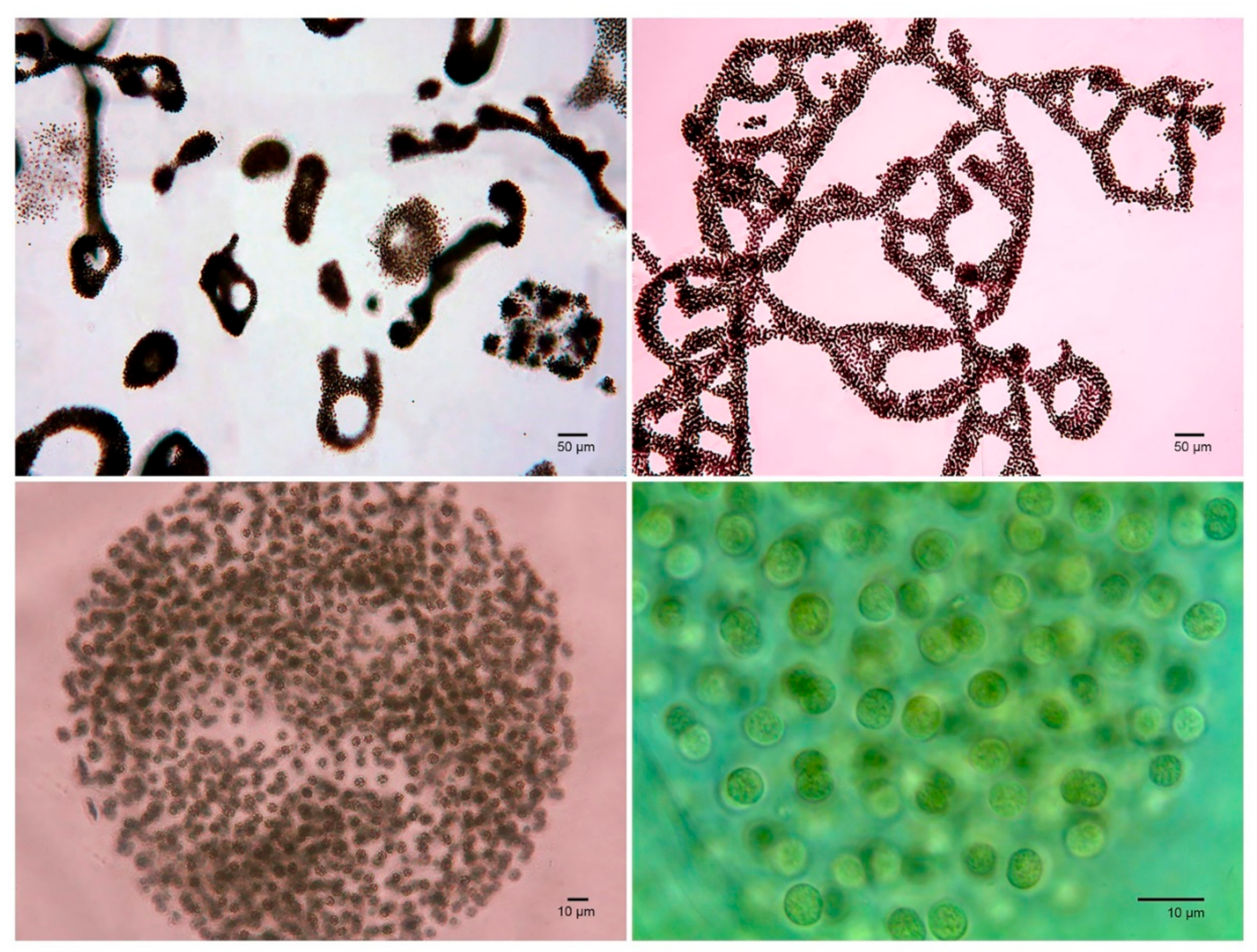
In the case of M. aeruginosa, only colonies were quantified because of the large and irregular size of the colonies (>50 μm in diameter), the abundance of M. aeruginosa cells in colonies and the difficulties for diluting the colonies matrix (Figure 3). Colony quantification was estimated in a Sedgewick Rafter chamber with a capacity of 1 mL.

3. Results

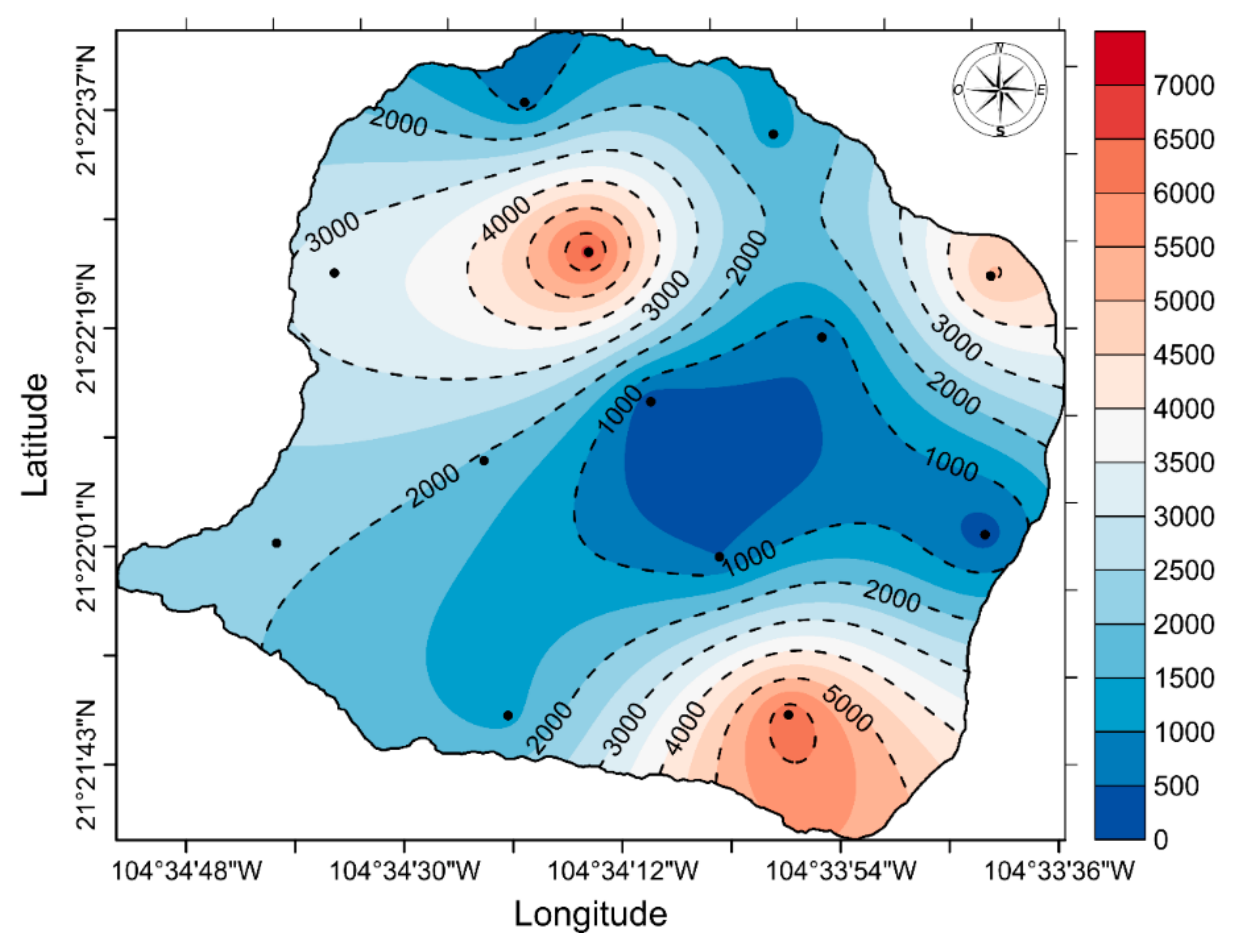
The results of this study demonstrate the presence of seven MC analogs in the algae scums of the SMO lake. The MC congeners identified were MC-WR, MC-LR, MC-LA, MC-HilR, MC-LF, MC-YR and MC-LY (Figure 4). In a weight basis, the total MC concentrations were low when compared with other zones [36,37] and ranged between 0.15 μg/kg and 6.84 μg/kg (Figure 5). The MC-WR, MC-LR and MC-LA congeners dominated the toxin profile, representing 94.5% of the total sample on a percentage concentration basis, with each of these congeners contributing 39.8%, 38.1% and 16.5%, respectively (Table 2).
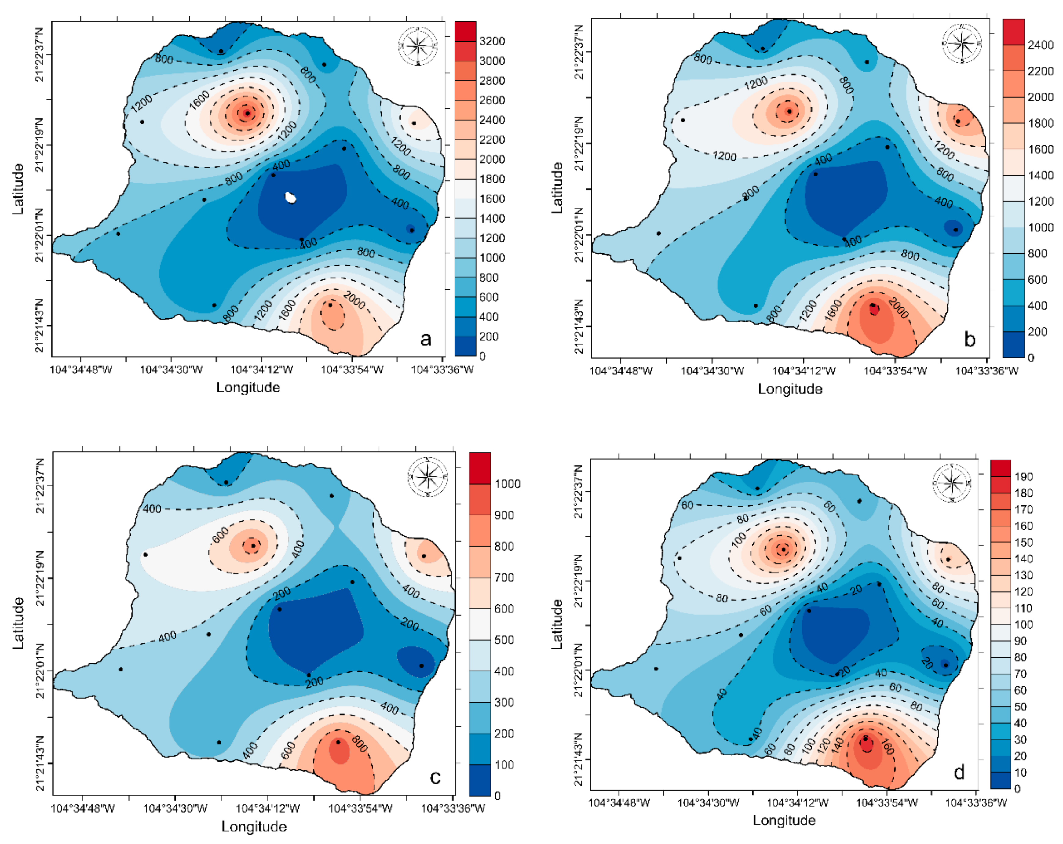
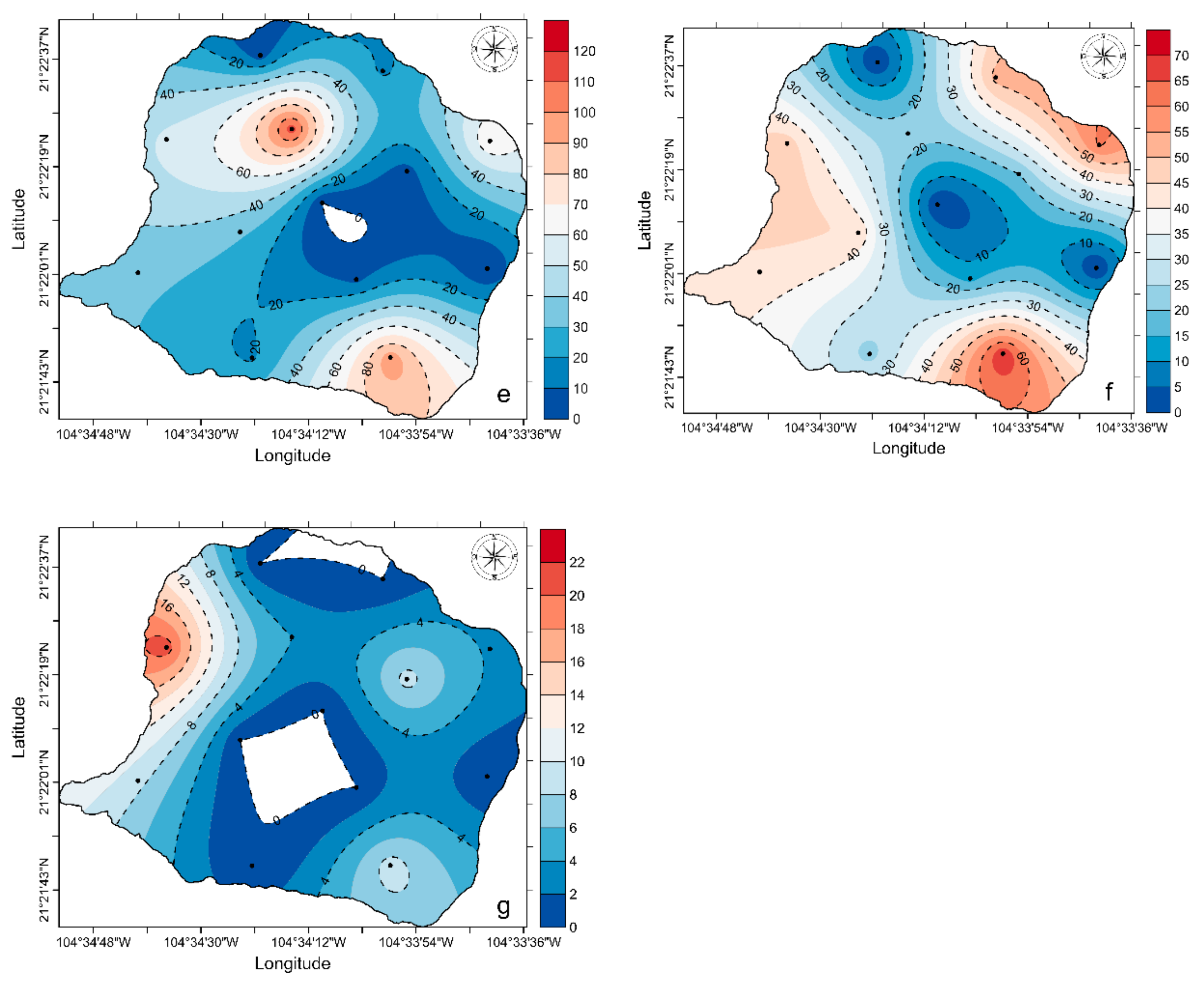
Horizontal surface distribution showed higher total microcystin values at the northern and southern part of the lake, and most of the MC congeners were detected with this general distribution pattern (Figure 6). These toxin distribution patterns can be explained by the modeled circulation cells. According to Serrano et al. [38], water circulation patterns in SMO lake are forced by the valley breeze circulation and the drift of currents from two circulating rings that have opposite directions: an anticyclonical direction on the northern part of the lake and a cyclonical pattern on the southern side. Thus, this circulation pattern may accumulate MCs in both rings, as demonstrated in this work.
HPLC fluorescence detector (HPLC-FLD) chromatograms from floating scums for PST analysis showed two peaks that have the same retention time as GTX3 and dcSTX (data not shown). However, these chromatographic peaks may correspond to other natural fluorescence co-extractives from the matrix, since reanalyses of this material by mass spectrometry confirm the absence of any considered PST analogs (data not shown). Neither MCs nor PST analogs were detected in the phytoplankton samples.
The bloom was confirmed to be caused by the cyanobacteria L. robusta and M. aeruginosa, although minor quantities of Lyngbya sp. were also recorded [21]. A lower cell density of L. robusta (43,139 cells/mL) and number of colonies of M. aeruginosa (2.9 colonies/mL) were recorded between stations 6 and 7, whereas the highest values were found between stations 8 and 9, with values of 50,936,326 cells/mL and 5300 colonies /mL for L. robusta and M. aeruginosa, respectively (Table 3).

4. Discussion

While occurrences of cyanobacteria related with hepatoxins and neurotoxins in freshwater have been recognized as a problem in many countries, mainly in temperate regions [39], a gap of information still exists regarding phytoplankton toxins in tropical zones [40]. The case of Mexico is not the exception, in spite of being a country with economically important freshwater systems. To our knowledge, MCs have been recorded in a few aquatic systems of Mexico City and the states of Mexico [41,42,43,44], Sinaloa [45] and Hidalgo [43]. However, microcystin presence has been quantified based on the ELISA technique and, therefore, there is no data available on the MC congeners present [46]. For some of these aquatic systems, matrix-assisted laser desorption/ionization time-of-flight mass spectrometry analysis was performed and showed the presence of MC-LR, MC-LR, MC-RR, MC-H4YR, MC-YR, MC-WR and MC-HtyR congeners [43]. However, no quantitative values were given. To our knowledge, this is the first quantitative report of an MC congener profile for a Mexican lake.
Neurotoxins, particularly PSTs, have been recorded only in Lago Catemaco, Veracruz [19]. In this work, we provided information regarding MCs and PSTs of the Santa María del Oro lake, Nayarit, with at least seven MC analogs and an absence of PSTs. The use of LC–MS/MS allowed us to confirm the presence of seven MC congeners (MC-WR, MC-LR, MC-LA, MC-HilR, MC-LF, MC-YR and MC LY). Previous studies using ELISA were unable to provide information on the presence MC congeners.
Although we only considered morphological characteristics to identify the cyanobacteria L. robusta and M. aeruginosa as the dominant species, these results agree with recent studies based on polyphasic analysis, which confirmed the presence of both species during the algal blooms in SMO lake [21,22]. It is plausible that, in our study, both species were correctly identified and that both species were present. However, L. robusta does not produce toxins, as revealed by the primer pairs DQmcyF/DQmcyR for microcystin/nodularin; sxtA F/sxtA–R for saxitoxin; and cyrF/cyrR for cylindrospermopsin [47]. The cyanobacteria M. aeruginosa is the only MC producer detected in the lake to date, so it is plausible to conclude that the MC profile found in this study corresponds to this species. Therefore, this could be the first report of the MC profile for a Mexican population of M. aeruginosa. Microcystis smithii, another MC producer, has been recorded in the lake [21,22]. However, we did not detect this species in our samples. Studies, including polyphasic analysis of the cyanobacterial community, are needed to confirm the presence of this species in the lake.
Although the number of MC structural variants in blooms is high, generally, just a few analogs contribute to the bulk share of the total of microcystin concentrations [48]. In our samples, MC-WR, MC-LR and MC-LA represented 94.5% of the bulk, which also agrees with other studies. For instance, a study of MC profiles in freshwater bloom samples from a tropical lake (Guatemala) demonstrated the dominance of MC-LR, MC-RR and MC-YR, accounting for 98% of the toxin burden in M. aeruginosa samples [49]. In other studies, high relative concentrations of MC-LR, MC-RR and MC-YR have also been reported from Microcystis samples [50,51]. Interestingly, two of the most potent MC congeners (LD50 of 50 μg kg−1 b,w, i.p. mouse [2]), MC-LR and MC-LA, represented more than 54% of the total MC congeners. Therefore, if these analogs increase in the future, their potential effects may result in ecosystem disruption and higher effects on animals and humans.
The genus Microcystis in the urban lakes of Mexico City is present when the temperature ranges between 20 °C and 25 °C, with a nitrate:phosphate proportion below 1 and a pH ≥ 10, similar to data referred to from other geographic regions [52,53]. However, in these artificial lakes, the supply of mineral nutrients is continuous and provided mainly by secondary wastewater treatment plants [10]. At SMO lake, blooms coincide with a transition from a mixed to a stratified water column that occurs during March–April [20,21]. Therefore, we hypothesize that nutrients may be supplied from water below the seasonal thermocline that is formed during the warm months. Hypolimnion anoxygenic conditions are present almost year-round in SMO lake [20]. These conditions might be a good environment for the remineralization of organic matter and nutrient production and, when the water column is mixed, these nutrients can fertilize the photic zone, providing adequate nutrient and light environments for the formation of the cyanobacterial bloom. Physicochemical and nutrient data seem to be undifferentiated during the period of complete vertical mixing [20]. Therefore, the whole lake may be a good environment for the growth of toxic cyanobacteria. According to Caballero et al. [20], SMO lake is a mesotrophic lake with relatively high soluble reactive phosphorus and silica levels. Nitrogen reaches its highest values during winter mixing (January), but becomes the limiting nutrient during stratification. These data provide additional support to our hypothesis.
The presence of M. aeruginosa in SMO lake seems to be a recently developed phenomenon. Previous phytoplankton analysis conducted in 2011 by colleagues of the National Institute of Ecology did not record this genus (G. Vázquez Hurtado pers. comm.). The risk of MC poisoning in humans and wildlife around the lake seems to be tangible since, according to [30], most of its inhabitants (64%) are considered scarce of resources, and some homes (6.5%) do not have bathroom facilities or access to drinking water (5.9%) [30]. Although there are no documented records of MC poisoning in the zone, it is important to note that, during the monitoring of the phytoplankton in the lake, colleagues and students reported skin rashes and intoxication symptoms such as headache, sore throat, dizziness and disorientation that lasted for more than four hours. In addition, local inhabitants commented on common nasal and digestive mucosa inflammation when recreational activities took place around or in the lake during cyanobacterial blooms. These symptoms are not likely to have been caused by MCs; other agents are far more likely for several reasons, one being the low MC concentration in the bloom, but more importantly, MCs do not cause these symptoms. Other yet unknown cyanobacterial co-occurring metabolites might be the cause, or microorganisms attached to the mucilage. Another metabolite not linked to the above-mentioned symptoms, but of human health concern, is the β-N-methylamino-41 L-alanine (BMAA), which may be also present in the lake, since this amino acid has been suggested to be a causative agent involved in several neurodegenerative diseases [54] and has been found in M. aeruginosa [55].
Clearly, MCs are present in Mexican freshwater ecosystems and should be considered as a public health problem by government authorities. As pointed out by Dörr et al. [56], reference laboratories are needed to validate results and to provide reference material, as well as the requirement for sophisticated and expensive equipment and high levels of technical expertise, which are the main limitations to improving our knowledge regarding MC occurrence, toxicity, profiles and human health impacts in Mexico.

Author Contributions

Conceptualization, O.U.H.-A.; data curation, C.A.R.-B. and E.J.N.-V.; formal analysis, J.J.B.-G., A.T. and O.U.H.-A.; funding acquisition, J.J.B.-G., O.U.H.-A. and C.J.B.-S.; investigation, O.U.H.-A.; methodology, A.T., C.A.R.-B., F.E.H.-S., E.J.N.-V. and Y.A.P.-H.; resources, J.J.B.-G. and O.U.H.-A.; writing—original draft, J.J.B.-G.; writing—review and editing, A.T., O.U.H.-A. and C.J.B.-S. All authors have read and agreed to the published version of the manuscript.

Funding

This research was funded by the institutional projects COSTA of PLAYCO from CIBNOR, SIP 2020-0571, and FORDECyT 260040.

Acknowledgments

We thank Enrique Arregui Alva of the Asociación de Colonos de la Cuenca de Santa María del Oro, and Eriberto Rodríguez Aguilar, Aurora Ramírez Castañeda, Ramona Castañeda Guerrero, and Blanca Estela Márquez Valdivia of the Asociación de hoteleros y restauranteros de Santa María del Oro for supporting the sampling campaigns. We also thank the Unidad de Tecnología de Alimentos and Laboratorio de Contaminación—UAN for providing laboratories and equipment, the thematic network on Harmful Algae Blooms-CONACyT (RedFAN) for the support and encouragement in this research field, and Eberto Novelo Maldonado and the Continental Algae Laboratory: Ecology and Taxonomy (LACET) for the aid in morphological identification of cyanobacteria species. CJBS is COFFA-IPN and EDI-IPN fellow.

Conflicts of Interest

The authors declare no conflict of interest.

References

  1. Anderson, D. HABs in a changing world: A perspective on harmful algal blooms, their impacts, and research and management in a dynamic era of climactic and environmental change. Harmful Algae 2012, 3–17. Available online: https://www.ncbi.nlm.nih.gov/pmc/articles/PMC4667985/ (accessed on 10 April 2020).
  2. Bouaïcha, N.; Miles, C.O.; Beach, D.G.; Labidi, Z.; Djabri, A.; Benayache, N.Y.; Nguyen-Quang, T. Structural diversity, characterization and toxicology of Microcystins. Toxins 2019, 11, 714. [Google Scholar] [CrossRef] [PubMed] [Green Version]
  3. Chen, L.; Xie, P. Mechanisms of Microcystin-induced cytotoxicity and apoptosis. Mini Rev. Med. Chem. 2016, 16, 1018–1031. [Google Scholar] [CrossRef] [PubMed]
  4. Kotak, B.G.; Zurawell, R.W. Cyanobacterial toxins in Canadian freshwaters: A review. Lake Reservoir Manag. 2007, 23, 109–122. [Google Scholar] [CrossRef]
  5. Funari, E.; Testai, E. Human health risk assessment related to cyanotoxins exposure. Crit. Rev. Toxicol. 2008, 38, 97–125. [Google Scholar] [CrossRef] [PubMed]
  6. Pearson, L.; Mihali, L.; Moffitt, M.; Kellmann, R.; Neilan, B. On the chemistry, toxicology and genetics of the cyanobacterial toxins, microcystin, nodularin, saxitoxin and cylindrospermopsin. Mar. Drugs. 2010, 8, 1650–1680. [Google Scholar] [CrossRef] [PubMed] [Green Version]
  7. Martins, A.; Vasconcelos, V. Use of qPCR for the study of hepatotoxic cyanobacteria population dynamics. Arch. Microbiol. 2011, 193, 615–627. [Google Scholar] [CrossRef]
  8. Williams, C.D.; Aubel, M.T.; Chapman, A.D.; D’Aiuto, P.E. Identification of cyanobacterial toxins in Florida’s freshwater systems. Lake Reser. Manag. 2007, 23, 144–152. [Google Scholar] [CrossRef]
  9. Winter, J.G.; DeSellas, A.M.; Flecher, R.; Heintsch, L.; Morley, A.; Nakamoto, L.; Utsumi, K. Algal blooms in Ontario, Canada: Increases in reports since 1994. Lake Reser. Manag. 2011, 27, 107–114. [Google Scholar] [CrossRef] [Green Version]
  10. Arzate-Cárdenas, M.A.; Olvera-Ramírez, R.; Martínez-Jerónimo, F. Microcystis toxigenic strains in urban lakes: A case of study in Mexico City. Ecotoxicology 2010, 19, 1157–1165. [Google Scholar] [CrossRef]
  11. Hu, Y.; Chen, J.; Fan, H.; Xiel, P.; He, J. A review of neurotoxicity of microcystins. Environ. Sci. Pollut. Res. 2016, 23, 7211–7219. [Google Scholar] [CrossRef]
  12. Wiese, M.; D’Agostino, P.M.; Mihali, T.K.; Moffitt, M.C.; Neilan, B.A. Neurotoxic alkaloids: Saxitoxin and its analogs. Mar. Drugs 2010, 8, 2185–2211. [Google Scholar] [CrossRef] [PubMed] [Green Version]
  13. Durán-Riveroll, L.M.; Cembella, A.D.; Band-Schmidt, C.J.; Bustillos-Guzmán, J.J.; Correa-Basurto, J. Docking simulation of the binding interactions of saxitoxin analogs produced by the marine dinoflagellate Gymnodinium catenatum to the voltage-gated sodium channel Nav1.4. Toxins 2017, 8, 129. [Google Scholar] [CrossRef] [PubMed]
  14. Codd, G.A.; Bell, S.G.; Kaya, K.; Ward, C.; Beattie, K.A.; Metcalf, J. Cyanobacterial toxins, exposure routes and human health. Eur. J. Phycol. 1999, 34, 405–415. [Google Scholar] [CrossRef]
  15. Aráoz, R.; Molgó, J.; Tandeau de Marsac, N. Neurotoxic cyanobacterial toxins. Toxicon 2010, 56, 813–828. [Google Scholar] [CrossRef]
  16. Zamyadi, A. Emerging Toxic Cyanobacterial Issues in Freshwater Sources: Influence of Climate Change. In Seafood and Freshwater Toxins: Pharmacology, Physiology, and Detection, 3rd ed.; Botana, L.M., Ed.; CRC Press: New York, NY, USA, 2014; pp. 105–120. [Google Scholar]
  17. Vale, P. Saxitoxin and Analogs: Ecobiology, Origin, Chemistry, and Detection. In Seafood and Freshwater Toxins: Pharmacology, Physiology, and Detection, 3rd ed.; Botana, L.M., Ed.; CRC Press: New York, NY, USA, 2014; pp. 991–1011. [Google Scholar]
  18. Band-Schmidt, C.J.; Bustillos-Guzmán, J.J.; López-Cortés, D.J.; Gárate-Lizárraga, I.; Núñez-Vázquez, E.J.; Hernández-Sandoval, F.E. Ecological and physiological studies of Gymnodinium catenatum in the Mexican Pacific: A review. Mar. Drugs. 2010, 8, 1935–1961. [Google Scholar] [CrossRef] [Green Version]
  19. Berry, J.P.; Lind, O. First evidence of ‘‘paralytic shellfish toxins’’ and cylindrospermopsin in a Mexican freshwater system, Lago Catemaco, and apparent bioaccumulation of the toxins in ‘‘tegogolo’’ snails (Pomacea patula catemacensis). Toxicon 2010, 55, 930–938. [Google Scholar] [CrossRef]
  20. Caballero, M.; Rodríguez, A.; Vilaclara, G.; Ortega, B.; Roy, P.; Lozano, S. Hydrochemistry, ostracods and diatoms in a deep, tropical, crater lake in Western Mexico. J. Limnol. 2013, 72, 512–523. [Google Scholar] [CrossRef]
  21. Ochoa-Zamora, G.G. Variación Espacio-Temporal de las Poblaciones de Cianobacterias Formadoras de Florecimientos en el Lago Cráter de Santa María del Oro, Nayarit. Bachelor’s Thesis, Universidad Autónoma de Nayarit, Tepic, Mexico, 2018. (In Spanish). [Google Scholar]
  22. Salazar-Alcaraz, I. Identificación y Aislamiento de Cianobacterias de un Lago Cráter Tropical. Master Thesis, Universidad Autónoma de Nayarit, Tepic, Mexico, 2018. (In Spanish). [Google Scholar]
  23. Vázquez-Castro, G.; Ortega-Guerrero, B.; Rodríguez, A.; Caballero, M.; Lozano-García, S. Mineralogía magnética como indicador de sequía en los sedimentos lacustres de los últimos ca. 2600 años de Santa María del Oro, occidente de México. Rev. Mex. Cienc. Geol. 2008, 25, 21–38. [Google Scholar]
  24. Serrano-Hernández, D.E. Procesos Termodinámicos en el Lago Volcánico de Santa María del Oro, Nayarit: México. Ph.D. Thesis, Universidad Nacional Autónoma de México, Tepic, Mexico, 2004. (In Spanish). [Google Scholar]
  25. Cardoso-Mohedano, A.; Sánchez-Cabeza, J.A.; Ruiz-Fernández, A.C.; Pérez-Bernal, L.H.; Lima-Rego, J.; Giralt, S. Fast deep water warming of a subtropical crater lake. Sci. Total Environ. 2019, 691, 1353–1361. [Google Scholar] [CrossRef]
  26. Berbery, E.H. Mesoscale moisture analysis of the North American monsoon. J. Clim. 2001, 14, 121–137. [Google Scholar] [CrossRef]
  27. Liebmann, B.; Bladé, I.; Bond, N.A.; Gochis, D.; Allured, D.; Bates, G.T. Characteristics of North American summertime rainfall with emphasis on the monsoon. J. Clim. 2008, 21, 1277–1294. [Google Scholar] [CrossRef]
  28. Castro, S.C.D. Variabilidad de los ciclones tropicales que afectan a México. Interciencia 2010, 35, 306–310. Available online: https://www.redalyc.org/pdf/339/33913156011.pdf (accessed on 20 June 2020).
  29. Rodríguez-Ramírez, A.; Caballero, M.; Roy, P.; Ortega, B.; Vázquez-Castro, G.; Lozano-García, S. Climatic variability and human impact during the last 2000 years in western Mesoamerica: Evidence of late Classic (AD 600 900) and Little Ice Age drought events. Clim. Past. 2015, 11, 1239–1248. [Google Scholar] [CrossRef] [Green Version]
  30. Órgano del Gobierno del Estado de Nayarit. Plan de desarrollo municipal de Santa María del Oro, Nayarit, 2017–2021; Periódico Oficial: Tepic, Mexico, 2017; Volume 130, 124p. (In Spanish)
  31. Valencia-Plascencia, M.A.; Jacome-Pérez, B.G. Cultivo Experimental de Tilapia (Oerochromis aureus) en Jaulas Flortantes de p.v.c. en el Lago de Santa María del Oro, Nay. Bachelor’s Thesis, Universidad Autónoma de Nayarit, Tepic, Mexico, 1993; 47p. (In Spanish). [Google Scholar]
  32. Órgano del Gobierno del Estado de Nayarit. Plan de desarrollo urbano del centro de población de Santa María del Oro, Nayarit; Periódico Oficial: Tepic, Mexico, 2005; Volume 63, 108p. (In Spanish)
  33. Turner, A.D.; Waack, J.; Lewis, A.; Edwards, C.; Lawton, L. Development and single laboratory validation of a UHPLC-MS/MS method for quantitation of microcystins and nodularin in natural water, cyanobacteria, shellfish and algal supplement tablet powders. J. Chromatogr. B. 2018, 1074–1075, 111–123. [Google Scholar] [CrossRef]
  34. Turner, A.D.; Dhanji-Rapkova, M.; O’Neill, A.; Coates, L.; Lewis, A.; Lewis, K. Analysis of microcystins in cyanobacterial blooms from freshwater bodies in England. Toxins 2018, 10, 39. [Google Scholar] [CrossRef] [Green Version]
  35. Yu, R.C.; Hummert, C.; Luckas, B.; Qian, P.Y.; Zhou, M.J. Modified HPLC method for analysis of PSP toxins in algae and shellfish from China. Chromatographia 1998, 48, 671–676. Available online: https://link.springer.com/article/10.1007/BF02467597 (accessed on 20 June 2020). [CrossRef]
  36. Fastner, J.; Wirsing, B.; Wiedner, C.; Heinze, R.; Neumann, U.; Chorus, I. Cyanotoxin occurrence in Germany: Microcystins and hepatocyte toxicity. In Cyanotoxins: Occurrence, Causes, Consequences; Chorus, I., Ed.; Springer: Berlin/Heidelberg, Germany, 2001; pp. 22–37. [Google Scholar]
  37. IARC Working Group. Ingested Nitrate and Nitrite, and Cyanobacterial Peptide Toxins. In Monographs on the Evaluation of Carcinogenic Risks to Humans; WHO, IARC: Geneva, Switzerland, 2010; Volume 94, p. 450. ISBN 978-92-832-1294-2. [Google Scholar]
  38. Serrano, D.; Filonov, A.; Tereshchenko, I. Dynamic response to valley breeze circulation in Santa María del Oro, a volcanic lake in Mexico. Geophys. Res. Lett. 2002, 29, 27–31. [Google Scholar] [CrossRef]
  39. Harke, M.J.; Steffen, M.M.; Glober, C.J.; Otten, T.G.; Wilhelm, S.W.; Wood, S.A.; Paerl, H.M. A review of the global ecology, genomics and biogeography of the toxic cyanobacterium, Microcystis spp. Harmful Algae 2016, 54, 4–20. [Google Scholar] [CrossRef] [Green Version]
  40. Mowe, M.A.D.; Mitrovic, S.M.; Lim, R.P.; Furey, A.; Yeo, D.C.J. Tropical cyanobacterial blooms: A review of prevalence, problem taxa, toxins and influencing environmental factors. J. Limnol. 2015, 7, 205–224. [Google Scholar] [CrossRef] [Green Version]
  41. Nandini, S.; Sánchez-Zamora, C.; Sarma, S.S.S. Toxicity of cyanobacterial blooms from the reservoir Valle de Bravo (Mexico): A case study on the rotifer Brachionus calyciflorus. Sci. Total Environ. 2019, 688, 1348–1358. [Google Scholar] [CrossRef]
  42. Zamora-Barrios, C.A.; Nandini, S.; Sarma, S.S.S. Effect of crude extracts from cyanobacterial blooms in Lake Texcoco (Mexico) on the population growth of Brachionus calyciflorus (Rotifera). Toxicon 2017, 13, 45–53. [Google Scholar] [CrossRef] [PubMed]
  43. Zamora-Barrios, C.A.; Nandini, S.; Sarma, S.S.S. Bioaccumulation of microcystins in seston, zooplankton and fish: A case study in Lake Zumpango, Mexico. Environ. Pollut. 2019, 249, 267–276. [Google Scholar] [CrossRef] [PubMed]
  44. Vasconcelos, V.; Martins, A.; Vale, M.; Antunes, A.; Azevedo, J.; Welker, M.; López, O.; Montejano, G. First report on the occurrence of microcystins in planktonic cyanobacteria from Central Mexico. Toxicon 2010, 56, 425–431. [Google Scholar] [CrossRef] [PubMed]
  45. Alonso-Rodríguez, R.; Núñez-Vázquez, E.J.; Cordero-Tapia, A.; Gárate-Lizárraga, I.; Hiller, S.A.; Ochoa, J.L. Proliferación de Microcystis aeruginosa y presencia de microcistinas en un lago artificial en Culiacán, Sin., México. In Book of Abstracts of the XIV Reunión Nacional de la Sociedad Mexicana de Planctología, A.C. y VII Reunión Internacional de Planctología; Sociedad Mexicana de Planctología: Mexico City, Mexico, 2006. [Google Scholar]
  46. Ueno, Y.; Nagata, S.; Tsutsumi, T.; Hasegawa, A.; Watanabe, M.F.; Park, H.D.; Chen, G.C.; Chen, G.; Yu, S.Z. Detection of microcystins, a blue-green algal hepatotoxin, in drinking water sampled in Haimen and Fusui, endemic areas of primary liver cancer in China, by highly sensitive immunoassay. Carcinogenesis 1996, 17, 1317–1321. [Google Scholar] [CrossRef] [PubMed]
  47. Komárek, J.; Zapomělová, E.; Šmarda, J.; Kopecký, J.; Rejmánková, E.; Woodhouse, J.; Neilan, B.; Komárková, J. Polyphasic evaluation of Limnoraphis robusta, a water–bloom forming cyanobacterium from Lake Atitlán, Guatemala, with a description of Limnoraphis gen. nov. Fottea Olomouc. 2013, 13, 39–52. [Google Scholar] [CrossRef] [Green Version]
  48. Li, J.; Li, R.; Li, J. Current research scenario for microcystins biodegradation–A review on fundamental knowledge, application prospects and challenges. Sci. Total Environ. 2017, 595, 615–632. [Google Scholar] [CrossRef]
  49. Romero-Oliva, C.S.; Contardo-Jara, V.; Block, T.; Pflugmacher, S. Accumulation of microcystin congeners in different aquatic plants and crops—A case study from lake Amatitlán, Guatemala. Ecotox. Environ. Safe. 2014, 102, 121–128. [Google Scholar] [CrossRef]
  50. Pekar, H.; Westerberg, E.; Bruno, O.; Lääne, A.; Persson, K.M.; Sundstrom, L.F.; Thim, A.M. Fast, rugged and sensitive ultra high pressure liquid chromatography tandem mass spectrometry method for analysis of cyanotoxins in raw water and drinking water—First findings of anatoxins, cylindrospermopsins and microcystin variants in Swedish source waters and infiltration ponds. J. Chromatogr. 2016, 1429, 265–276. [Google Scholar] [CrossRef]
  51. Zervou, S.K.; Christophoridis, C.; Kaloudis, T.; Triantis, T.M.; Hiskia, A. New SPE-LC-MS/MS method for simultaneous determination of multi-class cyanobacterial and algal toxins. J. Hazar. Mater. 2017, 321, 56–66. [Google Scholar] [CrossRef]
  52. Visser, P.; Ibelings, B.; Mur, L.R.; Walsby, A. 2005. The ecophysiology of the harmful cyanobacterium Microcystis: Features explaining its success and measures for its control. In Harmful Cyanobacteria; Huisman, J., Matthijs, H.C.P., Visser, P., Eds.; Springer: Berlin/Heidelberg, Germany, 2005; pp. 109–142. Available online: https://www.academia.edu/28066256/ (accessed on 5 May 2020).
  53. Kim, S.G.; Rhee, S.K.; Ahn, C.Y.; Ko, S.R.; Choi, G.G.; Bae, J.W.; Park, Y.H.; Oh, H.M. Determination of cyanobacterial diversity during algal blooms in Daechung Reservoir, Korea, on the basis of cpcBA intergenic spacer region analysis. Appl. Environ. Microbiol. 2006, 72, 3252–3258. [Google Scholar] [CrossRef] [Green Version]
  54. Banack, S.A.; Caller, T.A.; Stommel, E.W. The cyanobacteria derived toxin beta-N-Methylamino-L-Alanine and amyotrophic lateral sclerosis. Toxins 2010, 2, 2837–2850. [Google Scholar] [CrossRef] [PubMed]
  55. Esterhuizen-Londt, M.; Baik, S.; Kwon, K.-S.; Ha, M.-H.; Oh, H.-M.; Pflugmacher, S. Toxicity and toxin composition of Microcystis aeruginosa from Wangsong reservoir. Toxicol. Environ. Health Sci. 2018, 10, 179–185. [Google Scholar] [CrossRef] [Green Version]
  56. Dörr, F.A.; Pinto, E.; Soares, R.M.; Feliciano de Oliveira e Azevedo, S.M. Microcystins in South American aquatic ecosystems: Occurrence, toxicity and toxicological assays. Toxicon 2010, 56, 1247–1256. [Google Scholar] [CrossRef] [PubMed]
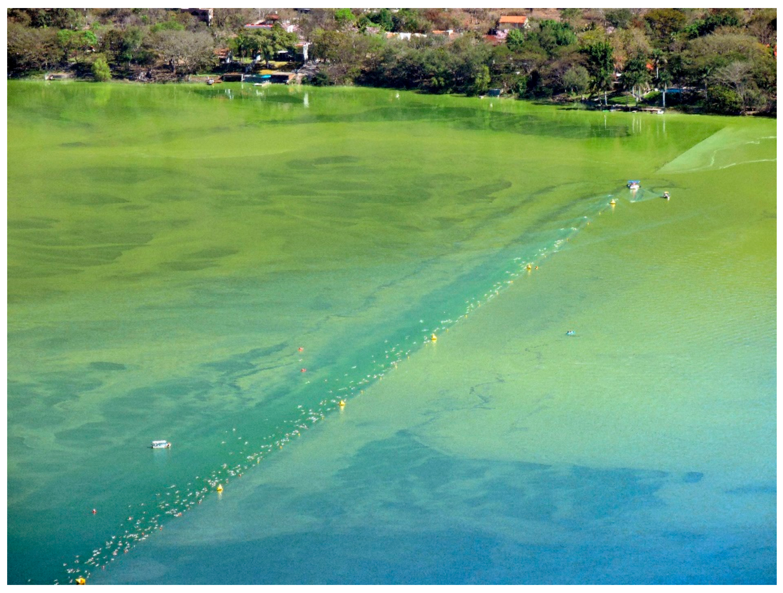
Figure 1. Swimming competition (Festival Acuático Santa María del Oro) in Santa María del Oro lake, Nayarit, during a cyanobacteria bloom in March 2018. Photo credit: Iván Salazar Alcaraz.
Applsci 10 06719 g001
Figure 2. Location of Santa María del Oro lake in the state of Nayarit (black point in the left figure) and sampling stations (numbers in the right figure).
Applsci 10 06719 g002
Figure 3. Diversity of colonies of Microcystis aeruginosa from the Santa María del Oro lake, Nayarit, during the cyanobacteria bloom. Photo credit: Génesis G. Ochoa Zamora.
Applsci 10 06719 g003
Figure 4. Ion chromatograms obtained following UHPLC-MS/MS, showing quantitative SRM transitions for detected microcystin congeners in (A) the high level calibration standard and (B) the algal scum sample from Station 4.
Applsci 10 06719 g004aApplsci 10 06719 g004b
Figure 5. Total microcystin distribution (μg/kg of dry weight) in algae scums in Santa María del Oro lake during March 2015.
Applsci 10 06719 g005
Figure 6. Surface distribution (μg/kg of dry weight) of microcystin congeners in algae mats in Santa María del Oro lake during March 2015. (a) MC-WR; (b) MC-LR; (c) MC-LA; (d) MC-HiIR; (e) MC-YR; (f) MC-LF; and (g) MC-LY distributions.
Applsci 10 06719 g006aApplsci 10 06719 g006b
Table 1. Selected reaction monitoring (SRM) transitions used for microcystin (MC) detection and quantitation.
AnalyteSRM TransitionsCone, VCE, eV
MC-RR519.9 > 134.9; 126.9; 102.83030; 50; 70
Nod825.5 > 135.1; 103.15560; 100
MC-LA910.1 > 135.1; 106.93570; 80
[Dha7]-MC-LR981.5 > 135.0; 106.87575; 80
Asp3-MC-LR981.5 > 134.9; 106.97570; 80
MC-LF986.5 > 213.0; 135.03560; 65
MC-LR995.6 > 135.0; 127.06070; 90
MC-LY1002.5 > 135.0; 106.94070; 90
MC-HilR1009.7 > 134.9; 126.9; 106.97575; 90; 80
MC-LW1025.5 > 134.9; 126.83565; 90
MC-YR1045.6 > 135.0; 126.97575; 90
MC-HtyR1059.6 > 134.9; 106.97570; 90
MC-WR1068.6 > 134.9; 106.98075; 100
CE = Collision energy.
Table 2. MC congener contribution (as a percentage) to the total MC concentration.
ToxinMean (%)SD
MC-WR39.97.65
MC-LR38.24.25
MC-LA16.54.25
MC-HilR2.70.44
MC-LF1.51.29
MC-YR1.10.61
MC-LY0.190.41
Table 3. Cyanobacteria density of L. robusta (cells/mL) and M. aeruginosa (colonies/mL) during sampling.
StationsL. robustaM. aeruginosa
1–24,504,067550
2–34,987,405133
3–4299,03218
4–53,962,30420
5–65,428,251214
6–743,1293
7–84,206,629356
8–950,936,3265300
9–10373,39125
10–113,691,423180
11–12992,17026
12–1354,601,1921700