
Clinical Validation of Fetal cfDNA Analysis Using Rolling-Circle-Replication and Imaging Technology in Osaka (CRITO Study)

 ,
, 
Abstract
1. Introduction
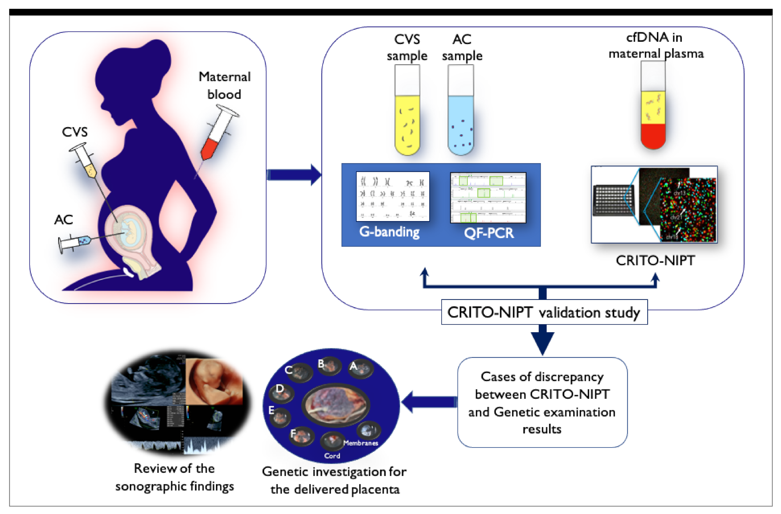
2. Materials and Methods
3. Results
3.1. Genetic Profile of 1218 Cases
3.2. Measures of CRITO-NIPT Diagnostic Accuracy
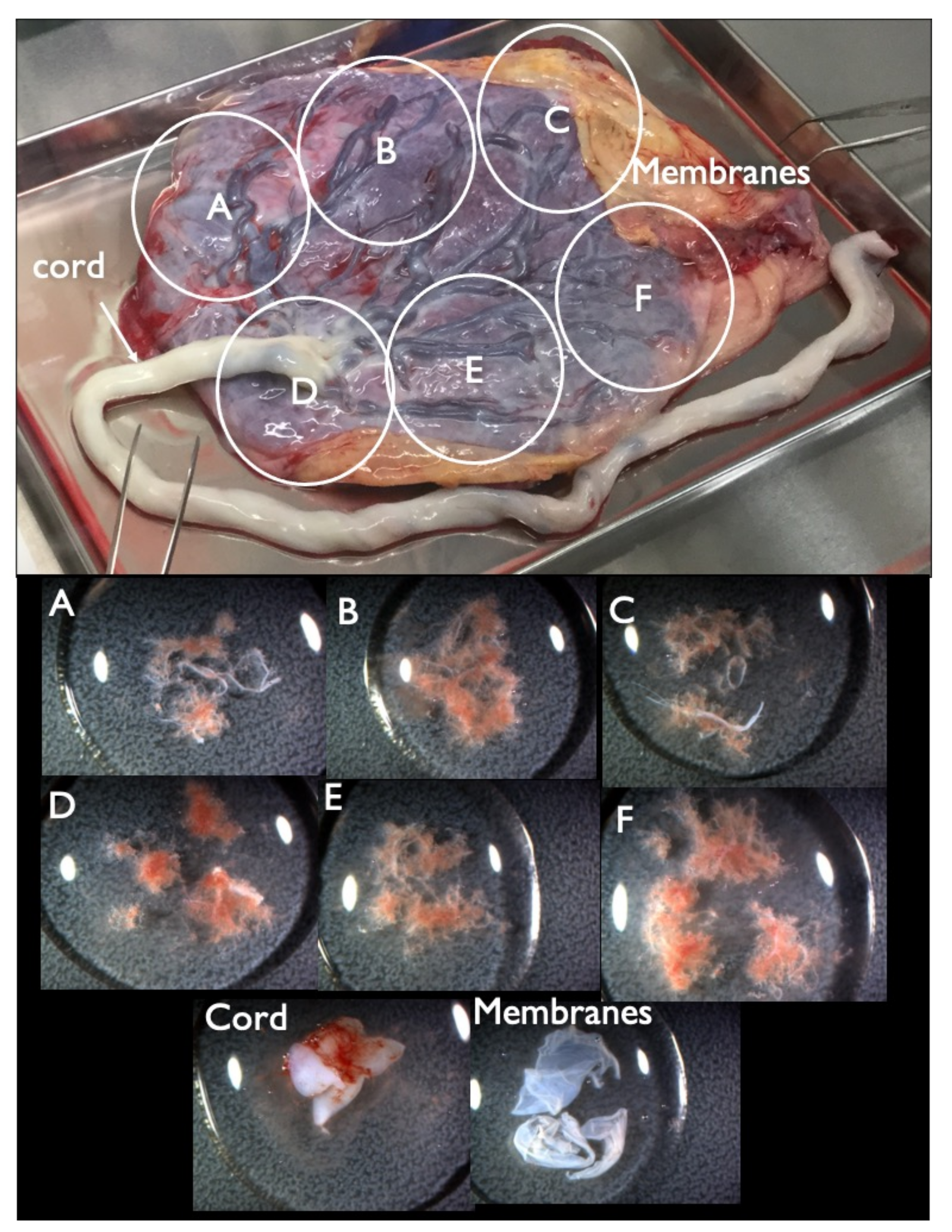
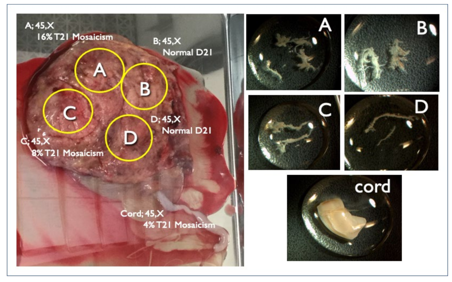
3.3. Placental Investigation Results in Cases with Discordancy between CRITO-NIPT and Genetic Results
3.3.1. False Positive (FP) CRITO-NIPT
3.3.2. False Negative CRITO-NIPT
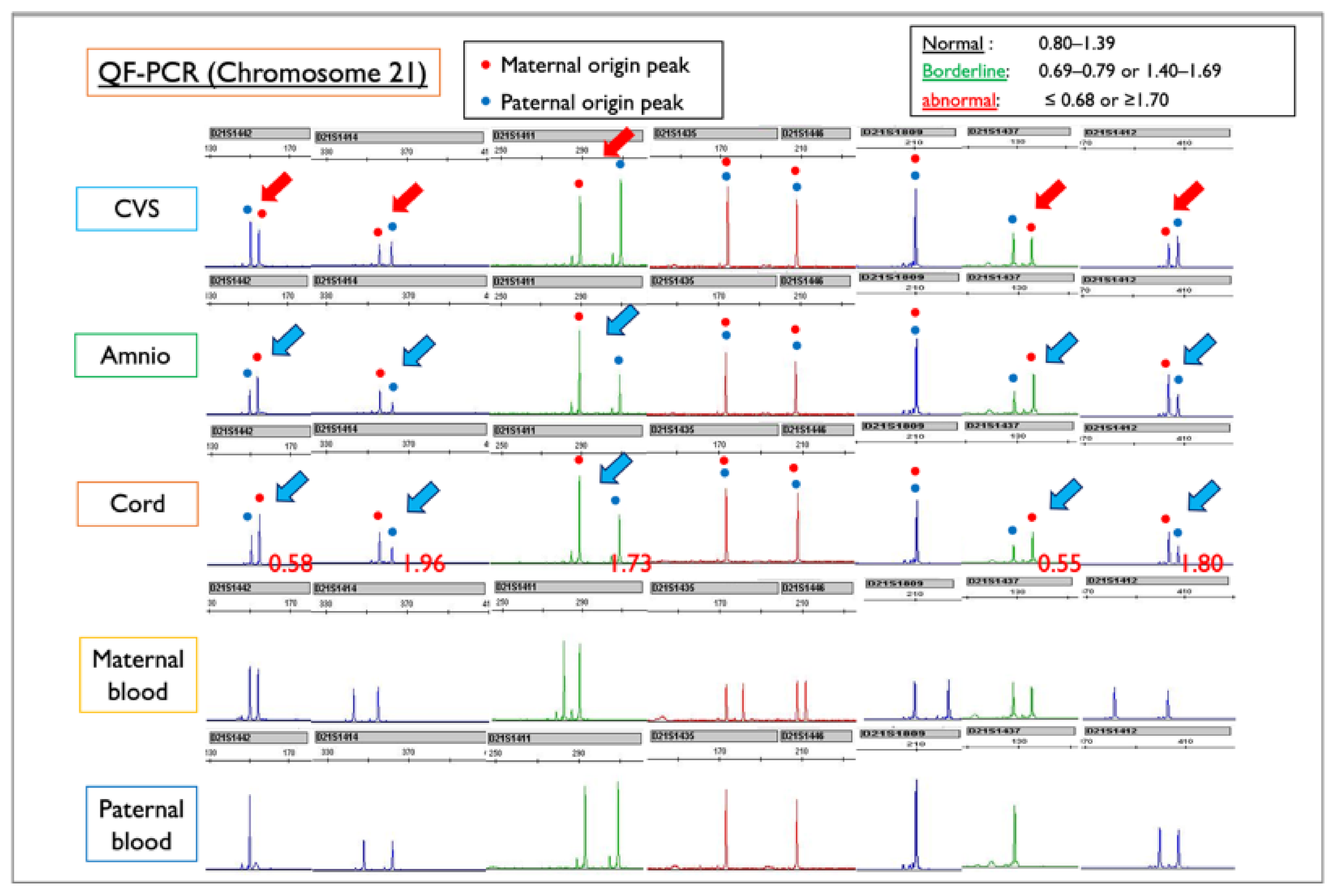
3.4. Investigation of Chromosome 21-, 18-, and 13-Relevant Mosaicism
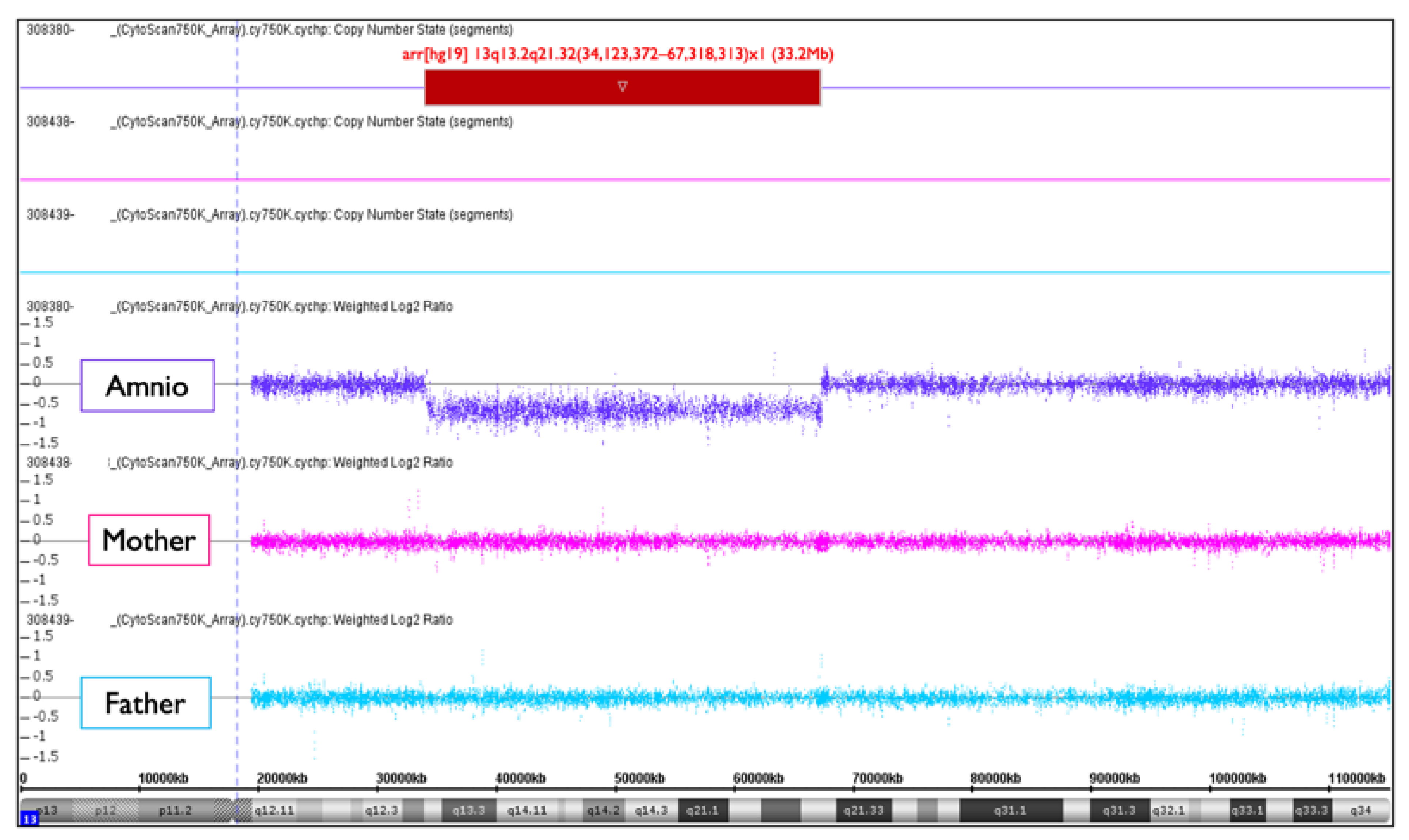
3.5. No-Call (NC) CRITO-NIPT
4. Discussion
4.1. Measures of CRITO-NIPT Diagnostic Accuracy
4.2. False Positive and False Negative CRITO-NIPT
4.3. Chromosome 21-, 18, and 13-Relevant Mosaicism
4.4. No-Call NIPT
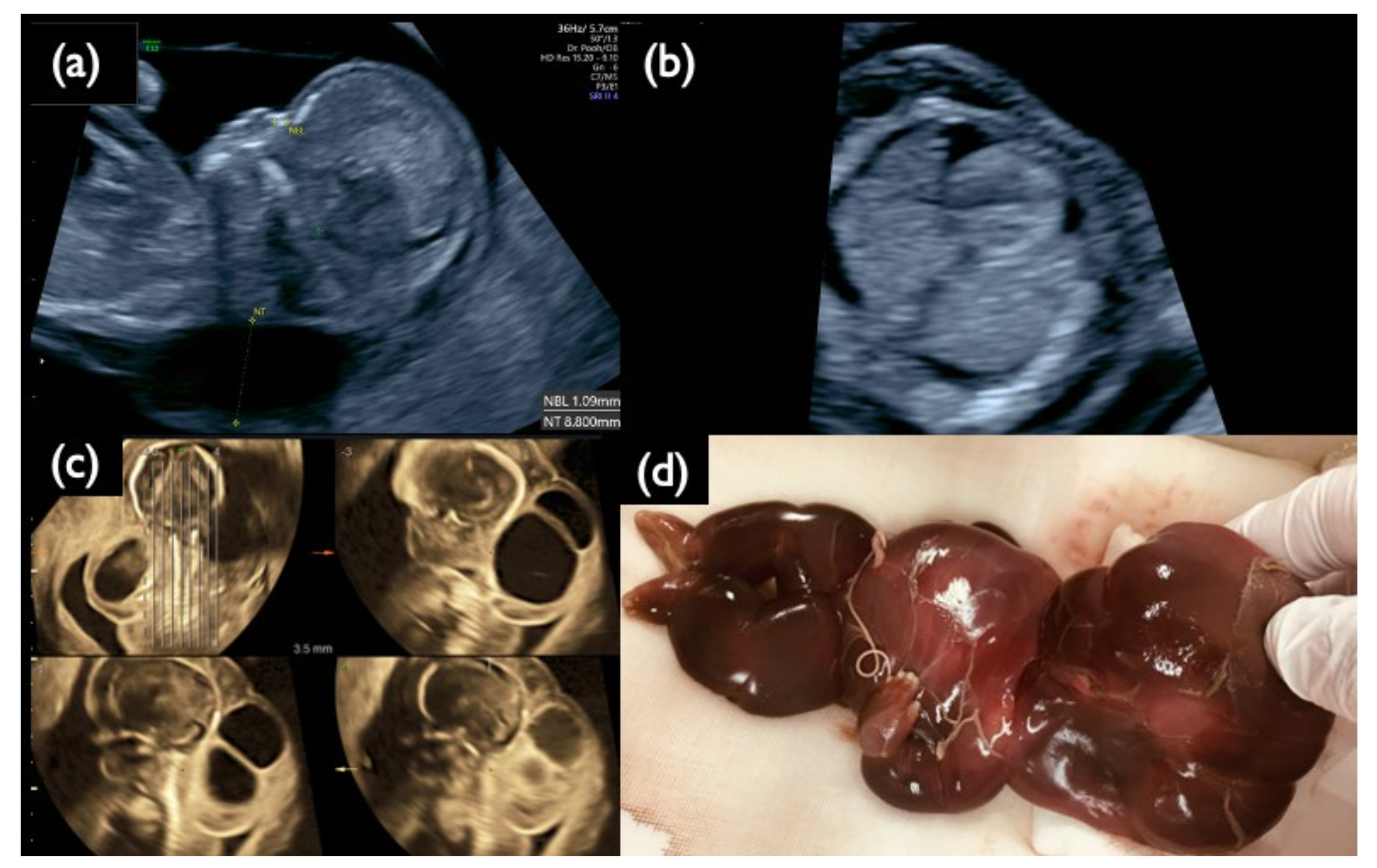
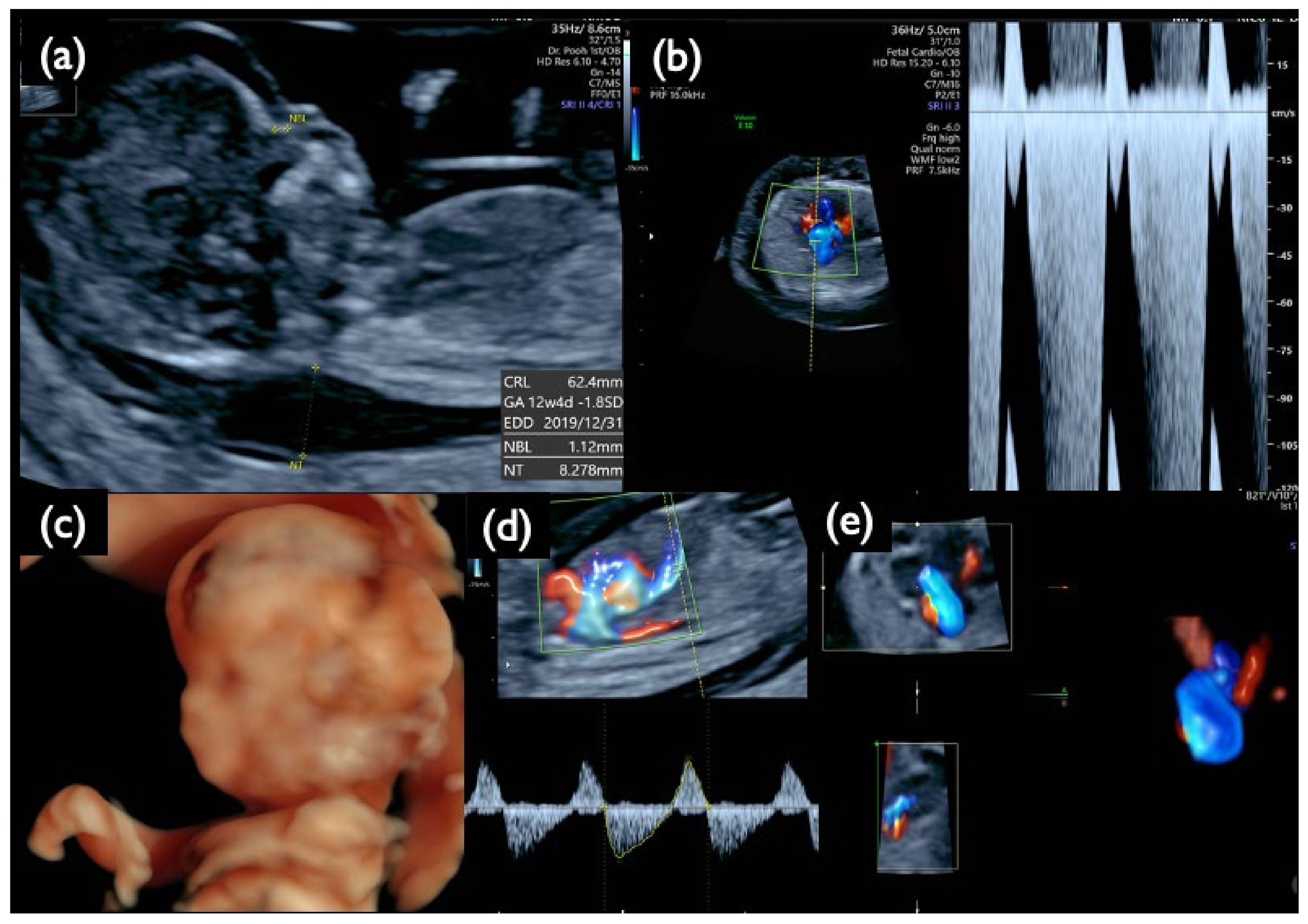
4.5. Significance of Fetal Sonography Prior to Genetic Testing
5. Conclusions
Author Contributions
Funding
Institutional Review Board Statement
Informed Consent Statement
Data Availability Statement
Acknowledgments
Conflicts of Interest
References
- Yin, A.H.; Peng, C.F.; Zhao, X.; Caughey, B.A.; Yang, J.X.; Liu, J.; Huang, W.W.; Liu, C.; Luo, D.H.; Liu, H.L.; et al. Noninvasive detection of fetal subchromosomal abnormalities by semiconductor sequencing of maternal plasma DNA. Proc. Natl. Acad. Sci. USA 2015, 112, 14670–14675. [Google Scholar] [CrossRef]
- Lefkowitz, R.B.; Tynan, J.A.; Liu, T.; Wu, Y.; Mazloom, A.R.; Almasri, E.; Hogg, G.; Angkachatchai, V.; Zhao, C.; Grosu, D.S.; et al. Clinical validation of a noninvasive prenatal test for genomewide detection of fetal copy number variants. Am. J. Obstet. Gynecol. 2016, 215, 227.e1–227.e16. [Google Scholar] [CrossRef]
- Zhang, J.; Li, J.; Saucier, J.B.; Feng, Y.; Jiang, Y.; Sinson, J.; McCombs, A.K.; Schmitt, E.S.; Peacock, S.; Chen, S.; et al. Noninvasive prenatal sequencing for multiple Mendelian monogenic disorders using circulating cell-free fetal DNA. Nat. Med. 2019, 25, 439–447. [Google Scholar] [CrossRef]
- Mellis, R.; Chandler, N.; Jenkins, L.; Chitty, L.S. The role of sonographic phenotyping in delivering an efficient noninvasive prenatal diagnosis service for FGFR3-related skeletal dysplasias. Prenat. Diagn. 2020, 785–791. [Google Scholar] [CrossRef]
- Yu, S.C.Y.; Jiang, P.; Chan, K.C.A.; Faas, B.H.W.; Choy, K.W.; Leung, W.C.; Leung, T.Y.; Lo, Y.M.D.; Chiu, R.W.K. Combined count- and size-based analysis of maternal plasma DNA for noninvasive prenatal detection of fetal subchromosomal aberrations facilitates elucidation of the fetal and/or maternal origin of the aberrations. Clin. Chem. 2017, 63, 495–502. [Google Scholar] [CrossRef][Green Version]
- Lo, K.K.; Karampetsou, E.; Boustred, C.; McKay, F.; Mason, S.; Hill, M.; Plagnol, V.; Chitty, L.S. Limited Clinical Utility of Noninvasive Prenatal Testing for Subchromosomal Abnormalities. Am. J. Hum. Genet. 2016, 98, 34–44. [Google Scholar] [CrossRef]
- Bianchi, D.W.; Chiu, R.W.K. Sequencing of Circulating Cell-free DNA during Pregnancy. N. Engl. J. Med. 2018, 379, 464–473. [Google Scholar] [CrossRef]
- Petersen, A.K.; Cheung, S.W.; Smith, J.L.; Bi, W.; Ward, P.A.; Peacock, S.; Braxton, A.; Van Den Veyver, I.B.; Breman, A.M. Positive predictive value estimates for cell-free noninvasive prenatal screening from data of a large referral genetic diagnostic laboratory. Am. J. Obstet. Gynecol. 2017, 217, 691.e1–691.e6. [Google Scholar] [CrossRef]
- Snyder, M.W.; Simmons, L.V.E.; Kitzman, J.O.; Coe, B.P.; Henson, J.M.; Daza, R.M.; Eichler, E.E.; Shendure, J.; Gammill, H.S. Copy-number variation and false positive prenatal aneuploidy screening results. N. Engl. J. Med. 2015, 372, 1639–1645. [Google Scholar] [CrossRef]
- Bianchi, D.W.; Chudova, D.; Sehnert, A.J.; Bhatt, S.; Murray, K.; Prosen, T.L.; Garber, J.E.; Wilkins-Haug, L.; Vora, N.L.; Warsof, S.; et al. Noninvasive prenatal testing and incidental detection of occult maternal malignancies. JAMA 2015, 314, 162–169. [Google Scholar] [CrossRef]
- Taylor-Phillips, S.; Freeman, K.; Geppert, J.; Agbebiyi, A.; Uthman, O.A.; Madan, J.; Clarke, A.; Quenby, S.; Clarke, A. Accuracy of noninvasive prenatal testing using cell-free DNA for detection of Down, Edwards and Patau syndromes: A systematic review and meta-analysis. BMJ Open 2016, 6, e010002. [Google Scholar] [CrossRef]
- Gil, M.M.; Accurti, V.; Santacruz, B.; Plana, M.N.; Nicolaides, K.H. Analysis of cell-free DNA in maternal blood in screening for aneuploidies: Updated meta-analysis. Ultrasound Obstet. Gynecol. 2017, 50, 302–314. [Google Scholar] [CrossRef] [PubMed]
- Dahl, F.; Ericsson, O.; Karlberg, O.; Karlsson, F.; Howell, M.; Persson, F.; Roos, F.; Stenberg, J.; Ahola, T.; Alftrén, I.; et al. Imaging single DNA molecules for high precision NIPT. Sci. Rep. 2018, 8, 1–8. [Google Scholar] [CrossRef]
- Ericsson, O.; Ahola, T.; Dahl, F.; Karlsson, F.; Persson, F.; Karlberg, O.; Roos, F.; Alftrén, I.; Andersson, B.; Barkenäs, E.; et al. Clinical validation of a novel automated cell-free DNA screening assay for trisomies 21, 13, and 18 in maternal plasma. Prenat. Diagn. 2019, 39, 1011–1015. [Google Scholar] [CrossRef] [PubMed]
- Brar, H.; Wang, E.; Struble, C.; Musci, T.J.; Norton, M.E. The fetal fraction of cell-free DNA in maternal plasma is not affected by a priori risk of fetal trisomy. J. Matern. Neonatal Med. 2013, 26, 143–145. [Google Scholar] [CrossRef]
- Suzumori, N.; Ebara, T.; Yamada, T.; Samura, O.; Yotsumoto, J.; Nishiyama, M.; Miura, K.; Sawai, H.; Murotsuki, J.; Kitagawa, M.; et al. Fetal cell-free DNA fraction in maternal plasma is affected by fetal trisomy. J. Hum. Genet. 2016, 61, 647–652. [Google Scholar] [CrossRef] [PubMed]
- Romero, R.; Mahoney, M.J. Noninvasive prenatal testing and detection of maternal cancer. JAMA 2015, 314, 131–133. [Google Scholar] [CrossRef]
- Hartwig, T.S.; Ambye, L.; Sorensen, S.; Jorgensen, F.S. Discordant noninvasive prenatal testing (NIPT)—A systematic review. Prenat Diagn 2017, 37, 527–539. [Google Scholar] [CrossRef]
- Zhang, H.; Gao, Y.; Jiang, F.; Fu, M.; Yuan, Y.; Guo, Y.; Zhu, Z.; Lin, M.; Liu, Q.; Tian, Z.; et al. Noninvasive prenatal testing for trisomies 21, 18 and 13: Clinical experience from 146 958 pregnancies. Ultrasound Obstet. Gynecol. 2015, 45, 530–538. [Google Scholar] [CrossRef]
- Mackie, F.L.; Hemming, K.; Allen, S.; Morris, R.K.; Kilby, M.D. The accuracy of cell-free fetal DNA-based noninvasive prenatal testing in singleton pregnancies: A systematic review and bivariate meta-analysis. BJOG An Int. J. Obstet. Gynaecol. 2017, 124, 32–46. [Google Scholar] [CrossRef]
- Nguyen, H.P.; Riess, A.; Krüger, M.; Bauer, P.; Singer, S.; Schneider, M.; Enders, H.; Dufke, A. Mosaic trisomy 21/monosomy 21 in a living female infant. Cytogenet. Genome Res. 2009, 125, 26–32. [Google Scholar] [CrossRef]
- Ashoor, G.; Syngelaki, A.; Poon, L.C.Y.; Rezende, J.C.; Nicolaides, K.H. Fetal fraction in maternal plasma cell-free DNA at 11-13 weeks’ gestation: Relation to maternal and fetal characteristics. Ultrasound Obstet. Gynecol. 2013, 41, 26–32. [Google Scholar] [CrossRef] [PubMed]
- Chan, N.; Smet, M.-E.; Sandow, R.; da Silva Costa, F.; McLennan, A. Implications of failure to achieve a result from prenatal maternal serum cell-free DNA testing: A historical cohort study. BJOG Int. J. Obstet. Gynaecol. 2018, 125, 848–855. [Google Scholar] [CrossRef]
- Kruckow, S.; Schelde, P.; Hatt, L.; Ravn, K.; Petersen, O.B.; Uldbjerg, N.; Vogel, I.; Singh, R. Does maternal body mass index affect the quantity of circulating fetal cells available to use for cell-based noninvasive prenatal test in high-risk pregnancies? Fetal Diagn. Ther. 2019, 45, 353–356. [Google Scholar] [CrossRef] [PubMed]
- Kater-Kuipers, A.; Bunnik, E.M.; De Beaufort, I.D.; Galjaard, R.J.H. Limits to the scope of noninvasive prenatal testing (NIPT): An analysis of the international ethical framework for prenatal screening and an interview study with Dutch professionals 11 Medical and Health Sciences 1117 Public Health and Health Services. BMC Pregnancy Childbirth 2018, 18, 1–14. [Google Scholar] [CrossRef]
- Haidar, H.; Vanstone, M.; Laberge, A.M.; Bibeau, G.; Ghulmiyyah, L.; Ravitsky, V. Implementation challenges for an ethical introduction of noninvasive prenatal testing: A qualitative study of healthcare professionals’ views from Lebanon and Quebec. BMC Med. Ethics 2020, 21, 1–11. [Google Scholar] [CrossRef] [PubMed]
- Zaami, S.; Orrico, A.; Signore, F.; Cavaliere, A.F.; Mazzi, M.; Marinelli, E. Ethical, legal and social issues (Elsi) associated with noninvasive prenatal testing: Reflections on the evolution of prenatal diagnosis and procreative choices. Genes 2021, 12, 204. [Google Scholar] [CrossRef]


| Chorionic Numbers Detected by Sonography | Case Number | % |
|---|---|---|
| Singleton | 1166 | 95.73% |
| DCDA twin | 22 | 1.81% |
| MCDA twin | 10 | 0.82% |
| MCDA vanishing twin with empty second amniotic sac | 3 | 0.25% |
| DCDA vanishing twin with an empty sac | 12 | 0.99% |
| DCDA vanishing twin with a nonviable fetus | 5 | 0.41% |
| Total | 1218 | 100% |
| Genetic Abnormality | Case Number | % |
|---|---|---|
| T21 | 60 | 32.6 |
| T18 | 47 | 25.5 |
| T13 | 12 | 6.5 |
| T21/T18/T13 Mosaicism | 7 | 3.8 |
| Sex Chromosome aneuploidy | 16 | 8.7 |
| Triploidy | 1 | 0.5 |
| Aneuploidy Mosaicism*1 | 12 | 6.5 |
| Structural abnormality*2 | 5 | 2.7 |
| Structural Mosaicism*3 | 1 | 0.5 |
| Pathogenic CNV*4 | 10 | 5.4 |
| Single gene mutation*5 | 13 | 7.1 |
| Total | 184 | 100 |
| Sensitivity | Specificity | PPV | NPV | |
|---|---|---|---|---|
| T21 | 98.31% (58/59) | 99.65% (1145/1149) | 93.55% (58/62) | 99.91% (1145/1146) |
| T18 | 100.00% (46/46) | 99.48% (1156/1162) | 88.46% (46/52) | 100.00% (1156/1156) |
| T13 | 100.00% (12/12) | 100.00% (1196/1196) | 100.00% (12/12) | 100.00% (1196/1196) |
| Case | CVS G–Band | CRITO Result | CRITO Z–Score | Placental Part | Cord | Membranes | Maternal Blood | Causal Factor of FP/FN Result | Increased NT | NT (mm) | Sonographic Findings | |||||
|---|---|---|---|---|---|---|---|---|---|---|---|---|---|---|---|---|
| A | B | C | D | E | F | |||||||||||
| FP1 | 45,X | T21 positive | 3.55 | MX+ T21 (16%) | MX+D21 | MX+ T21 (8%) | MX+D21 | – | – | MX+ T21 (4%) | 46,XX | CPM | + | 8.8 | Increased NT, CH, General edema, PE bilateral, Small NB, Tachycardia, Short FL/HL, Turner is strongly suspected | |
| FP2 | 46,XX | T21 positive | 4.18 | D21 | D21 | D21 | D21 | D21 | D21 | D21 | D21 | 46,XX | s/o nonviable twin with T21 | – | 2.2 | TR mild, DCDA vanishing twin with a nonviable fetus |
| FP3 | 46,XY | T21 positive | 7.52 | D21 | D21 | D21 | D21 | D21 | D21 | D21 | T21 (14–74%) * | 46,XX | nonviable twin with T21 | – | 1.4 | Small NB, TR mild, DCDA vanishing twin with a nonviable fetus |
| FP4 | 46,XY | T21 positive | 7.78 | D21 | D21 | D21 | D21 | T21 (15%) | D21 | D21 | D21 | 46,XX | CPM | – | 2.2 | Small NB, TR mild |
| FP5 | 46,XX | T18 positive | 4.20 | D18 | D18 | D18 | D18 | D18 | D18 | D18 | D18 | 46,XX | unclear | – | 2.1 | TR mild, DV reverse |
| FP6 | 46,XX | T18 positive | 12.76 | D18 | D18 | D18 | D18 | D18 | D18 | D18 | D18 | 46,XX | unclear | – | 2.2 | TR mild–moderate |
| FP7 | 46,XY | T18 positive | 5.49 | D18 | D18 | D18 | D18 | – | – | D18 | D18 | 46,XX | unclear | – | 1.6 | TR mild |
| FP8 | 46,XY | T18 positive | 3.56 | D18 | D18 | D18 | D18 | D18 | D18 | D18 | D18 | 46,XX | unclear | – | 2.0 | TR mild, SFD, SCH |
| FP9 | 46,XX | T18 positive | 3.22 | D18 | D18 | D18 | D18 | D18 | D18 | D18 | D18 | 46,XX | unclear | – | 1.2 | no particular findings |
| FP10 | 46,XX | T18 positive | 3.02 | – | – | – | – | – | – | – | – | 46,XX | unclear | – | 1.8 | TR mild |
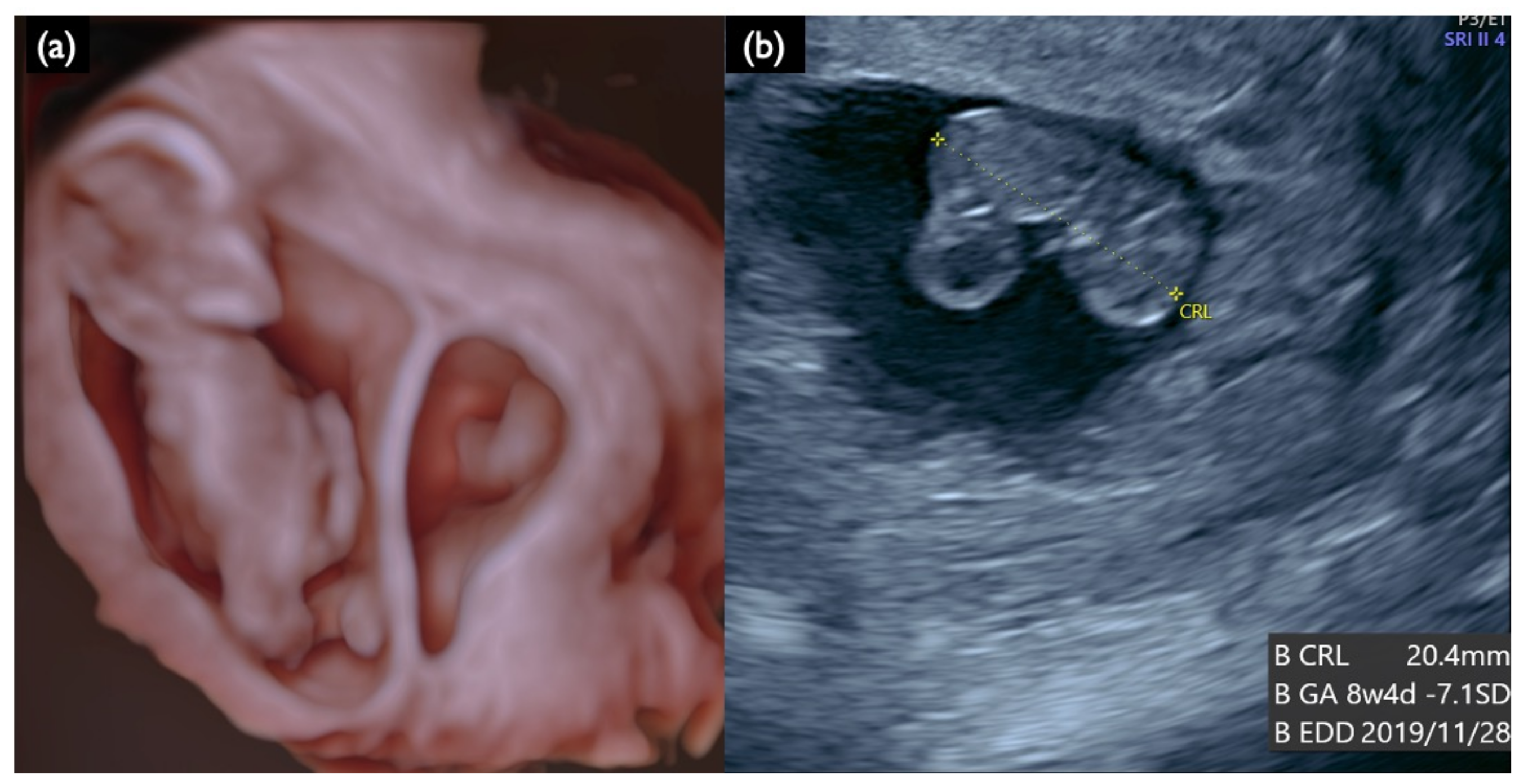
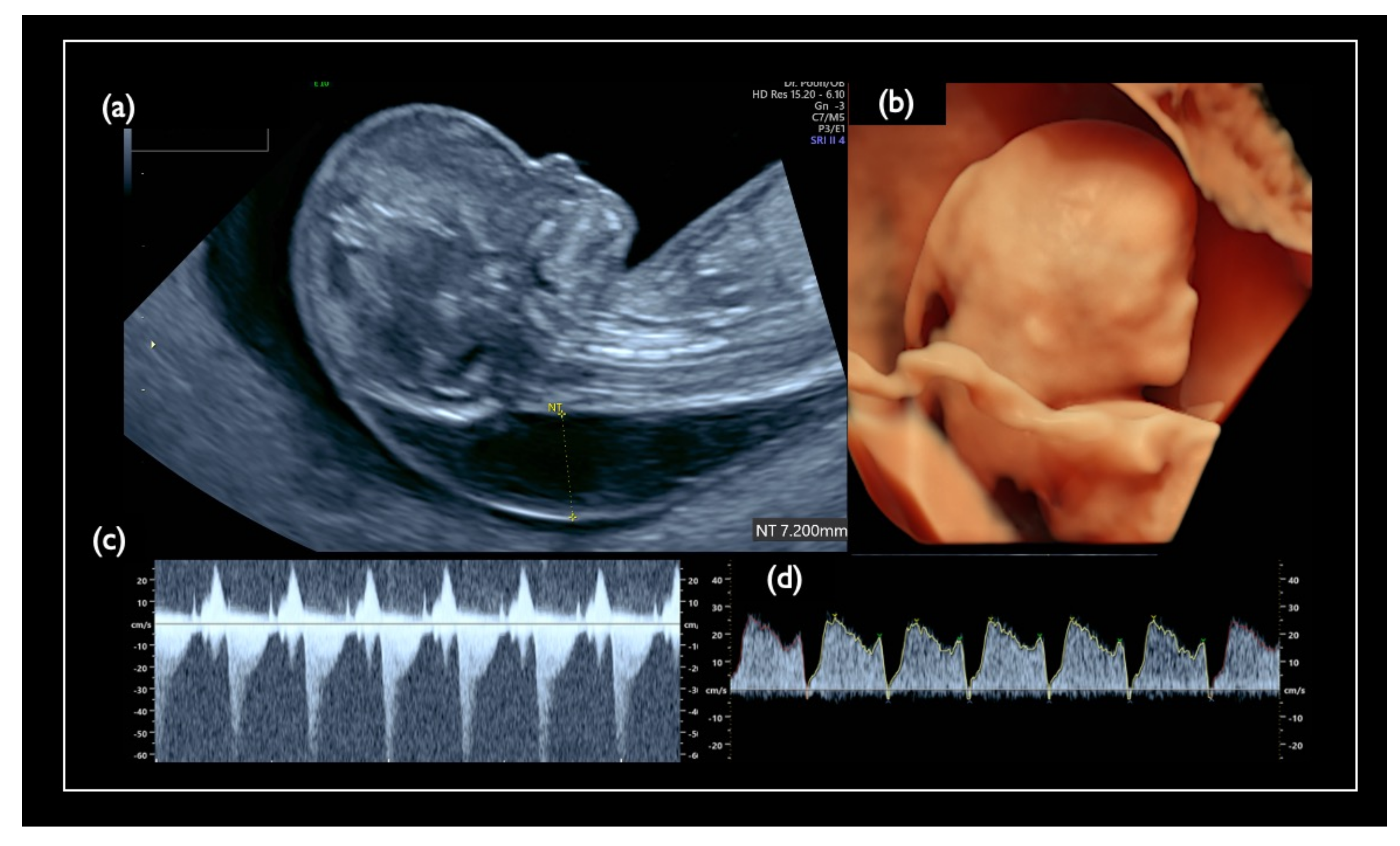
| FN1 | 47,XY,+21 | T21 negative | 1.07 | – | – | – | – | – | – | – | – | 46,XX | unclear | + | 7.2 | Increased NT, GE mild, NB defect, Lowset ear, TR mild, Small stomach, DV reverse, Tachycardia, T21 is strongly suspected |
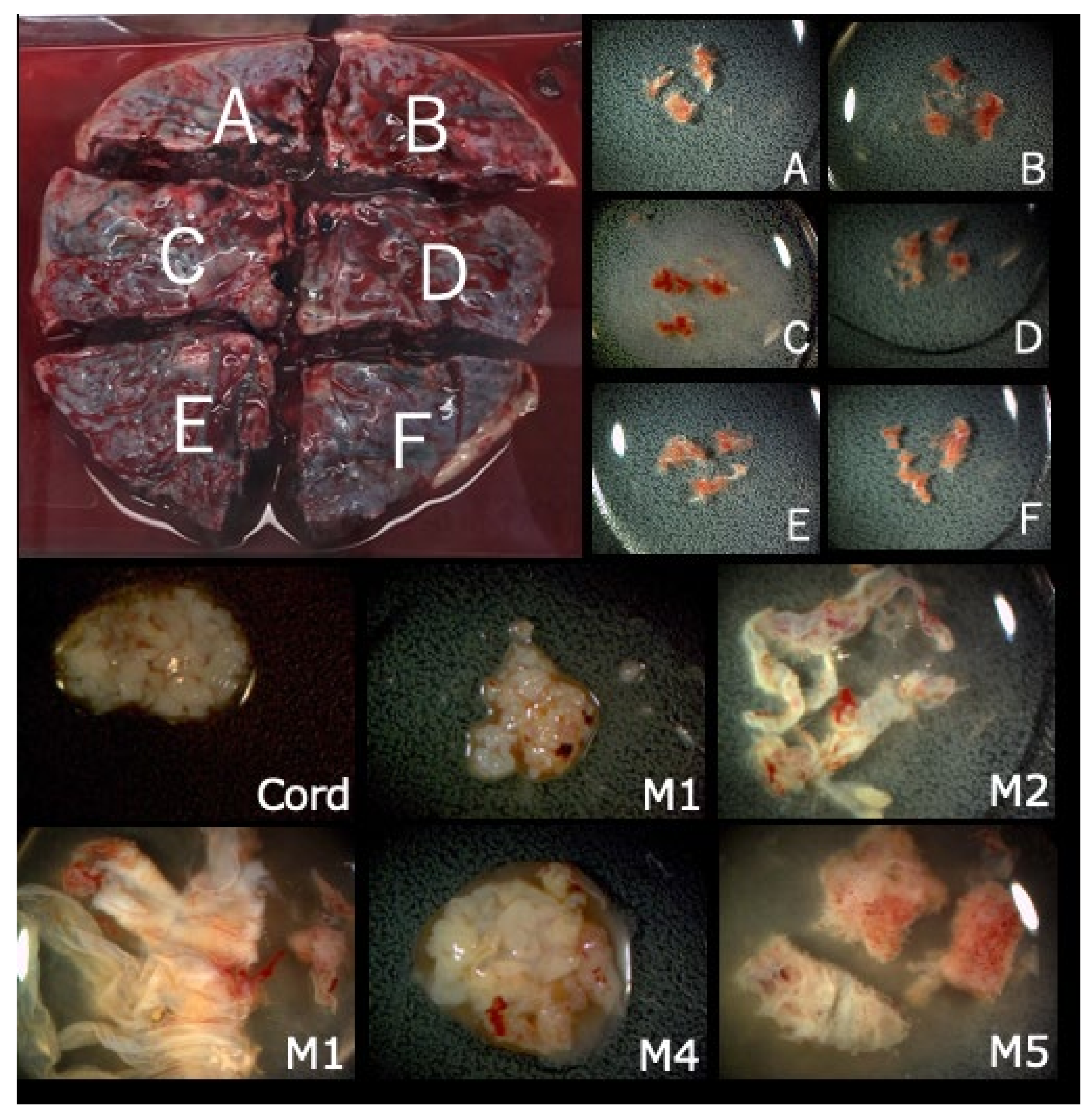
| Case FP3–Specimen | FISH | G–Band | |
|---|---|---|---|
| Disomy 21 | Trisomy 21 | ||
| Placenta–A | 100% | 0% | 46,XY |
| Placenta–B | 100% | 0% | culture failure |
| Placenta–C | 100% | 0% | 46,XY |
| Placenta–D | 100% | 0% | 46,XY |
| Placenta–E | 100% | 0% | 46,XY |
| Placenta–E | 100% | 0% | 46,XY |
| Cord | 100% | 0% | 46,XY |
| Membranes–1 | 86% | 14% | – |
| Membranes–2 | 90% | 10% | – |
| Membranes–3 | 16% | 74% | 47,XY,+21 |
| Membranes–4 | 100% | 0% | – |
| Membranes–5 | 100% | 0% | – |
| Case | GA (Week + Day) for US and CVS | CVS | AC | CRITO–NIPT | Fetal Ultrasonography | |||||||
|---|---|---|---|---|---|---|---|---|---|---|---|---|
| QF–PCR | Uncultured FISH Aneuploidy (%) | G–Band Karyotype | Uncultured FISH Aneuploidy (%) | G–Band Karyotype | Result | Z–Score | Increased NT | NT Thickness (mm) | Sonographic Findings | |||
| Chromosome 21 relevant mosaicism | Mo1 | 13 + 3 | XY,+21 Mosaicism | T21 (96%) | 47,XY,+21 (100%) | – | – | T21 positive | 22.05 | + | 4.6 | Increased NT, GE very mild, Micrognathia, Lowset ear, DS like profile, Large VSD, TR moderate, Hyperechoic bowel, DV reverse, Tachycardia, Straight cord, T21 is strongly suspected |
| Mo2 | 13 + 2 | XX,+21 Mosaicism | Tetrasomy 21 (20%) | 49,XX,+7,+21,+21 /46,XX (72%/28%) | – | – | T21 Negative | 1.11 | – | 2.1 | Small NB, Lowset ear, TR moderate | |
| Mo3 | 13 + 0 | XXY | T21 (2%) | 47,XXY /48,XXY,+21 (62%/38%) | T21 (2%) | 47,XXY | T21 Negative | –0.48 | + | 3.4 | Increased NT, Small NB, TR mild, SUA, Genetic disorder is suspected | |
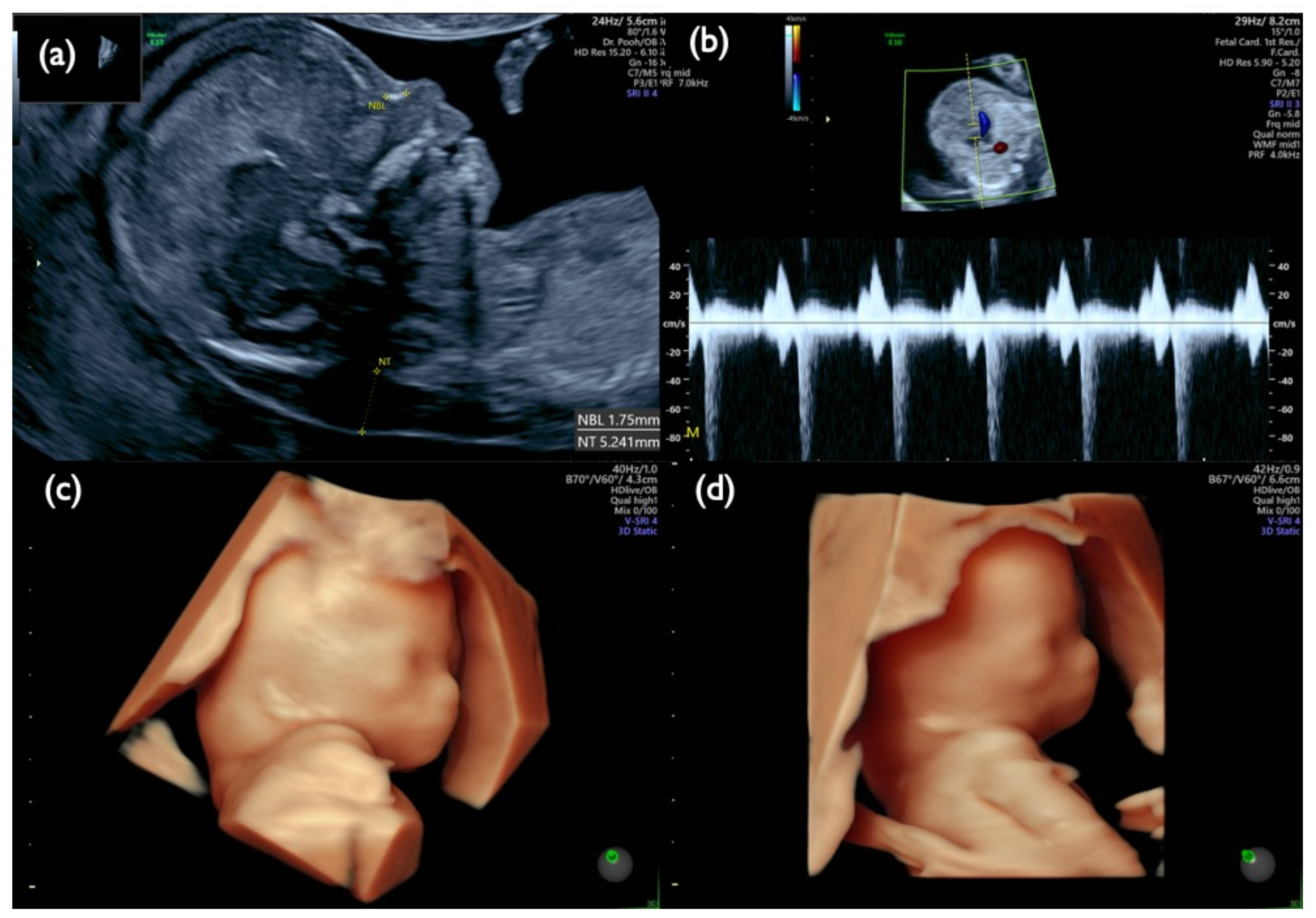
| Mo4 | 13 + 3 | XX,+21 Mosaicism | M21 (72%) T21 (8%) | 45,XX,–21 (100%) | M21 (1%) T21 (91%) | 46,XX,i(21)(q10) | T21 positive | 10.87 | + | 5.2 | Increased NT, Small NB, Micrognathia, Lowset ear, TR mild, DV defect, T21 is strongly suspected | |
| Chromosome 18 relevant mosaicism | Mo5 | 13 + 5 | XX,+18 Mosaicism | T18 (56%) | 47,XX,+18/46,XX (44%/56%) | – | – | T18 Negative | 2.09 | + | 8.3 | Increased NT, GE moderate, Small NB, Micrognathia, Lowset ear, T18 like profile, Wrist contracture bilateral, Cardiomegaly, Large VSD, TR severe, MR severe, Stomach invisible, Hyperechoic bowel, DV reversed flow, SUA, Umb.A.reverse, T18 is strongly suspected, |
| Mo6 | 13 + 0 | XY,T18 Mosaicism | T18 (88%) | 47,XY,+18 (100%) | – | – | T18 positive | 7.05 | + | 2.9 | Increased NT, NB defect, T18 like profile, Micrognathia, Lowset ear, Hypoplastic ear, Wrist contracture bilateral, Contracted lower extremities, Club foot (right), s/o DORV, RV>LV, TR moderate–severe, Omphalocele containing only bowel, DV reversed flow, FGR, T18 is strongly suspected | |
| Chromosome 13 relevant mosaicism | Mo7 | 12 + 3 | Normal XY | T13 (6%) | 46,XY (100%) | T13 (0%) | T13 (0%) | T13 Negative | 1.25 | – | 2.0 | TR mild |
| Case Mo2–Specimen | QF–PCR | FISH | G–Band | ||
|---|---|---|---|---|---|
| Trisomy 7 + Tetrasomy 21 | Disomy 7 + Disomy 21 | Disomy 7 + Monosomy 21 | |||
| CVS | s/o T21 mosaicism | 20% | 80% | 0% | 49,XX,+7,+21,+21/46,XX (72%/28%) |
| Placenta–A | s/o T21 mosaicism | 24% | 76% | 0% | 46,XX (100%) |
| Placenta–B | s/o T21 mosaicism | 0% | 90% | 10% | 49,XX,+7,+21,+21/46,XX (15%/85%) |
| Placenta–C | s/o T21 mosaicism | 10% | 80% | 10% | 49,XX,+7,+21,+21/45,XX,–21/46,XX (5%/10%/85 %) |
| Placenta–D | s/o T21 mosaicism | 20% | 70% | 10% | 49,XX,+7,+21,+21/46,XX (10%/90%) |
| Placenta–E | s/o T21 mosaicism | 24% | 64% | 12% | 49,XX,+7,+21,+21/46,XX (10%/90%) |
| Cord | Normal XX | 0% | 100% | 0% | 46,XX (100%) |
| Membranes | Inconclusive | 20% | 74% | 6% | 49,XX,+7,+21,+21/46,XX (5%/95%) |
| Cord blood | Normal XX | 0% | 100% | 0% | 46,XX (100%) |
| Case Mo4–Specimen | QF–PCR | FISH | G–Band | ||
|---|---|---|---|---|---|
| Monosomy 21 | Disomy 21 | Trisomy 21 | |||
| CVS | s/o low level T21 mosaicism | 72% | 20% | 8% | 45,XX,–21 (100%) |
| Amniotic cell | T21 | 1% | 8% | 91% | 46,XX,i(21)(q10) (100%) |
| Placenta–A | s/o low level T21 mosaicism | 56% | 10% | 34% | 45,XX,–21/46,XX,i(21)(q10) (95.5%/4.5%) |
| Placenta–B | s/o middle level T21 mosaicism | 68% | 18% | 14% | |
| Placenta–C | s/o low level T21 mosaicism | 60% | 14% | 26% | |
| Placenta–D | s/o low level T21 mosaicism | 64% | 8% | 28% | |
| Cord | s/o high level T21 mosaicism | 6% | 8% | 86% | 46,XX,i(21)(q10) (100%) |
| Membranes | s/o high level T21 mosaicism | 34% | 16% | 50% | 45,XX,–21/46,XX,i(21)(q10) (2%/98%) |
| Maternal blood | normal | – | – | – | 46,XX |
| Paternal blood | normal | – | – | – | 46,XY |
| Case | GA | Invasive Procedure | BMI | G–Band Result | CRITO-NIPT | Increased NT | NT (mm) | Sonographic Findings |
|---|---|---|---|---|---|---|---|---|
| NC1 | 30w0d | AC | 21.1 | 46,XY,del(13)(q13q21.3) | No Call | – | – | Thick prenasal skin, Low nasal bridge, Small NB, Micrognathia, Hypertrichosis, TR, Undescendant testis (left), Small mid phalanx of 5th digit (bilateral), Large head, Increased AF (AFI 23.68cm), T21 or Genetic disorder such as Costello or Cornelia de lange syndrome is suspected |
| NC2 | 12w2d | CVS | 22.8 | 47,XY,+21 | No Call | + | 5.3 | Increased NT, Small CH, GE, bilateral, PE, NB defect, Micrognathia, Lowset ear, s/o Large VSD, TR moderate, Levocardia, RV>LV, Hyperechoic bowel, DV reverse, Short FL, T 21 is strongly suspected |
| NC3 | 13w1d | CVS | 25.8 | 47,XY,+18 | No Call | + | 10.6 | Increased NT, CH, GE, Small NB, Micrognathia, Lowset ear, Hypoplastic ear, Cleft lip (left), Maxillary gap, Mild wrist contracture bilateral, RV>LV, TR, DV reverse, Bradycardia, T18 is strongly suspected |
Publisher’s Note: MDPI stays neutral with regard to jurisdictional claims in published maps and institutional affiliations. |
© 2021 by the authors. Licensee MDPI, Basel, Switzerland. This article is an open access article distributed under the terms and conditions of the Creative Commons Attribution (CC BY) license (https://creativecommons.org/licenses/by/4.0/).
Share and Cite
Pooh, R.K.; Masuda, C.; Matsushika, R.; Machida, M.; Nakamura, T.; Takeda, M.; Ohashi, H.; Kumagai, M.; Uenishi, K.; Roos, F.; et al. Clinical Validation of Fetal cfDNA Analysis Using Rolling-Circle-Replication and Imaging Technology in Osaka (CRITO Study). Diagnostics 2021, 11, 1837. https://doi.org/10.3390/diagnostics11101837
Pooh RK, Masuda C, Matsushika R, Machida M, Nakamura T, Takeda M, Ohashi H, Kumagai M, Uenishi K, Roos F, et al. Clinical Validation of Fetal cfDNA Analysis Using Rolling-Circle-Replication and Imaging Technology in Osaka (CRITO Study). Diagnostics. 2021; 11(10):1837. https://doi.org/10.3390/diagnostics11101837
Chicago/Turabian StylePooh, Ritsuko Kimata, Chika Masuda, Risa Matsushika, Megumi Machida, Takako Nakamura, Masayoshi Takeda, Hiroyasu Ohashi, Mami Kumagai, Kohtaro Uenishi, Fredrik Roos, and et al. 2021. "Clinical Validation of Fetal cfDNA Analysis Using Rolling-Circle-Replication and Imaging Technology in Osaka (CRITO Study)" Diagnostics 11, no. 10: 1837. https://doi.org/10.3390/diagnostics11101837
APA StylePooh, R. K., Masuda, C., Matsushika, R., Machida, M., Nakamura, T., Takeda, M., Ohashi, H., Kumagai, M., Uenishi, K., Roos, F., Persson, F., & Shimokawa, O. (2021). Clinical Validation of Fetal cfDNA Analysis Using Rolling-Circle-Replication and Imaging Technology in Osaka (CRITO Study). Diagnostics, 11(10), 1837. https://doi.org/10.3390/diagnostics11101837