Lung and Intercostal Upper Abdomen Ultrasonography for Staging Patients with Ovarian Cancer: A Method Description and Feasibility Study

 ,
,  ,
,  ,
, 
Abstract
1. Introduction
2. Materials and Methods
2.1. Patients
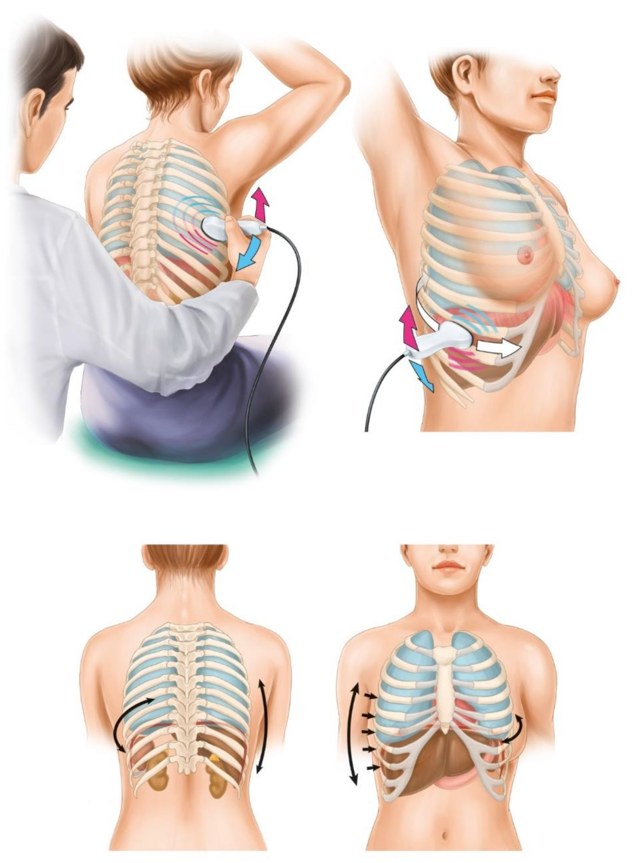
2.2. Imaging Technique
2.2.1. Lung and Intercostal upper Abdomen Ultrasonography
2.2.2. Transabdominal and Transvaginal Ultrasonography
2.3. Clinical Data
2.4. Feasibility Study
2.5. Statistical Analysis
3. Results
3.1. Patients
3.2. Imaging
3.3. Clinical Implementation
3.4. Feasibility Study
4. Discussion
5. Conclusions
Supplementary Materials
Author Contributions
Funding
Acknowledgments
Conflicts of Interest
Abbreviations
| CT | computed tomography |
| DLSK | diagnostic laparoscopy |
| ICAUS | intercostal upper abdomen ultrasonography |
| IDS | interval debulking surgery |
| LUS | lung ultrasonography |
| MRI | magnetic resonance imaging |
| NACT | neoadjuvant chemotherapy |
| OC | ovarian cancer |
| PDS | primary debulking surgery |
| TAS | transabdominal ultrasonography |
| TVS | transvaginal ultrasonography |
Appendix A
Appendix B

| No. | Age | PS | TAS/TVS | LUS/ICAUS | CT | Upstaging (IIIC → IV) with LUS/ICAUS Added to TAS/TVS? | Additional Procedures Planned after LUS/ICAUS? | LUS/ICAUS Added to TAS/TVS Changed Predicted Surgical Complexity [2]? | VATS Necessary? | Figure VS | Management /Comments |
|---|---|---|---|---|---|---|---|---|---|---|---|
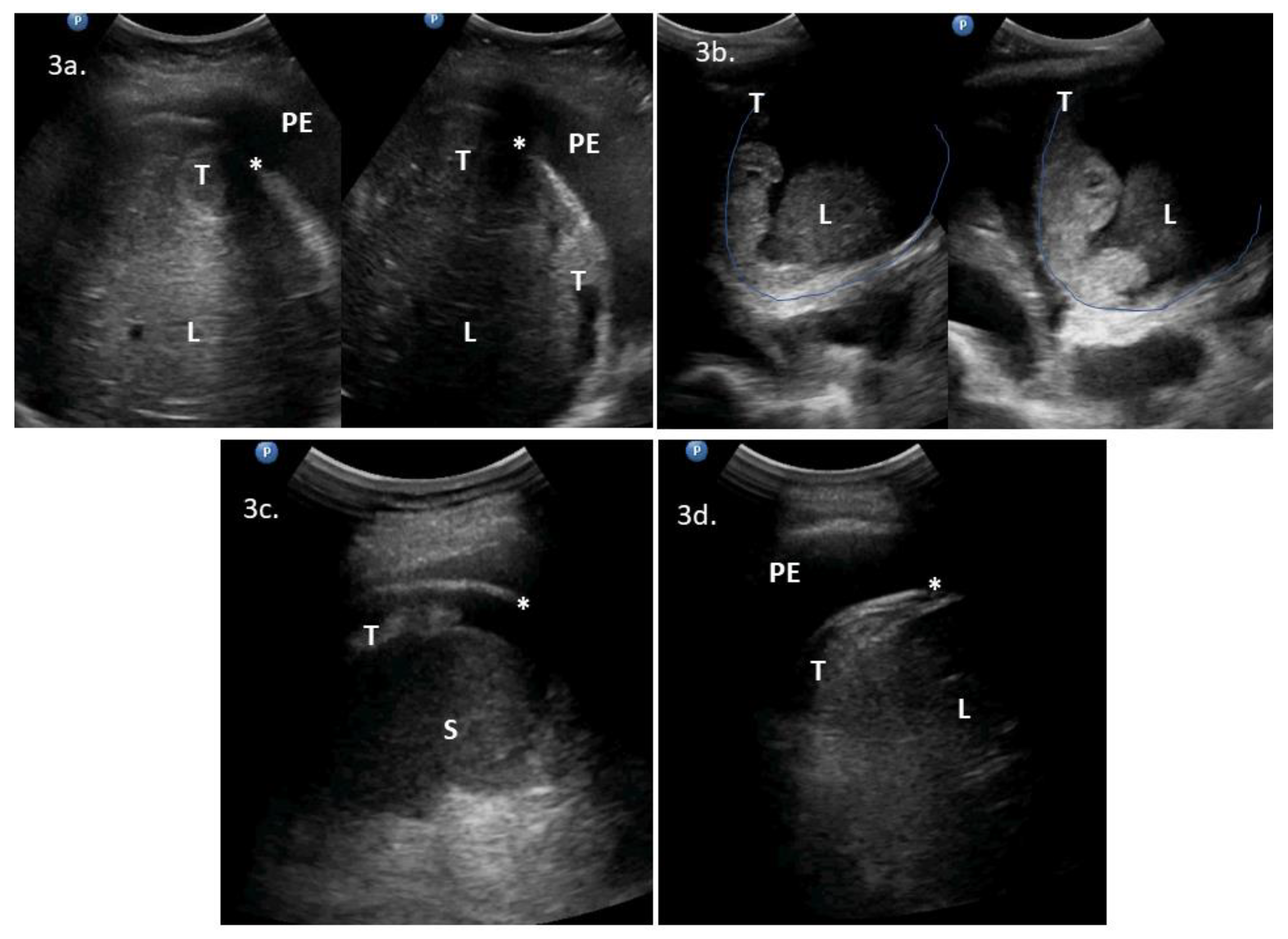
| 1 | 71 | 3 | ascites, massive pelvic involvement, omental involvement, spleen involvement | pleural diaphragm involvement *, pleural effusions | ascites, carcinomatosis, massive pelvic involvement, omental involvement, pleural effusions, spleen involvement | Yes | Yes | NoScore: 9 → 11 High → High | Could replace DLSK | 2a | DLSK, HGSOC, NACT |
| 2 | 62 | 1 | ascites, carcinomatosis, omental involvement, spleen involvement | abdominal diaphragm involvement **, pleural effusions, ligamentum teres of the liver involvement | - | Yes | Yes | YesScore: 6 → 8 Intermediate → High | Could precede PDS | 2b, VS1 | PDS attempt. HGSOC, R > 1 cm |
| 3 | 53 | 3 | ascites, massive pelvic involvement, omental involvement | abdominal diaphragm involvement **, pleural diaphragm involvement *, pleural effusions | - | Yes | Yes | YesScore: 6 → 8 Intermediate → High | No | 2c, 2d, 3a, VS2 | PDS, mucinous G3. R = microscopic |
| 4 | 48 | 3 | ascites, carcinomatosis, massive pelvic involvement, omental involvement | abdominal diaphragm involvement **, other *, pleural effusions | ascites, abdominal diaphragm involvement, carcinomatosis, massive pelvic involvement, other *, omental involvement, pleural effusions | Yes | Yes | YesScore: 7 → 9 Intermediate → High | No | 3b, 4c, 4d, VS3 | PDS attempt because of low bowel obstruction symptoms. HGSOC, R > 1 cm. * suspected enlarged cardiophrenic lymph nodes |
| 5 | 79 | 3 | ascites, carcinomatosis, omental involvement | abdominal diaphragm involvement **, pleural effusions, spleen involvement | - | Yes | Yes | Yes Score: 4 → 8 Intermediate → High | No | 3c, 3d, VS4 | DLSK, HGSOC, NACT |
| 6 | 60 | 1 | massive pelvic involvement, omental involvement | abdominal diaphragm involvement **, spleen involvement, | - | Yes | Yes | YesScore: 6 → 10 Intermediate → High | No | VS5, VS6 | PDS, HGSOC, R microscopic |
| 7 | 69 | 2 | ascites, bowel mesentery involvement, carcinomatosis, massive pelvic involvement, omental involvement | ligamentum teres of the liver involvement | - | Yes | Yes | NoScore: 8 → 10 High → High | No | VS7 | DLSK, HGSOC, NACT. |
| 8 | 74 | 3 | ascites, carcinomatosis, massive pelvic involvement, omental involvement | pleural diaphragm involvement *, lung parenchymal pathology, pleural effusions | ascites, carcinomatosis, massive pelvic involvement, omental involvement, pleural effusions, other | Yes | Yes | YesScore: 7 → 9 Intermediate → High | No | 4a, 4b, VS8 | DLSK, HGSOC. NACT. |
References
- National Comprehensive Cancer Network Clinical Practice Guidelines in Oncology. Ovarian Cancer Including Fallopian Tube Cancer and Primary Peritoneal Cancer. Version 2.2018. National Comprehensive Cancer Network: 2018. Available online: https://www2.tri-kobe.org/nccn/guideline/gynecological/english/ovarian.pdf (accessed on 3 February 2020).
- Aletti, G.D.; Dowdy, S.C.; Podratz, K.C.; Cliby, W.A. Relationship among surgical complexity, short-term morbidity, and overall survival in primary surgery for advanced ovarian cancer. Am. J. Obs. Gynecol. 2007, 197, 671–677. [Google Scholar] [CrossRef] [PubMed]
- Kumar, A.; Janco, J.M.; Mariani, A.; Bakkum-Gamez, J.N.; Langstraat, C.L.; Weaver, A.L.; McGree, M.E.; Cliby, W.A. Risk-prediction model of severe postoperative complications after primary debulking surgery for advanced ovarian cancer. Gynecol. Oncol. 2016, 140, 15–21. [Google Scholar] [CrossRef] [PubMed]
- Gill, S.E.; McGree, M.E.; Weaver, A.L.; Cliby, W.A.; Langstraat, C.L. Optimizing the treatment of ovarian cancer: Neoadjuvant chemotherapy and interval debulking versus primary debulking surgery for epithelial ovarian cancers likely to have suboptimal resection. Gynecol. Oncol. 2017, 144, 266–273. [Google Scholar] [CrossRef] [PubMed]
- Michielsen, K.; Dresen, R.; Vanslembrouck, R.; De Keyzer, F.; Amant, F.; Mussen, E.; Leunen, K.; Berteloot, P.; Moerman, P.; Vergote, I.; et al. Diagnostic value of whole body diffusion-weighted MRI compared to computed tomography for pre-operative assessment of patients suspected for ovarian cancer. Eur. J. Cancer 2017, 83, 88–98. [Google Scholar] [CrossRef] [PubMed]
- Torre, L.A.; Trabert, B.; DeSantis, C.E.; Miller, K.D.; Samimi, G.; Runowicz, C.D.; Gaudet, M.M.; Jemal, A.; Siegel, R.L. Ovarian cancer statistics, 2018. Ca A Cancer J. Clin. 2018, 68, 284–296. [Google Scholar] [CrossRef] [PubMed]
- Timmermans, M.; Sonke, G.S.; Van de Vijver, K.K.; Ottevanger, P.B.; Nijman, H.W.; van der Aa, M.A.; Kruitwagen, R. Localization of distant metastases defines prognosis and treatment efficacy in patients with FIGO stage IV ovarian cancer. Int. J. Gynecol. Cancer 2019, 29, 392–397. [Google Scholar] [CrossRef] [PubMed]
- Cohen-Mouly, S.; Badia, A.; Bats, A.S.; Barthes, F.; Bensaid, C.; Riquet, M.; Lecuru, F. Role of video-assisted thoracoscopy in patients with ovarian cancer and pleural effusion. Int. J. Gynecol. Cancer 2009, 19, 1662–1665. [Google Scholar] [CrossRef] [PubMed]
- Fischerova, D. Ultrasound scanning of the pelvis and abdomen for staging of gynecological tumors: A review. Ultrasound Obstet. Gynecol. 2011, 38, 246–266. [Google Scholar] [CrossRef]
- Fischerova, D.; Zikan, M.; Semeradova, I.; Slama, J.; Kocian, R.; Dundr, P.; Nemejcova, K.; Burgetova, A.; Dusek, L.; Cibula, D. Ultrasound in preoperative assessment of pelvic and abdominal spread in patients with ovarian cancer: A prospective study. Ultrasound Obstet. Gynecol. 2017, 49, 263–274. [Google Scholar] [CrossRef] [PubMed]
- Alcázar, J.L.; Caparros, M.; Arraiza, M.; Mínguez, J.Á.; Guerriero, S.; Chiva, L.; Jurado, M. Pre-operative assessment of intra-abdominal disease spread in epithelial ovarian cancer: A comparative study between ultrasound and computed tomography. Int. J. Gynecol. Cancer 2019, 29, 227–233. [Google Scholar] [CrossRef] [PubMed]
- Dietrich, C.F.; Mathis, G.; Cui, X.W.; Ignee, A.; Hocke, M.; Hirche, T.O. Ultrasound of the pleurae and lungs. Ultrasound Med. Biol. 2015, 41, 351–365. [Google Scholar] [CrossRef] [PubMed]
- Buda, N.; Kosiak, W.; Radzikowska, E.; Olszewski, R.; Jassem, E.; Grabczak, E.M.; Pomiecko, A.; Piotrkowski, J.; Piskunowicz, M.; Soltysiak, M.; et al. Polish recommendations for lung ultrasound in internal medicine (POLLUS-IM). J. Ultrason 2018, 18, 198–206. [Google Scholar] [CrossRef] [PubMed]
- Bugalho, A.; Ferreira, D.; Dias, S.S.; Schuhmann, M.; Branco, J.C.; Marques Gomes, M.J.; Eberhardt, R. The diagnostic value of transthoracic ultrasonographic features in predicting malignancy in undiagnosed pleural effusions: A prospective observational study. Respiration 2014, 87, 270–278. [Google Scholar] [CrossRef] [PubMed]
- Testa, A.C.; Ludovisi, M.; Mascilini, F.; Di Legge, A.; Malaggese, M.; Fagotti, A.; Fanfani, F.; Salerno, M.G.; Ercoli, A.; Scambia, G.; et al. Ultrasound evaluation of intra-abdominal sites of disease to predict likelihood of suboptimal cytoreduction in advanced ovarian cancer: A prospective study. Ultrasound Obstet. Gynecol. 2012, 39, 99–105. [Google Scholar] [CrossRef] [PubMed]
- Harris, P.A.; Taylor, R.; Thielke, R.; Payne, J.; Gonzalez, N.; Conde, J.G. Research electronic data capture (REDCap)--a metadata-driven methodology and workflow process for providing translational research informatics support. J. Biomed. Inf. 2009, 42, 377–381. [Google Scholar] [CrossRef] [PubMed]
- Arain, M.; Campbell, M.J.; Cooper, C.L.; Lancaster, G.A. What is a pilot or feasibility study? A review of current practice and editorial policy. Bmc Med. Res. Methodol. 2010, 10, 67. [Google Scholar] [CrossRef] [PubMed]
- (NETSCC), NIHRTASCC. Glossary: Feasibility Studies. Available online: https://www.nihr.ac.uk/glossary?letter=F&postcategory=-1 (accessed on 3 February 2020).
- Fischerova, D.; Pinto, P.; Kocian, R.; Slama, J.; Zikan, M.; Dundr, P.; Dusek, L.; Masek, M.; Burgetova, A.; Cibula, D. Preoperative staging of ovarian cancer: Comparison between ultrasound, CT and WB-DWI/MRI [abstract]. Ultrasound Obstet. Gynecol. 2018, 52, 16. [Google Scholar] [CrossRef]
- Juretzka, M.M.; Abu-Rustum, N.R.; Sonoda, Y.; Downey, R.J.; Flores, R.M.; Park, B.J.; Hensley, M.L.; Barakat, R.R.; Chi, D.S. The impact of video-assisted thoracic surgery (VATS) in patients with suspected advanced ovarian malignancies and pleural effusions. Gynecol. Oncol. 2007, 104, 670–674. [Google Scholar] [CrossRef] [PubMed]
- Hirche, T.O.; Hirche, H.; Cui, X.W.; Wagner, T.O.; Dietrich, C.F. Ultrasound evaluation of mediastinal lymphadenopathy in patients with sarcoidosis. Med. Ultrason 2014, 16, 194–200. [Google Scholar] [PubMed]
- Jenssen, C.; Annema, J.T.; Clementsen, P.; Cui, X.W.; Borst, M.M.; Dietrich, C.F. Ultrasound techniques in the evaluation of the mediastinum, part 2: Mediastinal lymph node anatomy and diagnostic reach of ultrasound techniques, clinical work up of neoplastic and inflammatory mediastinal lymphadenopathy using ultrasound techniques and how to learn mediastinal endosonography. J. Thorac. Dis. 2015, 7, E439–E458. [Google Scholar] [CrossRef] [PubMed]


| Sensitivity (95% CI) | Specificity (95% CI) | PPV (95% CI) | NPV (95% CI) | Overall Accuracy (95% CI) | TP (n (%)) | FP (n (%)) | FN (n (%)) | TN (n (%)) | AUC (95% CI) | p-Value | |
|---|---|---|---|---|---|---|---|---|---|---|---|
| Liver, parenchymal lesions | 100.0 (100.0;100.0) | 98.7 (96.1;100.0) | 66.7 (13.3;100.0) | 100.0 (100.0;100.0) | 98.7 (96.2;100.0) | 2 (2.6) | 1 (1.3) | 0 (0.0) | 74 (96.1) | 0.993 (0.976;1.000) | <0.0001 |
| Hepatic hilum | 41.7 (13.8;69.6) | 98.5 (95.5;100.0) | 83.3 (53.5;100.0) | 90.1 (83.2;97.1) | 89.6 (82.8;96.4) | 5 (6.5) | 1 (1.3) | 7 (9.1) | 64 (83.1) | 0.701 (0.509;0.892) | 0.0403 |
| Spleen, parenchymal lesions | 100.0 (100.0;100.0) | 100.0 (100.0;100.0) | 100.0 (100.0;100.0) | 100.0 (100.0;100.0) | 100.0 (100.0;100.0) | 1 (1.3) | 0 (0.0) | 0 (0.0) | 76 (98.7) | 1.00 (1.00;1.00) | <0.0001 |
| Spleen hilum | 90.0 (76.9;100.0) | 94.7 (88.9;100.0) | 85.7 (70.8;100.0) | 96.4 (91.6;100.0) | 93.5 (88.0;99.0) | 18 (23.4) | 3 (3.9) | 2 (2.6) | 54 (70.1) | 0.924 (0.840;1.000) | <0.0001 |
| Diaphragm, right side | 62.0 (48.6;75.5) | 88.9 (77.0;100.0) | 91.2 (81.6;100.0) | 55.8 (41.0;70.7) | 71.4 (61.3;81.5) | 31 (40.3) | 3 (3.9) | 19 (24.7) | 24 (31.2) | 0.754 (0.644;0.865) | <0.0001 |
| Diaphragm, left side | 16.7 (0.0;33.9) | 98.3 (95.0;100.0) | 75.0 (32.6;100.0) | 79.5 (70.2;88.7) | 79.2 (70.2;88.3) | 3 (3.9) | 1 (1.3) | 15 (19.5) | 58 (75.3) | 0.575 (0.414;0.736) | 0.3629 |
© 2020 by the authors. Licensee MDPI, Basel, Switzerland. This article is an open access article distributed under the terms and conditions of the Creative Commons Attribution (CC BY) license (http://creativecommons.org/licenses/by/4.0/).
Share and Cite
Stukan, M.; Bugalho, A.; Kumar, A.; Kowalewska, J.; Świetlik, D.; Buda, N.; Pietrzak-Stukan, M.; Dudziak, M. Lung and Intercostal Upper Abdomen Ultrasonography for Staging Patients with Ovarian Cancer: A Method Description and Feasibility Study. Diagnostics 2020, 10, 85. https://doi.org/10.3390/diagnostics10020085
Stukan M, Bugalho A, Kumar A, Kowalewska J, Świetlik D, Buda N, Pietrzak-Stukan M, Dudziak M. Lung and Intercostal Upper Abdomen Ultrasonography for Staging Patients with Ovarian Cancer: A Method Description and Feasibility Study. Diagnostics. 2020; 10(2):85. https://doi.org/10.3390/diagnostics10020085
Chicago/Turabian StyleStukan, Maciej, Antonio Bugalho, Amanika Kumar, Julita Kowalewska, Dariusz Świetlik, Natalia Buda, Małgorzata Pietrzak-Stukan, and Mirosław Dudziak. 2020. "Lung and Intercostal Upper Abdomen Ultrasonography for Staging Patients with Ovarian Cancer: A Method Description and Feasibility Study" Diagnostics 10, no. 2: 85. https://doi.org/10.3390/diagnostics10020085
APA StyleStukan, M., Bugalho, A., Kumar, A., Kowalewska, J., Świetlik, D., Buda, N., Pietrzak-Stukan, M., & Dudziak, M. (2020). Lung and Intercostal Upper Abdomen Ultrasonography for Staging Patients with Ovarian Cancer: A Method Description and Feasibility Study. Diagnostics, 10(2), 85. https://doi.org/10.3390/diagnostics10020085