Abstract
The structures of 2-(3-hydroxy-5-methylphenoxy)-N-(4-nitrophenyl)acetamide), B1, and 2-(3-hydroxy-5-methylphenoxy)-N-(4-(trifluoromethyl)phenyl)acetamide, B2, are crystallized in the crystal symmetry of the monoclinic structure and in the space group of P21/c. For the different structures of B1 and B2, B1 displays an absolute planarity conformation with torsion angles of 172.2–179.1. The two molecules of B1 are linked by a hydrogen bond of N–H…O–N–O, where a hydrogen atom of amide is bound to an oxygen atom of the nitro-substituted group. In the case of B2, the molecules are connected to each other through the hydrogen bonding interaction of C=O…H–O. DFT calculations reveal that the transition from HOMO to LUMO of B1 and B2 shows the absorption bands B1 and B2 at 314 and 240 nm, respectively. Upon the addition of an OH− ion, the absorption bands of B1 and B2 shift to a longer wavelength than the original bands of B1 and B2. According to the 1H-NMR results, the NH proton of B1 and B2 disappears due to the deprotonation process. The methylene group and aromatic region move to upfield shift when adding an OH− ion.
1. Introduction
Orcinolic derivatives are vital for many applications, such as organic synthesis, chemical sensors, enzyme inhibitors and antifungal agents [1,2]. The hydroxide ion (OH−) is an ion necessary for precipitation processes and environmental systems. Moreover, the degree strength of the hydroxide ion is used to deprotonate the NH- and OH- groups within molecules [3,4]. The nitro group (-NO2) and trifluoromethyl group (-CF3) within the compounds are used as electron-withdrawing substituents [5,6]. In many studies, the nitro group connecting to compounds displays antibacterial activity and is used in optical materials—for example, fluorescence probes, photonics and optoelectronics [7,8,9,10]. Compounds with trifluoromethyl groups play important roles as anticancer compounds and dye materials [11,12,13,14]. In relation to the photophysical properties, these substituents exist in polar solvents and basic conditions, leading to various mechanisms, such as internal charge transfer and pi–pi transition. The design and synthesis of compounds containing −NO2 and −CF3 groups are highly focused on their utilization in many applications. Nath et al. prepared a sulfadiazine derivative containing 4-nitroaniline that can be grown in the form of crystals. The crystals that were obtained were solvated by DMF, and the process of binding with anions, such as Cl−, F−, Br− and I−, was investigated by using UV–Vis and 1H NMR techniques, resulting in the loosening of the NH proton when F− was added due to the deprotonation process [15]. Nakayama et al. synthesized and studied the asymmetric N-phenyl imide derivatives acting as triboluminescence materials—for example, N-(4-trifluoromethylphenyl)phthalimide exhibited a twisted structure, and the blue light emission was also observed [11]. Lobatto et al. studied the inclusion complexes of triflurin derivatives and β-cyclodextrin by crystal X-ray and spectroscopic techniques, and observed that the 1:1 complex conformation of the 4-trifluoromethyl aniline derivative and β-cyclodextrin produced increases in the absorption band at 238 and 430 nm, and they calculated the association constant as 18 M−1 [16]. In the present study, we report on the synthesis and crystals of orcinolic derivatives. Additionally, the behavior of crystals in DMSO was studied using DFT calculations. Moreover, the crystals were investigated as OH− indicators by 1H-NMR and UV–Vis spectroscopy.
2. Experimental
2.1. Materials
In the experiment, all the chemical reagents were purchased from RCI Labscan Limited and Sigma-Aldrich Co. All the reagents for synthesis were used without purification.
2.2. Equipment
The 1H and 13C NMR spectra were obtained on a 400 MHz NMR spectrometer. UV–Vis spectra were recorded on a Perkin Elmer lambda 365. HRMS was performed on a Bruker microtof-Q III. Infrared spectra (4000–400 cm−1) were obtained by a Perkin Elmer system 2000 Fourier transform infrared spectrometer.
2.3. Methods
2.3.1. Crystallographic Methods
The X-ray diffraction data of the compounds, such as lattice constants and diffraction intensities for the crystals of the title compounds, were collected at 273.15 and 296.15 K with a Bruker D8 Quest CMOS diffractometer equipped with graphite monochromatic MoKα radiation. The structures were solved using the dual space method [17] with the olex2.solve structure solution program using Charge Flipping and refined by successive differential Fourier syntheses performed by the least-squares minimization method using the SHELXL refinement package [18]. Anisotropic thermal parameters were refined for all non-hydrogen atoms. The hydrogen atoms were isotopically treated with a mixture of independent and constrained refinements. The disordered fluorine atoms of CF3- substituent groups in compound B2 were refined with half occupancy. OLEX2 and Mercury 3.10.3 [19] software were used for graphical illustrations related to single crystal X-ray data. The supplementary crystallographic data for the structures of the compounds reported in the current article have been deposited in CCDC 2075678 and 2075682. These data can be obtained via http://www.ccdc.cam.ac.uk/conts/retrieving.html (accessed on 30 June 2021).
2.3.2. Computational Details
In the present study, all the theoretical calculations were performed for the isolated B1 and B2 molecules using density functional theory (DFT) to obtain further information concerning the structural and electronic properties. The electronic geometries for all molecules in the ground (S0) and the first excited state (S1) were fully optimized at the PBE0 level of theory [20] using the 6-311G(d,p) basis set. The electronic transitions were performed by time-dependent DFT (TD-DFT) with the PBE0 method using the 6-311G(d,p) basis set. The results for the solvent effects were analyzed with a polarizable continuum model (PCM) [21] of dimethyl sulfoxide (DMSO). All the calculations were performed using the Gaussian 09 program [22].
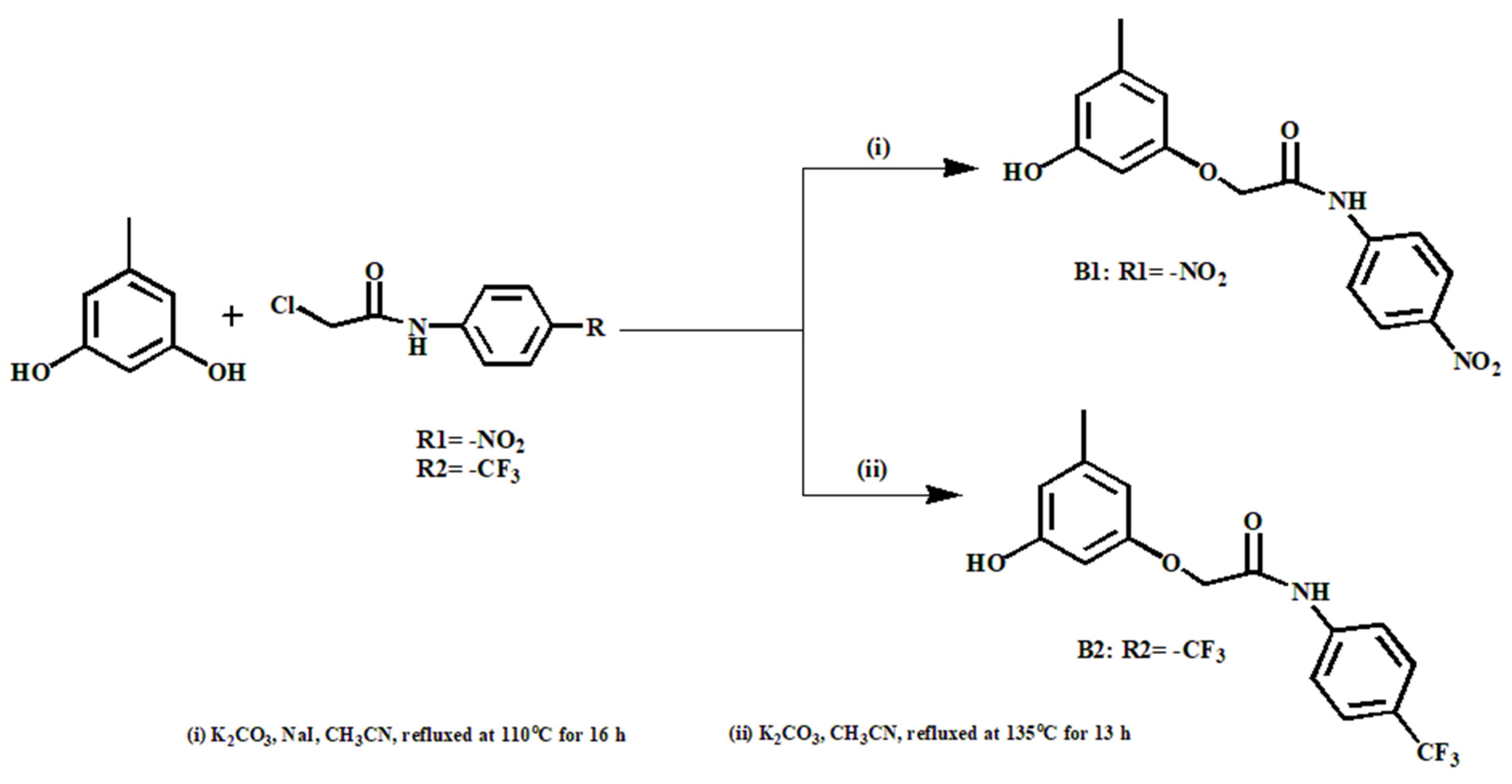
2.4. Synthesis of Compounds B1 and B2
2.4.1. Synthesis of 2-(3-hydroxy-5-methylphenoxy)-N-(4-nitrophenyl)acetamide), B1, and 2-(3-hydroxy-5-methylphenoxy)-N-(4-(trifluoromethyl)phenyl)acetamide, B2, as shown in Scheme 1
Scheme 1.
The synthesis of compounds B1 and B2.
2.4.1.1 2-(3-hydroxy-5-methylphenoxy)-N-(4-nitrophenyl)acetamide), B1
The compound B1 was synthesized in previous literature [23].
2.4.1.2 2-(3-hydroxy-5-methylphenoxy)-N-(4-(trifluoromethyl)phenyl)acetamide, B2
The B2 compound was synthesized using an orcinol as a starting material. The reaction mixture of 4-(trifluoromethyl) aniline (2.50 g, 16 mmol), chloroacetyl chloride (4.95 mL) and triethylamine (3.25 mL) in CH2Cl2 (10 mL) was stirred in an ice bath for 2 h. Moreover, the resultant reaction mixture was stirred at room temperature for 24 h, resulting in a brown precipitate of 2-chloro-N-(4-(trifluoromethyl)phenyl)acetamide (R2), with a yield of 85%. 1H NMR (400 MHz, CDCl3, ppm): δ = 8.41 (s, 1H), 7.72 (d, J = 8.6 Hz, 2H), 7.65 (d, J = 8.7 Hz, 2H), 4.25 (s, 2H). Then, the condensation reaction of 2-chloro-N-(4-(trifluoromethyl)phenyl)acetamide (R2) and 3,5-dihydroxytoluene (1.05 g, 8.42 mmol) in CH3CN (15 mL) was performed by stirring the mixture at 135 °C for 13 h under an N2 atmosphere. A total of 20 mL of deionized water was added to the resulting mixture. The aqueous layer was additionally extracted with 20 mL of CH2Cl2 three times and then dried over anhydrous Na2SO4. The reaction product was purified via column chromatography on silica gel using EtOAc/CH2Cl2 = 1:9 (v/v) as an eluent. The product of 2-(3-hydroxy-5-methylphenoxy)-N-(4-(trifluoromethyl)phenyl)acetamide was isolated as white crystals with a yield of 26.6%. 1H NMR (400 MHz, d6-DMSO, ppm): δ = 10.39 (s, 1H), 9.34. (s, 1H), 7.87 (d, J = 8.5 Hz, 2H), 7.69 (d, J = 8.6 Hz, 2H), 6.26 (s, 1H), 6.22 (s, 1H), 6.20 (s, 1H), 4.64 (s, 2H), 2.18 (s, 3H). 13C NMR (400 MHz, d6-DMSO, ppm): δ = 167.88, 159.26, 158.78, 142.49, 142.48, 140.00, 126.55, 126.51, 124.29, 120.02, 109.771, 106.69, 99.66, 67.52, 21.76. IR (cm−1); 3275, 3095, 1687, 1560, 1402, 826 HRMS (m/z): calculated for C16H14F3NO3+Na: 348.0823; found: 348.0815.
3. Results and Discussion
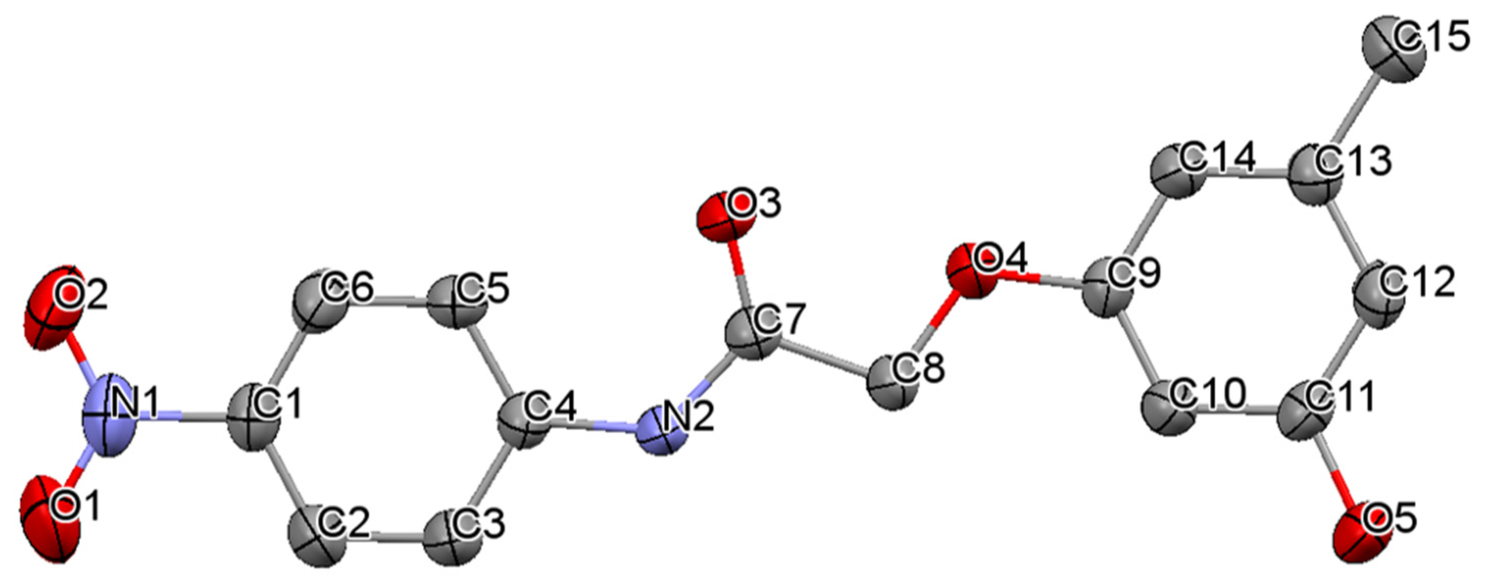
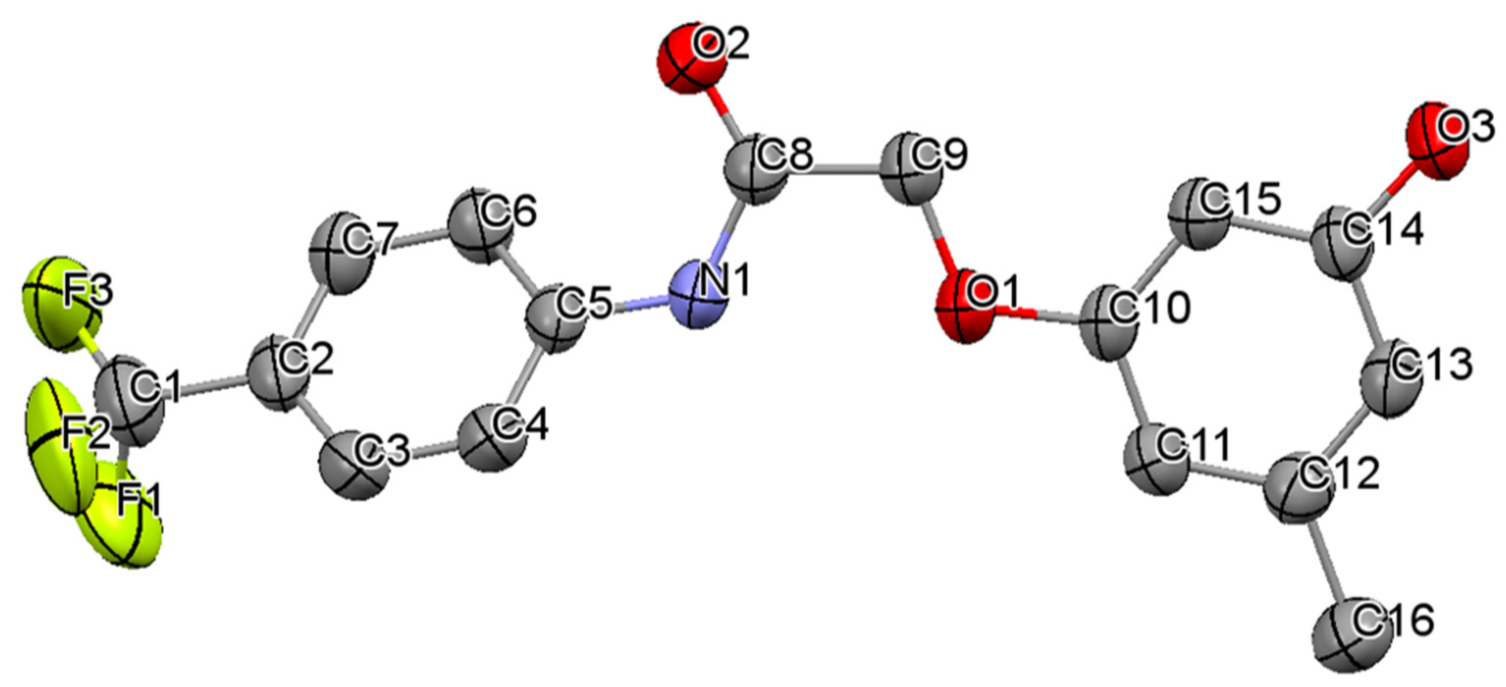
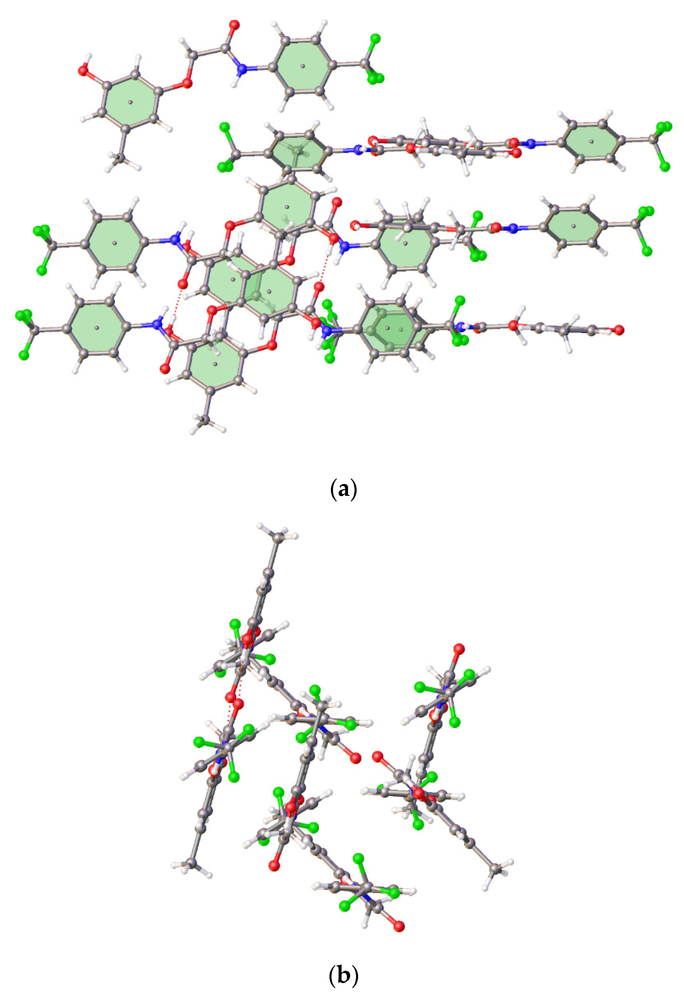
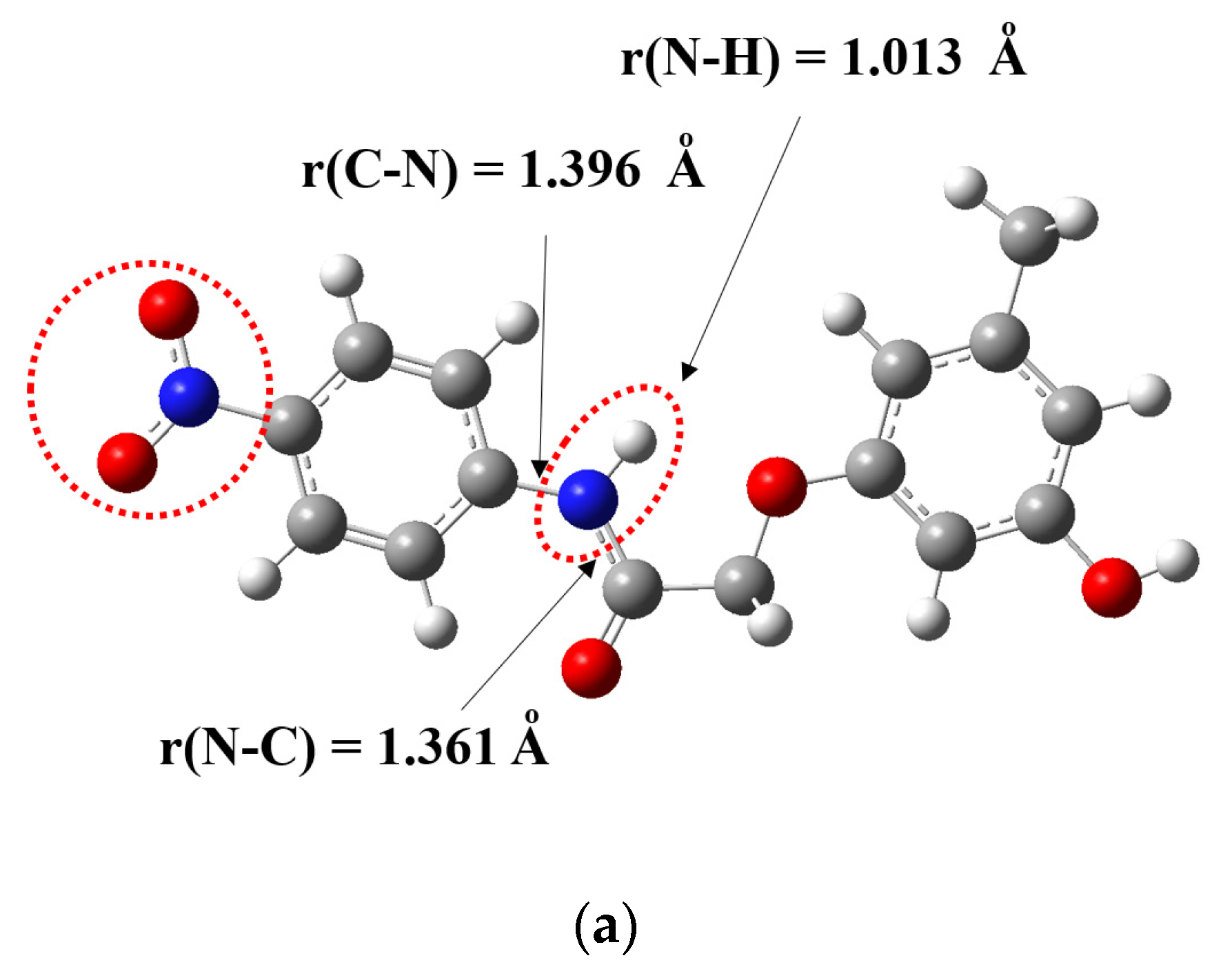
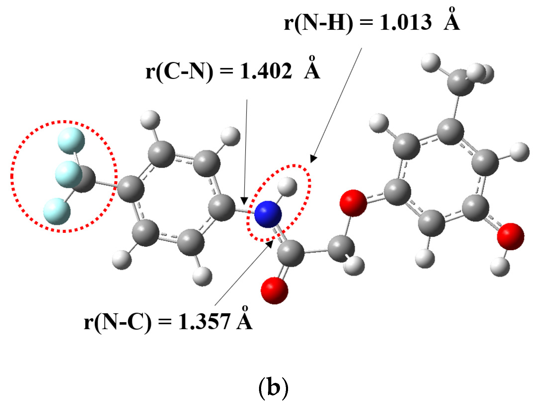
3.1. Crystal Structures of Compounds B1 and B2
The crystals of compounds B1 and B2 were grown in the solvent as EtOAc:CH2Cl2 = 1:9. The experimental parameters relating to the single-crystal X-ray analysis of compounds are presented in Table 1. Compound B1 was crystalized as a monoclinic structure with a P21/c space group, and its molecular structure with the E configuration for the N–C(=O)–C–O bond is presented in Figure 1. The structure of the compound was observed to present an almost absolute planarity conformation with the torsion angles of C4–N2–C7–O3; 0.9(5), C4–N2–C7–C8; 179.1(3), C9–O4–C8–C7; 174.2(2) and N2–C7–C8–O4; 172.7(2), respectively. Additionally, the torsion angles involved in the substituted benzene ring, with respect to the amide functional group, were discovered to be C7–N2–C4–C3; −178.6(3) and C7–N2–C4–C5; 1.8(5), which indicated a minor deviation from the planar of the structure. The packing images of compound B1 for the front and side views are presented in Figure 2. Moreover, the difference between each plane of the structure was 3.778(2) Å; however; the π–π interaction was not observed in such a compound.
Table 1.
Crystal data and structure refinement parameters for compounds B1 and B2.
Figure 1.
Molecular structure of compound B1 drawn at 50% thermal ellipsoid probability. Hydrogen atoms have been omitted for clarity.
Figure 2.
Packing images of compound B1: (a) front view and (b) side view.
The molecular configuration of B1 was stabilized by the intramolecular H-bonding of C=O…H, displayed in Figure 3. The molecules were linked to each other through C=O…H–O bonding by the carbonyl of amide and the hydroxy group from the methoxy phenol ring. Furthermore, the molecules were also connected by N–H…O–N–O, in which the hydrogen atom of amide is bound to the oxygen of the nitro-substituted group.
Figure 3.
Intramolecular and intermolecular hydrogen bonding in compound B1.
For compound B2, the molecule was also crystalized as a monoclinic structure with a P21/c space group, and its Z configuration concerning the N–C(=O)–C–O bond is presented in Figure 4. Remarkably, the structure of compound B2 was observed to be disordered at the position of fluorine atoms of the -CF3 functional group; therefore, the formula was presented as a double of the literal structure. Unlike compound B1, compound B2 was observed to be more diverged from the planarity condition, with the torsion angles of C8–C9–O1–C10; 172.8(1), C5–N1–C8–O2; 0.7(2), C5–N1–C8–C9; −179.6(1) and N1–C8–C9–O1; 1.7(2). Notably, the torsion angles of C8–N1–C5–C4 and C8–N1–C5–C6 related to the -CF3-substituted benzene ring aligned with the remaining structure were −142.3(2) and 38.2(2), respectively. The arrangement largely deviated in comparison to compound B1; accordingly, the packing images are exhibited as zigzag structures, as presented in Figure 5. According to this orientation, the intramolecular H-bonding of C=O…H was not observed in compound B2; however, the molecules were also linked to each other through C=O…H–O bonding, as presented in Figure 6. The H-bonding process according to the hetero atom of fluorine with N–H, including π–π interaction between each plane, was not observed; nonetheless, the difference between each centroid was detected with the value of 4.8447(15) Å.
Figure 4.
Molecular structure of compound B2 drawn at 50% thermal ellipsoid probability. Hydrogen atoms have been omitted for clarity.
Figure 5.
Packing images of compound B2: (a) front view and (b) side view.
Figure 6.
Intermolecular hydrogen bonding in compound B2.
In terms of the bond lengths, the comparison of bond distances involving the amide functional group demonstrated that N2–C7; 1.352(4) and C7–O3; 1.211(4) in compound B1 were relatively comparable to the bonds of N1–C8; 1.338(2) and C8–O2; 1.222(2) in compound B2. In addition, the bond that bound the nitrogen atom of the amide group and the phenyl ring was observed to be 1.408(4) and 1.419(2) in compounds B1 and B2, respectively. This was attributed to the related equivalent strength of the -CF3 electron-withdrawing group related to -NO2 that created an almost equal electron density in the bond formation, leading to the similar bond lengths of such compounds. Some important bond lengths, bond angles and torsion angles are presented in Table 2.
Table 2.
Selected bond lengths (Å), bond angles (°) and torsion angles (°) in B1 and B2 compounds.
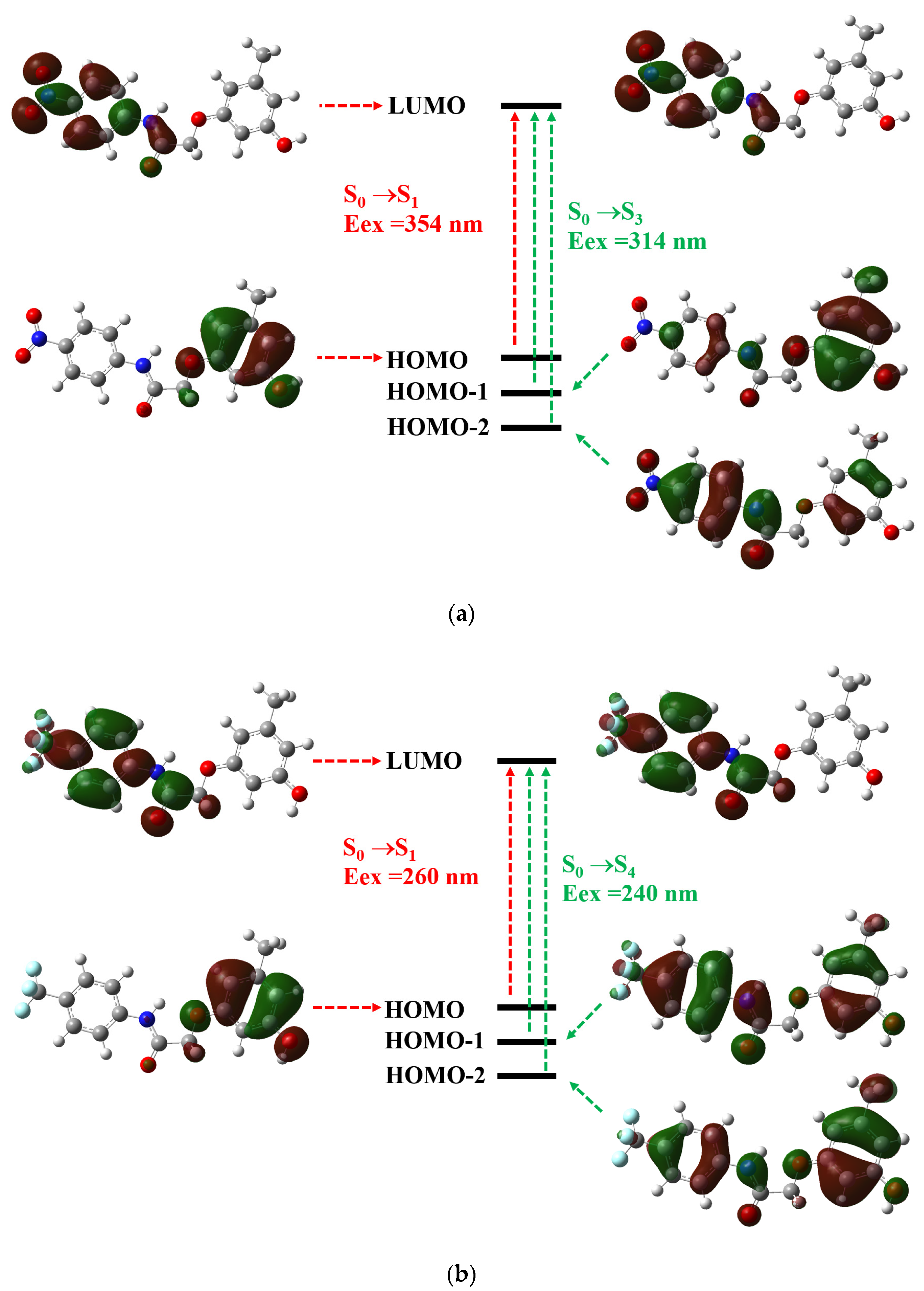
3.2. Theoretical Calculations
For the calculations, the structures of B1 and B2 were optimized, as illustrated in Figure 7. The bond lengths for the C-N, N-H and N-C distances of B1 and B2 were 1.396Å and 1.402 Å; 1.013 Å and 1.013 Å; 1.361 Å and 1.357 Å, respectively. There were no significant differences in the bond lengths’ values in the optimized geometries of B1 and B2. However, optimized structures of B1 and B2 exhibited the existence of a strong H-bond (1.013 Å) belonging to N-H.

Figure 7.
Optimized structure of (a) compound B1 and (b) compound B2.
The excitation energies, oscillator strengths (f) and transition states obtained from the TD-DFT calculations at the PBE0/6-311G(d,p) level using optimized ground state geometries for B1 and B2 are listed in Table 3. The calculated wavelengths with the maximum absorption values of B1 and B2 were 314 and 240 nm, respectively, which were slightly lower than the experimental results at 327 and 258 nm. According to the TD-DFT calculations, these strong absorptions corresponded to HOMO-1→LUMO (S0→S3 state) and HOMO-2→LUMO (S0→S4 state) transitions for B1 and B2, respectively. The results clearly show that the electronic transition of B1 with substituted (-NO2) is more effective than that of B2 with the trifluoromethyl (-CF3) group.
Table 3.
The main contributions of electronic transition using PCM-TD-PBE0 functional at 6-311G(d,p) basis set compared to the experimental absorption wavelengths of B1 and B2 in the DMSO solvent (Eex is the calculated vertical excitation energy, H corresponds to HOMO and L corresponds to LUMO).
The molecular orbitals were related to the electronic transitions for both compounds, as presented in Figure 8. The structures of compounds B1 and B2 explained the contributions of HOMO-1, HOMO-2 and LUMO excitations. The HOMO-1 to LUMO and HOMO-2 to LUMO excitations corresponded to the internal charge transfer (ICT) process for the electronic spectra of compounds B1 and B2.
Figure 8.
Energy level diagrams of HOMO and LUMO orbitals for compounds (a) B1 and (b) B2.
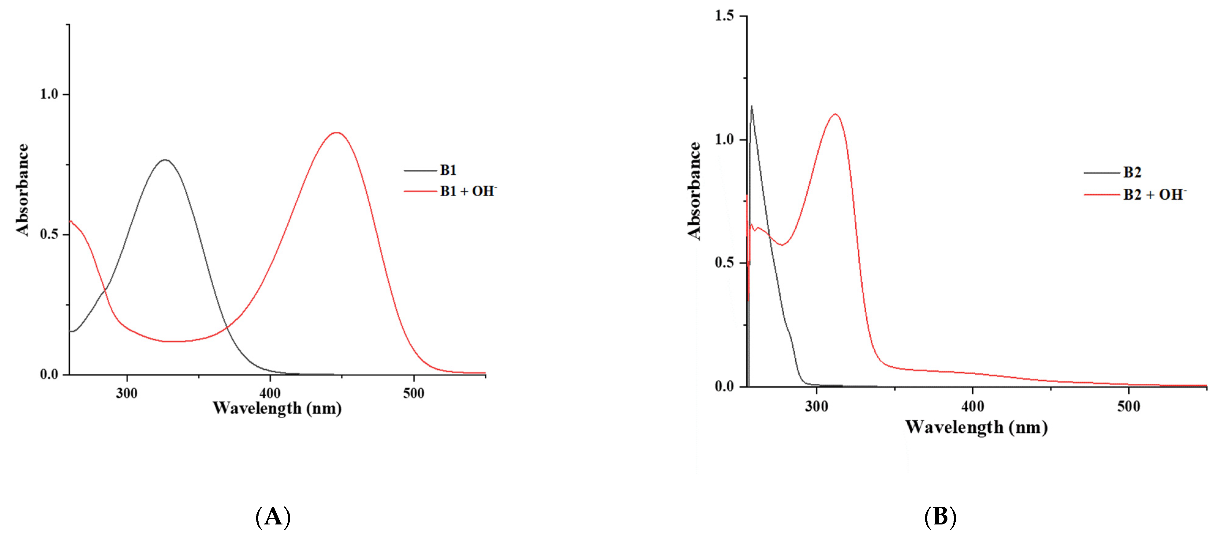
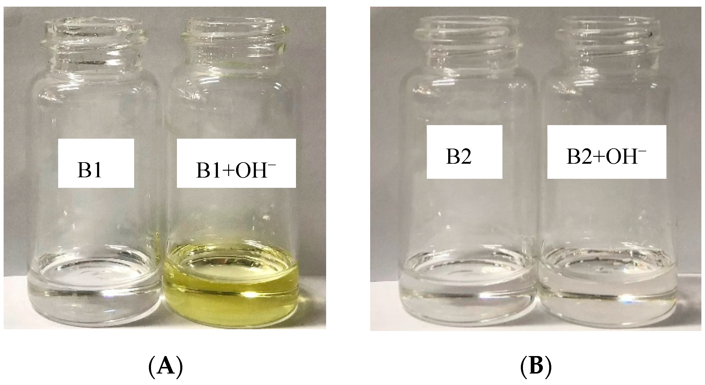
3.3. The Optical Properties of Compounds B1 and B2 by UV–Visible Spectroscopy
The maximum wavelength absorptions of compounds B1 and B2 in DMSO were at 327 and 258 nm, respectively. Upon the addition of the OH− ion to the B1 solution, an absorption wavelength of 327 nm disappeared. This indicated that a new band at 466 nm corresponded to the deprotonated species in the solution, as presented in Figure 9A. This mechanism was under the internal charge transfer process [24]. This phenomenon occurred under the π–π* transition that produced the red shift of the absorption wavelength. In the case of B2, the absorption wavelength at 258 nm decreased and presented a new absorption wavelength at 303 nm upon the addition of OH−, as presented in Figure 9B. The mechanism was similar to that of B1. An obvious change can be observed in B1, rather than B2, because of the NO2 group performing as an electron-withdrawing group, resulting in the NH of the amide functional group being a more acidic proton; therefore, the addition of OH− to the B1 solution led to the loss of a proton in the amide group. For the B2 compound, the CF3 moiety is less powerful than the NO2 group in B1, but B2 can lose the proton of the amide group. For the color change when adding OH−, as presented in Figure 10, the B1 solution changed from being colorless to an intense yellow color, but B2 did not change color. The color changes of the B1 and B2 solutions were consistent with the UV–Vis experiments.
Figure 9.
UV–Vis spectrum of (A) B1 (1 × 10⁻⁴ M) and B1 (1 × 10⁻⁴ M) with addition of 10 equiv. OH− in DMSO (B) B2 (1 × 10⁻⁴ M) and B2 (1 × 10⁻⁴ M) with addition of 10 equiv. OH− in DMSO.
Figure 10.
Naked-eye color change of (A) B1 (1 × 10⁻⁴ M) and B1 with addition of 10 equiv. OH− ion in DMSO (B) B2 (1 × 10⁻⁴ M) and B2 with addition of 10 equiv. OH− ion in DMSO.
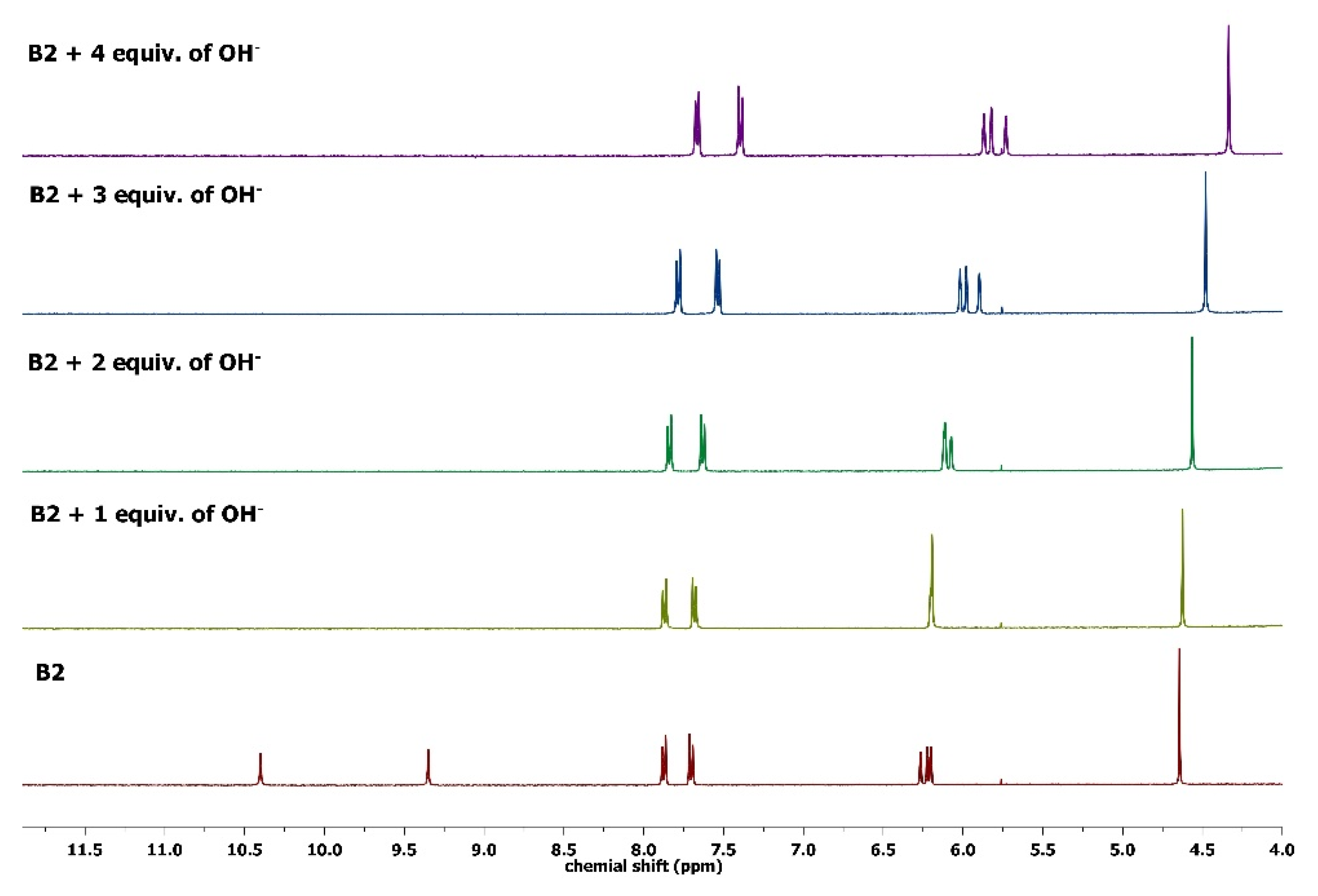
3.4. The Interactions between Compounds (B1 and B2) and OH− Ion by 1H NMR Spectroscopy
Additionally, the NH proton signals of B1 and B2 were 10.64 and 10.39 ppm, respectively. The OH proton signals of B1 and B2 are 9.33 and 9.34 ppm, as presented in Figure 11 and Figure 12. Upon addition of OH− in 1 equivalent, the NH and OH protons of both compounds disappeared due to the deprotonation process [25]. The CH2 and ArH protons of B1 and B2 exhibited an upfield shift because these protons gained an enhanced electron density. By adding 2–4 equivalents of OH− to the B1 and B2 solutions, the proton peaks presented greater upfield shift when compared to one equivalent of the OH− ion. This evidence indicates that the structures of B1 and B2 are changeable to a deprotonated form under basic conditions.
Figure 11.
1H NMR spectra of B1 (5 × 10⁻³ M) and B1 with different equivalents of OH− in d6-DMSO.
Figure 12.
1H NMR spectra of B2 (5 × 10⁻³ M) and B2 with different equivalents of OH− in d6-DMSO.
4. Conclusions
The orcinolic derivatives (B1 and B2) were synthesized and crystallized in the present study. The crystal structures of B1 and B2 were also stabilized by the hydrogen-bonding interaction. The π–π stacking interaction did not occur within intermolecular crystals. The DFT results indicate that the HOMO and LUMO of B1 and B2 are consistent with that of the UV–Vis results. The 1H-NMR results show that the structures of B1 and B2 changed the resonance signals of aromatic and methylene protons upon the addition of OH− ions. Therefore, B1 and B2 can be used as hydroxide indicators.
Author Contributions
Conceptualization, methodology, funding and writing of draft: B.W.; methodology and writing: S.S.; methodology: P.K., P.J., T.D., A.S. and K.C.; writing: P.H. and B.P. All authors have read and agreed to the published version of the manuscript.
Funding
The authors would like to thank the Kasetsart University Research and Development Institute (20420165810347000), the Department of Chemistry, Faculty of Science, Kasetsart University, and the Center of Excellence for Innovation in Chemistry (PERCH-CIC), the Ministry of Higher Education, Science, Research and Innovation, for the financial support.
Institutional Review Board Statement
Not applicable.
Informed Consent Statement
Not applicable.
Data Availability Statement
Not applicable.
Acknowledgments
The authors thank the Department of Chemistry, Faculty of Science, Kasetsart University for providing facilities and equipment.
Conflicts of Interest
The authors declare no conflict of interest.
References
- Cardullo, N.; Muccilli, V.; Pulvirenti, L.; Corrado Tringali, C. Natural isoflavones and semisynthetic derivatives as pancreatic lipase inhibitors. J. Nat. Prod. 2021, 84, 654–665. [Google Scholar] [CrossRef] [PubMed]
- Taborga, L.; Díaz, K.; Olea, A.F.; Reyes-Bravo, P.; Flores, M.E.; Peña-Cortés, H.; Espinoza, L. Effect of polymer micelles on antifungal activity of geranylorcinol compounds against botrytis cinerea. J. Agric. Food Chem. 2015, 63, 6890–6896. [Google Scholar] [CrossRef] [PubMed]
- Li, S.Y.; Zhanga, D.B.; Wang, J.Y.; Lua, R.M.; Zhenga, C.H.; Pua, S.Z. A novel diarylethene-hydrazinopyridine-based probe for fluorescent detection of aluminum ion and naked-eye detection of hydroxide ion. Sens. Actuators B 2017, 245, 263–272. [Google Scholar] [CrossRef]
- Mariappan, K.; Shrestha, P.K.; Hussain, A.; Sykes, A.G. A chemodosimeter for the detection of hydroxide using an anthraquinone-based receptor: Photophysical properties and X-ray crystallography. J. Mol. Struct. 2022, 1267, 133585. [Google Scholar] [CrossRef]
- Kuo, E.A.; Hambleton, P.T.; Kay, D.P.; Evans, P.L.; Matharu, S.S.; Little, E.; McDowall, N.; Jones, C.B.; Hedgecock, C.J.R.; Yea, C.M.; et al. Synthesis, Structure−Activity Relationships, and Pharmacokinetic Properties of Dihydroorotate Dehydrogenase Inhibitors: 2-Cyano-3-cyclopropyl-3-hydroxy- N-[3‘-methyl-4‘-(trifluoromethyl)phenyl]propenamide and Related Compounds. J. Med. Chem. 1996, 39, 4608–4621. [Google Scholar] [CrossRef] [PubMed]
- Aro-Heinilä, A.; Lepistö, A.; Äärelä, A.; Lönnberg, T.A.; Virta, P. 2 Trifluoromethyl-6-mercurianiline Nucleotide, a Sensitive 19F NMR Probe for Hg(II)-mediated Base Pairing. J. Org. Chem. 2022, 87, 137–146. [Google Scholar] [CrossRef]
- Zhou, Z.; Li, W.; Hao, X.; Redshaw, C.; Chen, L.; Sun, W.-H. 6-Benzhydryl-4-methyl-2-(1H-benzoimidazol-2-yl)phenol ligands and their zinc complexes: Syntheses, characterization and photoluminescence behavior. Inorg. Chim. Acta 2012, 392, 345–353. [Google Scholar] [CrossRef]
- Chauhan, P.; Yan, N. Novel nitroaniline-cellulose nanohybrids: Nitro radical photo-release and its antibacterial action. Carbohydr. Polym. 2017, 174, 1106–1113. [Google Scholar] [CrossRef]
- Mohan, B.; Choudhary, M.; Bharti, S.; Jana, A.; Das, N.; Muhammad, S.; Al-Sehemi, A.G.; Kumar, S. Syntheses, characterizations, crystal structures and efficient NLO applications of new organic compounds bearing 2-methoxy-4-nitrobenzeneamine moiety and copper (II) complex of (E)-N’-(3, 5-dichloro-2-hydroxybenzylidene) benzohydrazide. J. Mol. Struc. 2019, 1190, 54–67. [Google Scholar] [CrossRef]
- Prabukanthan, P.; Lakshmi, R.; Harichandran, G.; Kumar, C.S. Synthesis, structural, optical and thermal properties of N-methyl–N-aryl benzamide organic single crystals grown by a slow evaporation technique. J. Mol. Struc. 2018, 1156, 62–73. [Google Scholar] [CrossRef]
- Nakayama, H.; Nishida, J.-I.; Takada, N.; Hiroyasu Sato, H.; Yamashita, Y. Crystal Structures and Triboluminescence Based on Trifluoromethyl and Pentafluorosulfanyl Substituted Asymmetric N-Phenyl Imide Compounds. Chem. Mater. 2012, 24, 671–676. [Google Scholar] [CrossRef]
- Ahmad, I.; Rehman, Z.U.; Waseem, A.; Tariq, M.; Beth, C.M.; Bacsa, J.; Venkataraman, D.; Rajakumar, A.; Ullah, N.; Tabassum, S. Organotin (IV) derivatives of amide-based carboxylates: Synthesis, spectroscopic characterization, single crystal studies and antimicrobial, antioxidant, cytotoxic, anti-leishmanial, hemolytic, noncancerous, anticancer activities. Inorg. Chim. Acta 2020, 505, 119433. [Google Scholar] [CrossRef]
- Sawant, A.S.; Kamble, S.S.; Pisal, P.M.; Sawant, S.S.; Hese, S.V.; Bagul, K.T.; Pinjari, R.V.; Kamble, V.T.; Meshram, R.J.; Gacche, R.N. Synthesis and evaluation of N-(4-(substituted)-3-(trifluoromethyl) phenyl) isobutyramides and their N-ethyl analogous as anticancer, anti-angiogenic & antioxidant agents: In vitro and in silico analysis. Comput. Biol. Chem. 2021, 92, 107484. [Google Scholar] [PubMed]
- Tian, C.; Wang, Q.; Wang, X.; An, G.; Li, G. Visible-Light Mediated ortho-Trifluoromethylation of Aniline Derivatives. J. Org. Chem. 2019, 84, 14241–14247. [Google Scholar] [CrossRef] [PubMed]
- Nath, J.; Baruah, B.J. Self-Assemblies of Solvates, Ionic Cocrystals, and a Salt Based on 4 {[(4-Nitrophenyl)carbamoyl]amino} N (pyrimidin-2-yl)benzene-1- sulfonamide: Study in the Solid and Solution States. Cryst. Growth Des. 2021, 21, 5325–5341. [Google Scholar] [CrossRef]
- Lobatto, V.L.; Argüello, G.A.; Caira, M.R.; Bujan, E.I. Trifluralin and two of its photodegradation products: Crystal structures and phase solubility/UV studies with cyclodextrins. J. Phys. Org. Chem. 2019, 32, e4006. [Google Scholar] [CrossRef]
- Dolomanov, O.V.; Bourhis, L.J.; Gildea, R.J.; Howard, J.A.K.; Puschmann, H. OLEX2: A complete structure solution, refinement and analysis program. J. Appl. Cryst. 2009, 42, 339–341. [Google Scholar] [CrossRef]
- Sheldrick, G.M. Crystal structure refinement with SHELXL. Acta Cryst. 2015, C71, 3–8. [Google Scholar]
- Macrae, C.F.; Bruno, I.J.; Chisholm, J.A.; Edgington, P.R.; McCabe, P.; Pidcock, E.; Rodriguez-Monge, L.; Taylor, R.; Streek, J.V.D.; Wood, P.A. Mercury CSD 2.0-new features for the visualization and investigation of crystal structures. J. Appl. Cryst. 2008, 41, 466–470. [Google Scholar] [CrossRef]
- Yeh, M.-Y.; Lin, H.-C. Theoretical analysis of the intermolecular interactions in naphthalene diimide and pyrene complexes. Phys. Chem. Chem. Phys. 2014, 16, 24216–24222. [Google Scholar] [CrossRef]
- Mennucci, B. Polarizable continuum model. Wiley Interdiscip. Rev. Comput. Mol. Sci. 2012, 2, 386–404. [Google Scholar] [CrossRef]
- Frisch, M.J.; Trucks, G.W.; Schlegel, H.B.; Scuseria, G.E.; Robb, M.A.; Cheeseman, J.R.; Scalmani, G.; Barone, V.; Mennucci, B.; Petersson, G.A.; et al. Gussion 09, Revision A.02; Gaussian Inc.: Wallingford, CT, USA, 2013. [Google Scholar]
- Jansukra, P.; Duangthongyou, T.; Suramitr, S.; Kittipong Chainok, K.; Wannalerse, B. Synthesis, Crystal Structure and Optical Properties of 2-(3-(Hexyloxy)-5-Methylphenoxy)-N-(4-Nitrophenyl)acetamide for Anion Detection. Crystals 2021, 11, 671. [Google Scholar] [CrossRef]
- Bhat, H.R.; Gupta, P.S.S.; Satyaranjan Biswal, S.; Rana, M.K. Anion Sensing by Novel Triarylboranes Containing Boraanthracene: DFT Functional Assessment, Selective Interactions, and Mechanism Demonstration. ACS Omega 2019, 4, 4505–4518. [Google Scholar] [CrossRef] [PubMed]
- Yin, Y.; Sarma, T.; Wang, F.; Yuan, N.; Duan, Z.; Sessler, J.L.; Zhang, Z. Air-Stable N,N′ Dihydroporphycene: A Quinoxaline-Fused Tetrapyrrolic Macrocycle That Detects Fluoride Anion via Deprotonation. Org. Lett. 2019, 21, 1849–1852. [Google Scholar] [CrossRef] [PubMed]
Publisher’s Note: MDPI stays neutral with regard to jurisdictional claims in published maps and institutional affiliations. |
© 2022 by the authors. Licensee MDPI, Basel, Switzerland. This article is an open access article distributed under the terms and conditions of the Creative Commons Attribution (CC BY) license (https://creativecommons.org/licenses/by/4.0/).