




Next Issue
Volume 18, June
Previous Issue
Volume 18, February


Hematol. Rep., Volume 18, Issue 2 (April 2026) – 14 articles
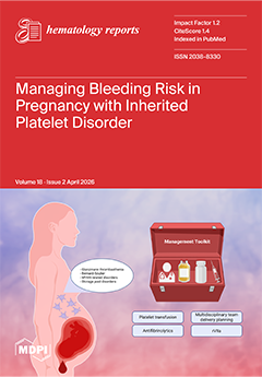
Cover Story (view full-size image): Inherited platelet disorders represent a heterogeneous group of rare bleeding conditions that pose unique challenges during pregnancy and delivery. Bleeding risk varies significantly across disorders, driven by differences in platelet function and number, and is further amplified by the hemostatic demands of the peripartum period. Individuals are at increased risk for postpartum hemorrhage, while neonates may also be affected, adding complexity to clinical management. Current strategies rely on individualized, multidisciplinary care incorporating antifibrinolytics, platelet transfusion, and bypassing agents, though evidence remains limited. This review synthesizes available data across major inherited platelet disorders and highlights the need for tailored approaches and improved evidence to guide management in this high-risk population. View this paper
  • Issues are regarded as officially published after their release is announced to the table of contents alert mailing list.
  • You may sign up for e-mail alerts to receive table of contents of newly released issues.
  • PDF is the official format for papers published in both, html and pdf forms. To view the papers in pdf format, click on the "PDF Full-text" link, and use the free Adobe Reader to open them.
Order results
Result details
Select all
Export citation of selected articles as:
15 pages, 1874 KB  
Article
The Prognostic Value of the CD8+PD-1+/CD4+PD-1+ (PERLS) Ratio for Leukemic Transformation in MDS
by Panagiotis Panagiotidis, Emmanuel Karavanis, Konstantinos Neanidis, Eleftherios Panteris and Maria Moysidou
Hematol. Rep. 2026, 18(2), 29; https://doi.org/10.3390/hematolrep18020029 - 15 Apr 2026
Viewed by 447
Abstract
Background/Objectives: Myelodysplastic syndromes (MDS) are associated with a significant risk of progression to acute myeloid leukemia (AML), affecting approximately 30% of patients. In high-risk MDS, leukemic transformation may occur within a short time frame, highlighting the need for early and reliable biomarkers of [...] Read more.
Background/Objectives: Myelodysplastic syndromes (MDS) are associated with a significant risk of progression to acute myeloid leukemia (AML), affecting approximately 30% of patients. In high-risk MDS, leukemic transformation may occur within a short time frame, highlighting the need for early and reliable biomarkers of disease progression. Increasing evidence suggests that immune dysregulation and cytotoxic T-cell dysfunction contribute to disease evolution. This study aimed to evaluate PD-1 and CD57 expressions on CD8+ T cells and to investigate the CD8+PD-1+/CD4+PD-1+ ratio (PERLS) as a potential immunological marker predictive of leukemic transformation. Methods: Thirty-one patients with MDS were prospectively followed over a 12-month period. At baseline, patients underwent routine clinical and laboratory evaluation, including multiparameter flow cytometric assessment of bone marrow blasts. An extended immunophenotypic analysis of bone marrow samples was performed at study entry to assess PD-1 and CD57 expression on CD8+ T cells. Cytogenetic and molecular analyses were conducted when clinical findings suggested disease progression. Patients who developed signs of progression were re-evaluated approximately one month later, during the progression phase, to assess dynamic immunological changes. Results: Of the thirty-one patients included, eighteen progressed to AML, whereas thirteen remained clinically stable. Patients who progressed demonstrated a significant increase in PD-1 and CD57 expression on CD8+ T cells compared with stable patients. Moreover, a markedly higher CD8+PD-1+/CD4+PD-1+ (PERLS) ratio was observed in patients who subsequently developed AML, particularly during the progression phase. Conclusions: Dynamic immunophenotypic monitoring reveals that increased PD-1 on CD8+ T cells and an elevated PERLS ratio are associated with imminent leukemic transformation in MDS. These findings support the incorporation of immune-based biomarkers, particularly the CD8+PD-1+/CD4+PD-1+ ratio, into routine risk assessment to enable earlier identification of disease progression and timely therapeutic intervention. Full article
Show Figures

Figure 1

11 pages, 230 KB  
Case Report
Asciminib in Philadelphia Chromosome-Positive Acute Lymphoblastic Leukemia: A Case Series and Review of Emerging Evidence
by Mostafa F. Mohammed Saleh, Abdulrahman Nasiri, Ahmed Kotb Abdrabou, Hadeel Samarkandi, Ayman Saad, Mahmoud Aljurf, Amr Hanbali and Ali Alahmari
Hematol. Rep. 2026, 18(2), 28; https://doi.org/10.3390/hematolrep18020028 - 13 Apr 2026
Viewed by 881
Abstract
Philadelphia chromosome-positive acute lymphoblastic leukemia (Ph+ ALL) remains a high-risk entity despite advances in tyrosine kinase inhibitors (TKIs), immunotherapy, and cellular therapies. Relapse driven by clonal evolution, central nervous system (CNS) sanctuary disease, and TKI resistance, particularly T315I mutations, continues to limit durable [...] Read more.
Philadelphia chromosome-positive acute lymphoblastic leukemia (Ph+ ALL) remains a high-risk entity despite advances in tyrosine kinase inhibitors (TKIs), immunotherapy, and cellular therapies. Relapse driven by clonal evolution, central nervous system (CNS) sanctuary disease, and TKI resistance, particularly T315I mutations, continues to limit durable disease control. Asciminib, a first-in-class allosteric BCR::ABL1 (STAMP) inhibitor, has demonstrated efficacy and favorable tolerability in chronic myeloid leukemia, but its optimal role in Ph+ ALL remains to be defined. We report a three-patient case series of Ph+ acute leukemia treated with asciminib across diverse high-risk clinical settings, including multiply relapsed disease, CNS involvement, T315I-mutated leukemia, post-CAR-T-cell relapses, and transplant bridging. Clinical outcomes are contextualized through a comprehensive review of emerging clinical trial data, real-world cohorts, and mechanistic studies evaluating asciminib in Ph+ ALL. Across all cases, asciminib was incorporated as part of combination or consolidation strategies rather than as monotherapy in active disease. Asciminib contributed to molecular disease control, CNS leukemia clearance, and successful bridging to allogeneic transplantation or cellular therapy, with acceptable tolerability and no major vascular toxicity. Integration of published evidence demonstrates that asciminib exhibits consistent biological activity in Ph+ ALL, with improved durability when used in rational combinations, particularly with immunotherapy or ATP-competitive TKIs. Preclinical data further support asciminib’s compatibility with antibody-based and cellular therapies through preservation of immune effector function. Asciminib represents a versatile but context-dependent therapeutic option in Ph+ ALL. Its greatest clinical value appears to lie in rational combination regimens, maintenance strategies, and bridging to definitive therapies rather than single-agent salvage. Emerging structural biomarkers and ongoing clinical trials are expected to further refine patient selection, sequencing, and optimal integration of asciminib, particularly in CNS-involved disease and post-CAR-T cell relapse. Full article
19 pages, 2966 KB  
Article
Metabolomic Signatures of Relapse and Survival in AML Patients Receiving Allogeneic Hematopoietic Stem Cell Transplantation
by Igor Novitzky-Basso, Changjiang Xu, Caden Chiarello, Julie A. Reisz, Angelo D’Alessandro, Gary D. Bader, Jonas Mattsson and Courtney Jones
Hematol. Rep. 2026, 18(2), 27; https://doi.org/10.3390/hematolrep18020027 - 7 Apr 2026
Viewed by 498
Abstract
Objectives: Allogeneic stem cell transplantation (HSCT) is curative in acute myeloid leukemia (AML) but is limited by relapse and non-relapse mortality (NRM). Metabolomic prognostic value is unclear. We assessed whether plasma metabolite profiles at diagnosis, pre-transplant, and post-transplant are associated with overall [...] Read more.
Objectives: Allogeneic stem cell transplantation (HSCT) is curative in acute myeloid leukemia (AML) but is limited by relapse and non-relapse mortality (NRM). Metabolomic prognostic value is unclear. We assessed whether plasma metabolite profiles at diagnosis, pre-transplant, and post-transplant are associated with overall survival (OS) and cause-specific mortality. Methods: We retrospectively analyzed plasma metabolites from 63 AML patients undergoing HSCT (263 samples). Results: Higher levels of valine (hazard ratio [HR] 24.454), citrulline (HR 20.478), 5-oxoproline (HR 11.766), and glutamine (HR 8.701) associated with higher NRM, while inosine diphosphate (HR 0.091) and pyridoxamine-5′-phosphate (HR 0.313) associated with lower NRM. For relapse-related mortality (RRM), higher levels of phenylalanine (HR 26.585), leucine/isoleucine (HR 10.755), indolepyruvate (HR 7.676), and creatinine (HR 13.874) were associated with higher RRM, while trans-4-hydroxy-L-proline (HR 0.101) was associated with lower RRM. Higher post-transplant ornithine (HR 0.063), 3-sulfocatechol (HR 0.590), and indole-3-acetate (HR 0.359) were associated with improved OS. Mixed-effects modelling identified lower dehydroascorbate and citrate in relapsed patients, with dehydroascorbate remaining significant after false discovery rate adjustment. Conclusions: Metabolomic profiling nominated candidate metabolites for validation in larger prospective studies and elucidated mechanistic pathways, potentially informing novel interventions or risk-adapted monitoring strategies in HSCT. Full article
Show Figures

Figure 1

9 pages, 1598 KB  
Case Report
The Utility of IgG4/IgG Ratio in the Diagnosis of Multicentric Castleman Disease: A Case Report of HHV8+ Castleman Disease in a Patient with Classical Hodgkin’s Lymphoma
by Adam Hagele, Philip Kay, Kevin Nishino, Akhil Mehta, Yan Liu, Anthony L. Nguyen and Eric Lau
Hematol. Rep. 2026, 18(2), 26; https://doi.org/10.3390/hematolrep18020026 - 3 Apr 2026
Viewed by 484
Abstract
Background/Objectives: Multicentric Castleman Disease (MCD) is a rare lymphoproliferative disorder that can mimic IgG4-related disease (IgG4-RD), particularly in patients presenting with elevated serum IgG4. Accurate diagnosis is crucial given differing treatments and prognoses. Case Presentation: We describe a 76-year-old male with fever, [...] Read more.
Background/Objectives: Multicentric Castleman Disease (MCD) is a rare lymphoproliferative disorder that can mimic IgG4-related disease (IgG4-RD), particularly in patients presenting with elevated serum IgG4. Accurate diagnosis is crucial given differing treatments and prognoses. Case Presentation: We describe a 76-year-old male with fever, lymphadenopathy, and elevated inflammatory markers. Labs revealed an elevated IgG4 of 133 mg/dL and total IgG of 1410 mg/dL, yielding an IgG4/IgG ratio of 9.43%. Lymph node biopsy showed nodular sclerosing classical Hodgkin lymphoma, for which he received five cycles of A + AVD. Persistent symptoms, elevated IL-6, and HHV8 viremia prompted repeat biopsy, which demonstrated HHV8-positive MCD. Rituximab was initiated, which resulted in clinical and radiographic resolution. Methods: We performed a systematic review of the English-language literature from 2000 to 2025, identifying 23 studies that contained MCD cases with individual-level serum IgG4 and IgG data. A total of 36 unique cases were included. Results: The mean IgG4/IgG ratio was 14.61%, which is substantially lower than ratios typically seen in IgG4-RD. To our knowledge, our case is the only reported instance of HHV8-associated MCD with elevated IgG4. Conclusions: A mildly elevated IgG4/IgG ratio may favor the diagnosis of MCD over IgG4-RD. Serum IgG4 and total IgG should be considered when suspecting Castleman Disease. Full article
Show Figures

Figure 1

12 pages, 1377 KB  
Article
Evaluation of mTOR, NFκB and BCL-2 Inhibitor Activity In Vitro in Karpas 1106P, a Primary Mediastinal B-Cell Lymphoma Cell Line
by Agata Majchrzak, Sylwia Mańka, Barbara Cebula-Obrzut, Paweł Robak, Damian Mikulski and Magdalena Witkowska
Hematol. Rep. 2026, 18(2), 25; https://doi.org/10.3390/hematolrep18020025 - 24 Mar 2026
Viewed by 484
Abstract
Introduction: PMBCL is an aggressive type of lymphoma characterized by high heterogeneity in clinical, molecular, and genetic features. In PMBCL, disturbances in the NFkB pathway and deregulation of BCL-2 and mTOR family proteins are observed, which may contribute to impaired apoptosis. Therefore, [...] Read more.
Introduction: PMBCL is an aggressive type of lymphoma characterized by high heterogeneity in clinical, molecular, and genetic features. In PMBCL, disturbances in the NFkB pathway and deregulation of BCL-2 and mTOR family proteins are observed, which may contribute to impaired apoptosis. Therefore, many strategies have been established to target the functioning of these pathways. Early clinical trials of mTOR, NFkB and Bcl-2 inhibitors suggest their activity in many hematological cancers, but their activity as monotherapy agents may still be insufficient; therefore, combinations of these compounds with other molecules acting on those active in a given cancer subtype are being sought. Materials and Methods: In vitro studies were conducted on a single PMBCL cell line, Karpas 1106P. We administered three novel drugs: AZD2014 (vistusertib), an inhibitor of the serine-threonine kinase mTOR; IMD-0354, an NFκB inhibitor; and ABT-199 (venetoclax), a highly selective inhibitor for BCL-2. Drugs were administered alone, in pairs and in combination of all three agents. Results: Based on the results of our own research, for the Karpas cell line individually, ABT-199 had the strongest pro-apoptotic effect on cancer cells, while in pairs the most potent induction of apoptosis occurred following treatment with AZD2014+ABT-199. The combination of three drugs did not have a stronger effect than either a single drug used alone or any two-drug combination. Conclusions: These results provide preliminary in vitro evidence that targeting the BCL-2 and mTOR pathways may enhance pro-apoptotic activity in a PMBCL cell model; however, further validation in additional cell lines and in vivo models is needed before translational implications can be considered. Full article
(This article belongs to the Special Issue Treatment and Prognosis of Hematological Malignancies)
Show Figures

Figure 1

9 pages, 2376 KB  
Case Report
Concomitant Clonal CBFB::MYH11 and PDGFRB Fusions in a Case of De Novo Acute Myeloid Leukemia
by Qiliang Ding, Natasha E. Lewis, Cody J. Artymiuk, Renee M. Olson, Rong He, Rhett P. Ketterling, David S. Viswanatha, Patricia T. Greipp and Cinthya J. Zepeda Mendoza
Hematol. Rep. 2026, 18(2), 24; https://doi.org/10.3390/hematolrep18020024 - 23 Mar 2026
Viewed by 455
Abstract
Background: Acute myeloid leukemia (AML) with CBFB::MYH11 fusion and myeloid/lymphoid neoplasms with eosinophilia and tyrosine kinase gene fusions (MLN-TK) are genetically defined and typically mutually exclusive entities. Case Presentation: We report a unique case of de novo AML harboring two clonal, [...] Read more.
Background: Acute myeloid leukemia (AML) with CBFB::MYH11 fusion and myeloid/lymphoid neoplasms with eosinophilia and tyrosine kinase gene fusions (MLN-TK) are genetically defined and typically mutually exclusive entities. Case Presentation: We report a unique case of de novo AML harboring two clonal, transcriptionally active class-defining fusions: CBFB::MYH11 and GOLGA4::PDGFRB. A 61-year-old woman presented with leukocytosis with neutrophilia, eosinophilia, and monocytosis; circulating blasts; and a markedly hypercellular marrow. Cytogenetic analysis revealed inv(16)(p13.1q22) and t(3;5)(p21;q32) in all 20 metaphases, and RNA sequencing confirmed expression of both CBFB::MYH11 and GOLGA4::PDGFRB fusions. In addition, an oncogenic WT1 frameshift variant was identified. Hematopathologic findings were largely consistent with AML with CBFB::MYH11 fusion but exhibited features reminiscent of PDGFRB-rearranged MLN-TK. The patient achieved complete remission following the standard 7 + 3 induction chemotherapy regimen for AML with gemtuzumab ozogamicin. Conclusions: This case illustrates the diagnostic challenges posed by concomitant class-defining alterations in hematologic neoplasms and underscores the importance of integrated genomic assessment. Full article
Show Figures

Figure 1

10 pages, 2681 KB  
Case Report
A Multidisciplinary Approach to the Diagnosis and Management of a Mammary Myofibroblastoma in a Male with a History of Diffuse Large B-Cell Lymphoma: A Case Report
by Carmen Montes Fernández, Norma C. Gutiérrez, Elena Alejo Alonso, Susana Gallego García, Luis Gonzaga Díaz-González, José Luis Revilla Hernández, María Ángeles Hernández García, Idalia González Morais, Miguel Ángel Cruz Sánchez, José María Sayagués and Luis Miguel Chinchilla-Tábora
Hematol. Rep. 2026, 18(2), 23; https://doi.org/10.3390/hematolrep18020023 - 17 Mar 2026
Viewed by 593
Abstract
Background and Clinical Significance: Diffuse Large B-Cell Lymphoma (DLBCL) is a morphologically and molecularly heterogeneous lymphoproliferative disorder that originates from a clonal B-cell ancestor. Patients usually present with rapidly enlarging lymph nodes or mass(es) at single or multiple sites. Generally, 18F-Fluorodeoxyglucose (18F-FDG) [...] Read more.
Background and Clinical Significance: Diffuse Large B-Cell Lymphoma (DLBCL) is a morphologically and molecularly heterogeneous lymphoproliferative disorder that originates from a clonal B-cell ancestor. Patients usually present with rapidly enlarging lymph nodes or mass(es) at single or multiple sites. Generally, 18F-Fluorodeoxyglucose (18F-FDG) positron emission tomography with computed tomography (PET-CT) is performed post-treatment to evaluate remission status, especially in radiologically residual tumors. Myofibroblastoma (MFB) is a benign mesenchymal tumor of the mammary stroma composed of fibroblasts and myofibroblasts. These entities do not often present concurrently. Case presentation: The patient was an 80-year-old man with a history of stage IV-BS Diffuse Large B-Cell Lymphoma (DLBCL) with a high-risk International Prognostic Index (IPI). The patient underwent treatment with a six-cycle R-CHOP regimen. Immediately after the last cycle, an 18F-Fluorodeoxyglucose (18F-FDG) positron emission tomography with computed tomography (PET-CT) scan revealed a nodular solid lesion with a faintly increased metabolic standardized uptake value (SUVmax) of 3 in the upper outer quadrant of his left breast. A biopsy of the breast lesion was performed, and it revealed a benign mesenchymal tumor, specifically a Myofibroblastoma. The patient has not presented any symptoms or complications since surgery (12 months) and remains in complete remission (CR). Conclusions: Given the potential diagnostic pitfalls and therapeutic implications of residual tumors in the context of DLBCL, a conscientious evaluation by a multidisciplinary team (MDT) is highly recommended. Full article
Show Figures

Figure 1

8 pages, 820 KB  
Case Report
Plasma Cell Granuloma Mimicking Plasmacytoma Illustrated by 18F-Fluorodeoxyglucose Positron Emission Tomography
by Osamu Imataki, Hiroaki Ide, Akihiro Takeuchi and Makiko Uemura
Hematol. Rep. 2026, 18(2), 22; https://doi.org/10.3390/hematolrep18020022 - 17 Mar 2026
Viewed by 538
Abstract
Background: Plasma cell granuloma is generally considered a pseudotumor formed by reactive, polyclonal plasma cells. Although most cases can show polyclonal gammaglobulin production, quite a minority may exhibit monoclonal gammopathy, which mimics plasma cell neoplasms such as multiple myeloma or plasmacytoma. Because of [...] Read more.
Background: Plasma cell granuloma is generally considered a pseudotumor formed by reactive, polyclonal plasma cells. Although most cases can show polyclonal gammaglobulin production, quite a minority may exhibit monoclonal gammopathy, which mimics plasma cell neoplasms such as multiple myeloma or plasmacytoma. Because of this overlap, distinguishing reactive monoclonal proliferation from true malignancy is clinically essential. Case report: A 79-year-old man was presented with an anterior chest wall mass that had grown during investigation for fever of unknown origin. 18F-fluorodeoxyglucose positron emission tomography (FDG-PET) revealed a sternal bone mass (SUVmax 9.04), aortic uptake of bifurcation (SUVmax 7.08), and Th7/8 soft tissue mass (SUVmax 5.32). Results from the FDG-PET revealed infectious reactions. A chest wall biopsy revealed high degree proliferation of plasma cells. Hematologists suspected plasmacytoma. The pathologist did not diagnose plasmacytoma; thus, there remains a possibility of reactive granuloma lesion. Lastly, the patient’s vertebral soft tissue mass culture yielded Staphylococcus aureus. The patient was treated with antimicrobials and responded well. Discussion: In the presented case, FDG-PET revealed an aortic mass with an aortic aneurysm, a sternal mass, and a vertebral mass, as multiple lesions. The abscess lesions that initially resembled multiple plasmacytomas were identified as plasma cell granuloma. The final diagnosis required demonstrating biopsy and definitive monoclonality. Light-chain restriction or monoclonal protein should be considered in the clinical context. Ultimately, this case highlights the diagnostic value of FDG-PET and the importance of differentiating reactive plasma cell granuloma from true plasma cell neoplasm to guide appropriate management. In conclusion, a reactive plasma cell granuloma associated with infectious aortitis can exhibit monoclonal gammopathy, mimicking plasma cell neoplasm. Careful pathological and clinical evaluation is essential to avoid misdiagnosis and ensure proper treatment. Full article
Show Figures

Figure 1

7 pages, 435 KB  
Article
Do Patients with Antiphospholipid Syndrome Present with More Significant Venous Thromboembolic Clot Burden? A Retrospective Single-Center Study
by Joseph Liput, Rahim Jiwani, Rachel DiLeo, Ryan Moll, Abigail Arrigo, Yazan Samhouri and Deep Shah
Hematol. Rep. 2026, 18(2), 21; https://doi.org/10.3390/hematolrep18020021 - 10 Mar 2026
Viewed by 597
Abstract
Background/Objectives: Venous thromboembolic disease (VTE) is the most common initial manifestation of antiphospholipid syndrome (APS). Determining which patients with VTE to test for APS can be a challenging clinical decision. We aimed to determine if patients with APS present with more significant [...] Read more.
Background/Objectives: Venous thromboembolic disease (VTE) is the most common initial manifestation of antiphospholipid syndrome (APS). Determining which patients with VTE to test for APS can be a challenging clinical decision. We aimed to determine if patients with APS present with more significant venous thromboembolic clot burden, as compared to patients with VTE without a diagnosis of APS. Methods: A multi-hospital single-institution retrospective cohort study was designed. Patients with a diagnosis of VTE who had been tested for APS from 1 December 2019 to 31 January 2022 were included. Patients were stratified based on the presence of APS (APS versus non-APS). Significant venous thromboembolic clot burden was defined as PE involving the main and/or lobar pulmonary arteries or DVT involving the iliofemoral veins. Assessment of clot burden was performed by review of radiology reports of the index clotting event. Results: We included 748 patients with a history of VTE who had been tested for APS; 75 patients (10%) were positive for APS. Significant clot burden was present in 29 (38.7%) APS patients and 269 (40.0%) non-APS patients (OR 0.95, 95% CI 0.58–1.56; p = 0.85). No predictors for significant clot burden were found on multivariable analysis. Triple-positive APS (OR 0.83, 95% CI 0.16–4.21; p = 0.82) and primary APS (OR 0.72, 95% CI 0.15–3.45; p = 0.68) were not associated with more significant clot burden. Conclusions: This retrospective single-institution analysis suggests that patients with APS may not present with more significant venous thromboembolic clot burden than patients with VTE without APS. Full article
Show Figures

Figure 1

16 pages, 279 KB  
Review
Pediatric Oral Iron Therapy: Choosing the Right Product for Your Patient
by Sonia Alexiadou, Emmanouela Tsouvala and Elpis Mantadakis
Hematol. Rep. 2026, 18(2), 20; https://doi.org/10.3390/hematolrep18020020 - 5 Mar 2026
Viewed by 2138
Abstract
In this narrative review, we address the prevention and therapy of iron deficiency anemia (IDA) with oral iron products in pediatric patients. Fortification of complementary foods with iron-containing micronutrient powders is the preferred method for the prevention of IDA in resource-limited settings. In [...] Read more.
In this narrative review, we address the prevention and therapy of iron deficiency anemia (IDA) with oral iron products in pediatric patients. Fortification of complementary foods with iron-containing micronutrient powders is the preferred method for the prevention of IDA in resource-limited settings. In developed countries, the prevention of sideropenia is through the consumption of iron-rich foods of animal origin. Regarding oral iron therapy, ferrous sulfate is the most widely used and cheapest product, but it is less well tolerated due to gastrointestinal side effects compared to complexes of ferric iron with polysaccharides, and complexes of iron with amino acids in casein, such as iron protein succinylate and iron acetyl aspartylate. These latter products are expensive and available only as single-dose vials with a fixed amount of elemental iron. Intermittent administration of ferrous sulfate, once or twice a week, is equally effective to daily therapy, with fewer side effects, and can be used in selected patients. Oral carbonyl iron has excellent bioavailability and the additional advantage of a high safety margin in cases of accidental overdose compared to iron salts, an important consideration given the potentially lethal consequences of iron overdose. Newer liposomal and sucrosomial iron products appear to have better intestinal tolerance and similar efficacy in the treatment of IDA, but limited pediatric data exist. In conclusion, all oral medicinal iron products are effective when prescribed for the treatment of IDA, if well-absorbed and taken consistently for 3 to 6 months. Physicians should be prepared to use alternative oral agents with better tolerance in case of gastrointestinal side effects. Full article
(This article belongs to the Special Issue Anaemia in Focus: Challenges and Solutions in Haematology)
11 pages, 1470 KB  
Case Report
Clinical Experience with Emicizumab and Rituximab as First-Line Treatment in a Case Series of Acquired Hemophilia A
by Hikari Ota, Kyohei Yasuda, Namie Toyota and Kazuhiro Masuoka
Hematol. Rep. 2026, 18(2), 19; https://doi.org/10.3390/hematolrep18020019 - 5 Mar 2026
Viewed by 762
Abstract
Background: Acquired hemophilia A (AHA) is a bleeding disorder caused by autoantibodies against coagulation factor VIII. Treatment includes controlling bleeding and eliminating the inhibitor. Emicizumab has been increasingly used to prevent bleeding in patients with AHA. Rituximab is used as a first-line immunosuppressive [...] Read more.
Background: Acquired hemophilia A (AHA) is a bleeding disorder caused by autoantibodies against coagulation factor VIII. Treatment includes controlling bleeding and eliminating the inhibitor. Emicizumab has been increasingly used to prevent bleeding in patients with AHA. Rituximab is used as a first-line immunosuppressive therapy (IST) for AHA, either in combination with corticosteroids in high-risk patients or as monotherapy in low-risk patients who cannot tolerate corticosteroids. However, evidence regarding concomitant emicizumab and rituximab as first-line treatment for AHA is limited. Case presentations: We present five cases of AHA diagnosed at a single institution. The first three high-risk AHA cases in the era before emicizumab resulted in poor outcomes due to bleeding (Cases 1 and 3) or infection (Case 2). The recent cases (Cases 4 and 5) were successfully treated with emicizumab and rituximab-containing IST without severe bleeding and infections. Since emicizumab effectively relieved pain in these patients, rehabilitation could be initiated promptly, resulting in earlier hospital discharge. Complete remission was achieved on Day 42 in Case 4 and on Day 22 in Case 5, respectively, and emicizumab was subsequently discontinued in both cases. Conclusions: Our case series suggests that early initiation of emicizumab for patients with AHA is effective in preventing severe bleeding and subsequent immobility, and it can be combined with rituximab-containing IST to achieve remission, potentially with fewer adverse effects than standard IST. Further studies are warranted to establish the optimal treatment protocol involving emicizumab and IST for AHA. Full article
(This article belongs to the Special Issue Hemophilia: The Paradigm Shift and the Unresolved Challenges)
Show Figures

Figure 1

9 pages, 2841 KB  
Case Report
Paraneoplastic Hepatitis Associated with Relapsed Nodular Lymphocyte-Predominant Hodgkin Lymphoma
by Jasmin Nelissen, Sandra Coenen, King Lam, Michael Doukas, Harry L. A. Janssen and Yasmina Serroukh
Hematol. Rep. 2026, 18(2), 18; https://doi.org/10.3390/hematolrep18020018 - 28 Feb 2026
Viewed by 650
Abstract
Background: Nodular lymphocyte-predominant Hodgkin lymphoma (NLPHL) is an indolent B-cell lymphoma with long-term survival and a tendency for late relapse. Hepatic manifestations of varying etiologies have been described in lymphoproliferative disorders. However, paraneoplastic hepatitis is rare, and reports typically describe acute presentations. [...] Read more.
Background: Nodular lymphocyte-predominant Hodgkin lymphoma (NLPHL) is an indolent B-cell lymphoma with long-term survival and a tendency for late relapse. Hepatic manifestations of varying etiologies have been described in lymphoproliferative disorders. However, paraneoplastic hepatitis is rare, and reports typically describe acute presentations. We describe an unusual case of paraneoplastic hepatitis with an indolent and progressive clinical course occurring in the setting of relapsed NLPHL. Case Presentation: A 32-year-old man with a history of NLPHL was found to have marked transaminase elevation with preserved liver function during routine follow-up. Extensive evaluation excluded viral, autoimmune, and metabolic causes of liver disease. Liver biopsy demonstrated confluent and bridging necrosis with lymphoplasmacytic infiltrates, without evidence of direct lymphoma involvement. Excisional biopsy of a cervical lymph node revealed relapse of NLPHL without histologic transformation. Treatment with corticosteroids resulted in partial biochemical improvement, and subsequent rituximab monotherapy achieved lymphoma remission. Despite this, low-grade transaminase elevation persisted, and follow-up imaging and liver biopsy demonstrated progression to fibrosis, suggesting a tendency towards chronicity. Conclusions: Paraneoplastic hepatitis should be considered in patients with NLPHL who present with unexplained liver abnormalities. This report illustrates a fibrosing form of paraneoplastic hepatitis associated with NLPHL and broadens the clinical spectrum of paraneoplastic hepatic injury. Early recognition, histological confirmation, and tailored immunosuppressive management are critical to optimizing hepatic and lymphoma-related outcomes. Full article
(This article belongs to the Special Issue Treatment and Prognosis of Hematological Malignancies)
Show Figures

Figure 1

11 pages, 1112 KB  
Case Report
Hb Thessaloniki, a Novel, Hyperunstable, Alpha Globin Variant Detected in Northern Greece
by Effrossyni Boutou, Nikos Papandreou, Genovefa Mantzou, Efthymia Vlachaki, Athanasios Vyzantiadis, Christos Chassanidis, Maria Dimopoulou, Angeliki Balassopoulou and Stamatia Theodoridou
Hematol. Rep. 2026, 18(2), 17; https://doi.org/10.3390/hematolrep18020017 - 26 Feb 2026
Viewed by 581
Abstract
Background: Haemoglobinopathies are the most common monogenic disorders both in Greece and worldwide. The most effective strategies against them are carrier detection and prenatal testing following genetic risk assessment consultation for couples on the likelihood of their offspring being affected. Case Presentation: A [...] Read more.
Background: Haemoglobinopathies are the most common monogenic disorders both in Greece and worldwide. The most effective strategies against them are carrier detection and prenatal testing following genetic risk assessment consultation for couples on the likelihood of their offspring being affected. Case Presentation: A novel alpha globin chain variant, named Hb Thessaloniki, was detected in Northern Greece. The underlying point variation HBA1:c.260T>C (ref. seq. NM_000558.5) was detected in the HBA1 gene, in heterozygosity, during a routinely performed population screening for haemoglobinopathies. The amino-acid residue Leu86 was replaced by a structure disrupting Pro residue, resulting in a hyperunstable product as shown by the isopropanol test and predicted by the Dynamut2 and Alphafold3 algorithms. The haematological phenotype, due to which genetic analysis was performed, presented with mild microcytosis and hypochromia and was also indicative of the presence of an unstable haemoglobin produced in small quantities (variant encoded by HBA1). Since the proband’s partner presented with a normal haematological phenotype, there is no risk of the couple giving birth to an affected offspring. Expanded analysis of the proband’s relatives identified biallelic variants (αParmaα/ααΤhessaloniki) in the proband’s mother, who presented with no apparent clinical findings, expect for slightly reduced haematological indices. Conclusions: The novel Hb Thessaloniki identified, although theoretically hyperunstable, seems to have minor effects on erythrocyte function, as indicated by haematological findings on the proband and his close relatives. Future identification of co-inheritance with HBA pathogenic point variations or deletions may provide further information regarding genetic counselling. In parallel, the usage of structure–function relation-calculating algorithms may enhance our prediction capability for novel variants. Full article
Show Figures

Figure 1

14 pages, 706 KB  
Review
Inherited Platelet Disorders During Pregnancy and Delivery: Overview of Management Strategies and Emerging Therapeutic Considerations
by Victor Zibara and Nicoletta Machin
Hematol. Rep. 2026, 18(2), 16; https://doi.org/10.3390/hematolrep18020016 - 26 Feb 2026
Viewed by 803
Abstract
Inherited platelet disorders (IPDs) comprise a heterogeneous group of rare conditions that present particular challenges during pregnancy, with bleeding risk increasing during labor and the immediate postpartum period. These disorders require coordinated, multidisciplinary management to mitigate maternal and neonatal bleeding risk. Although data [...] Read more.
Inherited platelet disorders (IPDs) comprise a heterogeneous group of rare conditions that present particular challenges during pregnancy, with bleeding risk increasing during labor and the immediate postpartum period. These disorders require coordinated, multidisciplinary management to mitigate maternal and neonatal bleeding risk. Although data remains limited, individuals with IPD, including Bernard–Soulier syndrome, Glanzmann thrombasthenia, MYH9-related disorders, Hermansky–Pudlak syndrome, and platelet storage pool disorders, are at an increased risk for obstetrical bleeding, with the degree of risk varying by underlying diagnosis. In severe inherited platelet disorders such as Glanzmann thrombasthenia, peripartum hemorrhage is common, with up to half of the deliveries in some series requiring red cell or platelet transfusion. Because these conditions are congenital, the fetus may also be affected, placing neonates at risk for serious bleeding complications, including intracranial hemorrhage, although available data is limited. Despite the considerable morbidity and mortality risk associated with inherited platelet disorders, management strategies during pregnancy and delivery remain poorly defined. This stands in contrast to other bleeding disorders, such as factor deficiencies, for which multiple therapeutic approaches have been evaluated in the peripartum setting. In this review, we summarize the available evidence and current management strategies for individuals with inherited platelet disorders during pregnancy and delivery. Full article
Show Figures

Graphical abstract

Previous Issue
Next Issue
Back to TopTop
