Abstract
Immunomodulatory biomaterials have the potential to stimulate an immune response able to promote constructive and functional tissue remodeling responses as opposed to persistent inflammation and scar tissue formation. As such, the controlled activation of macrophages and modulation of their phenotype through implant surface modification has emerged as a key therapeutic strategy. Methods: Online databases were searched for in vitro studies between January 1991 and June 2020 which examined the effect of titanium implant surface topography on the adherent macrophage phenotype at either the gene or protein level. Results: Thirty-nine studies were subsequently included for review. Although there was significant heterogeneity between studies, treatment of titanium surfaces increased the surface roughness or hydrophilicity, and hence increased macrophage attachment but decreased cell spreading. Physical coating of the titanium surface also tended to promote the formation of cell clusters. Titanium and titanium-zirconium alloy with a micro- or nano-scale rough topography combined with a hydrophilic surface chemistry were the most effective surfaces for inducing an anti-inflammatory phenotype in adherent macrophages, as indicated by significant changes in cytokine gene expression and or cytokine secretion profiles. Conclusions: The published data support the hypothesis that incorporation of specific topographical and physiochemical surface modifications to titanium can modulate the phenotypic response of adherent macrophages.
1. Introduction
Implants manufactured from titanium are a well-established treatment modality for the anchorage of prosthetic devices into bone in a process known as osseointegration. Titanium has been the material of choice for many medical devices such as hip, knee and dental implants due to its excellent mechanical properties and high degree of biocompatibility [1]. Biomaterials such as titanium however when implanted into the body, triggers a host immune response. In this regard, the early in vivo work by Donath and colleagues (1984), was the first to clearly demonstrate the importance of macrophages in this response and their subsequent role in peri-implant endosseous healing [2]. Macrophages, which are key early mediators of the host response, are a phenotypically heterogeneous population which following their arrival at sites of inflammation, become activated in response to signals present in the tissue to increase their production of cytokines, chemokines, and other molecules that contribute to the local inflammatory response.
Immune system biology has shown that within injured tissue including bone following implant placement, immune response mediators such as macrophages polarize into different phenotypes depending upon the signals received during their activation. These signals arise from their interaction with the titanium surface and thus differential macrophage responses may be critical in the overall osseointegration process given macrophages play dual roles as a major modulator of the initial healing response, as well as in the formation of osteoclasts involved in the later remodeling phase of bone homeostasis [3]. Biomaterial-induced modulation of macrophage function, phenotype and polarization to varying topography, has been a subject now of intense research for several decades [4] (for review). In this regard, Arron and Choi (2000) coined the term “osteoimmunology” to describe this interdisciplinary research field, that concentrates on the potential interplay between the skeletal and immune systems [5]. Moreover, osteoimmunology provides a heuristic concept to explore effective novel therapies for bone defect repair and regeneration [6,7].
The phenotype of macrophages present at the implant site can be broadly defined by their functional properties and are generally referred to as having either a M1 or an M2 phenotype mimicking the Th1/Th2 nomenclature described for T-helper cells [8]. The M1 macrophage phenotype is characterized by the expression of high levels of pro-inflammatory cytokines, high production of reactive nitrogen and oxygen intermediates, promotion of a Th1 response, and strong microbicidal and tumoricidal activity. In contrast, M2 macrophages are characterized by low levels of pro-inflammatory cytokines and high expression of anti-inflammatory cytokines which play a major role in promoting growth and regeneration. M2 macrophages have been further sub-classified (M2a, b, c and d), based on the type of stimulation and the subsequent expression of surface molecules and cytokines which reflect functional and molecular specializations [9].
For an implant to become osseointegrated, it therefore needs to trigger an overall osteo-formative response around the titanium surface at both cortical and cancellous bone levels. The interplay between M1 and M2 dominated microenvironments and the temporal modulation of the transition from M1 to M2 driven by interaction with the implant surface, may therefore be critical in determining the implant tissue response and ultimately the fate of the implant through the release of anti-inflammatory and proinflammatory cytokines, respectively.
Despite its superior mechanical properties compared to other biomaterials, titanium does not possess any osteoconductive or osteoinductive properties by itself. Hence, to try and improve the rate and or degree of bone formation around implants, significant research on the modification of titanium’s surface properties such as its topography (i.e., roughness) and physiochemistry (hydrophilicity and biofunctionalization with various polymers, peptides, etc) has been performed in this regard. For example, studies using topographically modified implants that aimed to reproduce the morphology of native bone, clearly showed that microtopography and or hydrophilic implants performed better clinically at influencing contact osteogenesis [10,11,12].
Following these attempts to further improve the rate and or degree of osseointegration, and thereby subsequently enhance the ultimate success rate of implants, there has been a paradigm shift in the development of titanium implants away from being classically inert, to being immunomodulatory, i.e., able to stimulate a host immune response that provides an osteogenesis-enhanced environment for bone producing cells. However, in vitro and in vivo studies using the same biomaterials have often been shown to yield varying results [13]. While significant progress has been made towards defining the systemic signaling pathways which underlie the different forms of macrophage activation, the influence of the titanium surface itself when in contact with macrophages has not yet been systematically studied. This study therefore aims to review the in vitro research data to determine whether surface modification of dental implant surfaces, promotes a regenerative associated phenotype in adherent macrophages.
2. Materials and Methods
2.1. Review Question
The review search question was formulated using the ‘PICO’ framework [14], with dental implants as the ‘Population (P)’ cohort, surface modification as the ‘Intervention (I)’, and macrophage phenotype as the ‘Outcome (O)’. No ‘Comparison (C)’ was defined. Hence, the formulated question was, “Does surface modification of dental implant surfaces, promote a regenerative macrophage phenotype?”. The PRISMA reporting guidelines [15] for systematic reviews were subsequently followed.
2.2. Search Strategy
Electronic searches of the PubMed, Ebsco, Embase, Scopus, Wiley Online and Ebsco Dent databases were performed. The search strategy used in PubMed, comprising free keywords and Medical Subject Headings is presented in Table 1. Manual search was conducted through screening the references of the included studies for identifying additional studies. All the experimental in vitro study designs were included. The search was limited to include studies published in English from 1 January 1991 to 1 June 2020. Conferences abstracts, letters to the editor, and studies that did not specifically assess the macrophage phenotype were excluded. Initial screening of the titles and abstracts was performed independently by one investigator (MP) and was reviewed by another investigator (ST). A list of potential papers was subsequently compiled, and the full text of these selected articles was reviewed to confirm their fulfilment of the inclusion criteria.

Table 1.
Search strategy used. PubMed database, “mh” represents MeSH terms and “all” represents search in all fields.
2.3. Inclusion and Exclusion Criteria
Articles were included for review if the studies considered the effect of titanium implant surfaces on macrophage phenotype at either the gene or protein level. Studies carried out entirely in vitro, or those in vivo studies which included some in vitro analysis, were included. Articles were excluded if implants other than dental implants were investigated, i.e., studies reporting on the effect of macrophages related to artificial joint replacement, plates for maxillofacial rehabilitation and other implants outside the oral cavity for purposes other than dental restoration. Studies on microorganisms which influence the regulation of the immune system and reactive lesions in patients that used dental implants except as a control group were also excluded. Review articles, letters, personal opinions, book chapters, conference abstracts and articles published in a language other than English were also excluded.
2.4. Data Extraction
All results retrieved from the searches were exported into reference management software and the duplicates removed. Those articles found to be appropriate through title and abstract screening were considered for further full-text review. Summary data from the selected full-text articles were extracted and split into six tables under the following subject headings: Reference, Author, Cell Type, Surface Modification, Surface Coating, Morphological Changes, Proliferation Changes, Osteogenic Activity, Gene and Cytokine Expression Changes and Study Conclusion(s). These subject heading were selected as the most consistent across the selected articles that broadly described (1) the methodology utilised (Cell Type, Surface Modification, Surface Coating); and (2) the macrophage phenotypic changes observed (Morphological Changes, Proliferation Changes, Osteogenic Activity, Gene and Cytokine Expression Changes). A glossary of abbreviations used is provided as Supplementary Material. Whilst a formal meta-analysis of the variables (described by subject headings) could not be performed due to the significant heterogeneity noted in the study methodologies and data reporting systems, where possible, a semiquantitative analysis was attempted.
2.5. Risk of Bias
To avoid any risk of bias in manuscript selection process, the ‘ToxRTOOL Ver. 1.0’ assessment tool was used to assess the data provided [16]. This tool evaluates the quality of toxicological data from in vitro studies by assigning a binary score of 1 or 0 for eighteen different ‘reliability criterion’. These criteria are then categorised with an overall ‘Klimisch category’ [17] of 1 (reliable without restrictions), 2 (reliable with restrictions) or 3 (not reliable) following simple addition. Category 1 is assigned for scores ≥ 15, category 2 for scores 11–14 and category 3 for scores < 11.
3. Results
3.1. Data Retrieval
A flow chart demonstrating the records retrieved from the database searches, included and excluded based on eligibility criteria is presented in Figure 1. Initial screening of the databases yielded a total of 606 articles. After elimination of duplicate records (108), screening of titles and abstracts resulted in 53 relevant studies were included for full-text review. Thirty-nine of these studies [13,18,19,20,21,22,23,24,25,26,27,28,29,30,31,32,33,34,35,36,37,38,39,40,41,42,43,44,45,46,47,48,49,50,51,52,53,54,55] which only assessed in vitro investigations were included in this systematic review.
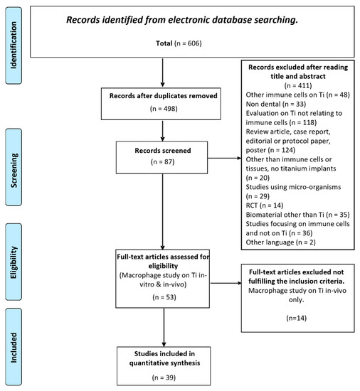
Figure 1.
PRISMA flowchart of the screening process for the selected databases.
3.2. Surface Topography
The most studied biomaterial was commercially available titanium alloy (Ti-6Al-4V) used in either a disc or screw shape of various diameters and dimensions with a range of surface modifications (Table 2). The surface modifications could be broadly grouped according to the methodology used in their production, i.e., (1) subtractive techniques (16 studies), e.g., sandblasting, acid etching; (2) additive techniques (11 studies), e.g., plasma spraying, nanotube formation, or (3) coating techniques (17 studies), e.g., covering the native titanium surface with inorganic or organic polymers. Of the subtractive techniques, a combination of sandblasting and acid etching were the most frequently used surface modifications [18,19,20,21,22,23,24,25,26,27,28,30,31,32] producing a hydrophobic microrough surface. Of these, five studies (36%) subsequently induced super-hydrophilicity to the already sandblasted and acid etched surface [19,21,23,28,31]. Of the additive techniques, the induction of nanoscale roughness or nanotube formation (internal diameters from 30–100 nm) by anodization at 5–100 volts were the most prevalent [13,22,33,34,35,36,37,38,39]. Micro-arc oxidation was used in a further two studies [40,41] to produce a micro-rough surface. Four studies included both subtractive or additive and coated surfaces for comparison [25,27,32,42]. Inorganic osteogenic materials, e.g., hydroxy apatite, chitosan and zirconia were the most frequently used coating materials [25,27,32,42,43,44,45,46,47,48,49,50,51], while a smaller number of studies examined the effects of natural compounds such as bacterial lipopolysaccharide (LPS) [52], interleukin-4 (IL-4) [53], silk fibroin [54] and collagen [55].
Table 2.
Summary data (Cell type, Surface Modification and Surface Coating) of studies included in review. Both primary bone-derived macrophages as well as commercial cell-lines were included for analysis. Given the articles were selected based on the titanium eliciting an immune (macrophage) response, surface modification techniques were relatively evenly split between subtractive (41%) additive (28%) and coated (44%). Note that some studies included more than one methodology. (NA) not applicable in this study, (NR) not recorded in this study.
Macrophages studied included both primary human (7 studies) and rodent cells (5 studies) and those from human (THP-1, 2 studies) and rodent monocytic cell lines, i.e., J774.A1 (3 studies) and RAW 264.7 (22 studies) (Table 2).
3.3. Cell Morphology
Cell shape changes have often been associated with different functional states of cells. McWhorter (2013) demonstrated that macrophages polarized toward different phenotypes in vitro also exhibited dramatic changes in cell shape, with M2 cells exhibiting an elongated shape compared with M1 cells [56]. As such, striking morphological differences in macrophages were also reported in many of the included studies cultured on the various surfaces (Table 3 and Table 4). Although not examined in a consistent manner, some studies reviewed showed oval shaped M1 associated cells were often seen on polished titanium surfaces, e.g., studies [13,23,38], whereas surface modification to include plasma spraying [13] and/or the addition of nanoscale features [13,26,34,38] was associated with significant cell spreading and elongation. Indeed, the study by Pan (2017) showed that the further addition of nanofeatures to an already plasma sprayed surface resulted in multi-directional elongation and spreading and 3D distribution of the cytoskeleton [13].
Table 3.
Morphological changes associated with an M2 macrophage phenotype were grouped according to whether this occurred on either a ‘smooth’ or ‘rough’ titanium surface. To facilitate this, given the multitude of ways the surfaces were prepared, a single roughness (Ra) value was used to group the data. M2 phenotype was significantly associated with attachment to the rough surfaces (chi-square statistic with Yates correction 29.301, p-value < 0.001).
Table 4.
Summary data (Morphological and Proliferative Changes) of studies included in review. (NA) not applicable in this study, (NR) not recorded in this study.
Overall, of the 39 studies selected in this review, 25 studies (64%) also reported morphological changes in cells that were attached to the test (modified) surface when compared to the control surface (Table 3). To help assess the possible significance of surface driven morphological changes, the various surface topographies used these studies have been broadly grouped as either ‘smooth’ (Ra < 100 nm) or ‘rough’ (blasted, etched etc where Ra > 100 nm) accordingly. 96% of the studies on a rough surface described morphological changes such as ‘increased cytoplasmic volume’, ‘granularity’ and ‘extension of pseudopodia’ following culture compared to only 15% in macrophages cultured on a smooth titanium surface. Three or 11% of studies described morphological changes such as the cell shape becoming more ‘spindle-shaped’ or ‘elongated’, developing ‘pseudopodia extensions’ or ‘cell-spreading’ or ‘fully spread lamellipodia interacting with the surfaces’ on both smooth and rough titanium surfaces [47,53,55]. Only one study (2.5%) however showed the changes in macrophage morphology were more prominent on the smooth compared to rough titanium surfaces [23].
Subsequent statistical analysis clearly supports the proposal that a rough titanium implant topography promotes the activation of an M2 macrophage phenotype in adherent cells (chi-square statistic with Yates correction 29.301, p-value < 0.001).
3.4. Cellular Response
Regardless of the type of surface modification fabricated on the titanium implants, almost all studies included in this review reported significant immunological responses by the macrophages (Table 5). Of the 39 studies selected, only 5% of studies did not report any measure of cytokine expression by macrophages [18,48]. In the remaining 37 studies, inflammatory cytokine gene expression (as measured by either fold change or relative expression levels) or secreted cytokine levels (measured by immunoassay) were upregulated on rough titanium surfaces compared to the smooth surfaces. E.g., the microrough surface topography produced by combined sand blasting with acid etching of the titanium surface [19,21,23,24,25,31,32], was shown to induce the upregulation of pro-inflammatory cytokine gene expression, or cytokine secretion of IL-1β, IL-6 and TNF-α in adherent macrophages. Further modification of rough surfaces to induce super-hydrophilicity [18,19,21,23,28,30,31,32,39] showed this modification could induce a switch in macrophage phenotype from pro-inflammatory to a regenerative ‘M2-like’ phenotype, i.e., upregulation of IL-4 and IL-10 expression and concurrent down-regulation of the pro-inflammatory markers.
Table 5.
Summary data (Osteogenic activity and Gene and Cytokine Expression changes) of studies included in review. (NR) not recorded in this study.
These studies (Table 5) suggest this surface modification, i.e., hydrophilicity, could potentially promote faster wound healing during osseointegration. In vitro evidence to support this hypothesis was clearly demonstrated in those studies [28,31,32] in which the osteogenic effects of this titanium surface-derived macrophage phenotypic changes on osteoblasts were examined using co-culture or conditioned media studies, i.e., the highest levels of mesenchymal stem cell (MSC) recruitment were seen with either M2 activated macrophages or macrophages on rough hydrophilic titanium [28]. Osteoblasts co-cultured with macrophages on modSLA titanium surfaces resulted in >2-fold increases in the expression of TGFβ / BMP signalling genes [31], and increased expression of RunX2, Sp7, Bglap, Alp, Bmp2 and Vegf was seen in co-cultured MSC’s [32].
Similar results of significant paracrine osteogenic effects in osteoblasts or osteoblast progenitors (Table 5) were also shown in many of the included studies [22,27,32,33,34,35,36,37,38,39,40,41,43], whereby the titanium surface modification resulted in a shift of macrophage phenotype (M1 to M2), e.g., increased β-catenin and osterix expression [27] and increased TGFβ1, BMP2, VEGF and decreased TRAP expression [40].
Further studies carried out on titanium surfaces modified at the nanoscale level showed that incorporating nanotubes or grooves onto the surface could also result in a favourable osteo-immunomodulatory microenvironment [26,34,35,36,37,38]. The importance of the physical dimensions of the nanotubes was further shown to be an important factor on the behaviour of macrophages whereby smaller diameter nanotubes were associated with an M1 macrophage phenotype characterized by high levels of secreted IL-1β, TNF-α and iNOS compared to larger diameter nanotubes which induced an anti-inflammatory M2-like macrophage phenotype in adherent macrophages with enhanced IL-10 and Arg-1 gene expression [34,35,36,37]. These results agree with those by Chamberlain where 70 nm diameter was demonstrated to be optimal for minimizing the macrophage inflammatory response [57]. In a further nanotube modified surface that also incorporated zinc [38], adherent macrophages showed enhanced gene and protein expression of the M2 markers TGF-β and heme oxygenase-1, whereas M1 markers TNF-α and IL-6 were moderately inhibited thus establishing an osteogenic microenvironment that would be conducive for bone formation. Similarly, other nanoscale modified titanium surfaces produced by etching [22], plasma-spraying [13] or nano-wire addition [33,39] also promoted immunomodulatory behavior that would favor osteogenesis and angiogenesis.
Of the studies using a surface coating on titanium to support the migration, proliferation and differentiation of macrophages and reduction in the inflammatory response, the application of hydroxyapatite significantly increased macrophage adhesion and downregulated pro-inflammatory mediators [43,44,46,47,49]. Similarly, various other biomaterial coatings, e.g., Bioglass [44], silk sericin [45], LPS [52], Chitosan [48], IL-4 [53], Zinc [42], GPTMS [50], wound-healing peptides [54], aspirin [55] and anti-osteoporosis drugs [51], showed functionalization of the surface while maintaining the underlying topography (Table 5). This could also significantly increase the initial attachment of immune cells and alter the immune cells response, although this result was dependent upon the biomimetic agent(s) used.
As for native titanium, hydrophilic modification of a zirconia-titanium alloy (RXD -Roxolid®, Straumann, Basel, Switzerland), similarly downregulated pro-inflammatory cytokines IL-1β and IL-6, producing an anti-inflammatory microenvironment by inducing macrophage activation similar to the anti-inflammatory M2-like state and increased levels of the cytokines IL-4 and IL-10 [23,25,28]. In fact, this hydrophilic implant (RXD-SLActive), when compared to other immune-modulatory titanium surfaces induced the highest level of osteogenic factor released from MSC’s and anti-inflammatory factors from macrophages with the lowest level of pro-inflammatory factors [32].
3.5. Risk of Bias
In this review, the thirty-nine studies were all found to have reliability scores of ≥15 (Klimisch category 1) indicating that data from these studies is reliable without any restrictions (Supplementary Table S1). None of the studies had any unreliable data (category 3).
4. Discussion
The findings of the studies (Table 6) included in this systematic review, clearly support the hypothesis that incorporation of titanium surface modifications increasing surface roughness and hydrophilicity with or without additional application of bioactive coatings, can promote a regenerative or M2-like phenotype in adherent macrophages which may then have the potential to enhance osteogenesis in BMSC’s in a paracrine manner.
Table 6.
Conclusions of studies included in review.
More specifically, titanium surface roughness is well known to increase the surface area of implants and ultimately enhance osseointegration when compared with smooth surfaced implants, however further modification of this rough surface to increase surface energy thus promoting super-hydrophilicity, not only down-regulates the initial pro-inflammatory response by macrophages, but up-regulates an anti-inflammatory phenotype able to further promote wound healing. Topography-directed macrophage polarization is therefore a biologically feasible mechanism to assist in the design of implant surfaces aimed at promoting osteogenesis and osseointegration. Unfortunately, this review of in vitro studies only does not allow any determination of whether an amelioration of inflammation or promotion of anti-inflammatory mediators may be responsible for improved osseointegration seen clinically with specific surface modified titanium implants.
Of the 39 papers reviewed in this study, only one, Morra et al. (2015), provided data that suggested some caution should be used when assessing the potential impact of surface modification on the subsequent crosstalk between cells of the immune and skeletal systems [52]. These authors showed commercially available dental implants induced variable levels of expression of endotoxin-stimulated pro-inflammatory genes such as IL-1, IL-6, TNF-α, MCP-1, COX-2, and MCSF in murine J774-A1 macrophages, sometimes above the level expected to promote bone resorption in vivo. Moreover, the results were unaffected by the specific surface treatment; rather, they more likely reflected the level of care in the cleaning and packaging protocols of the manufacturers. Evaluation of adherent endotoxin should therefore be reappraised and considered amongst the relevant surface properties of implantable biomaterials for proper understanding of the tissue response to implants.
The mechanism through which the presence of a rough, hydrophilic surface topography and chemistry affects the osteoimmune response of macrophages in comparison to other distinct topographies is not yet understood. Li et al. (2020) showed higher hydrophilicity and surface free energy in anisotropic nanowire-like textured titanium promoted the availability of RGD binding domains in fibronectin and fibrinogen adsorbed onto the titanium surface [39]. These tripepetide Arg-Gly-Asp (RGD) domains provided more α5β1-integrin-specific instructions to MSCs, enhancing cell spreading and osteogenic differentiation. Furthermore, the authors suggested the combination of integrin α5-induced cell spreading and suppression of the interaction between fibrinogen and the integrin αM subunit, could act synergistically to cause accumulation of M2 macrophages on the nanowire-like textured surface.
In a subsequent coculture model, MSCs on the nanostructured surface exerted greater effects on naïve and M1 macrophages, causing them to adopt a less inflammatory macrophage profile characterized by reduced expression of IL-6 and TNF-α and concurrent increased expression of IL-10 and Arg1.
Alternatively, Pan et al. (2017) suggested the immunomodulatory properties of a plasma sprayed nanotextured surface, whereby macrophages were found to switch to M2 phenotype with decreased levels of inflammatory gene expression as well as increased expression of anti-inflammatory genes, were probably regulated by the ‘decisive’ role of cytoskeleton tension induced by specific cell shape when macrophages were cultured on this surface [13].
Regardless of the precise mechanism(s) responsible, biomaterial surface cues from immuno-modulatory surfaces interpreted by macrophages, results in the secretion of distinct cytokine profiles that are able to modulate osteogenic gene expression in osteoblasts in a paracrine fashion. Previous studies by our group have shown this may occur as a result of upregulation of the TGF-β/BMP signalling pathway [31]. Sun et al. (2013) also showed that TiO2 nanotube layers could stimulate RAW 264.7 macrophages to secrete BMP-2 in contrast to smooth surfaces [58]. Increasing nanostructure tube diameter further stimulated BMP-2 secretion. Further mechanistic studies by Li et al. (2020) also demonstrated that BMSC’s cultured on plasma sprayed nanotextured titanium mediated this immunomodulation via a ROCK-medicated COX2 pathway to enhance PGE2 production, which in turn acted on macrophages through the EP4 receptor and partially abrogated the activation of proinflammatory factors, specifically IL-6 [39].
An unfortunate limitation of this study was that a meta-analysis of the included papers was not able to be performed due to the significant heterogeneity found in the methods and outcome data presented by the study authors. Semiquantitative analysis where possible however (Table 3), did support in the affirmative that surface modification of dental implant surfaces could promote a regenerative macrophage phenotype as proposed in the research question. Lack of data on the physicochemical and mechanical properties of the titanium used in the included studies also presented as a major limitation of this review. Similarly, while most studies included subsequent osteogenic analyses in osteoblasts using co-culture or conditioned media, few examined any macrophage driven effects on mineralisation. While this disparity in reported studies continues, a strategy that similar future systematic reviews without meta-analysis could follow in order to allow for later synthesis has been proposed [59]. These ‘SWiM’ (synthesis without meta-analysis) guidelines have been included as Supplementary Data (Supplementary Table S2).
Finally, to help establish the veracity of our PICO question without the complications of unknown or unaccounted for systemic effects, this review has focused only on data arising from in vitro studies. Given the positive outcome, further systematic assessment of appropriate in vivo studies is now required to delineate the role of the biomaterial surface on the modulation of macrophage phenotype on in vivo osteogenesis.
5. Conclusions
In attempts to try and mimic the native tissue microenvironment, surface-modified titanium has been shown to modulate the function of adherent macrophages. Whilst any implanted device will result in an initial inflammatory response, modification of the device’s physiochemical properties to make it hydrophilic, treatment to add nanotube structures to the surface, or the addition of bio-functional surface coatings such as hydroxyapatite may reduce this initial inflammatory response and up-regulate a more regenerative phenotype in adherent macrophages, as suggested by the selected papers reviewed in this study. In vivo studies are now required to determine if these various modifications of potential implant surfaces will facilitate an enhanced rate or degree of osteogenesis not only in healthy individuals but also in immune-compromised patients.
Supplementary Materials
The following supporting information can be downloaded at: https://www.mdpi.com/article/10.3390/ma15207314/s1, Table S1: ToxRTOOL assessment tool [16]. A binary score (1 or 0) was determined for each of eighteen reliability criteria; Table S2: Synthesis without Meta-analysis (SWiM) reporting items.
Author Contributions
Conceptualization, M.P., D.I., S.T. and S.H.; Methodology, M.P., D.I. and S.T; Data curation, M.P., D.I. and S.T; writing—original draft preparation, M.P.; writing—review and editing, S.T. and S.H.; Supervision, D.I. and S.H. All authors have read and agreed to the published version of the manuscript.
Funding
This research received no external funding.
Institutional Review Board Statement
Not applicable.
Informed Consent Statement
Not applicable.
Data Availability Statement
Not applicable.
Conflicts of Interest
The authors declare no conflict of interest.
References
- Kaur, M.; Singh, K. Review on titanium and titanium based alloys as biomaterials for orthopaedic applications. Mater. Sci. Eng. C Mater. Biol. Appl. 2019, 102, 844–862. [Google Scholar] [CrossRef] [PubMed]
- Donath, K.; Kirsch, A.; Osborn, J.F. Zelluläre Dynamik um enossale Titanimplantate. Fortschr. Zahnärztl. Implantol. 1984, 1, 55–58. [Google Scholar]
- Pajarinen, J.; Kouri, V.-P.; Jämsen, E.; Li, T.-F.; Mandelin, J.; Konttinen, Y.T. The response of macrophages to titanium particles is determined by macrophage polarization. Acta Biomater. 2013, 9, 9229–9240. [Google Scholar] [CrossRef] [PubMed]
- Sridharan, R.; Cameron, A.R.; Kelly, D.J.; Kearney, C.J.; O’Brien, F.J. Biomaterial based modulation of macrophage polarization: A review and suggested design principles. Mater. Today 2015, 18, 313–325. [Google Scholar] [CrossRef]
- Arron, J.R.; Choi, Y. Bone versus immune system. Nature 2000, 408, 535–536. [Google Scholar] [CrossRef] [PubMed]
- Cui, Y.; Li, H.; Li, Y.; Mao, L. Novel insights into nanomaterials for immunomodulatory bone regeneration. Nanoscale Adv. 2021, 4, 334–352. [Google Scholar] [CrossRef]
- Guder, C.; Gravius, S.; Burger, C.; Wirtz, D.C.; Schildberg, F.A. Osteoimmunology: A Current Update of the Interplay between Bone and the Immune System. Front. Immunol. 2020, 11, 58. [Google Scholar] [CrossRef]
- Mills, C.D.; Kincaid, K.; Alt, J.M.; Heilman, M.J.; Hill, A.M. M-1/M-2 Macrophages and the Th1/Th2 Paradigm. J. Immunol. 2000, 164, 6166–6173. [Google Scholar] [CrossRef]
- Mantovani, A.; Sica, A.; Locati, M. Macrophage Polarization Comes of Age. Immunity 2005, 23, 344–346. [Google Scholar] [CrossRef]
- Wennerberg, A.; Albrektsson, T. Effects of titanium surface topography on bone integration: A systematic review. Clin. Oral Implants Res. 2009, 20, 172–184. [Google Scholar] [CrossRef]
- Jennissen, H.P. Ultra-Hydrophilic Transition Metals as Histophilic Biomaterials. Macromol. Symp. 2005, 225, 43–70. [Google Scholar] [CrossRef]
- Schwarz, F.; Wieland, M.; Schwartz, Z.; Zhao, G.; Rupp, F.; Geis-Gerstorfer, J.; Schedle, A.; Broggini, N.; Bornstein, M.M.; Buser, D.; et al. Potential of chemically modified hydrophilic surface characteristics to support tissue integration of titanium dental implants. J. Biomed. Mater. Res. Part B Appl. Biomater. 2008, 88, 544–557. [Google Scholar] [CrossRef] [PubMed]
- Pan, H.; Xie, Y.; Zhang, Z.; Li, K.; Hu, D.; Zheng, X.; Tang, T. Immunomodulation effect of a hierarchical macropore/nanosurface on osteogenesis and angiogenesis. Biomed. Mater. 2017, 12, 45006. [Google Scholar] [CrossRef] [PubMed]
- Richardson, W.S.; Wilson, M.C.; Nishikawa, J.; Hayward, R.S. The well-built clinical question: A key to evidence-based decisions. ACP J. Club 1995, 123, A12–A13. [Google Scholar] [CrossRef]
- Johnson, B.T.; Hennessy, E.A. Systematic reviews and meta-analyses in the health sciences: Best practice methods for research syntheses. Soc. Sci. Med. 2019, 233, 237–251. [Google Scholar] [CrossRef]
- Schneider, K.; Schwarz, M.; Burkholder, I.; Kopp-Schneider, A.; Edler, L.; Kinsner-Ovaskainen, A.; Hartung, T.; Hoffmann, S. “ToxRTool”, a new tool to assess the reliability of toxicological data. Toxicol. Lett. 2009, 189, 138–144. [Google Scholar] [CrossRef]
- Klimisch, H.-J.; Andreae, M.; Tillmann, U. A Systematic Approach for Evaluating the Quality of Experimental Toxicological and Ecotoxicological Data. Regul. Toxicol. Pharmacol. 1997, 25, 1–5. [Google Scholar] [CrossRef]
- Milleret, V.; Tugulu, S.; Schlottig, F.; Hall, H. Alkali treatment of microrough titanium surfaces affects macrophage/monocyte adhesion, platelet activation and architecture of blood clot formation. Eur. Cells Mater. 2011, 21, 430–444. [Google Scholar] [CrossRef]
- Hamlet, S.; Alfarsi, M.; George, R.; Ivanovski, S. The effect of hydrophilic titanium surface modification on macrophage inflammatory cytokine gene expression. Clin. Oral Implants Res. 2011, 23, 584–590. [Google Scholar] [CrossRef]
- Barth, K.A.; Waterfield, J.D.; Brunette, D.M. The effect of surface roughness on RAW 264.7 macrophage phenotype. J. Biomed. Mater. Res. Part A 2013, 101A, 2679–2688. [Google Scholar] [CrossRef]
- Alfarsi, M.A.; Hamlet, S.M.; Ivanovski, S. Titanium surface hydrophilicity modulates the human macrophage inflammatory cytokine response. J. Biomed. Mater. Res. Part A 2013, 102, 60–67. [Google Scholar] [CrossRef] [PubMed]
- Nagasawa, M.; Cooper, L.; Ogino, Y.; Mendonca, G.; Liang, R.; Yang, S.; Uoshima, K. Topography Influences Adherent Cell Regulation of Osteoclastogenesis. J. Dent. Res. 2015, 95, 319–326. [Google Scholar] [CrossRef] [PubMed]
- Hotchkiss, K.M.; Reddy, G.B.; Hyzy, S.L.; Schwartz, Z.; Boyan, B.D.; Olivares-Navarrete, R. Titanium surface characteristics, including topography and wettability, alter macrophage activation. Acta Biomater. 2015, 31, 425–434. [Google Scholar] [CrossRef] [PubMed]
- Eger, M.; Sterer, N.; Liron, T.; Kohavi, D.; Gabet, Y. Scaling of titanium implants entrains inflammation-induced osteolysis. Sci. Rep. 2017, 7, 39612. [Google Scholar] [CrossRef]
- Hotchkiss, K.M.; Ayad, N.B.; Hyzy, S.L.; Boyan, B.D.; Olivares-Navarrete, R. Dental implant surface chemistry and energy alter macrophage activation in vitro. Clin. Oral Implant. Res. 2016, 28, 414–423. [Google Scholar] [CrossRef]
- Kianoush, F.; Nematollahi, M.; Waterfield, J.D.; Brunette, D.M. Regulation of RAW264.7 macrophage polarization on smooth and rough surface topographies by galectin-3. J. Biomed. Mater. Res. Part A 2017, 105, 2499–2509. [Google Scholar] [CrossRef] [PubMed]
- Choi, S.-M.; Park, J.-W. Multifunctional effects of a modification of SLA titanium implant surface with strontium-containing nanostructures on immunoinflammatory and osteogenic cell function. J. Biomed. Mater. Res. Part A 2018, 106, 3009–3020. [Google Scholar] [CrossRef]
- Hotchkiss, K.M.; Clark, N.M.; Olivares-Navarrete, R. Macrophage response to hydrophilic biomaterials regulates MSC recruitment and T-helper cell populations. Biomaterials 2018, 182, 202–215. [Google Scholar] [CrossRef] [PubMed]
- Yang, C.; Sun, Y.; Yu, W.; Yin, X.; Weng, J.; Feng, B. Modulation of macrophage phenotype through controlled release of interleukin-4 from gelatine coatings on titanium surfaces. Eur. Cells Mater. 2021, 36, 15–29. [Google Scholar] [CrossRef] [PubMed]
- Becker, M.; Quabius, S.; Kewitz, T.; Hansen, L.; Becker, G.; Kern, M.; Kersten, H.; Harder, S. In vitro proinflammatory gene expression changes in human whole blood after contact with plasma-treated implant surfaces. J. Cranio-Maxillofac. Surg. 2019, 47, 1255–1261. [Google Scholar] [CrossRef] [PubMed]
- Hamlet, S.M.; Lee, R.S.; Moon, H.; Alfarsi, M.A.; Ivanovski, S. Hydrophilic titanium surface-induced macrophage modulation promotes pro-osteogenic signalling. Clin. Oral Implant. Res. 2019, 30, 1085–1096. [Google Scholar] [CrossRef] [PubMed]
- Hotchkiss, K.M.; Sowers, K.T.; Olivares-Navarrete, R. Novel in vitro comparative model of osteogenic and inflammatory cell response to dental implants. Dent. Mater. 2019, 35, 176–184. [Google Scholar] [CrossRef] [PubMed]
- Zhu, W.-Q.; Shao, S.-Y.; Xu, L.-N.; Chen, W.-Q.; Yu, X.-Y.; Tang, K.-M.; Tang, Z.-H.; Zhang, F.; Qiu, J. Enhanced corrosion resistance of zinc-containing nanowires-modified titanium surface under exposure to oxidizing microenvironment. Nanobiotechnology 2019, 17, 55. [Google Scholar] [CrossRef]
- Ma, Q.-L.; Zhao, L.-Z.; Liu, R.-R.; Jin, B.-Q.; Song, W.; Wang, Y.; Zhang, Y.-S.; Chen, L.-H. Improved implant osseointegration of a nanostructured titanium surface via mediation of macrophage polarization. Biomaterials 2014, 35, 9853–9867. [Google Scholar] [CrossRef]
- Wang, J.; Qian, S.; Liu, X.; Xu, L.; Miao, X.; Xu, Z.; Cao, L.; Wang, H.; Jiang, X. M2 macrophages contribute to osteogenesis and angiogenesis on nanotubular TiO2 surfaces. J. Mater. Chem. B 2017, 5, 3364–3376. [Google Scholar] [CrossRef] [PubMed]
- Wang, J.; Meng, F.; Song, W.; Jin, J.; Ma, Q.; Fei, D.; Fang, L.; Chen, L.; Wang, Q.; Zhang, Y. Nanostructured titanium regulates osseointegration via influencing macrophage polarization in the osteogenic environment. Int. J. Nanomed. 2018, 13, 4029–4043. [Google Scholar] [CrossRef] [PubMed]
- Ma, Q.; Fang, L.; Jiang, N.; Zhang, L.; Wang, Y.; Zhang, Y.-M.; Chen, L.-H. Bone mesenchymal stem cell secretion of sRANKL/OPG/M-CSF in response to macrophage-mediated inflammatory response influences osteogenesis on nanostructured Ti surfaces. Biomaterials 2018, 154, 234–247. [Google Scholar] [CrossRef]
- Chen, B.; You, Y.; Ma, A.; Song, Y.; Jiao, J.; Song, L.; Shi, E.; Zhong, X.; Li, Y.; Li, C. Zn-Incorporated TiO2 Nanotube Surface Improves Osteogenesis Ability Through Influencing Immunomodulatory Function of Macrophages. Int. J. Nanomed. 2020, 15, 2095–2118. [Google Scholar] [CrossRef] [PubMed]
- Li, K.; Liu, S.; Hu, T.; Razanau, I.; Wu, X.; Ao, H.; Huang, L.; Xie, Y.; Zheng, X. Optimized Nanointerface Engineering of Micro/Nanostructured Titanium Implants to Enhance Cell–Nanotopography Interactions and Osseointegration. ACS Biomater. Sci. Eng. 2020, 6, 969–983. [Google Scholar] [CrossRef] [PubMed]
- Bai, L.; Liu, Y.; Du, Z.; Weng, Z.; Yao, W.; Zhang, X.; Huang, X.; Yao, X.; Crawford, R.; Hang, R.; et al. Differential effect of hydroxyapatite nano-particle versus nano-rod decorated titanium micro-surface on osseointegration. Acta Biomater. 2018, 76, 344–358. [Google Scholar] [CrossRef] [PubMed]
- Bai, L.; Du, Z.; Du, J.; Yao, W.; Zhang, J.; Weng, Z.; Liu, S.; Zhao, Y.; Liu, Y.; Zhang, X.; et al. A multifaceted coating on titanium dictates osteoimmunomodulation and osteo/angio-genesis towards ameliorative osseointegration. Biomaterials 2018, 162, 154–169. [Google Scholar] [CrossRef] [PubMed]
- Zhang, R.; Liu, X.; Xiong, Z.; Huang, Q.; Yang, X.; Yan, H.; Ma, J.; Feng, Q.; Shen, Z. The immunomodulatory effects of Zn-incorporated micro/nanostructured coating in inducing osteogenesis. Artif. Cells Nanomed. Biotechnol. 2018, 46, 1123–1130. [Google Scholar] [CrossRef]
- Takebe, J.; Ito, S.; Champagne, C.; Cooper, L.; Ishibashi, K. Anodic oxidation and hydrothermal treatment of commercially pure titanium surfaces increases expression of bone morphogenetic protein-2 in the adherent macrophage cell line J774A.1. J. Biomed. Mater. Res. Part A 2007, 80, 711–718. [Google Scholar] [CrossRef] [PubMed]
- Scislowska-Czarnecka, A.; Menaszek, E.; Szaraniec, B.; Kolaczkowska, E. Ceramic modifications of porous titanium: Effects on macrophage activation. Tissue Cell 2012, 44, 391–400. [Google Scholar] [CrossRef] [PubMed]
- Nayak, S.; Dey, T.; Naskar, D.; Kundu, S.C. The promotion of osseointegration of titanium surfaces by coating with silk protein sericin. Biomaterials 2013, 34, 2855–2864. [Google Scholar] [CrossRef] [PubMed]
- Wu, C.; Chen, Z.; Yi, D.; Chang, J.; Xiao, Y. Multidirectional Effects of Sr-, Mg-, and Si-Containing Bioceramic Coatings with High Bonding Strength on Inflammation, Osteoclastogenesis, and Osteogenesis. ACS Appl. Mater. Interfaces 2014, 6, 4264–4276. [Google Scholar] [CrossRef] [PubMed]
- Wu, C.; Chen, Z.; Wu, Q.; Yi, D.; Friis, T.; Zheng, X.; Chang, J.; Jiang, X.; Xiao, Y. Clinoenstatite coatings have high bonding strength, bioactive ion release, and osteoimmunomodulatory effects that enhance in vivo osseointegration. Biomaterials 2015, 71, 35–47. [Google Scholar] [CrossRef] [PubMed]
- Huang, L.; Luo, Z.; Hu, Y.; Shen, X.; Li, M.; Li, L.; Zhang, Y.; Yang, W.; Liu, P.; Cai, K. Enhancement of local bone remodeling in osteoporotic rabbits by biomimic multilayered structures on Ti6Al4V implants. J. Biomed. Mater. Res. Part A 2016, 104, 1437–1451. [Google Scholar] [CrossRef] [PubMed]
- Rydén, L.; Omar, O.; Johansson, A.; Jimbo, R.; Palmquist, A.; Thomsen, P. Inflammatory cell response to ultra-thin amorphous and crystalline hydroxyapatite surfaces. J. Mater. Sci. Mater. Med. 2016, 28, 9. [Google Scholar] [CrossRef] [PubMed]
- Araújo-Gomes, N.; Romero-Gavilán, F.; Zhang, Y.; Martinez-Ramos, C.; Elortza, F.; Azkargorta, M.; DE Llano, J.J.M.; Gurruchaga, M.; Goñi, I.; Beucken, J.V.D.; et al. Complement proteins regulating macrophage polarisation on biomaterials. Colloids Surfaces B Biointerfaces 2019, 181, 125–133. [Google Scholar] [CrossRef]
- Chen, M.; Huang, L.; Shen, X.; Li, M.; Luo, Z.; Cai, K.; Hu, Y. Construction of multilayered molecular reservoirs on a titanium alloy implant for combinational drug delivery to promote osseointegration in osteoporotic conditions. Acta Biomater. 2020, 105, 304–318. [Google Scholar] [CrossRef] [PubMed]
- Morra, M.; Cassinelli, C.; Bollati, D.; Cascardo, G.; Bellanda, M. Adherent Endotoxin on Dental Implant Surfaces: A Reappraisal. J. Oral Implant. 2015, 41, 10–16. [Google Scholar] [CrossRef] [PubMed]
- Zhang, Y.; Chen, S.E.; Shao, J.; van den Beucken, J.J.J.P. Combinatorial Surface Roughness Effects on Osteoclastogenesis and Osteogenesis. ACS Appl. Mater. Interfaces 2018, 10, 36652–36663. [Google Scholar] [CrossRef] [PubMed]
- He, Y.; Yang, X.; Yuan, Z.; Shen, X.; Xu, K.; Lin, C.; Tao, B.; Li, K.; Chen, M.; Hu, Y.; et al. Regulation of MSC and macrophage functions in bone healing by peptide LL-37-loaded silk fibroin nanoparticles on a titanium surface. Biomater. Sci. 2019, 7, 5492–5505. [Google Scholar] [CrossRef]
- Zhang, W.; Lu, X.; Yuan, Z.; Shen, M.; Song, Y.; Liu, H.; Deng, J.; Zhong, X.; Zhang, X. Establishing an osteoimmunomodulatory coating loaded with aspirin on the surface of titanium primed with phase-transited lysozyme. Int. J. Nanomed. 2019, 14, 977–991. [Google Scholar] [CrossRef]
- McWhorter, F.Y.; Wang, T.; Nguyen, P.; Chung, T.; Liu, W.F. Modulation of macrophage phenotype by cell shape. Proc. Natl. Acad. Sci. USA 2013, 110, 17253–17258. [Google Scholar] [CrossRef]
- Sun, S.J.; Yu, W.Q.; Zhang, Y.L.; Jiang, X.Q.; Zhang, F.Q. Effects of TiO2nanotube layers on RAW 264.7 macrophage behaviour and bone morphogenetic protein-2 expression. Cell Prolif. 2013, 46, 685–694. [Google Scholar] [CrossRef] [PubMed]
- Chamberlain, L.M.; Brammer, K.S.; Johnston, G.W.; Chien, S.; Jin, S. Macrophage Inflammatory Response to TiO2 Nanotube Surfaces. J. Biomater. Nanobiotechnol. 2011, 2, 293–300. [Google Scholar] [CrossRef]
- Campbell, M.; McKenzie, J.E.; Sowden, A.; Katikireddi, S.V.; Brennan, S.E.; Ellis, S.; Hartmann-Boyce, J.; Ryan, R.; Shepperd, S.; Thomas, J.; et al. Synthesis without meta-analysis (SWiM) in systematic reviews: Reporting guideline. BMJ 2020, 368, l6890. [Google Scholar] [CrossRef]
Publisher’s Note: MDPI stays neutral with regard to jurisdictional claims in published maps and institutional affiliations. |
© 2022 by the authors. Licensee MDPI, Basel, Switzerland. This article is an open access article distributed under the terms and conditions of the Creative Commons Attribution (CC BY) license (https://creativecommons.org/licenses/by/4.0/).