Article

First Verified Record and Morpho-Ecological Characterization of Bunodosoma cavernatum (Cnidaria: Actiniaria) in Cartagena Bay, Colombian Caribbean


by
M. Paula V-Boada
1,*,
Gabriel R. Navas-S
1 and
Lina M. Barrios
2,3
1
Biology Program, Universidad de Cartagena, Cra. 50 #24-120, Cartagena de Indias 130015, Colombia
2
Biodiversity- & Development-Solutions for Sustainability (B&D SOLUTIONS), Calle 72 No 13-23, piso 9, Edificio Nueva Granada, Bogotá D.C. 110231, Colombia
3
School of Biological and Marine Sciences, University of Plymouth, Drake Circus, Plymouth PL4 8AA, UK
*
Author to whom correspondence should be addressed.
Diversity 2026, 18(2), 118; https://doi.org/10.3390/d18020118
Submission received: 31 December 2025 / Revised: 9 February 2026 / Accepted: 9 February 2026 / Published: 12 February 2026
(This article belongs to the Special Issue Taxonomy, Phylogeny and Biogeography of Cnidaria)

Abstract

Bunodosoma cavernatum (Bosc, 1802) is a morphologically variable actinian species widely distributed in the Western Atlantic, yet poorly documented in Colombia. This study provides the first confirmed record of B. cavernatum in Cartagena Bay (Colombian Caribbean), extending its known distribution to a highly modified estuarine environment. The objectives were to (1) confirm the taxonomic identity of the species and characterize its external morphology, internal anatomy, histology and cnidome; (2) compare these traits with previously published Caribbean descriptions; (3) describe behavioral observations under controlled aquarium conditions; and (4) assess the abundance, microhabitat preferences and biological associations within the study area. Specimens were collected from shallow, sediment-rich substrates influenced by freshwater input from the Dique Channel. Diagnostic features were consistent with prior Caribbean descriptions. However, we report expanded cnidome size ranges and spirocysts in several tissues, not previously reported. Behavioral observations revealed rapid acclimation, consistent daily activity cycles, and sperm release associated with thermal variation (25–28 °C). Population density reached 32.2 individuals/m2, with an aggregated distribution around rocky or artificial substrates. These findings expand current knowledge of the ecological and reproductive plasticity of B. cavernatum in anthropogenically impacted tropical coastal ecosystems.

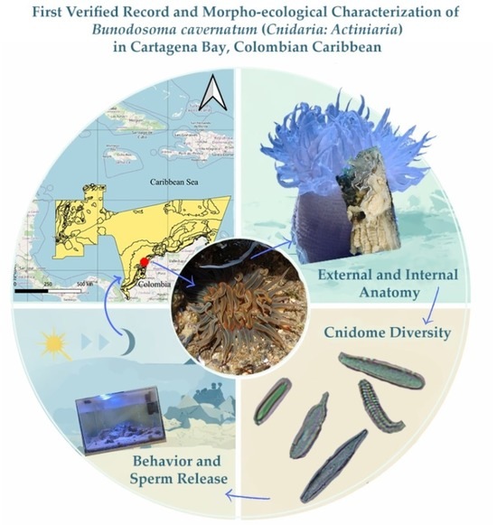
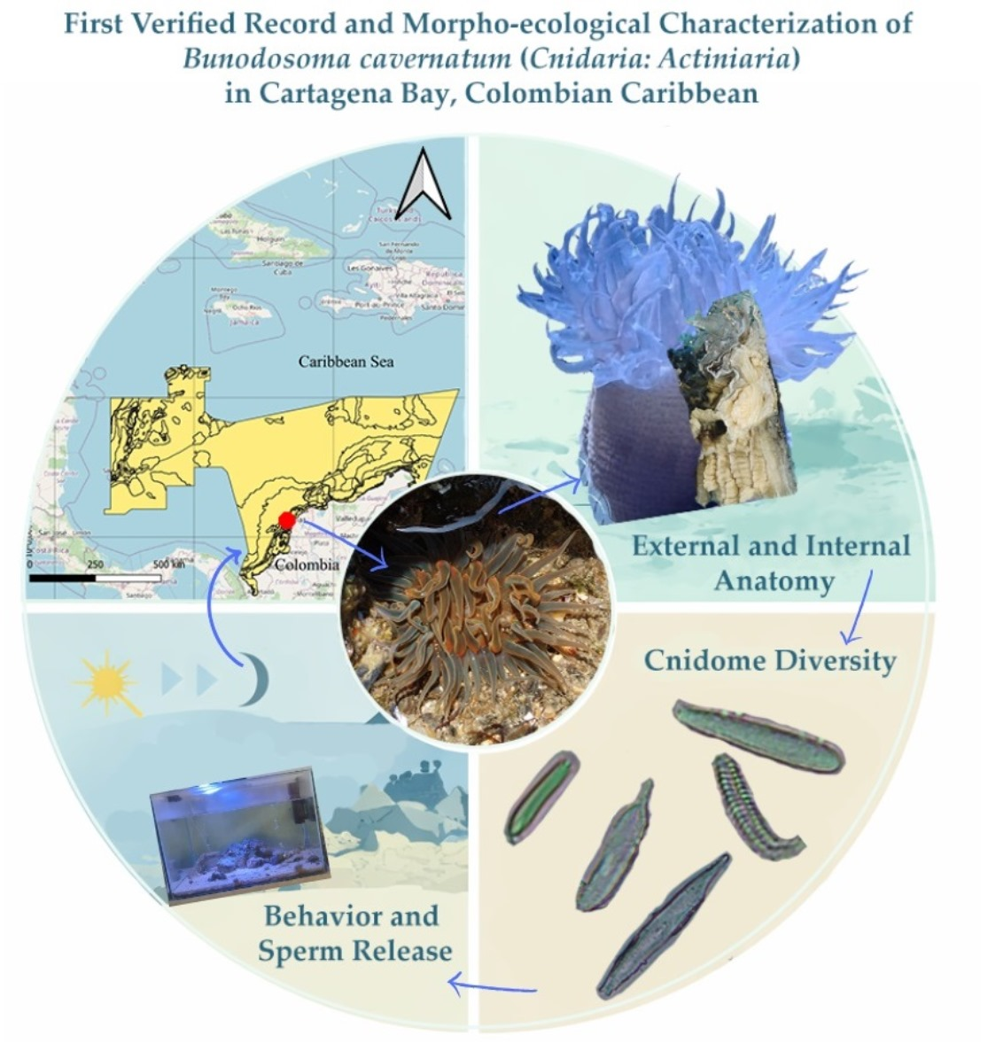
Graphical Abstract

1. Introduction

Sea anemones (Cnidaria: Anthozoa: Actiniaria) are among the most ecologically versatile and morphologically diverse benthic invertebrates [1,2]. These semi-sessile organisms exhibit a wide range of symbiotic interactions and environmental tolerances [3,4,5,6], playing key roles in coastal ecosystems. As habitat-forming species, actiniarians contribute to structural complexity and provide shelter for fishes and decapod crustaceans across a broad range of environments, from shallow coral reefs to deeper benthic zones [7]. Through the capture of particulate and dissolved organic matter, they contribute to nutrient cycling, support primary productivity, and sustain trophic networks within benthic communities [8,9]. Their trophic versatility and structural role allow actiniarians to form dense and heterogeneous assemblages across diverse substrates [10]. Moreover, several species—particularly those hosting symbiotic dinoflagellates—are sensitive to environmental stressors, making them valuable bioindicators of marine ecosystem health, particularly in response to thermal anomalies and anthropogenic pollution [9,11].
Despite their ecological relevance, sea anemones remain underrepresented in marine biodiversity inventories due to taxonomic complexity, cryptic morphology, and limited sampling effort [12,13,14]. This underrepresentation is particularly evident in Colombia [5,15,16,17], a region characterized by diverse coastal habitats, including mangroves, seagrass meadows, and coral reefs, which have been increasingly affected by urban development, industrial discharges, maritime traffic, and habitat degradation [18,19,20].
Cartagena Bay, located in the Department of Bolívar, is one of the Colombian coastal ecosystems most affected by human activities. The artificial estuarine system receives substantial inputs of freshwater and sediment from the Dique Channel, resulting in high turbidity, salinity fluctuations, and continuous deposition of organic and inorganic matter [21,22]. Nevertheless, the bay remains biologically productive and hosts a variety of benthic taxa, including actiniarians capable of withstanding variable environmental conditions.
Bunodosoma cavernatum (Bosc, 1802) is a sea anemone widely distributed throughout the Western Atlantic. It has been recorded from North Carolina to Barbados, the coasts of Texas (Puerto Aransas and Isabel), Louisiana, Beaufort, and the West Indies [3,23], the Yucatan Peninsula [24,25], the Caroline Islands, Micronesia [26] and the coasts of Nigeria [27]. In Colombia, the species was first reported in Caño Dulce (10°56′22″ N 75°01′42″ W) [28]; however, no confirmed record existed for Cartagena (10°24′34″ N 75°30′05″ W).
Despite its wide distribution, the identification of Bunodosoma cavernatum remains challenging due to extensive morphological overlaps with congeners, pronounced intraspecific variability, high chromatic variation, and tolerance to environments with elevated sedimentation and variable salinity [24,25,29]. This plasticity can obscure species boundaries, emphasizing the need for integrative approaches that combine external morphology, internal anatomy, cnidome composition, and ecological context for a reliable identification.
Considering the absence of confirmed records from Cartagena Bay and the limited national data on this species, the present study provides the first detailed record of B. cavernatum for the area. Specifically, it aims to confirm the identity of the species and (1) characterize its external and internal morphology, histology, and cnidome; (2) provide detailed descriptions of behavioral observations, including acclimatization, feeding, defensive responses, and sperm release in B. cavernatum maintained under controlled aquarium conditions; and (3) report on the local distribution, abundance, microhabitat preferences, and ecological associations of the species within the study area.

2. Materials and Methods

2.1. Study Area

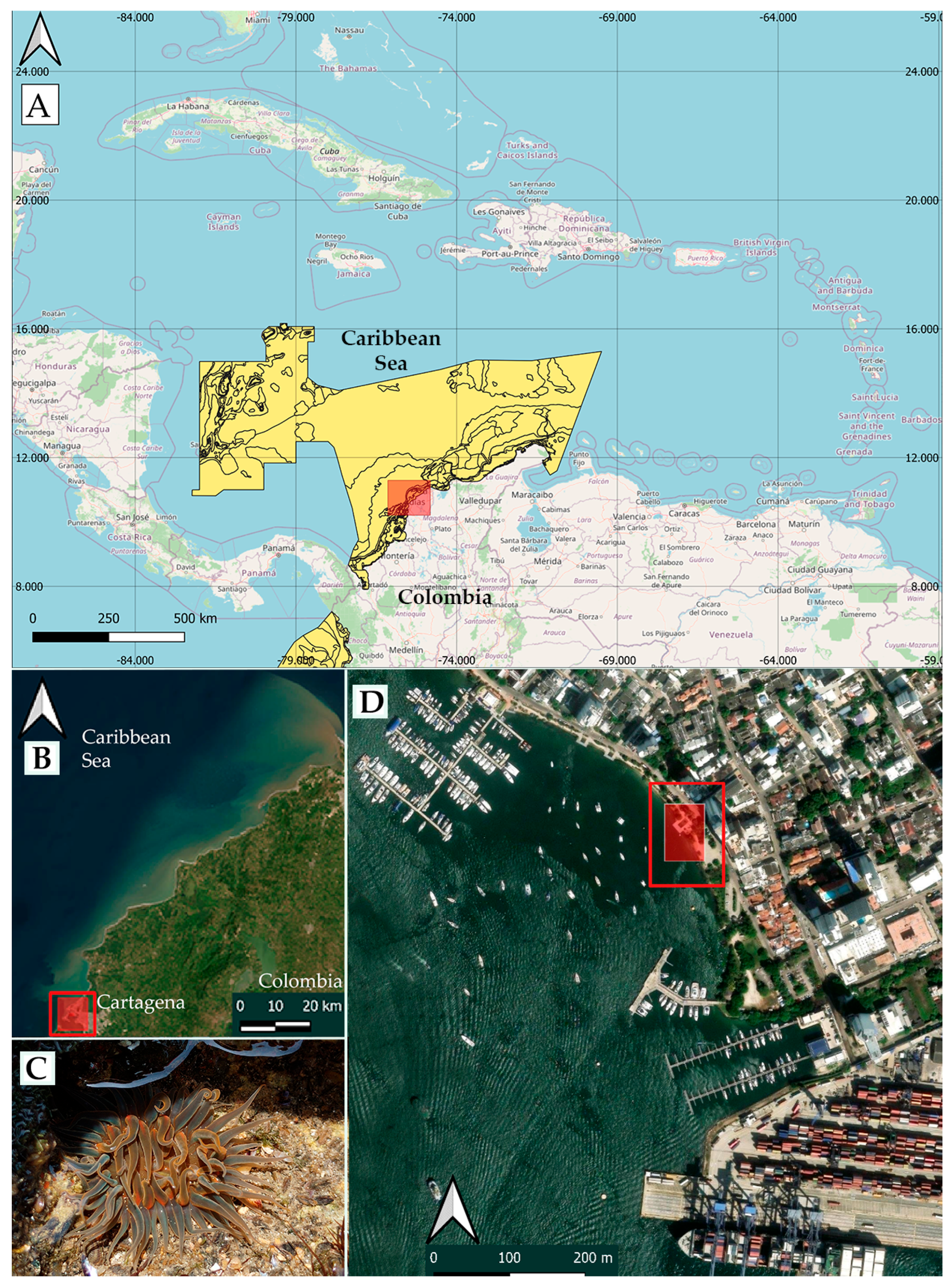
Specimens of Bunodosoma cavernatum were collected on 2 August 2024 in Cartagena Bay, part of the Bolívar Department in the Colombian Caribbean, at 10°24′37″ N and 75°32′20″ W (Figure 1). The sampling site was located at the transition between the mesolittoral and infralittoral zones, on a mixed substrate composed of compacted soft sediments and coral rock, under the direct hydrological influence of the Dique Channel.

2.2. Specimen Collection

Organisms occurring at depths between 0 and 0.50 m were located by direct observation while snorkeling and documented using an underwater camera and a dive torch. Individuals found on unconsolidated substrates were manually extracted following standardized protocols and placed in sealed polyethylene bags containing ambient seawater [30,31]. External morphology, behavioral traits, density, and spatial distribution were recorded in situ.

2.3. Estimation of Abundance and Distribution Pattern

Systematic sampling was performed in a zigzag arrangement using 20 quadrats (50 × 50 cm) placed at approximately 1.6 m intervals along a 30 m transect parallel to the coastline, at depths of 0–0.5 m (mesolittoral zone). The number of individuals per quadrant was recorded to calculate estimated density (ind/m2) and the distribution pattern was determined by descriptive statistics.

2.4. Analysis of Associated Organisms

To identify the organisms associated with Bunodosoma cavernatum, a qualitative microhabitat assessment was conducted by establishing a 15 cm radius around the oral disk of a randomly selected anemone in each quadrant. Biotic and abiotic components within the area were sampled following a microhabitat protocol adapted from [13,31].
The collected material was used for two complementary purposes. A representative portion was processed for taxonomic identification of associated organisms, while the remaining material was retained for subsequent use in the aquarium system to preserve natural sediment structure and local environmental conditions.
Organisms designated for analysis were manually separated under a stereomicroscope and identified using field guides and diagnostic keys. This assessment was qualitative and not intended to provide a quantitative inventory of the associated assemblage.

2.5. Material Examined

Thirteen adult specimens of B. cavernatum were collected in Manga Pier-Cartagena Bay (See Figure 1D). One specimen was fixed in 6% formalin for histological analysis. Three were preserved in 70% ethanol for morphological and anatomical examination, including transverse and longitudinal sections through the actinopharynx, column, and pedal disk. Additionally, two of these specimens were used for the characterization of the cnidome. The remaining nine individuals were maintained in aquarium conditions for behavioral observation and environmental tolerance studies; these specimens have not been released to date.

2.6. Aquarium Adaptation and Monitoring

In the laboratory, specimens were transferred to a 90 L aquarium system preconditioned for approximately 48 h using natural seawater collected from the sampling site. During this initial maturation phase, the system was equipped with continuous aeration, active filtration, a protein skimmer, and a controlled photoperiod (06:00 to 17:00), following the recommendations of [32].
After the system stabilization, specimens were introduced individually into a bare-bottom tank or placed directly onto the pebbles and cobbles to which they had been attached in the field. Adequate spacing between individuals was maintained to facilitate settlement and to minimize stress associated with physical contact or territorial interactions. The re-attachment process typically required 1–2 h, after which individuals were fully adhered to the substrate or tank bottom.
Natural substrate obtained during the microhabitat analysis of each collected anemone was added gradually to the aquarium. The substrate consisted of fine and coarse sand mixed with coral fragments, thereby preserving associated organisms and local sediment characteristics. And was added in successive thin layers and progressively increased to a final depth of approximately 5 cm, allowing individuals to acclimate between additions. This gradual process minimized disturbance and required 1 h.
Water quality parameters were monitored regularly to maintain stable physicochemical conditions. Salinity was measured weekly using a handheld refractometer, while pH, nitrite, nitrate, carbonate hardness, phosphate, KH, ammonia and calcium were assessed twice per month with the Reef Master Test Kit and Saltwater Master Test Kit (API Fish Care, Chalfont, PA, USA). A single aquarium was continuously monitored.
Organisms were fed every four days with shrimps and fresh fish, adjusting the portion size to each anemone. Behavioral observations were conducted by a single trained observer using direct visual observation throughout the study period. Observations were performed over 30 consecutive days, with four daily sessions corresponding to different phases of the photoperiod: before light onset, midday, end of the photoperiod and nighttime. Each session lasted 1 h, resulting in a total of 4 h of observations and an overall sampling effort of 120 h.
During each session, observations focused on the behavior of all individuals present in the aquarium. A combined observational approach was employed, consisting of continuous focal observation within each session and ad libitum recording of all relevant behaviors, particularly rare or sporadic events. Recorded behaviors included defensive response, territorial interactions, changes in posture or expansion, and a sperm-release event. All observations were performed under standardized aquarium conditions and without experimental manipulation.
Nighttime observations were conducted several hours after the end of the photoperiod. To minimize behavioral disturbance, observations were performed using very low indirect illumination and the camera’s night mode settings, allowing identification of structures and behaviors while avoiding strong light exposure. On the other hand, to check the reaction to light during the nighttime period on random nights, the main light was turned on.
The baseline state was defined as a fully extended posture maintained in the absence of identifiable external stimuli, including changes in light, tactile interactions, feeding events, or stressors. This baseline configuration served as a descriptive reference and was not treated as a quantitative response variable.
Postural states (PS) were defined as reference body configurations describing the morphological and muscular condition of B. cavernatum, independent of discrete stimulus-driven behaviors. These states were used as contextual references for interpreting stimulus-associated responses and were not considered behavioral categories for quantitative analysis.
PS1 (extended basal state) corresponds to a stable, fully turgid column with an open oral disk and extended tentacles oriented predominantly upward (~90°) toward the water current. The tentacles remain extended and sensitive to suspended particles, reflecting a functional disposition for feeding.
PS2 (fully contracted rigid state) describes a posture characterized by a contracted and rigid column, with the oral disk closed or partially hidden and the tentacles fully retracted. This state may precede or follow high-intensity events, such as defensive responses or the ingestion of large prey.
PS3 (relaxed expanded state) represents a low-activity postural configuration characterized by a lax expanded column with reduced muscular tone, doubling the length of the column observed in PS1, and extended tentacles, with minimal or no active movement. This state is most frequently observed under low-stimulation conditions, particularly during nocturnal phases, and is interpreted as a relaxed reference configuration distinct from the functionally receptive PS1 state. PS3 may constitute a transitional postural condition between postures.
Stimulus-dependent behaviors were recorded using a presence-absence matrix, in which each observation session constituted a sampling unit. For each individual and session, the presence (1) or absence (0) of each behavior category was documented. This approach allowed for the calculation of absolute and relative frequencies while preserving temporal consistency across individuals. Behavior frequencies were summarized.
During a spontaneous sperm release event, samples were collected directly from the oral disk using plastic Pasteur pipettes and stored in sterile 15 mL polypropylene tubes. Sperm emissions were taken at 4–10 min intervals with gentle suction to minimize agitation of the suspension. Eight samples (and 8 subsamples with ethanol, 4–10 min intervals) were obtained between 17:00 and 19:30 h, fixed with 70% ethanol (3:1 ratio) and refrigerated until microscopic analysis.
Microscopic observations were performed using two magnifications: 400× to count and estimate the density of spermatozoa across 40 fields of view, and 1000× to describe the morphology of spermatozoa and take measurements.

2.7. Anatomical and Histological Processing

Specimens were anesthetized in a 10% magnesium sulphate solution [13,33] and then fixed in 70% ethanol. Two individuals were dissected to examine external and internal anatomy through transverse sections at the actinopharynx and longitudinal sections along the column.
For histological analysis, tissues were fixed in 6% formalin and processed for dehydration using alcohol, followed by xylene and paraffin. The processing steps included sequential dehydration in graded alcohol: 70% alcohol (20 min.), 80% (20 min.), 90% (20 min.), 95% (20 min.), 95% (30 min.), 95% (40 min.), 100% (40 min.), 100% (100 min.); clearing in xylene (30 min.); and paraffin embedding: xylene-paraffin 1:1 (60 min.), paraffin at 58 °C (60 min.) and paraffin at 50 °C (60 min.); with resulting blocks measuring 2.5 × 2 × 1 cm. The subsequent 4 µm sections were stained with hematoxylin-eosin following [34], and the protocols of the Faculty of Medicine Histology Lab., at the University of Cartagena.
The resulting cross-sectional slices at the actinopharynx level and the three longitudinal sections from the actinopharynx to the pedal disk were used to document the mesenterial arrangement, siphonoglyphs, gonadal tissue and ectoderm structures.

2.8. Characterization of the Cnidome

Microslides were prepared from five anatomical regions: tentacles, column, acrorhagi, actinopharynx, and mesenterial filaments. Cnidocytes were observed under a compound optical microscope equipped with a digital imaging system at 400× and 500×; microphotographs were made with image analysis software HAYEAR (version x64.4.11.22070.20230204).
Measurements, identification, and classification of the nematocytes followed classical nomenclature [35,36,37,38], with a minimum of 20 and a maximum of 126 cnidocytes measured per type in each tissue, for both specimens examined.

3. Results

3.1. Sampling Area and Habitat

A total of 161 individuals of Bunodosoma cavernatum were recorded within the surveyed area, corresponding to an estimated density of 32.2 individuals/m2. The number of individuals per quadrant varied markedly, ranging from 0 to 30 individuals, indicating a highly heterogeneous spatial distribution.
The study area (Figure 1A–D) corresponds to a shallow estuarine zone of Cartagena Bay subject to strong anthropogenic influence. Common disturbances in the area include yachts, motorboat traffic, accumulation of organic waste and construction debris, and sediment discharge from the Dique Channel. These factors contribute to the estuarine characteristics of the bay, particularly in the mesolittoral zone (0–0.5 m depth), where various anthropogenic materials were observed on the seabed, including partially buried drainage pipes, rainwater outlets, bricks, concrete fragments, coral rock, glass bottles, and plastic waste. Several artificial structures, such as piers, were supported by wooden and PVC columns.
Fully expanded specimens of B. cavernatum—with tentacles extended and the oral disk open—were observed attached to compacted soft sediments, coral rubble, and artificial substrates, particularly in low-energy zones adjacent to the pier and other submerged structures (Figure 1). A progressive decline in abundance was noted with increasing distance from these structural elements.
Based on substrate type and spatial arrangement, two distinct microhabitats were identified. The first consisted of small clusters of individuals associated with dead coral fragments located away from artificial structures. The second comprised solitary, larger individuals occurring directly on unconsolidated sand lacking rock or hard substrate cover.

3.2. Associated Organisms

Bunodosoma cavernatum was found in close association with a diverse assemblage of benthic organisms spanning multiple taxonomic groups. The associated fauna included bivalves (Mytilidae, Ostreidae, Donacidae), gastropods (Calliostoma, Littorinidae, Nassariidae), and crustaceans (Cronius spp., Porcellanidae, Clibanarius tricolor, Clibanarius vittatus. Cirripedes, particularly Chthamalus spp., were frequently observed near the anemones, along with polychaetes belonging to the families Eunicidae and Sabellidae, and sponges (Porifera). In addition to invertebrate fauna, several algal groups were documented in the immediate vicinity of the sea anemones. These included filamentous algae (phyla Chlorophyta and Streptophyta) as well as other representatives of the phyla Phaeophyta and Chlorophyta, notably members of the family Caulerpaceae.

3.3. Taxonomy Account

Phylum Cnidaria
Class Hexacorallia
Order Actiniaria.
Family Actiniidae.
Genus Bunodosoma Verrill, 1899.
Actinia cavernata, Bosc, 1802.
Species: Bunodosoma cavernatum (Bosc, 1802)
Gonzales-Muñoz et al. [24] (pp. 90–92, Figure 5); Herrera-Bojorquez et al. [25] (p. 9, Figure 6); Duran et al. [28] (pp. 9–10, Figure 3); Daly et al. [39] (p. 5, Table 2); Gonzales-Muñoz et al. [40] (Table 1, Figure 2 pp. 13–14); Diez and Campos [41] (p. 3, Figures 8–10); De la Cruz and Gonzalez-Muñoz. [42] (p. 148); Fautin [43] (pp. 70; 192); De la Cruz et al. [44] (p. 7); Valdez et al. [45] (pp. 1–47, Figure 5).
Synonymy:
Actinia cavernata, Bosc, 1802
Records:
Bunodosoma cavernata
Bunodosoma cavernata sensu Philips [23] (pp. 2–5); Eno et al. [27] (pp. 2013–2019); McCommas [46] (pp. 169–172); Kasschau et al. [47] (pp. 156–158); Excoffon [48] (p. 188); Konya [49] (pp. 11–14); Maldonado [50] (pp. 27–39).
The literature diagnosis Refs. [24,25,28]:
Sea anemone with a flat, wide and smooth oral disk (8–33.1 mm in diameter, 54.5–56.9 mm when fully expanded). Coloration is olive-green, beige, pale-red, brown-yellowish or brown reddish, often with whitish or yellowish radial stripes in endocoelic spaces of the first two or three tentacular cycles; with reddish mouth. Deep fosse present. Margin with 48–100 rounded marginal projection forming acrorhagi containing holotrichs and basitrichs.
Tentacles are smooth, simple, conical, tapering distally, contractile, and hexameral arranged in five cycles (71–98, commonly 96), inner cycles longer than outer ones. Coloration is olive-green, gray, reddish, pale-orange, translucent, dark-blue or light yellow, often with yellow spots and orange, red or purple flashes.
The column is cylindrical (7–44 mm in height and 12–31 mm in diameter), densely covered with rounded vesicles arranged in longitudinal rows. Vesicles are beige to dark-toned with darker centers. The column and pedal disk are pale-orange to pale brown, but the pedal disk usually presents paler tones of the column.
Pedal disks are well developed. Mesenteries are hexamerously arranged in four cycles (48 pairs); the first, second and part of the third cycle are perfect. Two pairs of mesenteries are attached to well-developed siphonoglyphs; all mesenteries are fertile except the directive ones.

3.4. Comparison with Similar Species

Bunodosoma cavernatum is distinguished from Bunodosoma granuliferum (Le Sueur, 1817), as described by Herrera-Bojorquez et al. [25], by a consistent set of external and internal morphological characters.
In B. cavernatum, the margin bears up to 100 rounded marginal projections forming acrorhagi with holotrich and basitrich nematocysts, whereas in B. granuliferum, the acrorhagi are arranged in a more uniform marginal ring associated with a distinctly banded and granular column. Tentacles in B. cavernatum are smooth, conical, distally tapering and arranged in five hexameral cycles, numbering 71–98 (commonly 96), with inner tentacles longer than outer ones. In contrast, B. granuliferum typically exhibits a slightly higher tentacle count (83–100), also arranged in five hexameral cycles, producing a marked concentric appearance.
Coloration patterns further separate both species. B. cavernatum displays high chromatic variability, with tentacles and oral disk ranging from olive-green, gray, reddish, pale-orange, translucent, dark-blue and light yellow, often with yellow spots and orange, red or purple flashes. And shows the colors in the column and pedal disk are generally pale-orange to pale brown, and column, coloration is more restricted, predominantly reddish to greenish on the oral disk, with contrasting pigmentation around the mouth.
In contrast, B. granuliferum exhibits more structured and recurrent color patterns. The oral disk is typically olive-green, reddish-brown, dark red or dark green, sometimes presenting whitish or yellowish radial stripes marking the endocoelic spaces, with the mouth dark red, reddish brown or yellowish. Tentacles are predominantly olive-green, gray, white or translucent, frequently bearing yellowish circular spots on the oral face, pink, purple or dark-red flashes at the tips.
The column of B. cavernatum is cylindrical (7–44 mm in height; 12–31 mm in diameter) and densely covered with rounded vesicles arranged in longitudinal rows, lacking alternating band differentiation. In contrast, B. granuliferum presents a diagnostic column pattern of 24 alternating dark and light longitudinal bands. Which rows may be orange, green or light pink; darker bands bear approximately five rows of vesicles, whereas lighter bands bear about three, resulting in a conspicuous granular appearance.
The pedal disk is well developed in both species, but in B. granuliferum, it varies from orange to olive green or gray, brownish with bright-orange flashes, whereas in B. cavernatum, it generally matches the paler tones of the column.

3.5. Observed External Morphology

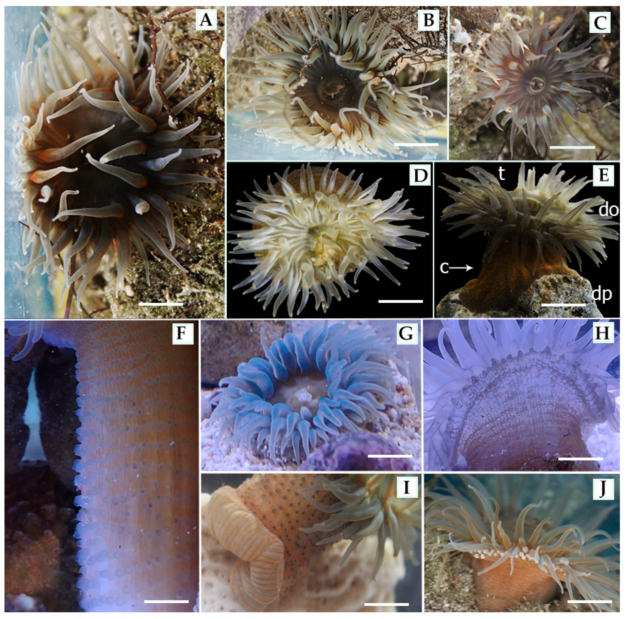
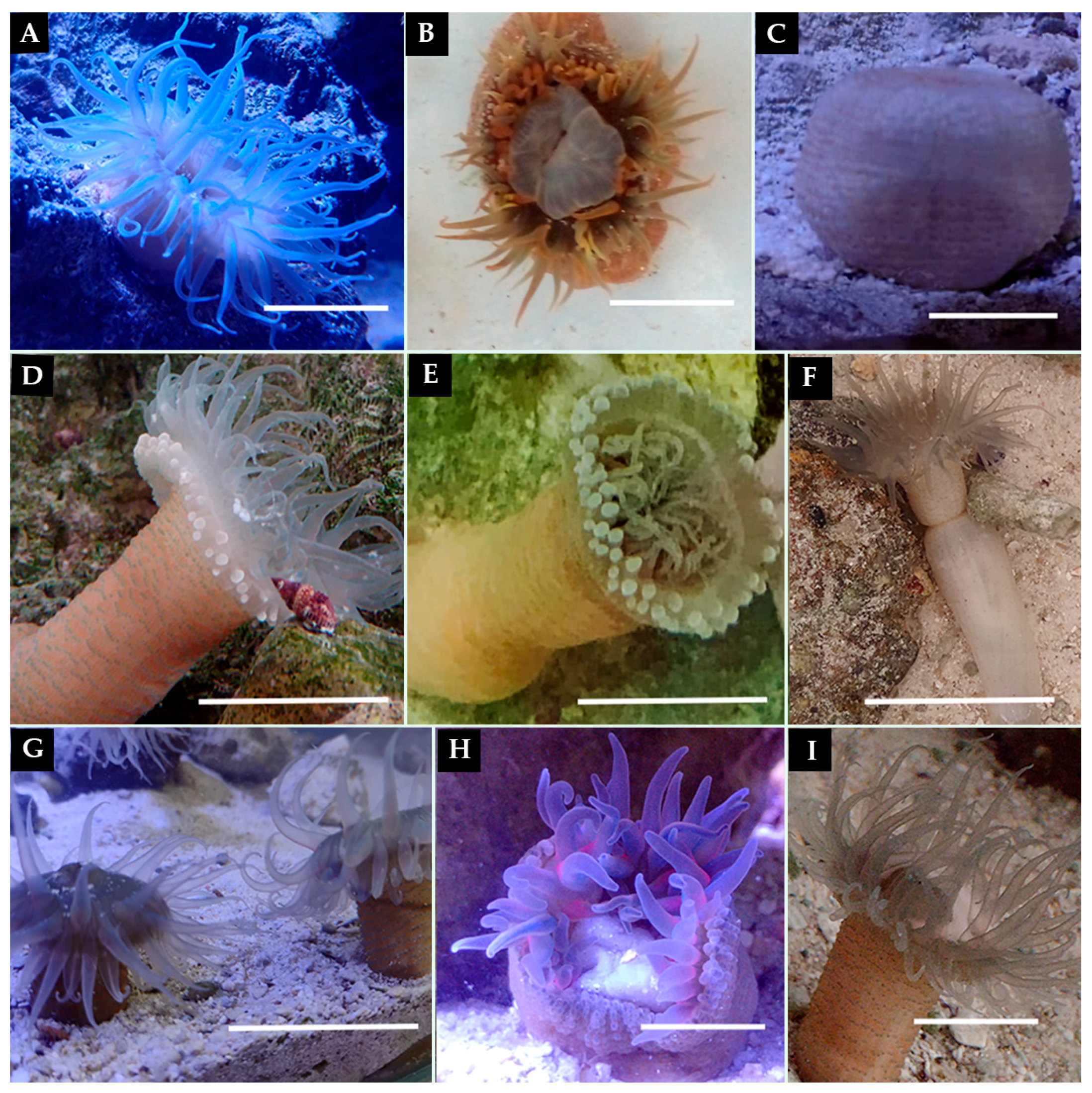
The oral disk of B. cavernatum is soft and smooth (Figure 2B–D,G), exhibiting coloration that varies among individuals, including olive green, reddish, brown, blue, and pale-yellow tones. Distinctive transparent radial lines and V-shaped bands are often visible at the bases of the tentacles and within the endocoelic sectors (Figure 2B–E; Table 1).
The number of tentacles ranges from 82 to 96. Tentacle coloration included olive green, translucent blue, and olive with red-orange spots. Tentacles are conical, soft, contractile, and capable of full retraction into the oral disk. They are arranged in five hexameral cycles, with approximately 12 tentacles per cycle in the first two to three cycles. Outer cycles include a greater number of tentacles due to their association with imperfect mesenteries. Inner tentacles (discal) are longer than outer (marginal) ones, contributing to a characteristic concentric pattern.
Acrorhagi fluctuated between 40 and 100. They are rounded, contractile, and typically olive or white in color. These structures are arranged in one or two marginal rows, although they are often inconspicuous or partially concealed in relaxed specimens (Figure 2H,I; Table 1).
Figure 2. External morphological characteristics of Bunodosoma cavernatum. (AE,G) Morphotypes observed in situ and under aquarium conditions, showing variation in the oral disk (do), tentacles (t), column (c) and pedal disk (dp) colorations. (A) Conical tentacles. (BE) Smooth oral disk with radial lines; (C) showing prominent V-shaped bands. (F) Column with transparent, non-adhesive vesicles. (G) Specimen with expanded tentacles and visible oral disk pattern. (H,I) Detail of acrorhagi: (H) relaxed state as small, transparent projections; (I) two marginal rows of acrorhagi. (J) Pedal disk displaying terracotta-colored longitudinal bands. (AJ) scale bars = 1 cm. (F) scale bars = 2 cm.
Diversity 18 00118 g002
The column is cylindrical and pale reddish-brown, fully covered with non-adhesive vesicles that are olive, transparent or terracotta colored. Vesicles are arranged in 40–50 vertical rows with 50–90 horizontal rows from the margin to the limb, and some vesicles display a central dark spot (Figure 2F, Table 1). In relaxed specimens, the column shows translucent orange or ochre tones with fine red longitudinal lines, which seem to correspond to longitudinal muscles (Figure 2F).
The pedal disk is broader than the column and exhibits a linear terracotta pigmentation pattern. When relaxed, the pedal disk becomes translucent and is clearly demarcated from the column base (Figure 2J).
Table 1. Comparative external morphological characteristics of Bunodosoma cavernatum. Features include the oral disk, tentacles, acrorhagi, column, and pedal disk. Data compares records from this study with the literature [24,25,28].
CharacteristicGonzáles-Muñoz et al. [24]Herrera-Bojorquez et al. [25]Durán et al. [28]Present Study
Oral DiskSmooth, with white or yellowish radial stripes in the endocellular spaces of the first two or three tentacular cycles (20–28 mm diameter). General color is yellowish brown, reddish or olive green.Flat, wide and smooth (8.28 mm diameter).Smooth (31.1 mm diameter), general color is olive green with yellow radial stripes.Wide, smooth and soft (28–45 mm diameter). General color is light brown, translucent or olive, with transparent radial lines and V-shaped bands that are often visible at the bases of the tentacles and within the endocoelics.
TentaclesArranged hexamerically, approximately 96, smooth conical, tapered distally, internal ones longer than external ones, contractile (12.5–22.5 mm long; 82–94 tentacles).Smooth, conical, distally narrowed, moderately long, contractile, in five cycles (71–98); internal longer than external.Approximately 96 arranged hexamerously in five cycles; simple, conical, and smooth (7.13–17.5 mm long).Contractile cones, longer on the inside than on the outside (96, 10–22.5 mm long).
AcrorhagiRounded endocellular acrorhagi, 80 to 100, with basitrichs and holotrichs. Olive green, reddish, orange, or yellowish on the oral side, sometimes with purple flashes.Ring of projections (around the edge of the column) with basitrichs and holotrichs. Gray, reddish or beige; red stripe on the aboral side of internal tentacles (not in later cycles).48, rounded acrorhagi, with basitrichs and holotrichs. Translucent, blue, brown, or yellow, with orange or red spots.Two rows of them, contractile, between 40 and 100. Bluish olive, light reddish or white.
ColumnCylindrical and rounded vesicles organized in 96 longitudinal rows (22–31 mm diameter; 20–50 mm length). Light brown, orange, reddish, yellowish, or olive green.Rounded vesicles arranged in longitudinal rows of alternating sizes (16–44 mm long; 14–31 mm wide). Beige or gray with a dark brown or black center.Densely covered with dark, rounded vesicles in 96 longitudinal rows (19.4–39.5 mm length). Orange or brown.Cylindrical, large number of vesicles in 50 columns × 46 rows (50–90 transverse rows; 40–50 longitudinal rows). Reddish brown, when spread out, it is light orange with red lines and black dots on the vesicles.
Pedal DiskWell developed (25.0–40 mm diameter). Light brown, orange, reddish, yellowish, or olive green.*Well developed (15.5–22.1 mm diameter). Pale orange or light brown.Strong and well developed. Pale orange when relaxed and with red longitudinal lines when contracted.
* The author does not present any information.

3.6. Internal Anatomy and Histology

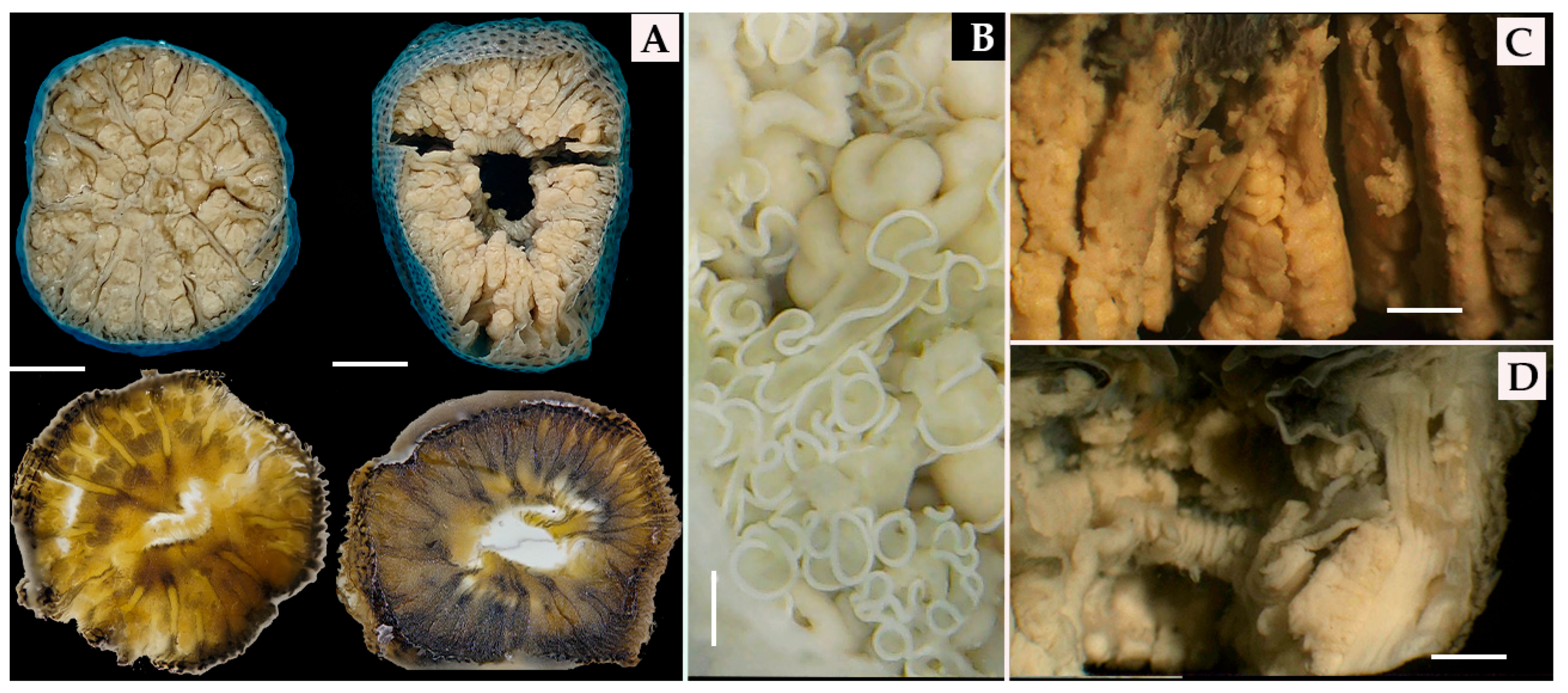
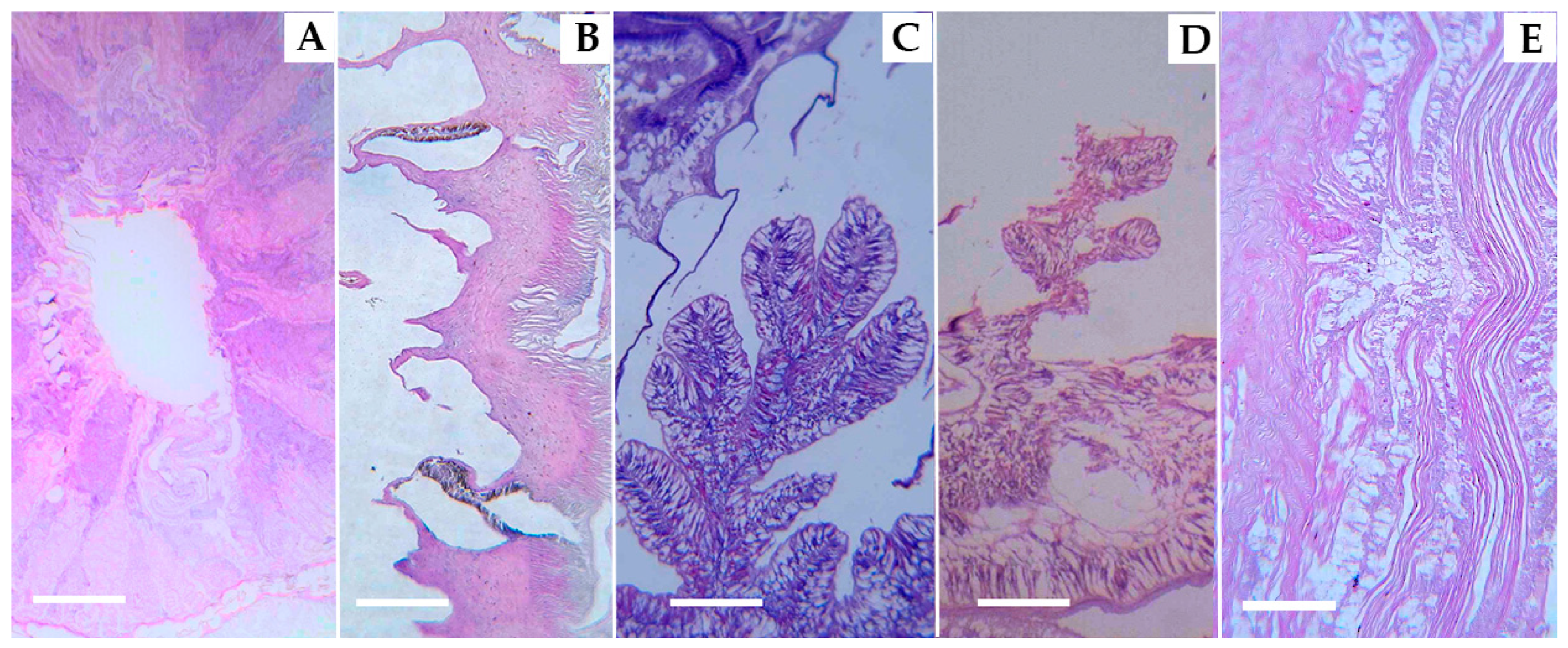
Dissections revealed a well-developed, tubular actinopharynx with prominent longitudinal folds. The mesenterial arrangement included both complete and incomplete pairs. Six pairs of perfect mesenteries were confirmed through cross-sections and were symmetrically associated with the siphonoglyphs (Figure 3A and Figure 4A). In turn, long white mesenteric filaments forming helical folds and gonads can be observed throughout the mesenteries and gastrovascular cavity (Figure 3B). These internal features are further documented through representative histological sections illustrated in Figure 4A,B,D.
Gonads were also located along the first cycle of complete mesenteries and exhibited a stacked, coin-like arrangement characteristic of the species. Histological analysis demonstrated the presence of nematocytes of varying sizes within the ectodermal layers of both the actinopharynx and the columnar non-adhesive vesicles (Figure 4B; Table 1 and Table 2). Longitudinal sections further revealed the gastrovascular architecture, the mesoglea layer and reproductive tissues with oocytes at different stages of development (Figure 3C and Figure 4C). The sphincter and string retractor muscle can be observed, as well as the folds associated with the siphonoglyphs and the pharynx (Figure 3D and Figure 4D).
The retractor muscle runs along the entire length of the column (Figure 4E). The pedal disk also contained prominent retractor muscles and was connected to mesenterial filaments arranged in helical folds (Figure 4C; Table 2). The free ends of the mesenteries bore flexible filaments, which appeared coiled in relaxed specimens. Fertile and directive mesenteries showed gonadal tissue containing developing gametes and mature oocytes (Figure 3B, Table 2).
Figure 3. Internal anatomy of Bunodosoma cavernatum. (A) Cross-anatomical sections obtained by dissection, illustrating internal organization prior to histological processing of the column, showing complete and incomplete mesenteries with stacked gonads near the pedal disk and actinopharynx (upper); paraffin-embedded sections illustrate mesenterial organization (lower). (B) Regurgitated actinopharynx exposing mesenterial filaments and associated mesenteries with helical folds. (C) Longitudinal section with gonadal structures in mesenterial, arranged in a stacked coins pattern. (D) Longitudinal section of the actinopharynx with longitudinal folds associated with siphonoglyphs, retractor muscle, and sphincter. Scale bars: (A) = 2 cm; (B) = 1 cm; (C,D) = 2 cm.
Diversity 18 00118 g003
Figure 4. Histological slices of Bunodosoma cavernatum. (A) Transverse histological section at the level of the actinopharynx, showing the central pharyngeal lumen, radially arranged complete mesenteries, well-developed mesoglea and associated gastrovascular compartments. (B) Longitudinal section of the ectoderm, detailing a non-adhesive vesicle containing densely packed nematocysts, associated with epitheliomuscular and glandular cells. (C) Longitudinal section of the pedal disk, showing a prominent gastrovascular cavity delimited by the gastrodermis, and mesenterial filament. (D) Longitudinal section of the actinopharynx displaying secondary gastrovascular spaces associated with mesenteries and pharyngeal folds. (E) Longitudinal section with long a mesenterial retractor muscle, showing longitudinally oriented epitheliomuscular fibers supported by the mesoglea, responsible for body retraction and postural control. Scale bars: (A) = 1 cm; (BE) = 100 µm.
Diversity 18 00118 g004
Table 2. Comparative internal anatomical and histological characteristics of B. cavernatum. Data compared dissections and histological sections with the Refs. [24,25]. Duran et al. [28] is not included in this table as it uses the same description as [24].
StructureGonzales-Muñoz et al. [24]Herrera-Bojorquez et al. [25]Present Study
MesenteriesArranged in four hexameral cycles (48 pairs) with perfect mesenteries in first, second and third cycles, others imperfect. Same number of distal and proximal mesenteries. All mesenteries, except the directive ones, are fertile. Two pairs of directive mesenteries, each joined to a well-developed siphonoglyphs. Gonochoric.Four hexameral in cycles (48 pairs), with perfect mesenteries in first, second and third cycles. Same number of distal mesenteries. Two pairs of directives, each joined to a well-developed siphonoglyph.Six pairs of perfect mesenteries arranged hexamerally; two directive pairs and two imperfect pairs. Only perfect mesenteries are fertile, each joined to a well-developed siphonoglyph.
GametesOocytes and spermatocytes are well developed in samples collected in January and May.Spermatocyst observed in the strongest mesenteries of first, second, and third cycles, less frequent in the directives.Presence of oocytes.
SiphonoglyphsWell developed.Well developed.Well developed.
MusclesRetractorsStrong and restricted.Strong and restricted.Strong.
ParietobasilarWell developed, with a relatively long free mesogleal nerve.Well developed, with a relatively long free mesogleal nerve.Well developed.
BasilarWell developed.Well developed.Well developed.
Marginal sphincterEndodermal, strong and circumscribed.Endodermal, strong and circumscribed.Endodermal, strong and circumscribed.
LongitudinalEctodermal in tentacles.Ectodermal in tentacles.Ectodermal in tentacles.
ZooxanthellaePresent.Present.Present.
CnidaeBasitrichs, microbasic b- and p-mastigophores, holotrichs and spirocysts.Basitrichs, microbasic b- and p-mastigophores, holotrichs, and spirocysts.Basitrichs, spirocysts, microbasic b- and p-mastigophores, holotrichs.

3.7. Cnidome

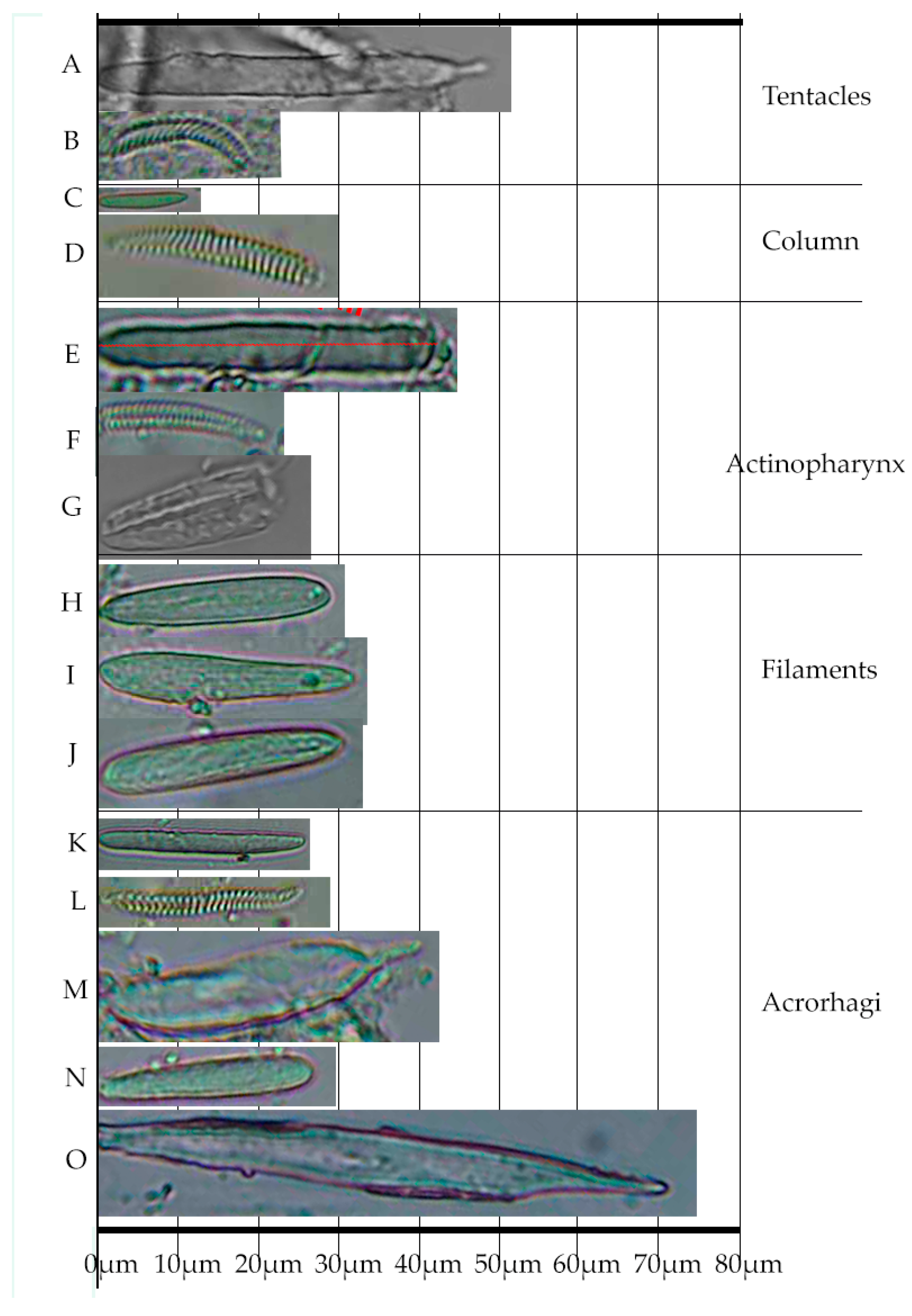
Five main types of nematocytes were identified across anatomical regions: basitrichs, spirocysts, microbasic p-mastigophores, microbasic b-mastigophores, and holotrichs (Figure 5, Table 3). All cnidocyst types described were present in the examined specimens. The number of capsules measured per cnidae type and tissue is indicated in Table 3.
Morphometric analysis revealed expanded size ranges for several cnidae types compared to previously published data. Basitrichs from the tentacles measured between 8.68 and 49.5 ± 12.42 (n = 126) µm in length, representing an extended range size of the one known for this type of capsules, which was 7.47–49.50 µm in length (see Table 3, proposed range between literature and new measurements with ±SD). Holotrichs observed in the acrorhagi ranged from 17.95 to 79.91 µm (Table 3). In the actinopharynx, spirocysts measured 6.51–21.86 µm, while those from the column ranged from 8.09 to 17.86 µm in length. In the acrorhagi region, two types of microbasic mastigophores were recorded: microbasic b-mastigophores (between 11.35 and 34.39 µm), and microbasic p-mastigophores (ranging from 8.72 to 36.16 µm). These measurements, along with capsule distribution across anatomical regions, contribute to the diagnostic cnidome profile of B. cavernatum and support its taxonomic differentiation from morphologically similar species.

3.8. Behavior

3.8.1. Ethogram and Behavioral Categories

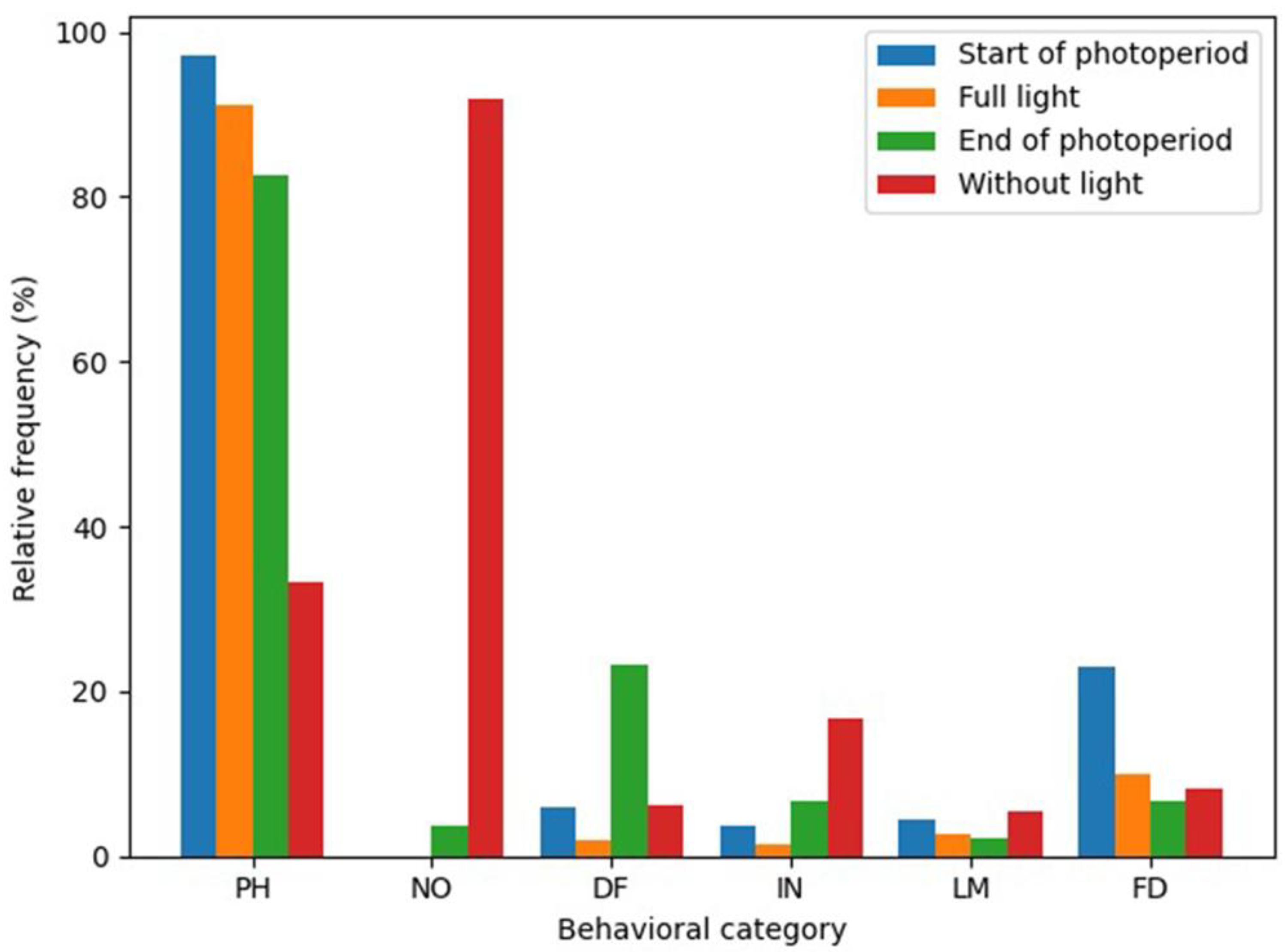
Behavioral expressions exhibited a clear diel structure, with photoperiod-associated behaviors dominating during light phases and nocturnal behaviors restricted to dark conditions (Figure 6). Defensive responses, feeding activity, locomotion and interspecific and intraspecific interactions occurred across all phases, but varied in frequency throughout the photoperiod. These behaviors were recorded across all diel phases and were not restricted to a single interval and are classified and coded in an ethogram (Table 4), which served as a reference framework for subsequent analyses.
Organisms acclimated rapidly to aquarium conditions. All individuals established stable attachment sites within the first day and exhibited consistent behavioral patterns throughout the one-month monitoring period. Environmental parameters in the aquarium ranged from 25 to 29 °C and 30–48 ‰ salinity, under which the anemones showed full tolerance with no mortality or visible signs of stress.

3.8.2. Diel Activity Patterns

The relative frequency of each stimulus-dependent behavioral code was quantified using a presence-absence matrix and expressed as the percentage of observation sessions in which each behavior was recorded (Figure 6). Behaviors associated with the photoperiod were the most frequent, with an expanded posture sustained during daylight (6:00–17:00). During this phase, organisms showed a fully expanded oral disk and tentacles (Figure 7A), with no signs of contraction or defensive posture (Figure 7B; PH1).
While some sea anemones remained fully expanded with asynchronous retraction of the tentacles and oral disk (PH2), others exhibited temporary tentacle and oral disk retraction into the column (Figure 7C). These contractions and retractions lasted from several minutes to over an hour and were followed by gradual re-expansion.
Exposure to artificial light outside the photoperiod consistently triggered immediate tentacle retraction and contraction, followed by slow re-extension (PH3).
Although PS1 and PH1 may be morphologically similar, both involving an expanded oral disk and extended tentacles, they are defined by different ethological criteria in this study. PS1 is a photoperiod-independent basal postural state of the individual, characterized by a stable, fully turgid column and a neutral, non-reactive tentacle configuration in the absence of discrete stimulus-driven responses. In contrast, PH1 represents a sustained expanded posture temporally associated with the photoperiod, expressed synchronously at the population level following light onset. Thus, while similar receptive postures may facilitate feeding or stimulus-related responses in both cases, PS1 is defined by body configuration as a baseline reference state, whereas PH1 is defined by its temporal coupling to light conditions.
Figure 7. Behavioral repertoire of B. cavernatum under laboratory conditions. (A) Basal state. (B) State of stress and actinopharynx eversion. (C) Fully contracted, water-filled posture. (D) Relaxed nocturnal posture following photoperiod or repeated intra/interspecific stimulation. (E) Nocturnal relaxation showing extended acrorhagi crown, tentacles retracted into the oral disk, and rhythmic column contractions. (F) Fully relaxed state before photoperiod onset, with visible column expansion and water intake. (G) Intraspecific interaction showing tactile contact between external tentacles of two individuals. (H) Feeding behavior with tentacles transferring food to the oral disk. (I) Sperm release through the relaxed oral disk. Scale bars: (AI) = 5 cm.
Diversity 18 00118 g007

3.8.3. Nocturnal Behavior

Behavior were consistently expressed under low-light conditions during the nocturnal phase. First, individuals exhibited complete expansion of the column and acrorhagi, often accompanied by slow, lateral undulations (Figure 7D; NO1). In the second behavior, tentacles displayed rhythmic extensions and retractions, with pulse-like contractions traveling from the oral disk toward the pedal disk, apparently associated with water uptake and circulation (Figure 7D–F; NO2).
In the third behavior, the column remained relaxed while tentacles extended fully, resembling a photoperiod posture (NO3). This stance persisted for prolonged periods throughout the night, sometimes until light onset. During these episodes, body size increased substantially, with some individuals extending their column more than 10 cm above the substrate and exceeding 5 cm in diameter (excluding tentacles) (Figure 7F).

3.8.4. Response to Physical Stimuli, Defensive and Intraspecific Interactions

Tactile sensitivity was evident. Light touch near the tentacles induced localized movement toward the stimulus without full contraction (DF1) and sustained contact caused column flexion and coordinated reorientation of the oral disk (DF2).
Contact near the column triggered defensive reactions, including acrorhagi extension, tentacle retraction, and full body contraction (Figure 7C). These responses were reversible, with gradual relaxation following cessation of stimulation.
Five distinct behavioral responses were recorded during interspecific and intraspecific interactions: 1. Conspecific contact prompted partial tentacle and column retraction, followed by mutual separation (Figure 7D,G; IN1). 2. Interaction with hermit crabs resulted in localized contraction; repeated contact induced full contraction and inflation of the column into a rigid, water-filled posture (Figure 7C; IN2). 3. Sustained contact led to full tentacle contraction and acrorhagi extension. 4. Persistent tactile stimulation triggered displacement across the substrate and 5. Gradual movement away from stimuli occurred without visible stress when interactions persisted (LM1).

3.8.5. Feeding and Locomotion

Feeding was initiated upon contact between food particles (e.g., shrimp fragments) and tentacles. The tentacles flexed inward, transferring food to the oral disk within seconds to minutes (FD1). While some individuals responded immediately, others displayed delayed ingestion.
Feeding was accompanied by complete tentacle contraction and thickening of the column, resulting in a rigid, inflated posture (Figure 7C). This state persisted for several minutes before relaxation (FD2). In the following 24–48 h, individuals frequently adopted expanded postures resembling nocturnal behavior, suggesting a post-feeding phase (Figure 7H).
Locomotion occurred through pedal disk-mediated displacement. Individuals initiated movement by expanding the anterior margin of the pedal disk while contracting the posterior margin, generating a slow dragging motion along the substrate (LM1). Once relocation was complete, the disk expanded fully to re-anchor the body. No detachment, floating, or swimming behavior was observed.

3.9. Sperm Release

A spontaneous sperm release event was recorded on 22 June 2025, during the waning quarter moon phase. Four of the nine individuals exhibited complete oral relaxation and released within sperm clouds through the oral cavity, without accompanying tentacle movement or defensive responses (Figure 7I; RP1). Emission occurred sequentially, beginning with a single individual and followed by others at intervals of 5–10 min.
Each episode lasted approximately five minutes, and the overall event extended from 17:00 to 19:00 h. Three additional individuals did not release visible gametes but displayed pronounced oral expansion and atypical tentacle activity following the second sperm emission, characterized by repeated cycles of full expansion and brief tentacle retraction at intervals of approximately five minutes (RP2). No oocytes were observed during this event in other individuals, precluding sex determination of the organisms.
Water parameters remained stable throughout the spawning period (Salinity: 36‰; temperature: 25–28 °C; pH: 8.4). By the end of the event, the water column contained a fine suspension of spermatozoa, visible as whitish particles homogeneously dispersed throughout the aquarium because of gamete release and water movement.
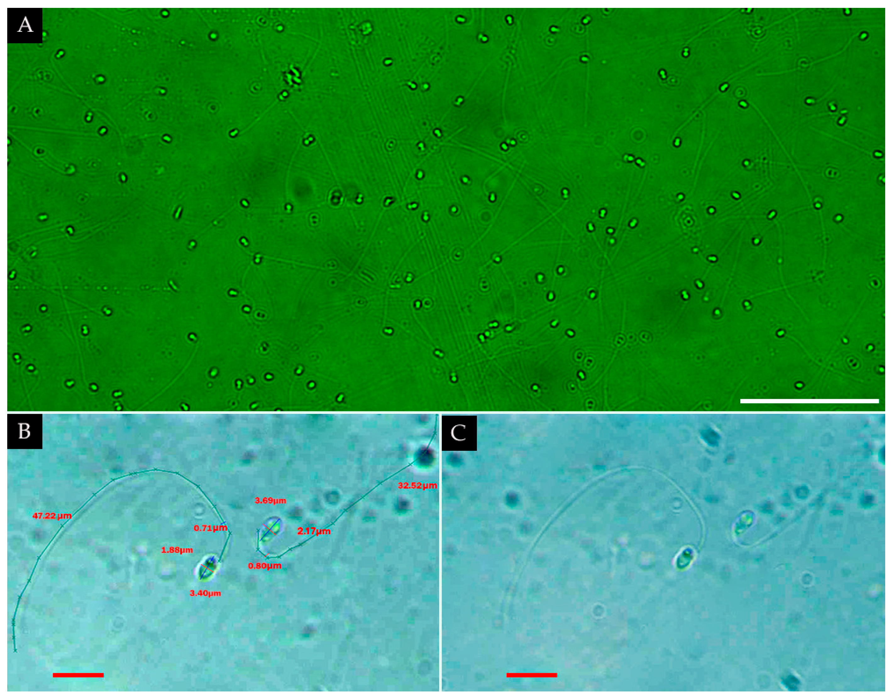
Microscopic analysis at 1000× revealed abundant spermatozoa (Figure 8A). Each cell possessed an ellipsoidal head (1–4 µm long, 1–2 µm wide) with a dense refractive nucleus. The acrosomal vesicle was not visible. The flagellum was filiform and undulating, measuring 30–50 µm in length and ~1 µm in width (Figure 8B). Mitochondrial structures and mucous matrices were not observed (Figure 8C).
Quantification across 40 fields of view at 400× yielded counts ranging from 121 to 689 spermatozoa per field (mean ± SD = 263.8 ± 113.4; n = 40). Spermatozoa were distributed homogeneously, occurring either as isolated cells or in a small aggregate.
Ethanol-fixed samples showed poor structural preservation, while fresh samples retained complete morphology. The best results were obtained from samples without any additives.
Figure 8. Sperm release in Bunodosoma cavernatum. (A) Fluorescence micrograph (400×) showing high spermatozoa density distributed across the field of view. Left (B): two spermatozoa imaged at 1000× magnification, illustrating size measurements; Right (C): two spermatozoa displaying overall morphology, including a thread-like flagellum and small ellipsoidal head with a collar-like structure, though internal features are indistinct. Scale bars: (A) = 50 µm; (B,C) = 5 µm.
Diversity 18 00118 g008

4. Discussion

4.1. Taxonomic Identity and Morphological Consistency

The present study expands the known distribution of B. cavernatum to Cartagena Bay and provides new morpho-ecological data for this species under estuarine conditions. The population analyzed exhibits a combination of diagnostic characters that are fully consistent with previous taxonomic descriptions from the Caribbean [24,25,28,40], including both external and internal morphology, as well as cnidome features. Diagnostic traits such as tentacles arranged in hexamerous cycles, the presence of V-shaped bands on the oral disk within endocoelic sectors, acrorhagi armed with basitrichs and holotrichs nematocysts, and a cylindrical column densely covered by non-adhesive vesicles, correspond closely with classical and modern descriptions of B. cavernatum and the genus Bunodosoma [23,48,51,52]. Minor variation was observed in tentacle number, vesicle arrangement, and the number of vesicle rows on the column, as well as in oral disk and column coloration.
Therefore, species identification in Bunodosoma cavernatum should be based on the congruence of external morphology, internal anatomy, and cnidome composition, rather than on a single isolated character.

4.2. Tentacle Arrangement and Mesenterial Development

In actiniarians, tentacle number is closely linked to the mesenterial development, although temporal mismatches between mesenteries and tentacles may occur during growth [53]. Our counts revealed a hexameric organization consistent with previous reports, but also asymmetry between cycles, explained by sequential mesenteries addition within the multiplicative chamber in the growth zone [54]. Accordingly, tentacles and mesenteries count follow a hexameric organization of cycles, with 12 corresponding to two or three mesenteries in the first cycle, while some of the marginal tentacles may be associated with developing cycles or imperfect cycles and conform to the general actiniarian pattern [1,2].

4.3. Internal Anatomy, Reproductive Structures and Mesenterial Stability

Internally, B. cavernatum exhibited gonads primarily associated with secondary and perfect mesenteries, a characteristic considered stable within the genus [55].
The coexistence of cysts and oocytes across perfect mesenterial cycles was also reported by Gonzáles-Muñoz et al. [24,25]. This suggests that while gonadal positions are conserved, gametogenic timing may be flexible and influenced by individual age, reproductive stage, or local environmental conditions.
Furthermore, the presence of six pairs of perfect mesenteries, two direct and two imperfect pairs, maintains the characteristic hexameric organization, although with a reduction compared to the four mesenteric cycles, indicating intraspecific plasticity rather than taxonomic divergence, caused by ontogenetic or environmental factors.
The presence of well-developed mesenterial filaments rich in nematocyst, paired siphonoglyphs and a consistent arrangement of perfect and imperfect mesenteries further emphasizes the stability of diagnostic traits [39,56]. Minor differences were observed in the number of fertile mesenteries, the development of a well-formed but relatively short mesoglea nerve in the parietobasilar muscle, and the proportion of perfect to imperfect mesenteries, all of which fall within known intraspecific variation in actiniarians.
The muscular system—comprising robust parietobasilar and basilar muscles, a strong endodermal marginal sphincter, and well-developed retractor muscles—along with persistent siphonoglyphs, ensures effective hydrostatic control, posture maintenance and resistance to hydrodynamic stress. This combination of features represents an adaptation that enables the species to persist and survive among rocky substrates in zones subjected to constant wave action and discontinuous water coverage.

4.4. Cnidome Variation and Functional Implications

The cnidome analysis across multiple tissues revealed an expansion of morphometric ranges previously reported for B. cavernatum, especially in basitrich and holotrich nematocysts. Basitrichs in the tentacles reached lengths of up to 49.50 µm, exceeding maximum values previously reported for the column and actinopharynx. Similarly, holotrichs in the acrorhagi reached 79.91 µm, nearly doubling the maximum size previously recorded for Colombian populations [28]. Additional cnidae types, including spirocysts, microbasic b-mastigophores, and microbasic p-mastigophores, also exhibited broad size ranges across tissues. This variability likely reflects adaptive responses to environmental factors such as prey availability, sediment load, predation pressure, and intraspecific competition. Larger holotrichs and mastigophores may confer functional advantages in defense and prey capture, particularly in structurally complex or disturbed habitats [52,57].

4.5. Coloration, Zooxanthellae and Environmental Influences

The wide chromatic variability observed in B. cavernatum aligns with previous reports and is likely influenced by multiple interacting factors, including zooxanthellae density, turbidity, light availability, and background substrate [21,45,49]. In estuarine environments such as Cartagena Bay, strong gradients in light penetration and suspended sediments may favor phenotypic plasticity in coloration, potentially enhancing camouflage and reducing predation risk.

4.6. Phylogenetic Context and Morphological Plasticity

Although the diagnostic traits observed are consistent with the genus Bunodosoma, recent phylogenetic analyses suggest that the genus is paraphyletic [39]. This implies that some shared morphological characters—such as color patterns or cnidome composition—may represent environmentally driven convergence or intraspecific variation rather than synapomorphies.
The combination of stable diagnostic traits and variable morphological features observed in B. cavernatum supports the view that morphological plasticity plays a significant role in the ecological success of these species across heterogeneous Caribbean environments.
From an ecological perspective, the tolerance of this sea anemone to wide salinity (30 to 48‰) and temperature (25–29 °C) ranges reflects a euryhaline and eurythermal behavior [46,47,58], an adaptation to the dynamic estuarine conditions of Cartagena Bay. Such plasticity is very important for species inhabiting tropical, sediment-laden habitats, where osmotic and thermal gradients fluctuate on diel and tidal scales. This is supported by the observations of [47], who demonstrated euryhaline behavior in ranges from 12 to 40‰, like a similar range (11–49‰ at 25 °C) is obtained during the essay of Ref. [58].

4.7. Reproductive Strategies and Gametogenesis

The genus exhibits multiple asexual strategies. In B. cangicum (Belem & Preslercravo, 1973), budding, fragmentation, pedal laceration and longitudinal fission have been previously documented, with reports of two fully formed individuals connected at the column, and budding after injury, in Carneiros Beach and Pacoti River estuary, Brazil, respectively [59,60]. In addition to this, 19 females, 29 males and one hermaphrodite (spermatozoa and small oocytes in the same mesentery), as well as longitudinal scars confirming a longitudinal fissure, and indicating the impossibility of knowing the exact gender of each anemone, even though four individuals could be identified as male as they had released sperm [61].
It was found in Jenninson [61] that the population was maturing again in May, with organisms containing sperm being found, while the females matured in March, April and May, with spawning occurring between October and November; moreover, this was also mentioned by Brandão [62].
In the present study, larvae were observed in August, and a spontaneous sperm release event was recorded in June of the following year. The coincidence of sperm release with the waning lunar phase suggests potential lunar entrainment of reproductive activity, as documented for other actiniarians [62,63]. Post-release behaviors observed in several individuals—such as sustained oral expansion and rhythmic tentacle contractions—may correspond to female receptivity or transitional reproductive phases, although this remains poorly documented for the species.
Following sperm release, several individuals exhibited behaviors compatible with potential female receptivity or female phase, including sustained oral expansion and a rhythmic tentacle contraction with oral disk closure, indicating possible synchronized reproduction, which remains poorly described for the species. The observations are consistent with similar descriptions for Entacmaea quadricolor (Leuckart in Ruppell & Leuckart, 1828) and Heteractis crispa (Hemprich & Ehrenberg in Ehrenberg, 1834), in which whitish gamete emissions through the oral cavity were accompanied by partial relaxation and occasional spasm [64].

4.8. Sperm Morphology and Preservation

Microscopic analyses confirmed abundant spermatozoa with ellipsoidal heads (1–4 µm long, 1–2 µm wide) and single, elongated flagella (30–50 µm long), consistent with class III–IV developmental stages typical of externally fertilizing benthic cnidarians [65,66,67,68]. This characteristic is preserved, as is the organization of the gonads, which were also located along the first cycle of complete mesenteries and had a stacked arrangement, like stacked coins [65].
The absence of a visible acrosomal vesicle may reflect its small size or limitations of light microscopy, reinforcing the need for ultrastructural studies using transmission electron microscopy [65,69].
The relatively long flagellum observed may represent an adaptive feature facilitating gamete dispersal under specific microhabitat conditions, as suggested for other actiniarians [66,70,71].
The poor preservation of spermatozoa in ethanol-fixed samples is consistent with previous findings in cnidarians, where ethanol induces osmotic damage, whereas aldehyde-based fixatives better preserve ultra-structural detail [72]. In these studies, ethanol induced osmotic shrinkage and flagellar collapse in cnidarian spermatozoa, whereas aldehyde-based fixatives (such as glutaraldehyde or Bouin’s solution) better preserved the ultrastructure of the nucleus, acrosome and mitochondria.

4.9. Habitat Use, Associated Fauna, and Ecological Role

The aggregated distribution under artificial structures and the high density (32.2 ind/m2) are consistent with reports for other tropical habitats [25,41]. These artificial structures are usually enriched in organic matter and sheltered from strong hydrodynamic forces, providing a favorable microenvironment for settlement and heterotrophic feeding [24]. Contrary to previous literature, where B. cavernatum is mainly reported in shallow reef areas [25], tide pools and rocky shores under fully marine conditions, as in the Lobos reef, Veracruz [42], this study confirms its presence in an estuarine environment, showing its adaptability to salinity changes.
The diversity of associated fauna and flora observed reinforces the role of B. cavernatum as a benthic structuring species. Its coexistence with mollusks, crustaceans, annelids, sponges, and macroalgae suggests commensal relationships and the provision of structural refuges, consistent with patterns reported for other Caribbean Sea anemones [13,42].

4.10. Behavioral Rhythms and Sensory Responses

The complex behavioral repertoire observed—including diel expansion–contraction cycles, nocturnal relaxation, and stimulus-dependent responses—expands the current knowledge of actiniarian behavior. Patterns of daytime expansion and nocturnal relaxation resemble those reported for Anemonia viridis (Forsskål, 1775) and Actinia equina (Linnaeus, 1758) [73]. The pulsatile nocturnal contractions and tentacle undulations observed in B. cavernatum suggest an integrated behavioral and physiological rhythm comparable to endogenous circadian control described in Nematostella vectensis (Stephenson, 1935) [74,75].
The alternation between photoperiod-driven expansion and nocturnal relaxation in B. cavernatum implies that these actinias possess a form of light-synchronized patterns adapted to estuarine habitats, where turbidity and light penetration fluctuate strongly. Similar diurnal metabolic oscillations have been linked to light-dark cycles in N. vectensis [75], and circadian and melatonin-mediated signaling pathways have been implicated in the maintenance of circadian rhythmicity in cnidarians [76,77].
The contraction observed around midday may represent a behavioral mechanism of thermoregulation or photoprotection, reducing metabolic expenditure or the risk of desiccation during periods of maximum irradiation, and following circadian behaviors similar in some respects to N. vectensis [76]. It is noteworthy that exposure to artificial light during the dark phase immediately induced retraction, supporting evidence that artificial night light (ALAN) can alter the rhythmic expansion-contraction patterns and feeding behavior in other sea anemones, such as Metridium senile (Linnaeus, 1761) [78]. Taken together, these findings reinforce the view that photic and thermal signals jointly regulate the behavior and physiology of anemones, even under stable laboratory conditions. On the other hand, movements related to feeding were like those reported in Sun [79].

4.11. Acrorhagi, Defense and Ecological Plasticity

Defensive and tactile responses in Bunodosoma cavernatum are consistent with a graded sensory and behavioral hierarchy characteristic of actiniarians, in which acrorhagi function as specialized structures involved in defense and competitive interactions [80,81]. Experimental and observational studies have demonstrated that acrorhagial responses mediate highly specific self–non-self-recognition and spatial competition through the targeted application of nematocyst-rich tissue following physical contact, rather than through generalized aggression [82,83,84]. In B. cavernatum and related taxa, these responses depend strongly on the identity of the interacting organism, indicating a finely tuned recognition system whose expression is context dependent [83].
Behavioral studies further indicate that the expression of acrorhagial aggression is modulated by ecological and demographic factors, including population density, frequency of physical contact, and competitive pressure [84,85]. Consequently, the absence or low frequency of observed acrorhagial interactions does not imply functional loss but rather reflects conditional deployment within a broader defensive repertoire.
The recurring nocturnal association between column relaxation, acrorhagi extension, partial tentacle retraction, and rhythmic column pulsations suggest a coordinated functional state distinct from both diurnal expansion and defensive responses. This behavioral configuration, accompanied by visible water circulation around the column and oral disk, is consistent with the hypothesis that nocturnal pulsatile movements may facilitate the passive interception of microorganisms of small macro-organisms in suspension under low light conditions or night. By retracting the tentacles while maintaining the acrorhagi expanded and generating a rhythmic flow of water, individuals may increase contact between potential food particles and cnidocyte-rich marginal surfaces. Although direct ingestion was not quantified, the repeated and structured occurrence of this combination of behaviors supports its interpretation as a functional state related to feeding, rather than random postural variation.
From an evolutionary and comparative perspective, phylogenetic and morphological analyses indicate that acrorhagi are ancestrally retained within Actiniidae and exhibit substantial variability in presence, morphology, and expression among taxa, populations, and even individuals [39,51]. Daly et al. [39] explicitly noted that acrorhagi-bearing anemones are most species rich in temperate regions with hard-substrate habitats and emphasized that historical and ecological contingencies cannot easily be disentangled. Within this framework, such habitats may favor the retention or re-evolution of acrorhagi or alternatively reflect lineage diversification associated with the presence of these structures, without implying uniform expression across ecological contexts.
In the present study, B. cavernatum consistently exhibited one to two marginal rows of acrorhagi, with counts ranging from approximately 46 to 100 structures, exceeding values typically reported in earlier taxonomic descriptions that document a single row with fewer acrorhagi. While this study does not directly test adaptive hypotheses, the increased number and arrangement of acrorhagi observed under estuarine conditions are congruent with the ecological scenarios proposed by Daly et al. [39], in which local environmental pressures may modulate the expression and retention of contact-mediated defensive structures.
Patterns documented across Actiniidae further support this interpretation. Sustained functional loss of marginal defensive structures is generally associated with clear morphological transformations, such as the conversion of adhesive verrucae into non-adhesive vesicles in Bunodosoma or the transformation of acrorhagi into pseudoacrorhagi in related taxa [51]. Additionally, the loss or reduction in specific nematocyst types, particularly holotrichs, may occur independently of the loss of the structure or the behavior in which they are deployed, indicating a modular pattern in the evolution of acrorhagial traits [39].
Taken together, available behavioral, morphological, and phylogenetic evidence supports the interpretation of acrorhagi in B. cavernatum as a retained and plastic defensive structure with facultative expression, rather than as a vestigial or non-functional remnant.

4.12. Ecological Tolerance and Implications

Additionally, their ecological importance has been highlighted in evolutionary discussions, considered critical to fitness and may have contributed to lineage diversification, some genera in Actiniidae are more diverse in temperate region with hard-substrate habitats [53] and used acrorhagi, raising the question of whether their persistence reflects an adaptative advantage [39], in this context, the presence of these structures in B. cavernatum inhabiting highly disturbed tropical estuarine systems suggest that these structures may also provide adaptive benefits under contrasting ecological pressures, reinforcing their role in the phenotypic plasticity of the species.
Despite the limited information on ethological reports for this species, its rapid adaptation to aquarium conditions without stress indicators of actinopharynx eversion supports the hypothesis of physiological and behavioral plasticity.
The successful establishment and active reproduction of B. cavernatum in Cartagena Bay—one of the most polluted estuaries in the Colombian Caribbean [86]—demonstrates remarkable ecological tolerance. The species thrives under fluctuating salinity, high sediment loads, and organic pollution associated with the Canal del Dique [21], contrasting with its occurrence in clearer reef environments [44]. These findings highlight B. cavernatum as a highly plastic, euryhaline species capable of maintaining stable populations across both marine and estuarine Caribbean ecosystems.

5. Conclusions

This study presents the first detailed morphological, histological, and expanded cnidome characterization and ethology of Bunodosoma cavernatum in Cartagena Bay, confirming its presence in this estuarine system and extending its known distribution within the southern Caribbean. These findings reinforce previous regional records and highlight the ecological plasticity of the species, particularly its ability to survive, reproduce, and maintain stable populations in environments with high sediment loads and anthropogenic influence.
The consistency of key diagnostic traits—such as tentacle arrangement, acrorhagi, and vesicle patterns—alongside the expanded size ranges of several nematocyst types and documented behavior, suggests local morpho-functional plasticity. Such variation may confer adaptive advantages under fluctuating environmental conditions, including salinity and turbidity shifts common to estuarine habitats.
Cnidome analyses documented expanded size ranges for several nematocyst types, particularly basitrichs and holotrichs, across multiple tissues. Together with variation in acrorhagi number and arrangement, these results indicate marked morpho-functional plasticity within the population, consistent with the range of variability reported for Actiniidae in heterogeneous environments.
Behavioral observations under controlled conditions revealed rapid acclimation, well-defined diel activity patterns, and a structured ethological repertoire quantified through an ethogram and frequency-based analysis. The predominance of photoperiod-associated expansion, recurrent nocturnal behaviors, and stimulus-dependent defensive responses reflects a flexible behavioral organization. The documentation of a spontaneous sperm release event, together with sperm morphology consistent with external fertilization, confirms reproductive viability under aquarium conditions and underscores the ecological resilience and adaptive capacity of B. cavernatum in one of Colombia’s most environmentally impacted estuaries.
The occurrence of B. cavernatum at high local density (32.2 individuals/m2) in Cartagena Bay, coupled with tolerance to wide salinity (30–48‰) and temperature (25–29 °C) ranges, demonstrates its capacity to persist, reproduce, and maintain stable populations under conditions of high sediment load and anthropogenic disturbance. Overall, these findings highlight pronounced morphological, behavioral, and physiological plasticity, supporting the ecological resilience of B. cavernatum in dynamic coastal environments.
This study, therefore, fills a critical gap in Colombian actiniarian research and provides a valuable baseline for future investigations into the morphological plasticity, reproductive ecology, and conservation relevance of B. cavernatum in dynamic coastal ecosystems.

6. Future Insights

Bunodosoma cavernatum emerges as a promising model for studies on ecological resilience in coastal environments. We recommend long-term field monitoring of populations across environmental gradients in Cartagena Bay to assess seasonal dynamics, reproductive cycles, and the impacts of eutrophication and artificial light on ethological and circadian rhythms.
A comparative phylogeographic and morphometric analysis across the Caribbean is also warranted to better understand the extent of intraspecific variability and resolve ongoing taxonomic ambiguities within the genus Bunodosoma, particularly in relation to Bunodosoma cavernatum.
Future studies should integrate molecular markers, such as nuclear ribosomal genes, with the morphological, histological, and cnidome framework presented here to further refine species delimitation within Bunodosoma. The incorporation of molecular data will be particularly valuable for assessing intraspecific variability and resolving taxonomic overlap.
To further consolidate the taxonomic and ecological understanding of B. cavernatum, future research should integrate long-term monitoring of reproductive and ecological dynamics, including seasonal gametogenesis, asexual reproductive strategies (e.g., pedal laceration or fission), and ultrastructural evaluations of cnidae and gametes. These approaches will be critical for elucidating the mechanisms underlying phenotypic plasticity and population persistence under environmental stress.
The combined documentation of morphological traits, cnidome variability, environmental tolerance, and behavioral patterns under controlled conditions offers a valuable baseline for future comparative and evolutionary studies, particularly in degraded or transitional habitats. In this context, histological staging of oogenesis and ultrastructural characterization of spermatozoa using transmission electron microscopy are recommended to refine the understanding of reproductive development and timing.
Finally, experimental investigations assessing physiological thresholds for salinity, temperature, and turbidity, together with controlled studies on nocturnal behavior, water circulation, and potential particle interception, would further clarify the adaptive capacity and behavioral plasticity of B. cavernatum. Evaluating its ecological interactions with co-occurring benthic fauna—such as mollusks, crustaceans, and annelids—will also help determine its role as a structural and potentially keystone species in tropical coastal ecosystems.

Author Contributions

Conceptualization, M.P.V.-B., G.R.N.-S. and L.M.B.; methodology, M.P.V.-B. and G.R.N.-S.; investigation, M.P.V.-B.; data curation, M.P.V.-B.; formal analysis, M.P.V.-B.; writing—original draft preparation; M.P.V.-B.; writing—review and editing, G.R.N.-S. and L.M.B.; supervision, G.R.N.-S. and L.M.B.; funding acquisition, G.R.N.-S. and L.M.B. All authors have read and agreed to the published version of the manuscript.

Funding

This research was funded by the Vice-Rectorate for Research at the University of Cartagena (Colombia), through the Strengthening Plan Towards a Transformative and Humanistic University, aimed at consolidating and ensuring the sustainability of research groups endorsed by the University of Cartagena and classified by the Ministry of Science, Technology, and Innovation (MINCIENCIAS). Founding was provided under Resolution No. 02047 (2023) and Commitment Act No. 033-2023.

Institutional Review Board Statement

All sampling activities were conducted under the collecting permit for Specimens of Wild Species of Biological Diversity for Non-commercial Scientific Research granted to the University of Cartagena by the Autoridad Nacional de Licencias Ambientales (ANLA), trough Resolución No. 001579 of 25 July 2024, and collections were carried out under the framework of the Hydrobiology Research Group (COL0111371), following institutional bioethical standards and best practices to minimize environmental impact.

Data Availability Statement

The data presented in this study, including complete morphometric measurements of nematocysts and presence-absence ethological matrices, are openly available in Zenodo repository at https://doi.org/10.5281/zenodo.18616537.

Acknowledgments

We extend our gratitude to the Histology Laboratory of the Faculty of Medicine of the University of Cartagena for their infinite patience and tireless work, and the Descriptive and Applied Biology and Hydrobiology Group for their availability and access to their equipment. We express our deepest appreciation to Richard Preziosi, Head School of Biological and Marine Sciences, (Faculty of Science and Engineering) University of Plymouth, for his continuous academic support, mentorship, and generous guidance across numerous stages of this and previous research efforts.

Conflicts of Interest

The authors declare no conflicts of interest.

References

  1. Carlgren, O. A survey of the Ptychodactiaria, Corallimorpharia and Actiniaria. Kungl. Sven. Vetensk.-Akad. Handl. 1949, 1, 1–121. [Google Scholar]
  2. Carlgren, O. Actiniaria from North America. Ark. För Zool. 1952, 3, 373–390. [Google Scholar]
  3. Hedgpeth, J.W. Anthozoa: The anemones. In Fisheries Bulletin of the Fish and Wildlife Service; U.S Government Printing Office: Washington, DC, USA, 1954; pp. 285–290. Available online: https://archive.org/details/fisherybulletino55usfi/page/285/mode/1up (accessed on 15 July 2025).
  4. Reyes, R.R. Macroinvertebrados Asociados a las Raices de Rhizophora Mangle Linaeus, 1753 (Mangle Rojo), en la Cienaga Grande de Santa Marta y en la Bahia de Chengue y Nenguange, Caribe Colombiano. Master’s Thesis, Universidad Nacional de Colombia, Santa Marta, Colombia, 1991. Available online: https://centrodocumentacion.invemar.org.co/cgi-bin/koha/opac-detail.pl?biblionumber=273 (accessed on 10 July 2025).
  5. Florez, P.; Santo-Domingo, N. Cnidaria: Corales Escleractinios, Antipatarios, Anemonas, Zoantideos, Octocorales e Hidroides. In Biodiversidad del Margen Continental del Caribe Colombiano; INVEMAR, Ed.; Serie de Publicaciones Especiales 20; INVEMAR: Santa Marta, Colombia, 2010; pp. 150–178. Available online: https://www.invemar.org.co/redcostera1/invemar/docs/8868Version_Final_Libro_Invemar-ANH.pdf (accessed on 10 July 2025).
  6. Cordova, M.D.A. Anemonas (Cnidaria, Anthozoa, Hexacorallia: Actiniaria, Corallimorpharia, Ceriantharia y Zoanthidea) del arrecife de Madagascar, Golfo de Mexico. Licentiate Thesis, Universidad Nacional Autonoma de Mexico, Mexico City, Mexico, 2013. Available online: https://ru.dgb.unam.mx/bitstream/20.500.14330/TES01000707301/3/0707301.pdf (accessed on 18 July 2025).
  7. Brooker, R.M.; Feeney, W.E.; Sih, T.L.; Ferrari, M.C.O.; Chivers, D.P. Comparative diversity of anemone-associated fishes and decapod crustaceans in a Belizean coral reef and seagrass system. Mar. Biodivers. 2019, 49, 2609–2620. [Google Scholar] [CrossRef]
  8. Lemieux, J.; Cusson, M. Effects of habitat-forming species richness, evenness, identity, and abundance on benthic intertidal community establishment and productivity. PLoS ONE 2014, 9, e109261. [Google Scholar] [CrossRef]
  9. Durand, A.; Valdivia-Chavez, D.; Aramayo, V. Biodiversity and distribution of sea anemones (Cnidaria, Anthozoa, Actiniaria) in Peru. J. Mar. Biol. Assoc. UK 2024, 104, e112. [Google Scholar] [CrossRef]
  10. Vanwonterghem, I.; Webster, N.S. Coral reef microorganisms in a changing climate. IScience 2020, 23, 100972. [Google Scholar] [CrossRef]
  11. Palladino, G.; Rampelli, S.; Galiá-Camps, C.; Scicchitano, D.; Trapella, G.; Nanetti, E.; Angelini, V.; Cleo, D.; Turroni, S.; Corinaldesi, S.; et al. Plasticity of the Anemonia viridis microbiota in response to different levels of combined anthropogenic and environmental stresses. Front. Mar. Sci. 2022, 9, 956899. [Google Scholar] [CrossRef]
  12. Reyes, J.; Barrios, L.M. Estado actual del conocimiento de las anémonas (Cnidaria: Anthozoa) en el Caribe y Pacífico colombianos. Bol. Ecotrópica Ecosist. Trop. 1998, 33, 39–43. [Google Scholar]
  13. Barrios, S.L.M.; Reyes, J.O.; Navas, G.R.; Garcia, C.B. Distribución de las anemonas (Anthozoa, Actiniaria y Corallimorpharia) en el área de Santa Marta, Caribe Colombiano. Cienc. Mar. 2002, 28, 37–48. [Google Scholar] [CrossRef]
  14. Acosta, A.; Casas, M.; Vargas, C.A.; Camacho, J.E. Lista de Zoantharia (Cnidaria: Anthozoa) del Caribe y de Colombia. Biota Colomb. 2005, 6, 147–162. [Google Scholar]
  15. Manjarres, A.G. Contribucion al conocimiento de las actinias en la region de Santa Marta. Boletín Investig. Mar. Costeras 1977, 9, 141–156. [Google Scholar] [CrossRef]
  16. Manjarres, A.G. Nuevos encuentros de Actinias (Hexacoralia) en la region de Santa Marta, Colombia. An. Inst. Investig. Mar. Punta Betin 1978, 10, 127–132. [Google Scholar] [CrossRef]
  17. Lattig, P. Porifera, Cnidaria y Annelida de la Franja Superior del Talud Continental (200–500 m) del Caribe Colombiano. Ph.D. Thesis, Pontificia Universidad Javeriana, Bogotá, Colombia, 2002. Reprint in Biodiversidad del Margen Continental del Caribe Colombiano; INVEMAR, Ed.; Serie de Publicaciones Especiales 20; INVEMAR: Santa Marta, Colombia, 2010; p. 458. Available online: https://www.invemar.org.co/redcostera1/invemar/docs/8868Version_Final_Libro_Invemar-ANH.pdf (accessed on 15 July 2025).
  18. IDEAM; Instituto Humboldt; INVEMAR; IGAC. Memoria Tecnica. Mapa de Ecosistemas Continentales, Costeros y Marinos de Colombia (MEC), Escala 1:100.000; IDEAM: Bogotá, Colombia, 2017; 170p, Available online: https://www.andi.com.co/Uploads/MapaEcosistemas2017.pdf (accessed on 10 July 2025).
  19. Aldana-Dominguez, J.; Montes, C.; Martinez, M.; Medina, N.; Hahn, J.; Duque, M. Biodiversity and Ecosystem Services Knowledge in the Colombian Caribbean: Progress and Challenges. Trop. Conserv. Sci. 2017, 10, 1940082917714229. [Google Scholar] [CrossRef]
  20. EAFIT; CARDIQUE. Programa de Rehabilitacion y Restauracion de Ecosistemas Marinos y Costeros Degradados, Conservacion de Especies y Biodiversidad Marina de la Bahia de Cartagena. Proyecto BASIC, Convenio No. 006 de 2021; CARDIQUE: Cartagena de Indias, Colombia, 2021; Available online: https://www.andi.com.co/Uploads/ANEXO2~1_637932388430917768.PDF (accessed on 14 July 2025).
  21. Tosic, M.; Restrepo, J.D.; Lonin, S.; Izquierdo, A.; Martins, F. Water and sediment quality in Cartagena Bay, Colombia: Seasonal variability and potential impacts of pollution. Estuar. Coast. Shelf Sci. 2019, 216, 187–203. [Google Scholar] [CrossRef]
  22. Tosic, M.; Martins, F.; Lonin, S.; Izquierdo, A.; Restrepo, J.D. A practical method for setting coastal water quality targets: Harmonization of land-based discharge limits with marine ecosystem thresholds. Mar. Policy 2019, 108, 103641. [Google Scholar] [CrossRef]
  23. Phillips, P.J. Protozoan Symbionts from the Anemone Bunodosoma cavernata from Galveston Island, Texas. Gulf Res. Rep. 1973, 4, 186–190. [Google Scholar] [CrossRef]
  24. Gonzalez-Muñoz, R.; Simoes, N.; Tello-Musi, J.L.; Rodriguez, E. Sea anemones (Cnidaria, Anthozoa, Actiniaria) from coral reefs in the southern Gulf of Mexico. ZooKeys 2013, 341, 77–106. [Google Scholar] [CrossRef] [PubMed]
  25. Herrera-Bojorquez, L.G.; Gonzalez-Muñoz, R.; Leon-Deniz, L.V.; Arjona-Torres, G.; Sima-Alvarez, R. Sea anemones (Anthozoa: Actiniaria) from the coast of Yucatan, Mexico. Rev. Mex. Biodivers. 2020, 91, e913195. [Google Scholar] [CrossRef]
  26. Bosc, L.A.G. Histoire Naturelle des Crustacés Contenant leur Description et leurs Moeurs; avec Figures Dessinées, d’Après Nature; Tome 2; Deterville: Paris, France, 1801; 1–296, pls. 9–18; Available online: https://www.biodiversitylibrary.org/page/27817320 (accessed on 1 August 2025).
  27. Eno, A.E.; Konya, R.S.; Ibu, J.O. Biological properties of a venom extract from the sea anemone, Bunodosoma cavernata. Toxicon 1998, 36, 2013–2020. [Google Scholar] [CrossRef]
  28. Duran Fuentes, J.; Gracia, C.A.; Gonzalez Muñoz, R. Sea anemones (Cnidaria, Anthozoa, Actiniaria) in high sedimentation environments influenced by the Magdalena River (Colombian Caribbean). An. Acad. Bras. Ciênc. 2022, 94, e20190862. [Google Scholar] [CrossRef]
  29. Braga Gomez, P. Estudio de la Vicarianza en Algunas Especies de Anemonas de Mar (Cnidaria: Actiniaria) del Intermareal de Brasil y Argentina Con el Uso de Datos Morfológicos y Genéticos. Ph.D. Thesis, Universidad de Buenos Aires, Buenos Aires, Argentina, 2001. Available online: https://bibliotecadigital.exactas.uba.ar/download/tesis/tesis_n3426_BragaGomes.pdf (accessed on 10 August 2025).
  30. Darrigran, G.; Vilches, A.; Lgarralde, T.; Damborenea, C. Guia Para el Estudio de Macroinvertebrados. I. Metodos de Colecta y Tecnicas de Fijación; Facultad de Ciencias Naturales y Museo, Universidad Nacional de La Plata: La Plata, Argentina, 2007; ISNN 1515-9329; Available online: https://api.naturalis.fcnym.unlp.edu.ar/server/api/core/bitstreams/81dedd63-ff34-4020-8ef4-3ef5f1d28a7c/content (accessed on 10 July 2024).
  31. SINAC-UNA. Protocolo Nacional Para el Monitoreo Ecologico (PRONAMEC) de los Arrecifes Rocosos; Escuela de Ciencias Biologicas, Universidad Nacional (UNA): San Jose, Costa Rica, 2021. Available online: https://www.sinac.go.cr/ES/docu/ASP/Protocolo-PRONAMEC-%20Arrecifes%20Rocosos.pdf (accessed on 10 July 2024).
  32. Delbreek, J.C.; Sprung, J. The Reef Aquarium; Ricordea Publishing: Miami, FL, USA, 1994. [Google Scholar]
  33. Spano, C.; Flores, V. Staining protocol for the histological study of sea anemones (Anthozoa: Actiniaria) with recommendations for anesthesia and fixation of specimens. Lat. Am. J. Aqua. Res. 2013, 41, 1019–1024. [Google Scholar] [CrossRef]
  34. Estrada-Flores, E.; Peralta, L.; Rivas, P. Manual de Tecnicas Histologicas; Universidad Autónoma de Aguascalientes: Aguascalientes, Mexico, 1982; ISBN 968-463-010-7. [Google Scholar]
  35. Östman, C. A guideline to nematocyst nomenclature and classification, and some notes on the systematic value of nematocysts. Sci. Mar. 2000, 64, 31–46. [Google Scholar] [CrossRef]
  36. Östman, C.; Kultima, J.R.; Karl, R. Acontia and Mesentery Nematocysts of the Sea Anemone Metridium Senile (Linnaeus, 1761) (Cnidaria: Anthozoa). Sci. Mar. 2010, 74, 483–497. [Google Scholar] [CrossRef]
  37. Muscatine, L.; Lenhoff, H.M. Coelenterate Biology, Reviews and New Perspectives; Academic Press: New York, NY, USA, 1974; Available online: https://api.pageplace.de/preview/DT0400.9780323147545_A23648005/preview-9780323147545_A23648005.pdf (accessed on 20 October 2024).
  38. Kass-Simons, G.; Scappaticci, A.A., Jr. The behavioral and developmental physiology of nematocyst. Can. J. Zool. 2002, 80, 1772–1794. [Google Scholar] [CrossRef]
  39. Daly, M.; Crowley, L.M.; Larson, P.; Rodriguez, E.; Heestand Saucier, E.; Fautin, D.G. Anthopleura and the phylogeny of Actinioidea (Cnidaria: Anthozoa: Actiniaria). Org. Divers. Evol. 2017, 17, 545–564. [Google Scholar] [CrossRef]
  40. Gonzalez-Muñoz, R.; Simões, N.; Guerra-Castro, E.J.; Hernández-Ortíz, C.; Carrasquel, G.; Mendez, E.; Lira, C.; Rada, M.; Hernández, I.; Pauls, S.M.; et al. Sea anemones (Cnidaria: Actiniaria, Corallimorpharia, Ceriantharia, Zoanthidea) from marine shallow-water environments in Venezuela: New records and an updated inventory. Mar. Biodivers. Rec. 2016, 9, 18. [Google Scholar] [CrossRef]
  41. Diez, Y.L.; Campos Castro, A. Soft corals (Anthozoa: Corallimorpharia, Actinaria and Zoantharia) from southeastern of Cuba, and its distribution in Marine Protected Areas. Rev. Investig. Mar. 2016, 36, 80–93. Available online: http://hdl.handle.net/1834/9860 (accessed on 7 February 2026).
  42. De La Cruz-Francisco, V.; Gonzalez-Muñoz, R. Las anemonas marinas de los sistemas arrecifales de Veracruz. In Estudios Cientificos en el Corredor Arrecifal del Suroeste del Golfo de Mexico; Granados Barba, A., Ortiz-Lozano, L., Gonzalez-Gandara, C., Salas-Monreal, D., Eds.; Universidad Autonoma de Campeche: Campeche, Mexico, 2019; pp. 139–159. [Google Scholar] [CrossRef]
  43. Fautin, D.G. Catalog to families, genera, and species of orders Actiniaria and Corallimorpharia (Cnidaria:Anthozoa). Zootaxa 2016, 4145, 1–449. [Google Scholar] [CrossRef]
  44. De la Cruz Francisco, V.; Orduña Medrano, R.; Paredes Flores, J.; Vasquez, E.R.; Gonzales Gonzalez, M.; Flores Galicia, L. Una aproximacion a la floristica y faunistica de la costa rocosa El Pulpo, Cazones, Veracruz, Mexico. CICIMAR Ocean. 2017, 32, 39–58. [Google Scholar] [CrossRef]
  45. Valdez Peralta, H.I. Integracion de Respuestas Fisiologicas y Bioquimicas Como Indicadores de Vulnerabilidad Ambiental en la Anemona Bunodosoma cavernatum (Bosc, 1802) (Cnidaria: Anthozoa: Actinaria). Master’s Thesis, Universidad Autonoma Metropolitana, México City, Mexico, 2022. Available online: https://bindani.izt.uam.mx/concern/tesiuams/vx021f392?locale=es (accessed on 10 August 2025).
  46. McCommas, S.A. Biochemical genetics of the sea anemone Bunodosoma cavernata and the zoogeography of the Gulf of Mexico. Mar. Biol. 1982, 68, 169–173. [Google Scholar] [CrossRef]
  47. Kasschau, M.R.; Ragland, J.B.; Pinkerton, S.A.; Chen, E.C.M. Time related changed in the free amino acid pool of the Sea Anemone, Bunodosoma cavernata, during salinity stress. Comp. Biochem. Physiol. A 1984, 79, 155–159. [Google Scholar] [CrossRef]
  48. Excoffon, A.C. Biología Reproductiva de Algunas Especies de la Tribu Thenaria (Cnidaria: Anthozoa) en el Litoral Marplatense. Ph.D. Thesis, Universidad Nacional de Mar de Plata, Mar de Plata, Argentina, 1993. Available online: https://aquadocs.org/bitstream/handle/1834/2957/Excoffon_1993.pdf?sequence=1 (accessed on 11 August 2025).
  49. Konya, R.S. Blocking actions of the Sea Anemone, Bunodosoma cavernata extract on histamine and acethylcholine, induced contractile actions of the wild grasscutter and rabbit ilea. J. Appl. Sci. Environ. Manag. 2004, 6, 11–16. [Google Scholar] [CrossRef]
  50. Maldonado Valenciano, D. Aislamiento de un Peptido Activo de la Anemona Anthopleura Texaensis de la Costa Yucateca. Licentiate Thesis, Universidad Nacional Autonoma de Mexico, Mexico City, Mexico, 2023. Available online: https://ru.dgb.unam.mx/bitstream/20.500.14330/TES01000836361/3/0836361.pdf (accessed on 10 August 2025).
  51. Daly, M. Phylogeny and biogeography of Anthopleura in the North Atlantic Ocean. Hydrobiologia 2004, 530–531, 241–248. [Google Scholar] [CrossRef]
  52. Fautin, D.G. Hexacorallians of the World Sea Anemones, Corals, and Their Allies. In Encyclopedia of Life. «Hexacorallians of the World». Zenodo 2013. [Google Scholar] [CrossRef]
  53. Malakhov, V.V. Symmetry and the tentacular apparatus in Cnidaria. Russ. J. Mar. Biol. 2016, 42, 287–298. [Google Scholar] [CrossRef]
  54. Grebelnyi, S.D.; Ivanova, N.Y. Significance of Symmetry Features for the Classification of Anthozoa. Paleontol. J. 2023, 57, 1226–1245. [Google Scholar] [CrossRef]
  55. Clark, W.H.; Dewel, W.C. The structure of the gonads, gametogenesis, and sperm-egg interactions in the Anthozoa. Integr. Comp. Biol. 1974, 14, 495–510. [Google Scholar] [CrossRef]
  56. Rodriguez, E.; Barbeitos, M.S.; Brugler, M.R.; Crowley, L.M.; Grajales, A.; Gusmão, L.; Häussermann, V.; Reft, A.; Daly, M. Hidden among sea anemones: The first comprehensive phylogenetic reconstruction of the order Actiniaria (Cnidaria, Anthozoa, Hexacorallia) reveals a novel group of hexacorals. PLoS ONE 2014, 9, e96998. [Google Scholar] [CrossRef]
  57. Fautin, D.G. Structural diversity, systematics, and evolution of cnidae. Toxicon 2009, 54, 1054–1064. [Google Scholar] [CrossRef]
  58. Benson-Rodenbough, B.; Ross, W.E. Responses of the Euryhaline Sea Anemone Bunodosoma cavernata (Bosc) (Anthozoa, Actinaria, Actiniidae) to Osmotic Stress. Comp. Biochem. Physiol. A 1982, 72, 731–735. [Google Scholar] [CrossRef]
  59. Da Silva Junior, J.G.A.; Brandao, R.A.; Gomes, P.B. First record of a sea anemone of genus Bunodosoma with two oral discs: A clue to asexual reproduction? An. Acad. Bras. Ciênc. 2022, 94, e20201003. [Google Scholar] [CrossRef] [PubMed]
  60. Aquino, G.G.D.E.S.; Martinelli Filho, J.E.; Stampar, S.N.; Ândrade, M. Asexual budding: A reproductive strategy observed in Bunodosoma cangicum after injury. J. Nat. Hist. 2025, 59, 1417–1422. [Google Scholar] [CrossRef]
  61. Jennison, B.L. Reproductive biology of three species of sea anemones from The Central Atlantic Coast of Florida. Florida Sci. 1983, 46, 179–186. [Google Scholar]
  62. Brandão, R.; Melo, Y.; Perez, C.; Braga Gomes, P. Sexual reproductive cycle and gametogenesis in sea anemones (Cnidaria: Anthozoa): A scope review. Ocean Coast. Res. 2025, 11, 73. [Google Scholar] [CrossRef]
  63. Chen, C.; Soong, K.; Chen, C.A. The Smallest Oocytes among Broadcast-Spawning Actiniarians and a Unique Lunar Reproductive Cycle in a Unisexual Population of the Sea Anemone, Aiptasia pulchella (Anthozoa: Actiniaria). Zool. Stud. 2008, 47, 37–45. [Google Scholar]
  64. Scott, A.; Harrison, P. Broadcast spawning of two species of sea anemone, Entacmaea quadricolor and Heteractis crispa, that host anemonefish. Invertebr. Reprod. Dev. 2007, 50, 163–171. [Google Scholar] [CrossRef]
  65. Dewel, W.C.; Clark, W.H. An ultrastructural investigation of spermiogenesis and mature sperm in the anthozoan Bunodosoma cavernata (cnidaria). J. Ultrastruct. Res. 1972, 40, 201–220. [Google Scholar] [CrossRef]
  66. Dewel, W.C.; Clark, W.H. A fine structural investigation of surface specializations and the cortical reaction in eggs of the cnidarian Bunodosoma cavernata. J. Cell Biol. 1975, 60, 78–91. [Google Scholar] [CrossRef]
  67. Bocharova, E.S.; Kozevich, I.A. Modes of reproduction in sea anemones (Cnidaria, Anthozoa). Biol. Bull. 2011, 38, 849–860. [Google Scholar] [CrossRef]
  68. Bocharova, E. Reproduction of sea anemones and other hexacorals. In The Cnidaria, Past, Present and Future; Goffredo, S., Dubinsky, Z., Eds.; Springer: Berlin/Heidelberg, Germany, 2016; pp. 1–25. [Google Scholar] [CrossRef]
  69. Carlisle, J.F.; Murphy, G.K.; Roark, A.M. Body size and symbiotic status influence gonad development in Aiptasia pallida anemones. Symbiosis 2017, 71, 121–127. [Google Scholar] [CrossRef]
  70. Hinsch, G.W. Comparative ultrastructure of cnidarian sperm. Am. Zool. 1974, 14, 457–465. [Google Scholar] [CrossRef]
  71. Glass, B.H.; Ashey, J.; Okongwu, A.R.; Putnam, H.M.; Barott, K.L. Characterization of a sperm motility signalling pathway in a gonochoric coral suggests conservation across cnidarian sexual systems. Proc. Biol. Sci. 2023, 290, 20230085. [Google Scholar] [CrossRef] [PubMed]
  72. Amaral, A.; Pastrana, Y.; Pereira, C.; Valente, W.; Godoy, L. Morphological assessment of free-spawned sperm in scleractinian coral: A relationship between cell morphology and motility. Mar. Biol. 2022, 169, 136. [Google Scholar] [CrossRef]
  73. Chintiroglou, C.; Koukouras, A. The feeding habits of three Mediterranean sea anemone species, Anemonia viridis (Forskål), Actinia equina (Linnaeus) and Cereus pedunculatus (Pennant). Helgoländer Meeresunters 1992, 46, 53–68. [Google Scholar] [CrossRef]
  74. Berger, C.A.; Tarrant, A.M. Sensory Conflict Disrupts Circadian Rhythms in the Sea Anemone Nematostella vectensis. Elife 2023, 12, E81084. [Google Scholar] [CrossRef]
  75. Maas, A.E.; Jones, I.T.; Reitzel, A.M.; Tarrant, A.M. Daily cycle in oxygen consumption by the sea anemone Nematostella vectensis Stephenson. Biol. Open 2016, 5, 161–164. [Google Scholar] [CrossRef]
  76. Hendricks, W.D.; Byrum, C.A.; Meyer-Bernstein, E.L. Characterization of Circadian Behavior in the Starlet Sea Anemone, Nematostella vectensis. PLoS ONE 2012, 7, e46843. [Google Scholar] [CrossRef]
  77. Peres, R.; Reitzel, A.M.; Passamaneck, Y.; Afeche, S.C.; Cipolla-Neto, J.; Marques, A.C.; Martindale, M.Q. Developmental and light-entrained expression of melatonin and its relationship to the circadian clock in the sea anemone Nematostella vectensis. EvoDevo 2014, 5, 26. [Google Scholar] [CrossRef]
  78. Lynn, K.D.; Quintanilla-Ahumada, D.; Duarte, C.; Quijón, P.A. Artificial light at night alters the feeding activity and two molecular indicators in the plumose sea anemone Metridium senile (L.). Mar. Pollut. Bull. 2024, 202, 116352. [Google Scholar] [CrossRef]
  79. Sun, J.; Hamel, J.F.; Shikon, V.; Cossignani, E.F.; Mercier, A. Trophic ecology, diet and feeding behaviour of three bathyal sea anemones (Actiniaria: Cnidaria) in the Northwest Atlantic. Deep Sea Res. Part I Oceanogr. Res. Pap. 2022, 179, 103678. [Google Scholar] [CrossRef]
  80. López-Victoria, M.; Barrios, L.M.; Kraus, H.; Osorio, L.A. New aspects on the symbiotic relationships between the bar-eye hermit Dardanus fucosus, the tricolor anemone Calliactis tricolor and the spotted porcelain crab Porcellana sayana. Bol. Investig. Mar. Cost. 2004, 33, 233–235. [Google Scholar]
  81. Lam, J.; Cheng, Y.W.; Chen, W.U.; Li, H.H.; Chen, C.S.; Peng, S.E. A detailed observation of the ejection and retraction of defense tissue acontia in sea anemone (Exaiptasia pallida). PeerJ 2017, 5, e2996. [Google Scholar] [CrossRef]
  82. Bigger, C.H. The Acrorhagial Response in Anthopleura Krebs I: Intraspecific and Interspecific Recognition. In Coelenterate Ecology and Behavior; Mackie, G.O., Ed.; Springer: Boston, MA, USA, 1976. [Google Scholar] [CrossRef]
  83. Bigger, C.H. Interspecific and intraspecific acrorhagial aggressive behavior among sea anemones: A recognition of self and not-self. Biol. Bull. 1980, 159, 117–134. [Google Scholar] [CrossRef]
  84. Bigger, C.H. The cellular basis of the aggressive acrorhagial response of sea anemones. J. Morphol. 1982, 173, 259–278. [Google Scholar] [CrossRef] [PubMed]
  85. Sebens, K.P. Agonistic Behavior in the Intertidal Sea Anemone Anthopleura xanthogrammica. Biol. Bull. 1984, 166, 457–472. [Google Scholar] [CrossRef]
  86. Tosic, M.; Restrepo, J.D. Sustainability impacts of sediments on the estuary, ports, and fishing communities of Cartagena Bay, Colombian Caribbean. WIREs Water 2024, 11, e1709. [Google Scholar] [CrossRef]
Figure 1. Study area. (A) Location of Colombia in the Caribbean, modified from IDEAM, 2017. (B) Cartagena Bay, Bolívar Department, Colombian Caribbean; note the sediment discharge from the mouth of the Magdalena River in the north and the Dique Channel in the south. (C) Bunodosoma cavernatum (Bosc, 1802) among coral rocks—its natural habitat—observed in the infralittoral. (D) Manga Pier in Cartagena, where the samples were collected, is surrounded by small boats and yachts, with the Harbour Association (Asociacion Portuaria) to a cargo ship to the south.
Diversity 18 00118 g001
Figure 5. Cnidome of Bunodosoma cavernatum. Tentacles: (A) Basitrichs, (B) Spirocysts. Column: (C) Basitrichs, (D) Spirocysts. Actinopharynx: (E) Basitrichs, (F) Spirocysts, (G) Microbasic p-Mastigophores. Filaments: (H) Basitrichs, (I) Microbasic p-Mastigophores, (J) Microbasic b-Mastigophores. Acrorhagi: (K) Basitrichs, (L) Spirocysts, (M) Microbasic b-Mastigophores, (N) Microbasic p-Mastigophores. (O) Holotrichs. Scale bars: 0–80 µm.
Diversity 18 00118 g005
Figure 6. Relative frequency (%) of behavior categories across diel phases in Bunodosoma cavernatum under laboratory conditions. Bars indicate the percentage of observation sessions in which each behavior category was recorded during the start of the photoperiod, full light, end of the photoperiod and dark conditions. Behavioral categories follow the ethogram defined in Table 4: photoperiod-associated behaviors (PH), nocturnal behaviors (NO), defensive and stress responses (DF), intra- and interspecific interactions (IN), locomotion (LM) and feeding behavior (FD). Frequencies were calculated from session-based presence-absence data.
Diversity 18 00118 g006
Table 3. Distribution and length ranges (in µm) of cnidae types identified in Bunodosoma cavernatum, based on specimens examined in this study. Comparative measurements from previous literature are also included for the reference literature [24,25,28]. * Herrera-Bojorquez et al. [25] is not included in the table to avoid redundancy with Gonzáles Muñoz et al. [24]. ** The authors do not present any information. Bold values indicate newly recorded cnidae types and their corresponding size ranges.
TissueCnidaeGonzáles-Muñoz et al. [24] *Durán et al. [28]Present StudyProposed Range
TentaclesBasitrich10.7–29.5 ± 4.77.47–12.54 ± 2.178.68–49.5 ± 12.42 (n = 126)7.47–49.50
15.81–24.83 ± 1.39
Spirocysts13.2–22.60 ± 2.38.26–21.37 ± 3.339.76–27.83 ± 3.42 (n = 37)8.26–27.83
ActinopharynxBasitrich21.0–27.2 ± 1.26.9–9.91 ± 1.136.51–50.49 ± 12.20 (n = 80)6.51–50.49
10.57–30.03 ± 5.52
Spirocysts****6.51–30.21 ± 5.29 (n = 38)6.51– 30.21
Microbasic p-mastigophore16.3–21.1 ± 1.516.06–18.89 ± 1.2410.74–28.83 ± 5.24 (n = 20)11.73–28.83
ColumnBasitrich14.7–19.8 ± 1.27.55–9.15 ± 1.24.43–28.85 ± 7.17 (n = 62)4.43–28.85
20.8–28.4 ± 1.611.94–21.83 ± 2.39
Spirocysts****8.09–25.66 ± 4.89 (n = 25)8.09–17.86
AcrorhagiBasitrich17.2–28.8 ± 3.56.85–22.52 ± 4.32.65–27.21 ± 4.70 (n = 112)2.65–28.8
Spirocysts**19.72–24.7 ± 2.0910.72–30.28 ± 4.44 (n = 63)10.72–30.28
Microbasic b-mastigophore****11.35–34.39 ± 6.58 (n = 32)11.35–36.16
Microbasic p-mastigophore****8.72–36.16 ± 7.95 (n = 39)8.72–36.16
Holotrich26.6–45.1 ± 3.717.95–43.54 ± 7.2528.40–79.91 ± 12.3 (n = 20)17.95–79.91
FilamentsBasitrich11.9–28.5 ± 4.79.67–16.15 ± 1.895.91–23.45 ± 3.17 (n = 68)5.91–28.50
Microbasic b-mastigophore20.5–37.4 ± 4.327.7–40.58 ± 3.0323.93–37.02 ± 3.23 (n = 25)20.50–40.58
Microbasic p-mastigophore14.4–23.1 ± 2.913.65–22.82 ± 1.3312.54–45.89 ± 8.93 (n = 22)12.54–45.89
Table 4. Ethogram of behavioral categories and associated codes observed in Bunodosoma cavernatum under laboratory conditions. Behavioral categories, codes, and brief descriptions of postural states and stimulus-associated behaviors, including related photoperiod, nocturnal behavior, defensive responses, locomotion, feeding, and reproductive behaviors recorded during the observation period.
Behavioral CategoryBehavior CodeDescription
AcclimatizationAC1Settlement and attachment to substrate post-transfer.
Postural StatesPS1Basal postural state, stable, turgid column and an open oral disk with tentacles extended but not lax and non-reactive. This state is independent of photoperiod or external stimuli and represents the physiological reference condition of the organism.
PS2Column strongly contracted, body shortened and rigid, oral disk closed or partially hidden, and tentacles fully retracted.
PS3Relaxed posture with expanded column, lax with reduced muscle tone and tentacles showing minimal or no movements. Most frequently during low stimulation in night conditions.
Photoperiod-associated BehaviorPH1Sustained expanded posture during photoperiod, characterized by population-level synchrony following light onset.
PH2Asynchronous tentacle and oral disk retraction during photoperiod, not associated with tactile stimulation or feeding.
PH3Rapid whole-body contraction triggered by exposure to artificial light outside the photoperiod.
Nocturnal BehaviorNO1Full column and acrorhagi extension accompanied by slow lateral undulations.
NO2Rhythmic tentacle contractions and pulse-like column movements associated with water circulation.
NO3Prolonged nocturnal full extension, without movements outside of stimuli.
Defensive and Stress ResponsesDF1Localized tentacle movement toward a tactile stimulus
DF2Full contraction with acrorhagi extension
DF3Column flexion and oral disk reorientation toward stimulus
DF4Actinopharynx eversion or regurgitation under stress
Intra- and Interspecific InteractionsIN1Partial tentacle and column retraction upon contact with conspecifics
IN2Displacement or separation following prolonged tactile interaction
LocomotionLM1Pedal disk-mediated displacement through alternating expansion and contraction
Feeding BehaviorFD1Tentacle flexion and prey transfer to the oral disk
FD2Full tentacle contraction and column inflation during ingestion
FD3Post-feeding nocturnal-like expansion within 24–48 h
Reproductive BehaviorRP1Sperm release through the relaxed oral disk
RP2Repetitive oral expansion and tentacle cycling following sperm emission
Disclaimer/Publisher’s Note: The statements, opinions and data contained in all publications are solely those of the individual author(s) and contributor(s) and not of MDPI and/or the editor(s). MDPI and/or the editor(s) disclaim responsibility for any injury to people or property resulting from any ideas, methods, instructions or products referred to in the content.