Review

Perineuronal Nets in the CNS: Architects of Memory and Potential Therapeutic Target in Neuropsychiatric Disorders

by
Xue Li
1,2,†,
Xianwen Wu
3,†,
Tangsheng Lu
1,2,
Chenyan Kuang
4,
Yue Si
1,2,
Wei Zheng
5,
Zhonghao Li
1,2 and
Yanxue Xue
1,*
1
National Institute on Drug Dependence, Peking University, Beijing 100191, China
2
School of Basic Medical Sciences, Peking University Health Science Center, Beijing 100191, China
3
Department of Laboratory Animal Sciences, Peking University Health Sciences Center, Beijing 100191, China
4
College of Forensic Medicine, Hebei Key Laboratory of Forensic Medicine, Collaborative Innovation Center of Forensic Medical Molecular Identification, Hebei Medical University, Shijiazhuang 050017, China
5
Peking-Tsinghua Centre for Life Sciences, PKU-IDG/McGovern Institute for Brain Research, Peking University, Beijing 100871, China
*
Author to whom correspondence should be addressed.
These authors contributed equally to this work.
Int. J. Mol. Sci. 2024, 25(6), 3412; https://doi.org/10.3390/ijms25063412
Submission received: 31 January 2024 / Revised: 8 March 2024 / Accepted: 12 March 2024 / Published: 18 March 2024
(This article belongs to the Section Molecular Neurobiology)

Abstract

:
The extracellular matrix (ECM) within the brain possesses a distinctive composition and functionality, influencing a spectrum of physiological and pathological states. Among its constituents, perineuronal nets (PNNs) are unique ECM structures that wrap around the cell body of many neurons and extend along their dendrites within the central nervous system (CNS). PNNs are pivotal regulators of plasticity in CNS, both during development and adulthood stages. Characterized by their condensed glycosaminoglycan-rich structures and heterogeneous molecular composition, PNNs not only offer neuroprotection but also participate in signal transduction, orchestrating neuronal activity and plasticity. Interfering with the PNNs in adult animals induces the reactivation of critical period plasticity, permitting modifications in neuronal connections and promoting the recovery of neuroplasticity following spinal cord damage. Interestingly, in the adult brain, PNN expression is dynamic, potentially modulating plasticity-associated states. Given their multifaceted roles, PNNs have emerged as regulators in the domains of learning, memory, addiction behaviors, and other neuropsychiatric disorders. In this review, we aimed to address how PNNs contribute to the memory processes in physiological and pathological conditions.

1. Introduction

In the intricate milieu of the brain, the extracellular matrix (ECM) serves as a pivotal piece of architecture, orchestrating both physiological processes and pathological aberrations [1,2]. Among the ECM’s specialized structures, perineuronal nets (PNNs) are reticular structures composed of aggregations of ECM molecules. Ubiquitous across the mammalian central nervous system’s (CNS) cells, PNNs mostly surround the soma and dendrites of GABAergic neurons, particular the fast-spiking parvalbumin interneurons (PV cells). The architecture provides both a physical barrier and an anionic shield which preserves the integrity of synaptic junctions and insulates them from potentially damaging neurochemical stimuli. PNNs have been shown to define the critical period plasticity and early neural trajectories, influencing processes from neuron migration and differentiation to axonal guidance, especially within visual and motor systems [3,4]. An intriguing temporal dynamic characterizes PNNs, with their emergence occurring in a particular experience-dependent manner during postnatal stages, reaching its expression peaks in adulthood, while being conspicuously minimal in newborn and aged animals [5]. Pizzorusso offered seminal insight by highlighting PNNs as gatekeepers in determining the closure of the critical period for ocular dominance plasticity [2]. Interestingly, this experience-dependent plasticity associated with PNNs could be reinitiated in adulthood by removing PNNs enzymatically [2]. It is also worth noting that synaptic plasticity underpins memory throughout the life’s critical junctures, from developmental stages to learning epochs, and post exposure to drug abuse, as well as during the aging process [6,7,8,9]. Alterations in PNNs invariably influence the synaptic framework, manifesting in memory, behaviors, and even susceptibility to psychiatric conditions [10,11,12,13]. An accumulating number of findings suggest PNNs as a key regulator for learning, memory, and information processing both in healthy individuals and in a plethora of pathological conditions, including brain damage, Alzheimer’s disease (AD), epilepsy, autism, and drug addiction [14]. To date, many studies of PNNs have focused on their role in plasticity. However, how PNNs affect memory by regulating synaptic plasticity is not fully understood. In this review, we will briefly describe the composition and physiological function of PNNs, and then dissect the multifaceted roles of PNNs in the mammalian CNS, casting a spotlight on their influence across the spectrum of memory and memory-related dysfunctions, from the conditions of spatial memory, fear memory, drug addiction, and neurodegeneration. Deciphering the nexus between PNNs, synaptic plasticity, and memory may lead to therapeutic intervention against neurodegenerative and neuropsychiatric disorders, including PTSD, AD, addiction, and age-associated cognitive impairments.

2. PNNs’ Structure and Functions

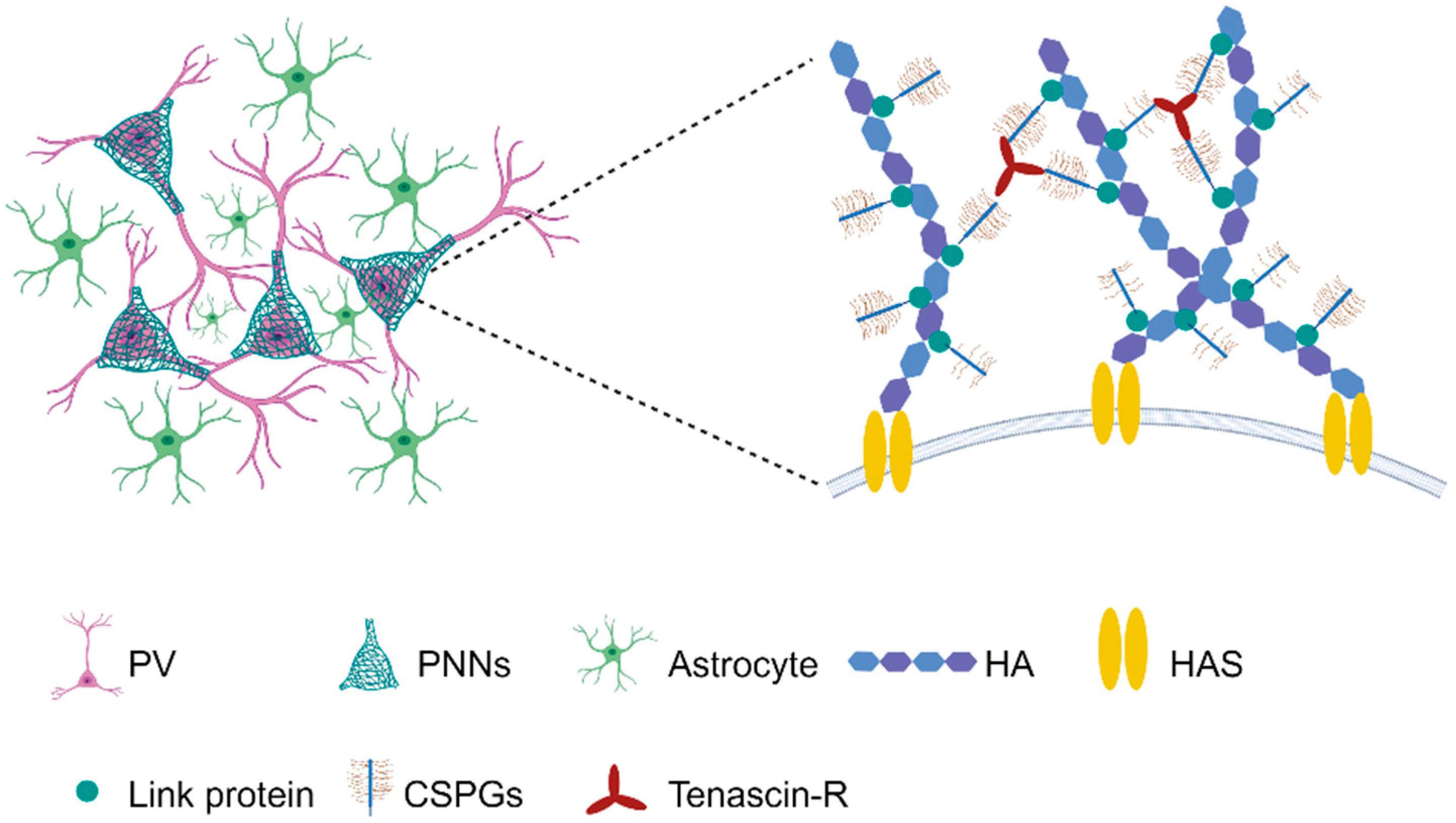
2.1. Composition and Distributions

Approximately 10–20% of the brain volume is occupied by the ECM, which is a dense network of proteins and glycans that provides anchorage points for nerve and glial cells and contributes to their normal physiology [15]. The cerebral ECM has been recognized as a key player in synaptic plasticity, especially regarding the structures of PNNs. PNNs were first described as reticular structures by Golgi in the late 1800s [16], and since then their appearances has been confirmed across a wide range of species, from frogs and birds to mammals, including humans [17]. The nets are intricate assemblies of highly diverse proteoglycans (PGs) and chondroitin sulphate glycosaminoglycan chains (CS-GAG) attached to chondroitin sulfate proteoglycans (CSPG). These components are anchored on a foundation of hyaluronan (HA) glycosaminoglycan backbones and proteins, which condense through specific interactions around certain neurons [11,12]. Although PNNs in the adult CNS are generally stable structures, various endogenous and exogenous stressors are able to cause their breakdown. For example, matrix metalloproteases (MMPs), produced by various cell types, are involved in remodeling the PNNs and, when disrupted, indiscriminately destroy laminin, collagens, and CSPG [18]. Therefore, the function and stability of PNNs are influenced by the constituent of CS-GAGs, which consist of repeating disaccharide units of sulfated glucuronic acid (GlcA) and N-acetylgalactosamine (GalNac) [11]. The PGs interact with morphogens, growth factors, cytokines, cell receptors, cell adhesion molecules, and neurotrophic peptides to facilitate regulatory roles in embryonic neural development [19]. HA is the only non-sulfated GAG and is fixed in hyalectan CSPGs aggregates in perineural structures so that it has important biophysical properties which are important for the segregation and hydration of PNNs to provide neuroprotection and neural plasticity, as well as to regulate memory and cognition [19] (Figure 1).
PNNs are formed by a cartilage-like structure as mesh-like webs, with link proteins (Crtl1/Hapln1 and Bral2/Hapln4) to stabilize the binding of various CSPGs to a HA backbone [20,21]. The CSPG components including lecticans, neurocan, versican, aggrecan, phosphacan, hyaluronan, tenascin-R, and link proteins, which are vulnerable to enzymatic degradation, primarily by chondroitinase-ABC (ChABC) [22]. Several animal KO and transgenic mouse models have been developed to prevent or reduce the formation of PNNs around neurons [23,24,25] (Figure 2). The cellular origin of CSPGs within PNNs has been a longstanding topic of debate. Investigations have shown that while certain CSPGs are produced and expressed by neurons, others have glial origins, and some even derive from both, ultimately converging into the extracellular space to assemble into PNNs [26,27,28]. In addition, aggrecan and neurocan are expressed by neurons, while brevican is expressed by both neurons and astrocytes [22]. Histologically, PNNs’ visualization is predominantly achieved through lectin wisteria floribunda agglutinin (WFA) staining, due to its affinity for N-acetylgalactosamine in the polysaccharide chain of most PNNs [29,30], although alternative methods have also been used [31,32]. It is worth noting that there are also WFA-negative PNNs. The PNNs that surround cortical output neurons do not bind to WFA and are instead recognized by aggregative proteoglycan antibodies [33].
PNNs play a key role in synaptic stabilization by acting as a physical barrier, as well as in the integration and generation of neuronal electrical activity. They provide a continuous microenvironment that facilitates the flow of cations across the membrane, thereby contributing to the electrical properties of neurons [34,35]. As mentioned earlier, the vast majority of the mammalian CNS cells are endowed with PNNs. Meanwhile, PNNs surround various types of nerve cell and exhibited a conserved distribution across mammalian species, from rodents to primates and human [36]. PNNs mostly surround the soma and dendrites of GABAergic neurons, especially around fast-spiking parvalbumin interneurons (PV cells) in various brain regions, like the forebrain, midbrain, and cerebellum [20,35,37,38,39]. Notably, PNNs envelop a smaller number of excitatory neurons [40], which can either express PV or not [41,42]. Furthermore, they also wrap around neurons pivotal to fast transmission, including glycinergic output neurons in the medial nucleus of the trapezoid body (MNTB) at the calyx of Held synapse, and excitatory neurons in the deep cerebellar nucleus [43]. In the hippocampus, PNNs are typically formed around inhibitory interneurons [44], yet in the CA2 region, PNNs have been observed surrounding both inhibitory and excitatory neurons [45]. Intriguingly, the expression of PNNs exhibit sexual dimorphism in certain regions. For instance, it has shown that PNNs are numerous and well developed in hippocampal CA1 of adult male rats but are lower in juvenile and possibly in adult females. Such differences, however, are not evident in other regions, like CA3 or the adjacent neocortex [46].

2.2. Physiological Functions

PNNs emerge during postnatal development, reaching its expression peaks in adulthood and presenting minimally in both newborn and aged animals [5]. The appearance of PNNs in juveniles coincides with the closing of critical developmental periods [2,47]. In the meanwhile, it is known that disruptions of either sulfate supply or sulfase can affect brain development and have long-lasting effects on brain function [48]. The principal reason affecting the CNS functions mentioned above is that PNNs strategically influence the development and stabilization of synaptic connections [49,50]. The functions of PNNs actually include various forms of plasticity. Numerous studies have shown that PNNs appear on PV inhibitory neurons, reaching mature levels by the end of the critical period during postnatal development. In adult animals, disrupted PNNs and associated signaling restore the visual cortical plasticity [2]. Fast-spiking, calcium-dependent, parvalbumin-positive (PV+) GABAergic interneurons are the most significant cells enwrapped by PNNs and they play an important role in maintaining plasticity and timing during critical periods [51]. When PNNs in sensory cortex are mature ahead of time, plasticity in the local network is greatly reduced. However, experimental removal of PNNs in adult animals can restore plasticity levels in young animals by increasing the structural plasticity and reducing the inhibitory spiking [2,52]. The correct pattern and assembly of PNNs is a key step in the maturation of PV+ GABAergic interneurons, and in the regulation of synaptic plasticity [53]. These finding suggest that the deposition of PNNs onto PV inhibitory neurons applied the brakes to critical period plasticity [54]. In this context, GABA antagonists reactivate the critical period plasticity and reduce the number of PNNs, whereas GABA agonists restore PV/PNN expression and limit plasticity in the aged cortex [55,56]. Mirroring these findings, chemogenetic inactivation of PV+ interneurons is sufficient to reinstate the critical period plasticity in the adult auditory cortex, and, in this process, PNNs undergo the same anatomical changes with PV+ [57]. Along with diminished neuronal and glial elements, Elise et al. (2021) found atypical PNNs that regulate plasticity in the CA2 of BTBR mice (social dysfunction) [58]. By diminishing PNNs in the CA2 of BTBR mice to control levels, they observed a partial restoration of social memory [58]. In conclusion, degradation of PNNs not only restores critical period plasticity but also modifies neuronal plasticity in response to a strong stimulus.
Structurally, PNNs restrict neurite growth and synapse development. At the synaptic level, PNNs compartmentalize the neuronal surface and restrict glutamate receptor mobility [59], supporting synaptic plasticity and stabilizing synapses. Preventing AMPA receptor mobility reduces short-term plasticity in rat primary neurons, indicating a potential role of PNNs in memory formation. Several PNN components regulate synaptic plasticity. Neurocan deficiency reduces late-phase LTP stability [60], brevican ablation significantly impairs LTP [61], and depletion of its binding partner tenascin-R also reduces LTP [62]. Knockout of the related glycoprotein tenascin-C results in a complete failure in LTD induction and impaired LTP development, likely due to reduced L-type VDCC channel signaling [63,64].
PNNs are crucial for protecting neurons from oxidative stress [23,65,66]. CS and HA are highly charged and rich in anions. Therefore, PNNs can bind a large number of cations than other ECM components, such as Fe3+, participate in local ion homeostasis maintenance, and reduce oxidative stress in the CNS [67]. Modified PNNs, stemming from exposure to a variety of substances, like high-fat diets [68,69] and cocaine, might impair cognition and memory [70,71]. For instance, a high-fat, high-sugar diet reduced PV neurons, and altered the coexpression of PNNs in adolescent rats, resulting in impaired social memory in the medial prefrontal cortex (mPFC) [69]. PNNs also have a neuroprotective function by protecting PV neurons from oxidative stress, because the PV neurons without a PNN structure are highly vulnerable to oxidative stress, as revealed by a previous study [72]. In particular, PV neurons are relevant to the physiological state of an increased metabolic activity rate and promote the production of reactive oxygen species. When the antioxidant function of PNNs are deficient, the excessive production of reactive oxygen in PV neurons leads to cell death [73].
PNNs have been implicated in controlling various forms of memory [30,74,75]. Inhibitory neurons, especially PV cells, across many regions play a pivotal role in shaping the memory engram [76,77,78,79]. Supporting this, disruptions in PNNs or their components has been observed in a number of psychiatric disorders related to learning, memory, and information processing [80]. It is now well established that PNNs ensure the stability of existing synapses and prevent the formation of new ones on mature PV interneurons [81]. As PNNs stabilize excitatory inputs, they lead to an increase in the maximum firing rate of PV interneurons, and in PV protein expression [44,82]. More recently, PNNs are shown to act to stabilize synaptic contacts, to limit further plasticity, to restrict the synaptic changes, and to help to solidify active synaptic networks, which are the basis for memory acquisition [81,83,84]. It has also been demonstrated that the absence of PNNs increases terminal axonal sprouting, synaptic plasticity, and memory retention in adult mice and restores memory in an Alzheimer’s model [71,85]. Post-learning sharp wave ripple (SWR) activity involves the sequential replay of training-related neuronal assemblies and is critical for system-level memory consolidation [86]. Sun et al. (2018) found that pretreatment with chondroitinase cleaved PNNs sidechains and increased SWR frequency [87]. Therefore, this reduction in PNNs would allow neurons to respond to changes in the environment. Indeed, degrading PNNs with ChABC promotes the assembly of new inhibitory synapses onto PV cells, which would promote new learning [88] and could even erase consolidated memories [89].
PNNs also regulate neural regeneration in adulthood by surrounding and stabilizing synaptic contacts. After brain injuries in mammals, a scar often forms, consisting of fibrous tissue in the lesion core and glial tissue, which creates a chemical barrier containing various ECM molecules [90,91]. Notably, these chemical barrier molecules, mainly CSPGs and CS-GAGs, limit the ability of axons to regrow over long distances and inhibit new contact with descending axons [91,92]. Previous studies revealed that the administration of ChABC after spinal cord injury removed CS-GAGs from PNNs at the lesion site, thus promoting plasticity and nerve regeneration in long tracts entering the lesion site. In goldfish with a spinal hemisection, plasticity in the spinal neurons might be restored by downregulation of CS in the PNNs in the regeneration process [93]. Moreover, the administration of ChABC degrades CSPGs in the PNNs and appears to boost the plasticity of spinal neurons and to promote the sprouting of spared long tract axons [94]. Nevertheless, it is important to note that thorough digestion of CSPG GAG chains through intracortical ChABC injection might worsen motor deficits and hinder axonal sprouting in the corticospinal tract above the lesion in spinal cord injury models. These findings indicate that CSPGs modulation requires meticulous control to yield functional benefits [95]. It is well-established that in the mature nervous system, motoneurons located in the ventral horn of the spinal cord are surrounded by structures known as PNNs [96]. Recent research suggests that preserving motoneuron PNNs and minimizing synaptic stripping through exercise could facilitate the maintenance of the spinal circuitry and have beneficial effects on functional recovery following peripheral nerve injury [97]. It suggests that PNNs in the spinal cord can be differentially regulated by peripheral injury and activity when compared to brain injury. Based on the aforementioned information, it is evident that regeneration in the CNS, or the absence thereof, is tightly regulated by PNN formation. In a word, PNNs are related to stabilizing synaptic connectivity and protecting neurons by forming a physical barrier. PNNs protect the integrity of synaptic junctions and insulate them from potentially damaging stimuli, such as oxidative stress. Regulation of PNNs could promote or inhibit synapse formation to modify the timing and precision of information processing, memory, and cognition [12].

3. Roles of PNNs in Learning and Memory

3.1. Object Recognition (OR) Memory

Object recognition (OR) memory refers to an organism’s ability to recognize and remember previously encountered objects as familiar, distinguishing them from novel objects. OR memory tests, often using rodents, assess cognitive function by measuring the time spent exploring novel objects versus familiar ones, with a preference for the novel indicating intact memory [98,99]. The removal of major structural components of PNNs in the perirhinal cortex—a brain region important for object recognition—via ChABC in two mouse models exhibiting a significant impairment in OR memory effectively restores their OR memory and synaptic transmission to levels comparable to normal mice after one week [71,85]. A study emphasized the functional significance of neurocan in supporting perisomatic GABAergic inhibition, temporal order recognition memory, and cognitive flexibility. These functions are crucial cognitive resources that are often depleted in neuropsychiatric disorders [100]. Similarly, depletion of PNNs in the perirhinal cortex increases long-term object recognition memory as measured by spontaneous object recognition [85]. Systemic oral administration of 4-methylumbelliferone (4-MU) for 6 months reduces PNN formation around neurons and enhances memory retention in mice in spontaneous object recognition test [101]. Furthermore, downregulation of PNNs either through ChABC digestion or genetic knockdown can improve object recognition memory for a period up to 48 h [102]. PTPσ+/− mice, having a lower number of PNN receptors, display a disrupted PNN network consolidation function in their brain, which bolsters their short-term memory performance but impairs the long-term memory performance [103]. Interestingly, the cognitive and behavioral effects of ChABC vary depending on the location and time of treatment [104]. For example, the infusion of ChABC into the perirhinal cortex enhances recognition memory in an object recognition paradigm. However, the memory-enhancing effect of ChABC treatment attenuates overtime, suggesting that the regeneration of PNNs gradually restores the levels of plasticity [85]. In contrast, infusion of ChABC into the mPFC impairs cross-modal object recognition and object oddity tests in rats [105]. Carulli et al. (2020) [106] provided evidence of dynamic modulation of PNNs in response to delayed eyeblink conditioning (EBC), which is an associative learning paradigm.
Modulation of PNNs could change the balance between excitatory and inhibitory inputs to deep cerebellar nuclei (DCN) neurons which regulate the acquisition and retention of memory [106]. In addition, it has been known that PV interneurons, which predominantly contribute to the generation of GBO, are surrounded by PNNs [107]. Vitamin D deficiency (VDD) could increase the spontaneous GBO and decrease the evoked GBO, reminiscent of the aberrant GBO in schizophrenia, and, as such, decrease PNNs via this process [108]. Moreover, patients with depression often also suffer from cognitive impairments. Short-term object recognition memory was decreased, and PNNs’ expression was increased in the hippocampal CA1 region of depressed rats. Intracranial injection of ChABC into hippocampus CA1 could restore PNNs expression, LTP, hippocampal inhibitory tone, and memory performance in object recognition tests [109]. Therefore, enzymatic digestion of PNNs improves learning, but intact PNNs are necessary for memory retention.

3.2. Fear Memory

Fear conditioning induces a permanent memory in adult animals, often examined by training an animal to associate the presentation of a cue, typically a tone, with a footshock [110]. In early postnatal development, especially in rats younger than 3 weeks, extinction of conditioned fear leads to memory erasure. In contrast, in adult animals, extinction training creates a new memory but does not erase the original memory, suggesting that fear memories are actively protected in adults [89]. This protection is conferred by CSPGs, components of PNNs. The presence of PNNs enables original fear memory and extinction memories to coexist, protecting the former from erasure. Injection of ChABC in adult mice before acquisition of fear memory makes the rate of fear extinction similar to that observed in juvenile mice, indicating a greater response to extinction training. However, injection prior to extinction but after fear training was ineffective. Therefore, PNNs may be involved in the initial encoding process of fear memory and reinforcing their resistance to extinction, rather than in directly regulating the extinction process [111]. ChABC-dependent degradation of PNNs promoted the elimination of fear memory, underscoring the role of intact PNNs in erasure-resistant fear memories [89]. Accordingly, the transplantation of immature interneurons reduced the expression of PNNs and offered an expanded capacity of plasticity in response to the facilitation of extinction memory [112].
Beyond memory extinction, memory encompasses the processes of formation, consolidation, and reconsolidation [37,113]. PNNs have also been implicated in these three processes, fundamentally rooted in neural plasticity. The acquisition of auditory fear memory is associated with an increase in PNNs in such regions as the hippocampus, auditory cortices, and anterior cingulate cortex. Reinhard et al. (2019) found impaired tone-associated fear memory formation in the mouse model of FXS (Fragile X syndrome, Fmr1 KO mice), and this was paralleled by impaired regulation of PNNs in the superficial layers of auditory cortex in Fmr1 KO mice [114]. The elimination of PNNs impairs the inhibitory PV neurons, which are critical for the consolidation of auditory fear memories [115]. However, in the amygdala, PNNs were mainly expressed around excitatory neurons, which were recruited during auditory fear conditioning and memory retrieval [70]. Intriguingly, Evans et al. (2022) conducted post-learning manipulation of neurogenesis through voluntary exercise, and observed a reduction in PNN density in the CA1, accompanying a decrease in contextual fear memory retrieval [116]. More studies focus on the role of PNNs in memory consolidation and retention. Increased PNN expression in both the hippocampus and the ACC enhances the recall and reconsolidation of both recent and remote fear memory, whereas removal of PNNs impairs the consolidation and reconsolidation of both recent and remote fear memory by regulating the feedback inhibition of PV interneurons [37]. However, digestion of PNNs using ChABC in the secondary visual cortex (V2L) interrupts the recall of long-term fear memory but not recent fear memory in rats [117]. Several studies prior to this work also showed similar remote fear memory recall impairment when PNNs were disrupted in various regions of the brain [89,118]. Similarly, injection of hyaluronidase in the hippocampus to destroy another ingredient of PNNs attenuated fear responses one day after conditioning, and a combination of hyaluronidase plus ChABC injected into the hippocampus also attenuated long-term fear memory [118,119]. The elimination of PNNs in CA1 disrupted GABA release and long-term contextual fear memory retention [120]. Jovasevic et al. (2021) demonstrated that depleting primary cilia in the hippocampal CA1 subfield was accompanied by the disruption of PNNs in CA1 and played an important role in the persistence of fear memory. Therefore, the severe downregulation of the primary cilium-associated genes, as well as several genes encoding important component of PNNs, may lead to the failure of lasting memory [121]. Intriguingly, diurnal and circadian rhythms of PNNs were found in several brain regions involved in emotional memory processing. For example, sleep deprivation prevented the daytime decrease in PNNs in the hippocampus, BLA, and central amygdala (CeA) and enhanced fear memory extinction. Thus, rhythmic modification of PNNs may contribute to memory consolidation during sleep [122]. Collectively, PNNs play an important role in many facets of fear memory, and this functionality relies on myriad mechanisms that appear to be rhythmic and brain region-dependent.

3.3. Spatial Memory

The maturation of PNNs in the local network is known to greatly reduce the plasticity of the sensory cortex. It is interesting to note that the development time of PNNs coincides with the development of grid cell firing, which is an essential part of the network supporting navigation and spatial memory [123]. Once the critical period is over, PNNs play a crucial role in maintaining the stability of established synaptic connections, thereby ensuring both the network’s integrity and the spatiotemporal interplay among grid cells [124]. This stability is particularly important in maintaining consistent grid cells when revisiting previous environments [124]. Neuropeptide Y (NPY) is involved in regulating various physiological functions, including learning and memory abilities. Studies have demonstrated that Npy1rrfb mutant mice exhibited a significant slowdown in spatial learning, which was associated with an intense increase in PNN expression. Interestingly, enzymatic digestion of PNNs restored their learning abilities [125]. Tajerian et al. (2018) reported aberrant hippocampal LTP and a reduction in specialized PNNs around inhibitory interneurons, correlating with deficits in location memory [126]. Iron is a necessary substrate for neuronal function throughout life. Iron deficiency can impair spatial memory and result in disorganized apical dendrite structure accompanied by altered PV and PNN expression, along with reduced BDNF levels [127]. These findings suggest the crucial roles of PNNs in maintaining plasticity in the sensory cortex and spatial memory and highlight the potential for interventions targeting PNN expression to improve cognitive function.

4. Roles of PNNs in Memory-Related Dysfunctions

4.1. Addiction

Drug addiction modifies brain plasticity, leading to persistent drug reward memory. These alterations in plasticity and memories are believed to produce aberrant motivation and reinforcement, contributing to addiction. While much research has focused on the effect of drug abuse on pre- and postsynaptic cells and astrocytes [110], recent studies have highlighted PNNs and their individual constituents as important regulators of memories linked to addiction-related behaviors in animals. Emerging evidence, including our own, has shown that degradation of PNNs affects extinction and attenuates cocaine-conditioned place preference (cocaine-CPP) and cocaine self-administration [128,129]. PNNs were altered in specific brain regions when exposed to several drugs of abuse (cocaine, heroin, nicotine, and alcohol) [129]. Cocaine induces neuroplasticity in the ECM, evident in both cocaine-dependent humans [130] and rodent models of cocaine addiction [131]. Meanwhile, cocaine also induces metaplasticity [132], which alters the ability of natural stimuli to further alter plasticity. In this case, the removal of PNNs and/or regulation of other PNN-binding molecules may alter cell firing and homeostatic plasticity [133] so as to prevent the drug-induced changes, possibly by restoring the excitatory/inhibitory balance [65].
Modulation of the expression of PNN components seems to be influenced by the type of drug, the duration of drug exposure and withdrawal, and the brain region [129]. Key areas, like the BLA, hippocampus, and prefrontal cortex (PFC), played a critical role in acquiring and maintaining the drug-related memory. These regions notably overlap with brain regions essential for fear-conditioned memory [134]. Golgi neurons are vital for the modulation of activity and plasticity in the cerebellum, and PNNs surrounding Golgi interneurons play a role in consolidating drug-related memories [135]. Fully condensed PNNs around Golgi interneurons can label synaptic alignments that represent drug-cue associations [135]. A recent study showed that cocaine-induced conditioned preference increased neural activity and upregulated PNNs around Golgi interneurons in the posterior cerebellar cortex. However, a reduction in PNN protein expression around Golgi cells disrupted the consolidation process [20]. The inactivation of the IL (infralimbic cortex), but not the PrL (prelimbic cortex), increased posterior cerebellar cortex activity, and upregulated PNNs’ expression around Golgi interneurons. This can further intensify the preference for cocaine-related cues and the acquisition of cocaine-induced memory [136]. PNNs within the mPFC are also required for the maintenance of cocaine-associated memories [137], such as when PNN removal in the mPFC attenuated the acquisition and reconsolidation of cocaine-associated memory in a CPP task [138]. Cocaine memory reactivation significantly altered the synaptic and electrical properties of the PNN+ neuron in mPFC and its related proteins that regulate pyramidal output and influence the drug-seeking behavior [139]. Removed PNNs within the BLA or CeA enhanced extinction training and changed plasticity prior to extinction training of morphine or cocaine-induced CPP and heroin self-administration [140]. Increased densities of hippocampal PNNs, coupled with decreased ECM proteolytic genes and altered synaptic markers, support preclinical studies indicating that PNNs may stabilize reward memories. PNNs and ECM molecules may be promising targets for addressing cue-induced relapse in SUD [141]. In summary, PNN removal appears to create a blank slate for neuroplasticity that has the potential to combat drug-induced maladaptive plasticity and create new adaptive response, suggesting that targeting PNNs may be promising for the treatment of addiction.

4.2. Aging

As the nervous system ages, there is a gradual decline in its plasticity, as evidenced by the reduced capability of the CNS in the ability of learning, as well as the decrease in the ability to adapt to environmental changes and compensate for damage. Elevated microglial activity in aged brains negatively impacts cognition, in part through mechanisms that alter PNN assembly in memory-associated brain regions [142]. As mentioned before, CSPGs in PNNs are central to regulating synaptic plasticity [143]. Thus, PNNs have crucial roles in memory in the aging brain, and deterioration of PNNs contributes to age-dependent brain dysfunction [19]. The accumulation of CSPG-associated ECM correlates with age-dependent decline in striatum-related cognitive functions, including motor learning and working memory. Removal of CSPG-associated ECM by ChABC in aged mice significantly improved motor learning, suggesting that changes in the neural ECM’s CSPGs can regulate striatal plasticity [144]. The CSPGs of PNNs also have important functional roles in protecting against the development of AD. Cortical regions with abundant levels of ECM CSPGs seem to be less vulnerable to degenerative features associated with the development of AD [145,146]. The decline in PNNs, marked by the loss of lectin labeling of the PNN-associated CS-GAGs [147,148], has been reported in patients with neurocognitive and neuropsychiatric disorders, including AD [149]. Furthermore, PNN-associated CS-GAG sulfation patterns are also altered in AD brains [150,151]. Brain-specific lectican brevican were significantly elevated in AD patients, which suggests the loss of synaptic plasticity before cell death.
PNNs seem to provide protection against the formation of neurofibrillary tangles [152]. The colocalization of some proteoglycans with Aβ and tangles may reflect a pathogenic role of the PNNs in AD [153]. Consistently, it has been shown that disordered quantification of PV and PNNs in the hippocampus of Tg2576 mice are early events of amyloid pathology [154]. The application of ChABC significantly reduce amyloid β-peptide (Aβ) burden and increase synaptic density [155]. When ChABC is injected into the perirhinal cortex to digest the existing PNNs, the Tau mice show OR memory and synaptic transmission comparable to those of control animals, suggesting that PNNs is related with memory loss in neurodegenerative disorders, like AD [71]. Of the two mono-sulphated CSs [156], C6S is known to facilitate axon growth and plasticity, while C4S’ influence is the opposite [157]. In AD animal models, using a C4S-blocking antibody have been shown to alleviate the pathology-associated memory loss [71,158]. A decline in C6S is linked to premature memory loss, while an increase in C6S not only prevents age-related memory deficits but can also restore existing memory impairments [159]. An enriched environment might improve cognition in AD mouse models by modulating the PNN levels, which has been proven to have long-lasting beneficial effects on memory in AD subjects [154]. In conclusion, the change in PNN GAG sulfation renders the PNNs more inhibitory, which leads to a decrease in plasticity and adversely affects memory in aged brains [156]. By modulating PNNs, it is possible to restore neural plasticity to a juvenile-like state, which results in the restoration of cognition in both AD-affected and aged mouse models [14,102,158].

4.3. Other

Consistent with working memory impairments in schizophrenia, multiple post-mortem studies have demonstrated morphological and/or molecular alterations in the dorsolateral prefrontal cortex (DLPFC) layer 3 pyramidal cells (PCs) and PV neurons in individuals with schizophrenia [160]. These alterations include reduced dendritic spine density on PCs, lower levels of PV mRNA, and reduced PV protein in PV basket cell terminals. The lower levels of PV mRNA and protein could be explained by a loss of PV neurons [161]. However, studies investigating PV cell density in schizophrenia have provided mixed results. A lower density of perineuronal nets (PNNs) in schizophrenia could result from several different pathological processes. Firstly, it could be the result of a lower density of PV neurons for PNNs to surround. Secondly, fewer PNNs could indicate a failure in PNN formation and/or maintenance, which would be characterized by a loss of multiple PNN markers and a lower proportion of PV neurons surrounded by PNNs, without a reduction in PV cell density. Thirdly, the detectability of PNNs could be reduced due to alterations in their composition, such as lower glycosylation of aggrecan. Given the roles of PNNs in regulating PV cellular physiology, the identified alterations in PV neurons and their PNNs could contribute to dysfunction in schizophrenia [162]. In Fmr1 knockout (KO) mice, a model for autism, there is a reduced density of PNNs in the amygdala and auditory cortex under all conditions. Additionally, there is a decrease in PNN intensity in the CA2 region of the hippocampus. Importantly, there appears to be a positive correlation between tone-associated memory and the density of PNNs in the amygdala and auditory cortex [114].

5. The Challenges and Prospects

Recent work has revealed the importance of PNNs in the control of CNS plasticity [19]. The removal of PNNs provides an opportunity to alter plasticity for CNS repair after injury and to facilitate learning and memory in aging and CNS disorders. However, the therapeutic potential of targeting PNNs remain in its nascent stage.
Firstly, the current understanding on the roles of PNNs in memory and associated psychiatric conditions has been developed through a large number of behavioral studies involving proteolytic cleavage and knockout models [163]. Genetic modifications, like KO models, are frequently used to abolish or attenuate PNNs. While these methods are very useful to establish the proof of concept that modulating PNNs can profoundly influence plasticity and memory, their direct clinic application remains challenging. Future studies should examine the potential genetic associations of other genes encoding PNN components in memory [129]. For more refined alteration in neuronal plasticity, strategies that specifically target and modify the molecular composition of PNNs are essential [164].
Secondly, the connection between PNNs and the modulation of learning and memory primarily revolve around four genes encoding PNNs components: Hapln1, TnR, TnC, and Bcan. However, the involvement of these and other PNNs components in long-term memory mechanisms is largely unclear and is currently a focus of research [165]. A majority of studies used a global disruption method, targeting not only the PNNs, but also the entire ECM and other neurons. Future studies need more specific approaches to target the PNNs and to verify the effects of these studies [110].
Thirdly, there is ambiguity surrounding the specific cell types responsible for in the production of PNNs [165]. For instance, astrocytes are able to package HA-based pericellular matrices in vitro [166], whereas CSPGs, such as aggrecan, brevican, and phosphacan, are produced by neurons [167]. Intriguingly, other evidence suggests that the neurons themselves seem to be able to create a PNN-like structure in a dissociated culture in the absence of glial cells [167].
Lastly, it is imperative to resolve how the PNNs actually affect the activity of individual neurons in the neuronal system. This necessitates in vivo electrophysiological signals or image calcium perfusion to distinguish between the cells that are enwrapped by PNNs and those that are not. However, a significant challenge is the current lack of tools for the in vivo staining of PNNs [168]. It is also worth noting that manipulation of PNNs probably causes neuronal circuits to experience hyperplasticity or might affect synapses vulnerable to neurotoxic stimuli, which could have a noxious effect on cognition [12]. While PNNs may not supersede neurons as the primary entity of research, their growing prominence in decoding brain functions and therapeutic potential cannot be overlooked [104].

6. Conclusions

In this review, we have explored the intricate molecular compositions and functions of PNNs. The emerging evidence suggests that PNNs have a vital role in controlling diverse facets of memory. Notably, PNNs appear to influence the capacity for new learning and/or memory formation, especially during both extinction and reversal learning, by restricting the critical-period plasticity. PNNs are also implicated in a spectrum of neuropsychiatric disorders related to memory, encompassing schizophrenia, drug addiction, and neurodegeneration. As we look forward, it is crucial for future studies to discover the detailed dynamic molecule changes occurring in PNNs in order to understand how PNNs and PNN-surrounded neurons influence memory processes. In conclusion, the diverse protein elements of PNNs present promising therapeutic avenues, potentially for aiding in erasing detrimental memories that drive relapses into drug abuse, rejuvenating neural plasticity and restoring cognition in neurological disorders, like AD.

Author Contributions

Conceptualization, X.L. and X.W.; writing—original draft preparation, X.L. and X.W.; writing—review and editing, T.L., C.K., Y.S., W.Z. and Z.L.; supervision, Y.X.; funding acquisition, Y.X. All authors have read and agreed to the published version of the manuscript.

Funding

This work was funded by several grants from the National Natural Science Foundation of China (32161143022, 81871046), STI2030-Major Projects (2022ZD0214500) as well by as Scientific Project of Beijing Life Science Academy (2023300CB0100).

Acknowledgments

Figure 1 and Figure 2 were created with BioRender.com. http://www.biorender.com/ (accessed on 30 January 2024).

Conflicts of Interest

The authors declare no conflicts of interest.

References

  1. Vargová, L.; Syková, E. Astrocytes and extracellular matrix in extrasynaptic volume transmission. Philos. Trans. R. Soc. London Ser. B Biol. Sci. 2014, 369, 20130608. [Google Scholar] [CrossRef]
  2. Pizzorusso, T.; Medini, P.; Berardi, N.; Chierzi, S.; Fawcett, J.; Maffei, L. Reactivation of ocular dominance plasticity in the adult visual cortex. Science 2002, 298, 1248–1251. [Google Scholar] [CrossRef]
  3. Massey, J.; Hubscher, C.; Wagoner, M.; Decker, J.; Amps, J.; Silver, J.; Onifer, S. Chondroitinase ABC digestion of the perineuronal net promotes functional collateral sprouting in the cuneate nucleus after cervical spinal cord injury. J. Neurosci. Off. J. Soc. Neurosci. 2006, 26, 4406–4414. [Google Scholar] [CrossRef]
  4. Barritt, A.; Davies, M.; Marchand, F.; Hartley, R.; Grist, J.; Yip, P.; McMahon, S.; Bradbury, E. Chondroitinase ABC promotes sprouting of intact and injured spinal systems after spinal cord injury. J. Neurosci. Off. J. Soc. Neurosci. 2006, 26, 10856–10867. [Google Scholar] [CrossRef]
  5. Yamada, J.; Jinno, S. Spatio-temporal differences in perineuronal net expression in the mouse hippocampus, with reference to parvalbumin. Neuroscience 2013, 253, 368–379. [Google Scholar] [CrossRef]
  6. O’Reilly, K.; Perica, M.; Fenton, A. Synaptic plasticity/dysplasticity, process memory and item memory in rodent models of mental dysfunction. Schizophr. Res. 2019, 207, 22–36. [Google Scholar] [CrossRef]
  7. Wanat, M.; Sparta, D.; Hopf, F.; Bowers, M.; Melis, M.; Bonci, A. Strain specific synaptic modifications on ventral tegmental area dopamine neurons after ethanol exposure. Biol. Psychiatry 2009, 65, 646–653. [Google Scholar] [CrossRef] [PubMed]
  8. Martin, S.; Grimwood, P.; Morris, R. Synaptic plasticity and memory: An evaluation of the hypothesis. Annu. Rev. Neurosci. 2000, 23, 649–711. [Google Scholar] [CrossRef] [PubMed]
  9. Jeong, Y.; Cho, H.; Kim, M.; Oh, J.; Kang, M.; Yoo, M.; Lee, H.; Han, J. Synaptic plasticity-dependent competition rule influences memory formation. Nat. Commun. 2021, 12, 3915. [Google Scholar] [CrossRef]
  10. Ruzicka, J.; Dalecka, M.; Safrankova, K.; Peretti, D.; Jendelova, P.; Kwok, J.; Fawcett, J. Perineuronal nets affect memory and learning after synapse withdrawal. Transl. Psychiatry 2022, 12, 480. [Google Scholar] [CrossRef] [PubMed]
  11. Testa, D.; Prochiantz, A.; Di Nardo, A. Perineuronal nets in brain physiology and disease. Semin. Cell Dev. Biol. 2019, 89, 125–135. [Google Scholar] [CrossRef] [PubMed]
  12. Reichelt, A.; Hare, D.; Bussey, T.; Saksida, L. Perineuronal Nets: Plasticity, Protection, and Therapeutic Potential. Trends Neurosci. 2019, 42, 458–470. [Google Scholar] [CrossRef] [PubMed]
  13. Bitanihirwe, B.; Woo, T. Perineuronal nets and schizophrenia: The importance of neuronal coatings. Neurosci. Biobehav. Rev. 2014, 45, 85–99. [Google Scholar] [CrossRef] [PubMed]
  14. Sorg, B.; Berretta, S.; Blacktop, J.; Fawcett, J.; Kitagawa, H.; Kwok, J.; Miquel, M. Casting a Wide Net: Role of Perineuronal Nets in Neural Plasticity. J. Neurosci. Off. J. Soc. Neurosci. 2016, 36, 11459–11468. [Google Scholar] [CrossRef] [PubMed]
  15. Bosiacki, M.; Gąssowska-Dobrowolska, M.; Kojder, K.; Fabiańska, M.; Jeżewski, D.; Gutowska, I.; Lubkowska, A. Perineuronal Nets and Their Role in Synaptic Homeostasis. Int. J. Mol. Sci. 2019, 20, 4108. [Google Scholar] [CrossRef]
  16. Celio, M.; Spreafico, R.; De Biasi, S.; Vitellaro-Zuccarello, L. Perineuronal nets: Past and present. Trends Neurosci. 1998, 21, 510–515. [Google Scholar] [CrossRef]
  17. Li, X.; Li, N.; Zhao, P.; Ren, D.; Luo, B.; Zhou, T. Perineuronal Nets: From Structure to Neurological Disorders. Curr. Med. Chem. 2024, 31. [Google Scholar] [CrossRef]
  18. Yong, V.W.; Power, C.; Forsyth, P.; Edwards, D.R. Metalloproteinases in biology and pathology of the nervous system. Nat. Rev. Neurosci. 2001, 2, 502–511. [Google Scholar] [CrossRef]
  19. Hayes, A.; Melrose, J. Neural Tissue Homeostasis and Repair Is Regulated via CS and DS Proteoglycan Motifs. Front. Cell Dev. Biol. 2021, 9, 696640. [Google Scholar] [CrossRef]
  20. Carbo-Gas, M.; Moreno-Rius, J.; Guarque-Chabrera, J.; Vazquez-Sanroman, D.; Gil-Miravet, I.; Carulli, D.; Hoebeek, F.; De Zeeuw, C.; Sanchis-Segura, C.; Miquel, M. Cerebellar perineuronal nets in cocaine-induced pavlovian memory: Site matters. Neuropharmacology 2017, 125, 166–180. [Google Scholar] [CrossRef]
  21. Mörgelin, M.; Heinegård, D.; Engel, J.; Paulsson, M. The cartilage proteoglycan aggregate: Assembly through combined protein-carbohydrate and protein-protein interactions. Biophys. Chem. 1994, 50, 113–128. [Google Scholar] [CrossRef] [PubMed]
  22. Galtrey, C.M.; Fawcett, J.W. The role of chondroitin sulfate proteoglycans in regeneration and plasticity in the central nervous system. Brain Res. Rev. 2007, 54, 1–18. [Google Scholar] [CrossRef] [PubMed]
  23. Carulli, D.; Pizzorusso, T.; Kwok, J.; Putignano, E.; Poli, A.; Forostyak, S.; Andrews, M.; Deepa, S.; Glant, T.; Fawcett, J. Animals lacking link protein have attenuated perineuronal nets and persistent plasticity. Brain A J. Neurol. 2010, 133, 2331–2347. [Google Scholar] [CrossRef] [PubMed]
  24. Yoshioka, N.; Miyata, S.; Tamada, A.; Watanabe, Y.; Kawasaki, A.; Kitagawa, H.; Takao, K.; Miyakawa, T.; Takeuchi, K.; Igarashi, M. Abnormalities in perineuronal nets and behavior in mice lacking CSGalNAcT1, a key enzyme in chondroitin sulfate synthesis. Mol. Brain 2017, 10, 47. [Google Scholar] [CrossRef] [PubMed]
  25. Geissler, M.; Gottschling, C.; Aguado, A.; Rauch, U.; Wetzel, C.; Hatt, H.; Faissner, A. Primary hippocampal neurons, which lack four crucial extracellular matrix molecules, display abnormalities of synaptic structure and function and severe deficits in perineuronal net formation. J. Neurosci. Off. J. Soc. Neurosci. 2013, 33, 7742–7755. [Google Scholar] [CrossRef] [PubMed]
  26. Carulli, D.; Rhodes, K.E.; Brown, D.J.; Bonnert, T.P.; Pollack, S.J.; Oliver, K.; Strata, P.; Fawcett, J.W. Composition of perineuronal nets in the adult rat cerebellum and the cellular origin of their components. J. Comp. Neurol. 2006, 494, 559–577. [Google Scholar] [CrossRef] [PubMed]
  27. Oohashi, T.; Edamatsu, M.; Bekku, Y.; Carulli, D. The hyaluronan and proteoglycan link proteins: Organizers of the brain extracellular matrix and key molecules for neuronal function and plasticity. Exp. Neurol. 2015, 274, 134–144. [Google Scholar] [CrossRef] [PubMed]
  28. Giamanco, K.A.; Matthews, R.T. Deconstructing the Perineuronal Net: Cellular Contributions and Molecular Composition of the Neuronal Extracellular Matrix. Neuroscience 2012, 218, 367–384. [Google Scholar] [CrossRef]
  29. Miyata, S.; Nadanaka, S.; Igarashi, M.; Kitagawa, H. Structural Variation of Chondroitin Sulfate Chains Contributes to the Molecular Heterogeneity of Perineuronal Nets. Front. Integr. Neurosci. 2018, 12, 3. [Google Scholar] [CrossRef]
  30. Duncan, J.; Foster, R.; Kwok, J. The potential of memory enhancement through modulation of perineuronal nets. Br. J. Pharmacol. 2019, 176, 3611–3621. [Google Scholar] [CrossRef]
  31. Mulligan, K.A.; Vanbrederode, J.F.M.; Hendrickson, A.E. The Lectin Vicia-Villosa Labels a Distinct Subset of Gabaergic Cells in Macaque Visual-Cortex. Vis. Neurosci 1989, 2, 63–72. [Google Scholar] [CrossRef]
  32. Giamanco, K.A.; Morawski, M.; Matthews, R.T. Perineuronal net formation and structure in aggrecan knockout mice. Neuroscience 2010, 170, 1314–1327. [Google Scholar] [CrossRef] [PubMed]
  33. Matthews, R.; Kelly, G.; Zerillo, C.; Gray, G.; Tiemeyer, M.; Hockfield, S. Aggrecan glycoforms contribute to the molecular heterogeneity of perineuronal nets. J. Neurosci. Off. J. Soc. Neurosci. 2002, 22, 7536–7547. [Google Scholar] [CrossRef] [PubMed]
  34. Brückner, G.; Szeöke, S.; Pavlica, S.; Grosche, J.; Kacza, J. Axon initial segment ensheathed by extracellular matrix in perineuronal nets. Neuroscience 2006, 138, 365–375. [Google Scholar] [CrossRef] [PubMed]
  35. Morris, N.; Henderson, Z. Perineuronal nets ensheath fast spiking, parvalbumin-immunoreactive neurons in the medial septum/diagonal band complex. Eur. J. Neurosci. 2000, 12, 828–838. [Google Scholar] [CrossRef] [PubMed]
  36. Brückner, G.; Härtig, W.; Kacza, J.; Seeger, J.; Welt, K.; Brauer, K. Extracellular matrix organization in various regions of rat brain grey matter. J. Neurocytol. 1996, 25, 333–346. [Google Scholar] [CrossRef] [PubMed]
  37. Shi, W.; Wei, X.; Wang, X.; Du, S.; Liu, W.; Song, J.; Wang, Y. Perineuronal nets protect long-term memory by limiting activity-dependent inhibition from parvalbumin interneurons. Proc. Natl. Acad. Sci. USA 2019, 116, 27063–27073. [Google Scholar] [CrossRef] [PubMed]
  38. Morino-Wannier, P.; Fujita, S.; Jones, E. GABAergic neuronal populations in monkey primary auditory cortex defined by co-localized calcium binding proteins and surface antigens. Exp. Brain Res. 1992, 88, 422–432. [Google Scholar] [CrossRef] [PubMed]
  39. Adams, I.; Brauer, K.; Arélin, C.; Härtig, W.; Fine, A.; Mäder, M.; Arendt, T.; Brückner, G. Perineuronal nets in the rhesus monkey and human basal forebrain including basal ganglia. Neuroscience 2001, 108, 285–298. [Google Scholar] [CrossRef]
  40. Wegner, F.; Härtig, W.; Bringmann, A.; Grosche, J.; Wohlfarth, K.; Zuschratter, W.; Brückner, G. Diffuse perineuronal nets and modified pyramidal cells immunoreactive for glutamate and the GABA(A) receptor alpha1 subunit form a unique entity in rat cerebral cortex. Exp. Neurol. 2003, 184, 705–714. [Google Scholar] [CrossRef]
  41. Mészár, Z.; Girard, F.; Saper, C.; Celio, M. The lateral hypothalamic parvalbumin-immunoreactive (PV1) nucleus in rodents. J. Comp. Neurol. 2012, 520, 798–815. [Google Scholar] [CrossRef]
  42. Horii-Hayashi, N.; Sasagawa, T.; Hashimoto, T.; Kaneko, T.; Takeuchi, K.; Nishi, M. A newly identified mouse hypothalamic area having bidirectional neural connections with the lateral septum: The perifornical area of the anterior hypothalamus rich in chondroitin sulfate proteoglycans. Eur. J. Neurosci. 2015, 42, 2322–2334. [Google Scholar] [CrossRef]
  43. Balmer, T. Perineuronal Nets Enhance the Excitability of Fast-Spiking Neurons. eNeuro 2016, 3, 0112. [Google Scholar] [CrossRef]
  44. Yamada, J.; Ohgomori, T.; Jinno, S. Perineuronal nets affect parvalbumin expression in GABAergic neurons of the mouse hippocampus. Eur. J. Neurosci. 2015, 41, 368–378. [Google Scholar] [CrossRef]
  45. Carstens, K.; Phillips, M.; Pozzo-Miller, L.; Weinberg, R.; Dudek, S. Perineuronal Nets Suppress Plasticity of Excitatory Synapses on CA2 Pyramidal Neurons. J. Neurosci. Off. J. Soc. Neurosci. 2016, 36, 6312–6320. [Google Scholar] [CrossRef] [PubMed]
  46. Griffiths, B.B.; Madden, A.M.K.; Edwards, K.A.; Zup, S.L.; Stary, C.M. Age-dependent sexual dimorphism in hippocampal cornu ammonis-1 perineuronal net expression in rats. Brain Behav. 2019, 9, e01265. [Google Scholar] [CrossRef] [PubMed]
  47. Cornez, G.; Jonckers, E.; Ter Haar, S.; Van der Linden, A.; Cornil, C.; Balthazart, J. Timing of perineuronal net development in the zebra finch song control system correlates with developmental song learning. Proc. Biol. Sci. 2018, 285, 20180849. [Google Scholar] [CrossRef] [PubMed]
  48. Purushothaman, A.; Sugahara, K.; Faissner, A. Chondroitin sulfate “wobble motifs” modulate maintenance and differentiation of neural stem cells and their progeny. J. Biol. Chem. 2012, 287, 2935–2942. [Google Scholar] [CrossRef]
  49. Zhang, Z.; Dawson, P.; Piper, M.; Simmons, D. Postnatal N-acetylcysteine administration rescues impaired social behaviors and neurogenesis in Slc13a4 haploinsufficient mice. EBioMedicine 2019, 43, 435–446. [Google Scholar] [CrossRef] [PubMed]
  50. Sharmin, S.; Pradhan, J.; Zhang, Z.; Bellingham, M.; Simmons, D.; Piper, M. Perineuronal net abnormalities in Slc13a4 mice are rescued by postnatal administration of N-acetylcysteine. Exp. Neurol. 2021, 342, 113734. [Google Scholar] [CrossRef] [PubMed]
  51. Levelt, C.; Hübener, M. Critical-period plasticity in the visual cortex. Annu. Rev. Neurosci. 2012, 35, 309–330. [Google Scholar] [CrossRef]
  52. Lensjø, K.; Lepperød, M.; Dick, G.; Hafting, T.; Fyhn, M. Removal of Perineuronal Nets Unlocks Juvenile Plasticity through Network Mechanisms of Decreased Inhibition and Increased Gamma Activity. J. Neurosci. Off. J. Soc. Neurosci. 2017, 37, 1269–1283. [Google Scholar] [CrossRef]
  53. Sigal, Y.; Bae, H.; Bogart, L.; Hensch, T.; Zhuang, X. Structural maturation of cortical perineuronal nets and their perforating synapses revealed by superresolution imaging. Proc. Natl. Acad. Sci. USA 2019, 116, 7071–7076. [Google Scholar] [CrossRef] [PubMed]
  54. Bradshaw, K.P.; Figueroa Velez, D.X.; Habeeb, M.; Gandhi, S.P. Precocious deposition of perineuronal nets on Parvalbumin inhibitory neurons transplanted into adult visual cortex. Sci. Rep. 2018, 8, 7480. [Google Scholar] [CrossRef] [PubMed]
  55. Harauzov, A.; Spolidoro, M.; DiCristo, G.; De Pasquale, R.; Cancedda, L.; Pizzorusso, T.; Viegi, A.; Berardi, N.; Maffei, L. Reducing Intracortical Inhibition in the Adult Visual Cortex Promotes Ocular Dominance Plasticity. J. Neurosci. 2010, 30, 361–371. [Google Scholar] [CrossRef] [PubMed]
  56. Cisneros-Franco, J.M.; Ouellet, L.; Kamal, B.; de Villers-Sidani, E. A Brain without Brakes: Reduced Inhibition Is Associated with Enhanced but Dysregulated Plasticity in the Aged Rat Auditory Cortex. Eneuro 2018, 5, 0051. [Google Scholar] [CrossRef] [PubMed]
  57. Cisneros-Franco, J.M.; de Villers-Sidani, É. Reactivation of critical period plasticity in adult auditory cortex through chemogenetic silencing of parvalbumin-positive interneurons. Proc. Natl. Acad. Sci. USA 2019, 116, 26329–26331. [Google Scholar] [CrossRef] [PubMed]
  58. Cope, E.; Zych, A.; Katchur, N.; Waters, R.; Laham, B.; Diethorn, E.; Park, C.; Meara, W.; Gould, E. Atypical perineuronal nets in the CA2 region interfere with social memory in a mouse model of social dysfunction. Mol. Psychiatry 2021, 27, 3520–3531. [Google Scholar] [CrossRef] [PubMed]
  59. Frischknecht, R.; Heine, M.; Perrais, D.; Seidenbecher, C.I.; Choquet, D.; Gundelfinger, E.D. Brain extracellular matrix affects AMPA receptor lateral mobility and short-term synaptic plasticity. Nat. Neurosci. 2009, 12, 897–904. [Google Scholar] [CrossRef] [PubMed]
  60. Zhou, X.-H.; Brakebusch, C.; Matthies, H.; Oohashi, T.; Hirsch, E.; Moser, M.; Krug, M.; Seidenbecher, C.I.; Boeckers, T.M.; Rauch, U. Neurocan is dispensable for brain development. Mol. Cell. Biol. 2001, 21, 5970–5978. [Google Scholar] [CrossRef]
  61. Brakebusch, C.; Seidenbecher, C.I.; Asztely, F.; Rauch, U.; Matthies, H.; Meyer, H.; Krug, M.; Böckers, T.M.; Zhou, X.; Kreutz, M.R. Brevican-deficient mice display impaired hippocampal CA1 long-term potentiation but show no obvious deficits in learning and memory. Mol. Cell. Biol. 2002, 22, 7417–7427. [Google Scholar] [CrossRef]
  62. Bukalo, O.; Schachner, M.; Dityatev, A. Modification of extracellular matrix by enzymatic removal of chondroitin sulfate and by lack of tenascin-R differentially affects several forms of synaptic plasticity in the hippocampus. Neuroscience 2001, 104, 359–369. [Google Scholar] [CrossRef]
  63. Evers, M.R.; Salmen, B.; Bukalo, O.; Rollenhagen, A.; Bösl, M.R.; Morellini, F.; Bartsch, U.; Dityatev, A.; Schachner, M. Impairment of L-type Ca2+ channel-dependent forms of hippocampal synaptic plasticity in mice deficient in the extracellular matrix glycoprotein tenascin-C. J. Neurosci. 2002, 22, 7177–7194. [Google Scholar] [CrossRef]
  64. Lev-Ram, V.; Palida, S.; Deerinck, T.J.; Bushong, E.A.; Toyoma, B.H.; Perez, A.; McClatchy, D.B.; Prichard, D.R.; Savas, J.N.; Ellisman, M.H. Do perineuronal nets stabilize the engram of a synaptic circuit? bioRxiv 2023, bioRxiv:2023.2004.2009.536164. [Google Scholar]
  65. Cabungcal, J.; Steullet, P.; Morishita, H.; Kraftsik, R.; Cuenod, M.; Hensch, T.; Do, K. Perineuronal nets protect fast-spiking interneurons against oxidative stress. Proc. Natl. Acad. Sci. USA 2013, 110, 9130–9135. [Google Scholar] [CrossRef] [PubMed]
  66. Morawski, M.; Brückner, M.; Riederer, P.; Brückner, G.; Arendt, T. Perineuronal nets potentially protect against oxidative stress. Exp. Neurol. 2004, 188, 309–315. [Google Scholar] [CrossRef] [PubMed]
  67. Suttkus, A.; Rohn, S.; Weigel, S.; Glöckner, P.; Arendt, T.; Morawski, M. Aggrecan, link protein and tenascin-R are essential components of the perineuronal net to protect neurons against iron-induced oxidative stress. Cell Death Dis. 2014, 5, e1119. [Google Scholar] [CrossRef] [PubMed]
  68. Dingess, P.; Harkness, J.; Slaker, M.; Zhang, Z.; Wulff, S.; Sorg, B.; Brown, T. Consumption of a High-Fat Diet Alters Perineuronal Nets in the Prefrontal Cortex. Neural Plast. 2018, 2018, 2108373. [Google Scholar] [CrossRef] [PubMed]
  69. Reichelt, A.; Gibson, G.; Abbott, K.; Hare, D. A high-fat high-sugar diet in adolescent rats impairs social memory and alters chemical markers characteristic of atypical neuroplasticity and parvalbumin interneuron depletion in the medial prefrontal cortex. Food Funct. 2019, 10, 1985–1998. [Google Scholar] [CrossRef]
  70. Morikawa, S.; Ikegaya, Y.; Narita, M.; Tamura, H. Activation of perineuronal net-expressing excitatory neurons during associative memory encoding and retrieval. Sci. Rep. 2017, 7, 46024. [Google Scholar] [CrossRef] [PubMed]
  71. Yang, S.; Cacquevel, M.; Saksida, L.; Bussey, T.; Schneider, B.; Aebischer, P.; Melani, R.; Pizzorusso, T.; Fawcett, J.; Spillantini, M. Perineuronal net digestion with chondroitinase restores memory in mice with tau pathology. Exp. Neurol. 2015, 265, 48–58. [Google Scholar] [CrossRef] [PubMed]
  72. Ueno, H.; Suemitsu, S.; Murakami, S.; Kitamura, N.; Wani, K.; Okamoto, M.; Aoki, S.; Ishihara, T. Postnatal development of GABAergic interneurons and perineuronal nets in mouse temporal cortex subregions. Int. J. Dev. Neurosci. 2017, 63, 27–37. [Google Scholar] [CrossRef]
  73. Morishita, H.; Cabungcal, J.; Chen, Y.; Do, K.; Hensch, T. Prolonged Period of Cortical Plasticity upon Redox Dysregulation in Fast-Spiking Interneurons. Biol. Psychiatry 2015, 78, 396–402. [Google Scholar] [CrossRef]
  74. Tsien, R. Very long-term memories may be stored in the pattern of holes in the perineuronal net. Proc. Natl. Acad. Sci. USA 2013, 110, 12456–12461. [Google Scholar] [CrossRef]
  75. McRae, P.A.; Porter, B.E. The perineuronal net component of the extracellular matrix in plasticity and epilepsy. Neurochem. Int 2012, 61, 963–972. [Google Scholar] [CrossRef]
  76. Goode, T.; Tanaka, K.; Sahay, A.; McHugh, T. An Integrated Index: Engrams, Place Cells, and Hippocampal Memory. Neuron 2020, 107, 805–820. [Google Scholar] [CrossRef]
  77. Giorgi, C.; Marinelli, S. Roles and Transcriptional Responses of Inhibitory Neurons in Learning and Memory. Front. Mol. Neurosci. 2021, 14, 689952. [Google Scholar] [CrossRef]
  78. Morrison, D.; Rashid, A.; Yiu, A.; Yan, C.; Frankland, P.; Josselyn, S. Parvalbumin interneurons constrain the size of the lateral amygdala engram. Neurobiol. Learn. Mem. 2016, 135, 91–99. [Google Scholar] [CrossRef] [PubMed]
  79. Barron, H.; Vogels, T.; Behrens, T.; Ramaswami, M. Inhibitory engrams in perception and memory. Proc. Natl. Acad. Sci. USA 2017, 114, 6666–6674. [Google Scholar] [CrossRef]
  80. Pantazopoulos, H.; Berretta, S. In Sickness and in Health: Perineuronal Nets and Synaptic Plasticity in Psychiatric Disorders. Neural Plast. 2016, 2016, 9847696. [Google Scholar] [CrossRef]
  81. Kwok, J.; Dick, G.; Wang, D.; Fawcett, J. Extracellular matrix and perineuronal nets in CNS repair. Dev. Neurobiol. 2011, 71, 1073–1089. [Google Scholar] [CrossRef]
  82. Favuzzi, E.; Marques-Smith, A.; Deogracias, R.; Winterflood, C.; Sánchez-Aguilera, A.; Mantoan, L.; Maeso, P.; Fernandes, C.; Ewers, H.; Rico, B. Activity-Dependent Gating of Parvalbumin Interneuron Function by the Perineuronal Net Protein Brevican. Neuron 2017, 95, 639–655.e610. [Google Scholar] [CrossRef]
  83. Dityatev, A.; Brückner, G.; Dityateva, G.; Grosche, J.; Kleene, R.; Schachner, M. Activity-dependent formation and functions of chondroitin sulfate-rich extracellular matrix of perineuronal nets. Dev. Neurobiol. 2007, 67, 570–588. [Google Scholar] [CrossRef]
  84. Donato, F.; Chowdhury, A.; Lahr, M.; Caroni, P. Early- and late-born parvalbumin basket cell subpopulations exhibiting distinct regulation and roles in learning. Neuron 2015, 85, 770–786. [Google Scholar] [CrossRef]
  85. Romberg, C.; Yang, S.; Melani, R.; Andrews, M.; Horner, A.; Spillantini, M.; Bussey, T.; Fawcett, J.; Pizzorusso, T.; Saksida, L. Depletion of perineuronal nets enhances recognition memory and long-term depression in the perirhinal cortex. J. Neurosci. Off. J. Soc. Neurosci. 2013, 33, 7057–7065. [Google Scholar] [CrossRef] [PubMed]
  86. Buzsáki, G. Hippocampal sharp wave-ripple: A cognitive biomarker for episodic memory and planning. Hippocampus 2015, 25, 1073–1188. [Google Scholar] [CrossRef]
  87. Sun, Z.; Bozzelli, P.; Caccavano, A.; Allen, M.; Balmuth, J.; Vicini, S.; Wu, J.; Conant, K. Disruption of perineuronal nets increases the frequency of sharp wave ripple events. Hippocampus 2018, 28, 42–52. [Google Scholar] [CrossRef]
  88. Donato, F.; Rompani, S.; Caroni, P. Parvalbumin-expressing basket-cell network plasticity induced by experience regulates adult learning. Nature 2013, 504, 272–276. [Google Scholar] [CrossRef] [PubMed]
  89. Gogolla, N.; Caroni, P.; Lüthi, A.; Herry, C. Perineuronal nets protect fear memories from erasure. Science 2009, 325, 1258–1261. [Google Scholar] [CrossRef]
  90. Kawano, H.; Kimura-Kuroda, J.; Komuta, Y.; Yoshioka, N.; Li, H.; Kawamura, K.; Li, Y.; Raisman, G. Role of the lesion scar in the response to damage and repair of the central nervous system. Cell Tissue Res. 2012, 349, 169–180. [Google Scholar] [CrossRef] [PubMed]
  91. Snow, D.; Lemmon, V.; Carrino, D.; Caplan, A.; Silver, J. Sulfated proteoglycans in astroglial barriers inhibit neurite outgrowth in vitro. Exp. Neurol. 1990, 109, 111–130. [Google Scholar] [CrossRef] [PubMed]
  92. Laabs, T.; Wang, H.; Katagiri, Y.; McCann, T.; Fawcett, J.; Geller, H. Inhibiting glycosaminoglycan chain polymerization decreases the inhibitory activity of astrocyte-derived chondroitin sulfate proteoglycans. J. Neurosci. Off. J. Soc. Neurosci. 2007, 27, 14494–14501. [Google Scholar] [CrossRef] [PubMed]
  93. Takeda, A.; Shuto, M.; Funakoshi, K. Chondroitin Sulfate Expression in Perineuronal Nets after Goldfish Spinal Cord Lesion. Front. Cell. Neurosci. 2018, 12, 63. [Google Scholar] [CrossRef] [PubMed]
  94. Starkey, M.; Bartus, K.; Barritt, A.; Bradbury, E. Chondroitinase ABC promotes compensatory sprouting of the intact corticospinal tract and recovery of forelimb function following unilateral pyramidotomy in adult mice. Eur. J. Neurosci. 2012, 36, 3665–3678. [Google Scholar] [CrossRef] [PubMed]
  95. Orlando, C.; Raineteau, O. Integrity of cortical perineuronal nets influences corticospinal tract plasticity after spinal cord injury. Brain Struct. Funct. 2015, 220, 1077–1091. [Google Scholar] [CrossRef] [PubMed]
  96. Takahashi-Iwanaga, H.; Murakami, T.; Abe, K. Three-dimensional microanatomy of perineuronal proteoglycan nets enveloping motor neurons in the rat spinal cord. J. Neurocytol. 1998, 27, 817–827. [Google Scholar] [CrossRef] [PubMed]
  97. Arbat-Plana, A.; Torres-Espín, A.; Navarro, X.; Udina, E. Activity dependent therapies modulate the spinal changes that motoneurons suffer after a peripheral nerve injury. Exp. Neurol. 2015, 263, 293–305. [Google Scholar] [CrossRef]
  98. Leger, M.; Quiedeville, A.; Bouet, V.; Haelewyn, B.; Boulouard, M.; Schumann-Bard, P.; Freret, T. Object recognition test in mice. Nat. Protoc. 2013, 8, 2531–2537. [Google Scholar] [CrossRef]
  99. Polydoro, M.; Acker, C.; Duff, K.; Castillo, P.; Davies, P. Age-dependent impairment of cognitive and synaptic function in the htau mouse model of tau pathology. J. Neurosci. Off. J. Soc. Neurosci. 2009, 29, 10741–10749. [Google Scholar] [CrossRef]
  100. Baidoe-Ansah, D.; Mirzapourdelavar, H.; Carceller, H.; Perez, M.; Strackeljan, L.; Garcia, B.; Seidenbecher, C.I.; Kaushik, R.; Nacher, J.; Dityatev, A. Depletion of neurocan in the prefrontal cortex impairs temporal order recognition, cognitive flexibility and perisomatic GABAergic innervation. bioRxiv 2023, bioRxiv:2023.2004.2018.537277. [Google Scholar]
  101. Dubisova, J.; Burianova, J.; Svobodova, L.; Makovicky, P.; Martinez-Varea, N.; Cimpean, A.; Fawcett, J.; Kwok, J.; Kubinova, S. Oral treatment of 4-methylumbelliferone reduced perineuronal nets and improved recognition memory in mice. Brain Res. Bull. 2022, 181, 144–156. [Google Scholar] [CrossRef]
  102. Rowlands, D.; Lensjø, K.; Dinh, T.; Yang, S.; Andrews, M.; Hafting, T.; Fyhn, M.; Fawcett, J.; Dick, G. Aggrecan Directs Extracellular Matrix-Mediated Neuronal Plasticity. J. Neurosci. Off. J. Soc. Neurosci. 2018, 38, 10102–10113. [Google Scholar] [CrossRef]
  103. Lesnikova, A.; Casarotto, P.; Moliner, R.; Fred, S.; Biojone, C.; Castrén, E. Perineuronal Net Receptor PTPσ Regulates Retention of Memories. Front. Synaptic Neurosci. 2021, 13, 672475. [Google Scholar] [CrossRef]
  104. Shen, H. Core Concept: Perineuronal nets gain prominence for their role in learning, memory, and plasticity. Proc. Natl. Acad. Sci. USA 2018, 115, 9813–9815. [Google Scholar] [CrossRef]
  105. Paylor, J.; Wendlandt, E.; Freeman, T.; Greba, Q.; Marks, W.; Howland, J.; Winship, I. Impaired Cognitive Function after Perineuronal Net Degradation in the Medial Prefrontal Cortex. eNeuro 2018, 5, 1–15. [Google Scholar] [CrossRef]
  106. Carulli, D.; Broersen, R.; de Winter, F.; Muir, E.; Mešković, M.; de Waal, M.; de Vries, S.; Boele, H.; Canto, C.; De Zeeuw, C.; et al. Cerebellar plasticity and associative memories are controlled by perineuronal nets. Proc. Natl. Acad. Sci. USA 2020, 117, 6855–6865. [Google Scholar] [CrossRef]
  107. Buzsáki, G.; Wang, X. Mechanisms of gamma oscillations. Annu. Rev. Neurosci. 2012, 35, 203–225. [Google Scholar] [CrossRef] [PubMed]
  108. Yu, S.; Park, M.; Kang, J.; Lee, E.; Jung, J.; Kim, T. Aberrant Gamma-Band Oscillations in Mice with Vitamin D Deficiency: Implications on Schizophrenia and its Cognitive Symptoms. J. Pers. Med. 2022, 12, 318. [Google Scholar] [CrossRef] [PubMed]
  109. Riga, D.; Kramvis, I.; Koskinen, M.; van Bokhoven, P.; van der Harst, J.; Heistek, T.; Jaap Timmerman, A.; van Nierop, P.; van der Schors, R.; Pieneman, A.; et al. Hippocampal extracellular matrix alterations contribute to cognitive impairment associated with a chronic depressive-like state in rats. Sci. Transl. Med. 2017, 9, eaai8753. [Google Scholar] [CrossRef]
  110. Slaker, M.; Blacktop, J.M.; Sorg, B.A. Caught in the Net: Perineuronal Nets and Addiction. Neural Plast. 2016, 2016, 7538208. [Google Scholar] [CrossRef]
  111. Nabel, E.; Morishita, H. Regulating critical period plasticity: Insight from the visual system to fear circuitry for therapeutic interventions. Front. Psychiatry 2013, 4, 146. [Google Scholar] [CrossRef]
  112. Yang, W.; Liu, T.; Cao, J.; Chen, X.; Liu, X.; Wang, M.; Su, X.; Zhang, S.; Qiu, B.; Hu, W.; et al. Fear Erasure Facilitated by Immature Inhibitory Neuron Transplantation. Neuron 2016, 92, 1352–1367. [Google Scholar] [CrossRef]
  113. Fawcett, J.; Oohashi, T.; Pizzorusso, T. The roles of perineuronal nets and the perinodal extracellular matrix in neuronal function. Nat. Rev. Neurosci. 2019, 20, 451–465. [Google Scholar] [CrossRef]
  114. Reinhard, S.; Rais, M.; Afroz, S.; Hanania, Y.; Pendi, K.; Espinoza, K.; Rosenthal, R.; Binder, D.; Ethell, I.; Razak, K. Reduced perineuronal net expression in Fmr1 KO mice auditory cortex and amygdala is linked to impaired fear-associated memory. Neurobiol. Learn. Mem. 2019, 164, 107042. [Google Scholar] [CrossRef]
  115. Banerjee, S.; Gutzeit, V.; Baman, J.; Aoued, H.; Doshi, N.; Liu, R.; Ressler, K. Perineuronal Nets in the Adult Sensory Cortex Are Necessary for Fear Learning. Neuron 2017, 95, 169–179.e163. [Google Scholar] [CrossRef] [PubMed]
  116. Evans, A.; Terstege, D.; Scott, G.; Tsutsui, M.; Epp, J. Neurogenesis mediated plasticity is associated with reduced neuronal activity in CA1 during context fear memory retrieval. Sci. Rep. 2022, 12, 7016. [Google Scholar] [CrossRef] [PubMed]
  117. Thompson, E.; Lensjø, K.; Wigestrand, M.; Malthe-Sørenssen, A.; Hafting, T.; Fyhn, M. Removal of perineuronal nets disrupts recall of a remote fear memory. Proc. Natl. Acad. Sci. USA 2018, 115, 607–612. [Google Scholar] [CrossRef] [PubMed]
  118. Hylin, M.; Orsi, S.; Moore, A.; Dash, P. Disruption of the perineuronal net in the hippocampus or medial prefrontal cortex impairs fear conditioning. Learn. Mem. 2013, 20, 267–273. [Google Scholar] [CrossRef] [PubMed]
  119. Kochlamazashvili, G.; Henneberger, C.; Bukalo, O.; Dvoretskova, E.; Senkov, O.; Lievens, P.; Westenbroek, R.; Engel, A.; Catterall, W.; Rusakov, D.; et al. The extracellular matrix molecule hyaluronic acid regulates hippocampal synaptic plasticity by modulating postsynaptic L-type Ca(2+) channels. Neuron 2010, 67, 116–128. [Google Scholar] [CrossRef] [PubMed]
  120. Liu, L.; Zhang, Y.; Men, S.; Li, X.; Hou, S.; Ju, J. Elimination of perineuronal nets in CA1 disrupts GABA release and long-term contextual fear memory retention. Hippocampus 2023, 33, 862–871. [Google Scholar] [CrossRef]
  121. Jovasevic, V.; Zhang, H.; Sananbenesi, F.; Guedea, A.; Soman, K.; Wiktorowicz, J.; Fischer, A.; Radulovic, J. Primary cilia are required for the persistence of memory and stabilization of perineuronal nets. iScience 2021, 24, 102617. [Google Scholar] [CrossRef]
  122. Pantazopoulos, H.; Gisabella, B.; Rexrode, L.; Benefield, D.; Yildiz, E.; Seltzer, P.; Valeri, J.; Chelini, G.; Reich, A.; Ardelt, M.; et al. Circadian Rhythms of Perineuronal Net Composition. eNeuro 2020, 7, 1–21. [Google Scholar] [CrossRef]
  123. Bjerknes, T.; Moser, E.; Moser, M. Representation of geometric borders in the developing rat. Neuron 2014, 82, 71–78. [Google Scholar] [CrossRef]
  124. Christensen, A.; Lensjø, K.; Lepperød, M.; Dragly, S.; Sutterud, H.; Blackstad, J.; Fyhn, M.; Hafting, T. Perineuronal nets stabilize the grid cell network. Nat. Commun. 2021, 12, 253. [Google Scholar] [CrossRef] [PubMed]
  125. Bertocchi, I.; Mele, P.; Ferrero, G.; Oberto, A.; Carulli, D.; Eva, C. NPY-Y1 receptor signaling controls spatial learning and perineuronal net expression. Neuropharmacology 2021, 184, 108425. [Google Scholar] [CrossRef] [PubMed]
  126. Tajerian, M.; Hung, V.; Nguyen, H.; Lee, G.; Joubert, L.; Malkovskiy, A.; Zou, B.; Xie, S.; Huang, T.; Clark, J. The hippocampal extracellular matrix regulates pain and memory after injury. Mol. Psychiatry 2018, 23, 2302–2313. [Google Scholar] [CrossRef] [PubMed]
  127. Fretham, S.; Carlson, E.; Wobken, J.; Tran, P.; Petryk, A.; Georgieff, M. Temporal manipulation of transferrin-receptor-1-dependent iron uptake identifies a sensitive period in mouse hippocampal neurodevelopment. Hippocampus 2012, 22, 1691–1702. [Google Scholar] [CrossRef] [PubMed]
  128. Blacktop, J.; Todd, R.; Sorg, B. Role of perineuronal nets in the anterior dorsal lateral hypothalamic area in the acquisition of cocaine-induced conditioned place preference and self-administration. Neuropharmacology 2017, 118, 124–136. [Google Scholar] [CrossRef] [PubMed]
  129. Lasek, A.; Chen, H.; Chen, W. Releasing Addiction Memories Trapped in Perineuronal Nets. Trends Genet. TIG 2018, 34, 197–208. [Google Scholar] [CrossRef] [PubMed]
  130. Mash, D.; ffrench-Mullen, J.; Adi, N.; Qin, Y.; Buck, A.; Pablo, J. Gene expression in human hippocampus from cocaine abusers identifies genes which regulate extracellular matrix remodeling. PLoS ONE 2007, 2, e1187. [Google Scholar] [CrossRef]
  131. Smith, A.; Kupchik, Y.; Scofield, M.; Gipson, C.; Wiggins, A.; Thomas, C.; Kalivas, P. Synaptic plasticity mediating cocaine relapse requires matrix metalloproteinases. Nat. Neurosci. 2014, 17, 1655–1657. [Google Scholar] [CrossRef]
  132. Lee, B.; Dong, Y. Cocaine-induced metaplasticity in the nucleus accumbens: Silent synapse and beyond. Neuropharmacology 2011, 61, 1060–1069. [Google Scholar] [CrossRef] [PubMed]
  133. Frischknecht, R.; Chang, K.; Rasband, M.; Seidenbecher, C. Neural ECM molecules in axonal and synaptic homeostatic plasticity. Prog. Brain Res. 2014, 214, 81–100. [Google Scholar] [CrossRef] [PubMed]
  134. Peters, J.; Kalivas, P.W.; Quirk, G.J. Extinction circuits for fear and addiction overlap in prefrontal cortex. Learn Mem. 2009, 16, 279–288. [Google Scholar] [CrossRef]
  135. Melchor-Eixea, I.; Guarque-Chabrera, J.; Sanchez-Hernandez, A.; Ibáñez-Marín, P.; Pastor, R.; Miquel, M. Putting forward a model for the role of the cerebellum in cocaine-induced pavlovian memory. Front. Syst. Neurosci. 2023, 17, 1154014. [Google Scholar] [CrossRef] [PubMed]
  136. Guarque-Chabrera, J.; Gil-Miravet, I.; Olucha-Bordonau, F.; Melchor-Eixea, I.; Miquel, M. When the front fails, the rear wins. Cerebellar correlates of prefrontal dysfunction in cocaine-induced memory in male rats. Prog. Neuro-Psychopharmacol. Biol. Psychiatry 2022, 112, 110429. [Google Scholar] [CrossRef]
  137. Sorg, B.A.; Wingert, J.C.; Ramos, J.D.; Reynolds, S.X.; Gonzalez, A.E.; Rose, R.M.; Hegarty, D.M.; Aicher, S.A.; Bailey, L.G.; Brown, T.E. Perineuronal nets in the rat medial prefrontal cortex alter hippocampal-prefrontal oscillations and reshape cocaine self-administration memories. bioRxiv 2024, bioRxiv:2024.2002.2005.577568. [Google Scholar]
  138. Slaker, M.; Churchill, L.; Todd, R.; Blacktop, J.; Zuloaga, D.; Raber, J.; Darling, R.; Brown, T.; Sorg, B. Removal of perineuronal nets in the medial prefrontal cortex impairs the acquisition and reconsolidation of a cocaine-induced conditioned place preference memory. J. Neurosci. Off. J. Soc. Neurosci. 2015, 35, 4190–4202. [Google Scholar] [CrossRef]
  139. Jorgensen, E.; Gonzalez, A.; Harkness, J.; Hegarty, D.; Thakar, A.; Burchi, D.; Aadland, J.; Aicher, S.; Sorg, B.; Brown, T. Cocaine memory reactivation induces functional adaptations within parvalbumin interneurons in the rat medial prefrontal cortex. Addict. Biol. 2021, 26, e12947. [Google Scholar] [CrossRef]
  140. Xue, Y.; Xue, L.; Liu, J.; He, J.; Deng, J.; Sun, S.; Han, H.; Luo, Y.; Xu, L.; Wu, P.; et al. Depletion of perineuronal nets in the amygdala to enhance the erasure of drug memories. J. Neurosci. Off. J. Soc. Neurosci. 2014, 34, 6647–6658. [Google Scholar] [CrossRef]
  141. Valeri, J.; Stiplosek, C.; O’Donovan, S.M.; Sinclair, D.; Grant, K.A.; Bollavarapu, R.; Platt, D.M.; Stockmeier, C.A.; Gisabella, B.; Pantazopoulos, H. Extracellular matrix abnormalities in the hippocampus of subjects with substance use disorder. Transl. Psychiatry 2024, 14, 115. [Google Scholar] [CrossRef]
  142. Gray, D.T.; Khattab, S.; Meltzer, J.; McDermott, K.; Schwyhart, R.; Sinakevitch, I.; Härtig, W.; Barnes, C.A. Retrosplenial cortex microglia and perineuronal net densities are associated with memory impairment in aged rhesus macaques. Cereb. Cortex 2023, 33, 4626–4644. [Google Scholar] [CrossRef]
  143. Erickson, C.A.; Barnes, C.A. The neurobiology of memory changes in normal aging. Exp Gerontol 2003, 38, 61–69. [Google Scholar] [CrossRef]
  144. Richard, A.; Tian, X.; El-Saadi, M.; Lu, X. Erasure of striatal chondroitin sulfate proteoglycan-associated extracellular matrix rescues aging-dependent decline of motor learning. Neurobiol. Aging 2018, 71, 61–71. [Google Scholar] [CrossRef]
  145. Brückner, G.; Hausen, D.; Härtig, W.; Drlicek, M.; Arendt, T.; Brauer, K. Cortical areas abundant in extracellular matrix chondroitin sulphate proteoglycans are less affected by cytoskeletal changes in Alzheimer’s disease. Neuroscience 1999, 92, 791–805. [Google Scholar] [CrossRef]
  146. Morawski, M.; Brückner, G.; Jäger, C.; Seeger, G.; Matthews, R.; Arendt, T. Involvement of perineuronal and perisynaptic extracellular matrix in Alzheimer’s disease neuropathology. Brain Pathol. 2012, 22, 547–561. [Google Scholar] [CrossRef]
  147. Baig, S.; Wilcock, G.; Love, S. Loss of perineuronal net N-acetylgalactosamine in Alzheimer’s disease. Acta Neuropathol. 2005, 110, 393–401. [Google Scholar] [CrossRef]
  148. Kobayashi, K.; Emson, P.; Mountjoy, C. Vicia villosa lectin-positive neurones in human cerebral cortex. Loss in Alzheimer-type dementia. Brain Res. 1989, 498, 170–174. [Google Scholar] [CrossRef] [PubMed]
  149. Reichelt, A. Is loss of perineuronal nets a critical pathological event in Alzheimer’s disease? EBioMedicine 2020, 59, 102946. [Google Scholar] [CrossRef] [PubMed]
  150. Alonge, K.; Logsdon, A.; Murphree, T.; Banks, W.; Keene, C.; Edgar, J.; Whittington, D.; Schwartz, M.; Guttman, M. Quantitative analysis of chondroitin sulfate disaccharides from human and rodent fixed brain tissue by electrospray ionization-tandem mass spectrometry. Glycobiology 2019, 29, 847–860. [Google Scholar] [CrossRef] [PubMed]
  151. Alonge, K.; Mirzadeh, Z.; Scarlett, J.; Logsdon, A.; Brown, J.; Cabrales, E.; Chan, C.; Kaiyala, K.; Bentsen, M.; Banks, W.; et al. Hypothalamic perineuronal net assembly is required for sustained diabetes remission induced by fibroblast growth factor 1 in rats. Nat. Metab. 2020, 2, 1025–1033. [Google Scholar] [CrossRef] [PubMed]
  152. Härtig, W.; Klein, C.; Brauer, K.; Schüppel, K.; Arendt, T.; Bigl, V.; Brückner, G. Hyperphosphorylated protein tau is restricted to neurons devoid of perineuronal nets in the cortex of aged bison. Neurobiol. Aging 2001, 22, 25–33. [Google Scholar] [CrossRef]
  153. Díaz-Nido, J.; Wandosell, F.; Avila, J. Glycosaminoglycans and beta-amyloid, prion and tau peptides in neurodegenerative diseases. Peptides 2002, 23, 1323–1332. [Google Scholar] [CrossRef]
  154. Cattaud, V.; Bezzina, C.; Rey, C.; Lejards, C.; Dahan, L.; Verret, L. Early disruption of parvalbumin expression and perineuronal nets in the hippocampus of the Tg2576 mouse model of Alzheimer’s disease can be rescued by enriched environment. Neurobiol. Aging 2018, 72, 147–158. [Google Scholar] [CrossRef] [PubMed]
  155. Howell, M.; Bailey, L.; Cozart, M.; Gannon, B.; Gottschall, P. Hippocampal administration of chondroitinase ABC increases plaque-adjacent synaptic marker and diminishes amyloid burden in aged APPswe/PS1dE9 mice. Acta Neuropathol. Commun. 2015, 3, 54. [Google Scholar] [CrossRef] [PubMed]
  156. Foscarin, S.; Raha-Chowdhury, R.; Fawcett, J.; Kwok, J. Brain ageing changes proteoglycan sulfation, rendering perineuronal nets more inhibitory. Aging 2017, 9, 1607–1622. [Google Scholar] [CrossRef]
  157. Miyata, S.; Komatsu, Y.; Yoshimura, Y.; Taya, C.; Kitagawa, H. Persistent cortical plasticity by upregulation of chondroitin 6-sulfation. Nat. Neurosci. 2012, 15, 414–422. [Google Scholar] [CrossRef]
  158. Yang, S.; Hilton, S.; Alves, J.; Saksida, L.; Bussey, T.; Matthews, R.; Kitagawa, H.; Spillantini, M.; Kwok, J.; Fawcett, J. Antibody recognizing 4-sulfated chondroitin sulfate proteoglycans restores memory in tauopathy-induced neurodegeneration. Neurobiol. Aging 2017, 59, 197–209. [Google Scholar] [CrossRef]
  159. Yang, S.; Gigout, S.; Molinaro, A.; Naito-Matsui, Y.; Hilton, S.; Foscarin, S.; Nieuwenhuis, B.; Tan, C.; Verhaagen, J.; Pizzorusso, T.; et al. Chondroitin 6-sulphate is required for neuroplasticity and memory in ageing. Mol. Psychiatry 2021, 26, 5658–5668. [Google Scholar] [CrossRef]
  160. Curley, A.A.; Lewis, D.A. Cortical basket cell dysfunction in schizophrenia. J. Physiol. 2012, 590, 715–724. [Google Scholar] [CrossRef]
  161. Glausier, J.; Fish, K.; Lewis, D. Altered parvalbumin basket cell inputs in the dorsolateral prefrontal cortex of schizophrenia subjects. Mol. Psychiatry 2014, 19, 30–36. [Google Scholar] [CrossRef]
  162. Enwright, J.F.; Sanapala, S.; Foglio, A.; Berry, R.; Fish, K.N.; Lewis, D.A. Reduced labeling of parvalbumin neurons and perineuronal nets in the dorsolateral prefrontal cortex of subjects with schizophrenia. Neuropsychopharmacology 2016, 41, 2206–2214. [Google Scholar] [CrossRef]
  163. Dityatev, A.; Seidenbecher, C.; Morawski, M. Brain extracellular matrix: An upcoming target in neurological and psychiatric disorders. Eur. J. Neurosci. 2021, 53, 3807–3810. [Google Scholar] [CrossRef]
  164. van ‘t Spijker, H.; Kwok, J. A Sweet Talk: The Molecular Systems of Perineuronal Nets in Controlling Neuronal Communication. Front. Integr. Neurosci. 2017, 11, 33. [Google Scholar] [CrossRef]
  165. Chelyshev, Y.; Kabdesh, I.; Mukhamedshina, Y. Extracellular Matrix in Neural Plasticity and Regeneration. Cell. Mol. Neurobiol. 2022, 42, 647–664. [Google Scholar] [CrossRef] [PubMed]
  166. Maleski, M.; Hockfield, S. Glial cells assemble hyaluronan-based pericellular matrices in vitro. Glia 1997, 20, 193–202. [Google Scholar] [CrossRef]
  167. Hayashi, N.; Mizusaki, M.; Kamei, K.; Harada, S.; Miyata, S. Chondroitin sulfate proteoglycan phosphacan associates with parallel fibers and modulates axonal extension and fasciculation of cerebellar granule cells. Mol. Cell. Neurosci. 2005, 30, 364–377. [Google Scholar] [CrossRef] [PubMed]
  168. Krishnaswamy, V.; Benbenishty, A.; Blinder, P.; Sagi, I. Demystifying the extracellular matrix and its proteolytic remodeling in the brain: Structural and functional insights. Cell. Mol. Life Sci. CMLS 2019, 76, 3229–3248. [Google Scholar] [CrossRef] [PubMed]
Figure 1. Structure and composition of PNNs. The hyaluronan (HA) forms a mesh-like structure to which other PNN molecules can bind. HA is attached to the cell surface through hyaluronan synthase (HAS). The chondroitin sulfate proteoglycans (CSPG) components, including lecticans, neurocan, versican, and aggrecan are all able to attach to chains of HA through link proteins. Tenascin-R can conjugate up to three CSPG, enhancing the overall rigidity of the PNNs.
Ijms 25 03412 g001
Figure 2. Summary of the current methods for modulating PNNs. PNNs can be modulated using several methods, including enzymatic degradation and genetic manipulation. Enzymatic degradation involves the use of enzymes, such as ChABC, to digest the glycosaminoglycan chains of PNNs, leading to their partial or complete removal (Top). Genetic manipulation techniques can be used to selectively delete or modify genes involved in PNN formation or maintenance, allowing for the study of specific molecular pathways (Bottom).
Ijms 25 03412 g002
Disclaimer/Publisher’s Note: The statements, opinions and data contained in all publications are solely those of the individual author(s) and contributor(s) and not of MDPI and/or the editor(s). MDPI and/or the editor(s) disclaim responsibility for any injury to people or property resulting from any ideas, methods, instructions or products referred to in the content.

