Article

Snail Transcriptionally Represses Brachyury to Promote the Mesenchymal-Epithelial Transition in Ascidian Notochord Cells

1
Fang Zongxi Center for Marine EvoDevo, MoE Key Laboratory of Marine Genetics and Breeding, College of Marine Life Sciences, Ocean University of China, Qingdao 266003, China
2
Laboratory for Marine Biology and Biotechnology, Qingdao Marine Science and Technology Center, Qingdao 266237, China
3
MoE Key Laboratory of Evolution and Marine Biodiversity, Institute of Evolution and Marine Biodiversity, Ocean University of China, Qingdao 266003, China
*
Author to whom correspondence should be addressed.
Int. J. Mol. Sci. 2024, 25(6), 3413; https://doi.org/10.3390/ijms25063413
Submission received: 9 February 2024 / Revised: 11 March 2024 / Accepted: 13 March 2024 / Published: 18 March 2024
(This article belongs to the Section Molecular Biology)

Abstract

:
Mesenchymal-epithelial transition (MET) is a widely spread and evolutionarily conserved process across species during development. In Ciona embryogenesis, the notochord cells undergo the transition from the non-polarized mesenchymal state into the polarized endothelial-like state to initiate the lumen formation between adjacent cells. Based on previously screened MET-related transcription factors by ATAC-seq and Smart-Seq of notochord cells, Ciona robusta Snail (Ci-Snail) was selected for its high-level expression during this period. Our current knockout results demonstrated that Ci-Snail was required for notochord cell MET. Importantly, overexpression of the transcription factor Brachyury in notochord cells resulted in a similar phenotype with failure of lumen formation and MET. More interestingly, expression of Ci-Snail in the notochord cells at the late tailbud stage could partially rescue the MET defect caused by Brachyury-overexpression. These results indicated an inverse relationship between Ci-Snail and Brachyury during notochord cell MET, which was verified by RT-qPCR analysis. Moreover, the overexpression of Ci-Snail could significantly inhibit the transcription of Brachyury, and the CUT&Tag-qPCR analysis demonstrated that Ci-Snail is directly bound to the upstream region of Brachyury. In summary, we revealed that Ci-Snail promoted the notochord cell MET and was essential for lumen formation via transcriptionally repressing Brachyury.

1. Introduction

Mesenchymal-epithelial transition (MET), as the reverse process of epithelial-mesenchymal transition (EMT), has been shown to occur widely in embryonic morphogenesis and organogenesis [1], as well as in the malignant process of tumors for spreading and extravasation to colonize the metastatic site [2]. Cells that undergo MET exhibit dramatic shape changes from the motile and non-polarized mesenchymal state to an immotile and polarized epithelial state, which would form a physical and functional barrier between the organism and the external environment, as well as closed cavities [1,3]. In addition to the establishment of epithelial polarity, increasing evidence has established their presence linked to cell fate changes, metabolic switching, and epigenetic modifications [4].
The key EMT-associated transcriptional repressor Snail was first identified in Drosophila embryos [5]. It acted as a boundary repressor to regulate mesodermal differentiation by down-regulating ectodermal gene expression within the mesoderm [6]. Its conserved functions were also presented in insects, urochordates, and vertebrates [7,8]. There are three Snail family proteins in vertebrates, namely Snail1 (Snail), Snail2 (Slug), and Snail3 (Smuc). Snail family proteins are characterized by a SNAG box in their N terminus required for transcriptional repression through recruiting co-repressors such as the histone deacetylase family [9]. The DNA binding domains in the C terminus show specificity for sequences centered on the 5′-CACCTG-3′-core. It has been shown that the vertebrate Snail family members Snail and Slug are involved in the regulation of the EMT processes by down-regulating mesenchymal markers (Fibronectin, Vimentin) and up-regulating cell junction-associated proteins (E-cadherin, Claudin, and Plakoglobin) [10]. However, only one Snail protein was identified in Ciona without a SNAG box [7], and its function remains to be elucidated.
The T-box gene Brachyury is a core regulatory transcription factor required for the Ciona notochord formation [11] and is restricted to be expressed in notochord cells by the action of the transcriptional repressor Snail, which leads to the subdivision of embryonic mesoderm into muscle and notochord lineages, separately [12]. During Ciona embryogenesis, ten presumptive notochord cells undergo two divisions, then develop into a rod-like structure composed of 40 loosely packed cells along the midline of the body [13]. Next, at the late tailbud stage (Stages 23 to 25), notochord cells undergo MET, in which the apical domains initially appear at the center of lateral domains of each notochord cell, and then extracellular lumen is deposited between adjacent notochord cells and expanded continuously [14,15]. During this process, each notochord cell transforms into an atypical epithelial cell with two apical domains [16]. However, the underlying mechanism of notochord cell MET is unclear.
In this study, we found that Ci-Snail played an important role in Ciona notochord cell MET. Both the knockout of Ci-Snail and the overexpression of Brachyury in notochord cells resulted in the failure of notochord cell MET, indicating the inverse correlation between Brachyury and Ci-Snail. We verified that expression of Ci-Snail in late tailbud-staged notochord cells triggered MET occurrence via down-regulating Brachyury expression, based on the facts that overexpression of Ci-Snail could rescue MET defect caused by Brachyury overexpression.

2. Results

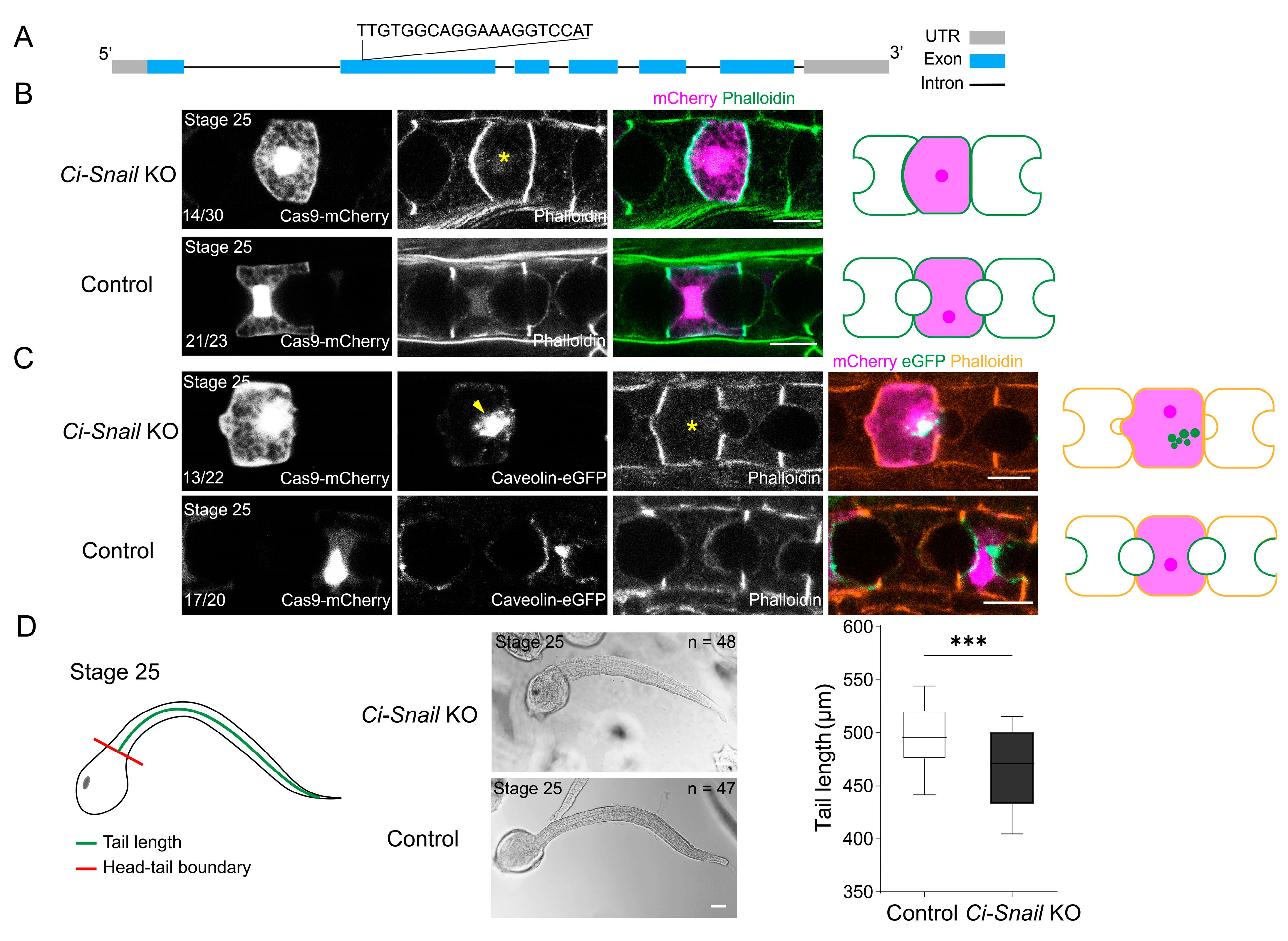
2.1. Snail Plays a Critical Role in Ciona Notochord MET

Our previous works searching for functionally important transcription factors related to notochord lumen formation using ATAC-seq have identified Ci-Snail for its open chromatin region [17] (Figure S1). The subsequent Smart-Seq of notochord cells revealed Ci-Snail expression during the lumenogenesis period (Figure S1). Phylogenetic analysis showed that the Ci-Snail was closely related to the vertebrate Snail, but amino acid sequence alignment indicated that Ci-Snail lacked the SNAG domain, which was characteristic of vertebrate Snail family members [18] (Figure S2). Ci-Snail antibody staining indicated that Ci-Snail was localized in the nuclei of notochord cells from Stage 23 to 25 when notochord cell MET occurs (Figure S1). To explore whether Ci-Snail plays an essential role in notochord lumen formation, knockout of Ci-Snail was carried out by CRISPR/Cas9 in notochord cells. The sgRNA targeted the second exon was selected for Ci-Snail knockout (Ci-Snail KO) after efficiency evaluation of sgRNA using co-electroporation of plasmid EF1α>NLS::Cas9::NLS::P2A-mCherry, together with Ci-Snail sgRNA or control sgRNA (Figure 1A and Figure S3). Then, notochord-expressing Cas9 through the construction of plasmid Brachyury (1 kb)>NLS::Cas9::NLS::P2A-mCherry, together with Ci-Snail KO sgRNA or control sgRNA was introduced into Ciona embryos to observe Ci-Snail KO-induced phenotypes. It was found that embryos with Ci-Snail KO displayed lumen deficiency at Stage 25 compared with the control group, which showed a developed lumen (Figure 1B).
The biological process of notochord lumen formation indicated that notochord cells went through MET in Ciona [15,16], which was characterized by apical membrane occurrence and expansion of notochord lumen. We thus further detected whether Ci-Snail KO influenced the MET occurrence. Caveolin, as the marker of caveolae, was recruited to the apical membrane of notochord cells during the MET process [19]. We examined the subcellular localization of Caveolin-mCherry in Ci-Snail KO notochord cells, and the results showed that Caveolin-mCherry proteins were diffused in the cytoplasm, while it normally localized at the apical membrane of notochord cells (Figure 1C). This result suggested that Ci-Snail affected the MET process in the notochord. MET defect in the Ciona notochord cell affected the following cell extension, which would be reflected in the tail dimension. We measured the tail length as a quantitative indicator for the Ci-Snail KO consequence and found that the embryo tail length was significantly shorter in the Ci-Snail KO group compared to the control group (Figure 1D). In conclusion, Ci-Snail expression in the notochord cells was essential for notochord cell MET.

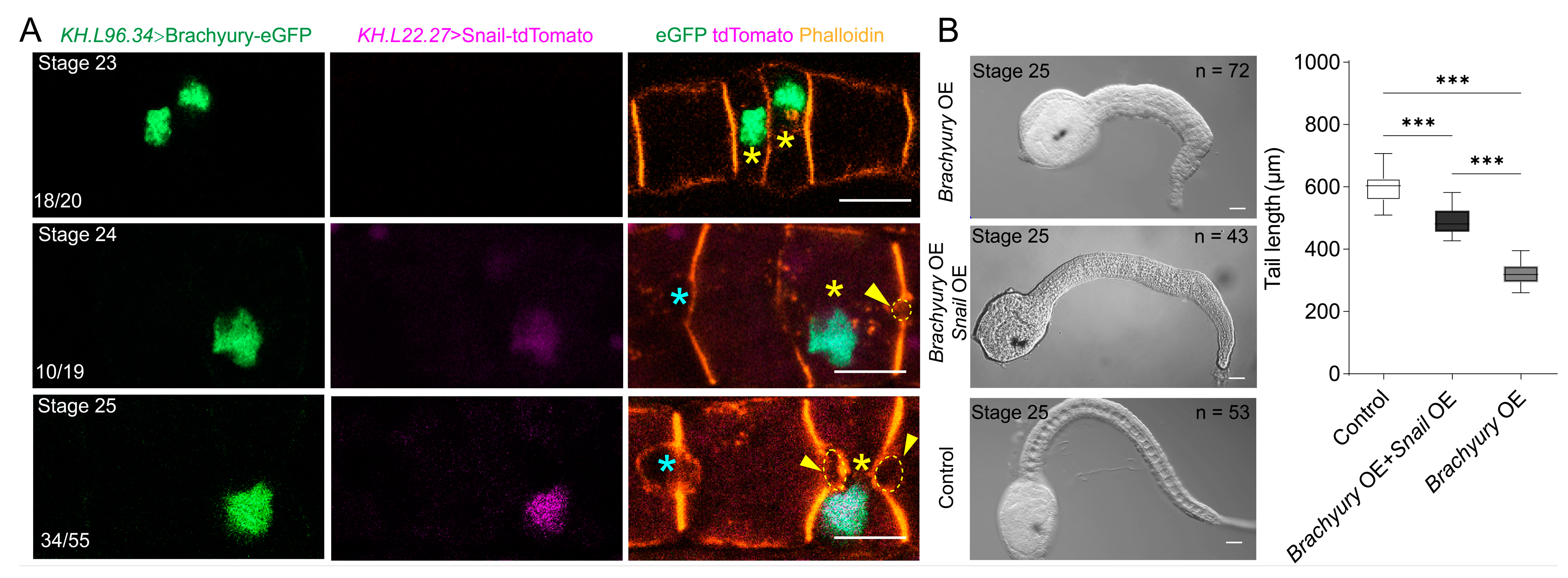
2.2. Brachyury Overexpression Affects Ciona Notochord Cell MET

In Brachyury-deficient early Ciona embryos, notochord cell differentiation is severely impaired [20]; in this study, we carried out Brachyury overexpression (Brachyury OE) to examine the effects of Brachyury on notochord development. We have utilized the promoters of genes KH.L96.34 (Rab11a) and KH.L22.27 (chitin synthase-like) to drive the expression of Brachyury. These two genes are specifically expressed in notochord cells at the initial tailbud and late tailbud stage [21], respectively, based on the Ciona single-cell database (https://singlecell.broadinstitute.org/single_cell) [22] (accessed on 11 October 2021) (Figure 2A). Brachyury-eGFP overexpression driven by both KH.L96.34 and KH.L22.27 promoters affected the notochord lumen formation compared with the control group (Figure 2B).
To further explore the influence of Brachyury OE on the MET process, we co-electroporated plasmids KH.L22.27>Brachyury-eGFP together with Brachyury (1 kb)>Caveolin-mCherry to examine the subcellular localization of caveolin proteins. It was found that Brachyury OE resulted in caveolin distribution in the cytoplasm other than at the apical membrane at the late tailbud stage, while caveolin was recruited at the apical membrane of notochord cells in the control group (Figure 2C). This result suggested that Brachyury OE caused MET defect in the notochord. Taken together, the downregulation of Brachyury expression at the late stage of development facilitates notochord cell MET.

2.3. Ci-Snail OE Partially Rescue the Delayed MET Caused by Brachyury OE

Since Ci-Snail proteins were essential for notochord MET, we intend to rescue MET defect caused by Brachyury OE through overexpression of Ci-Snail. The notochord MET defect phenotype was intended through overexpressing Brachyury driven by the KH.L96.34 promoter from the initial tailbud stage. KH.L22.27 promoter was chosen to drive the expression of Ci-Snail from the late tailbud stage. To experimentally verify the possibility, we co-electroporated KH.L96.34>Brachyury-eGFP and KH.L22.27>Snail-tdTomato together into the fertilized eggs and sequentially sampled from stages 23 to 25. It was found that no lumen formed between adjacent notochord cells at stage 23 when Brachyury-eGFP was detectable but no Ci-Snail overexpression; embryos with strong Brachyury-eGFP expression but weak Ci-Snail overexpression showed small lumen between adjacent notochord cells at Stage 24; in contrast, enlarged lumen between adjacent notochord cells were present with both robust Ci-Snail and Brachyury-eGFP expression at Stage 25 (Figure 3A). We further assessed the tail length of Ciona embryos. The results showed that embryos with both strong expression of Ci-Snail and Brachyury had significantly longer tails than groups with only Brachyury OE but shorter tails than wild-type embryos (Figure 3B).

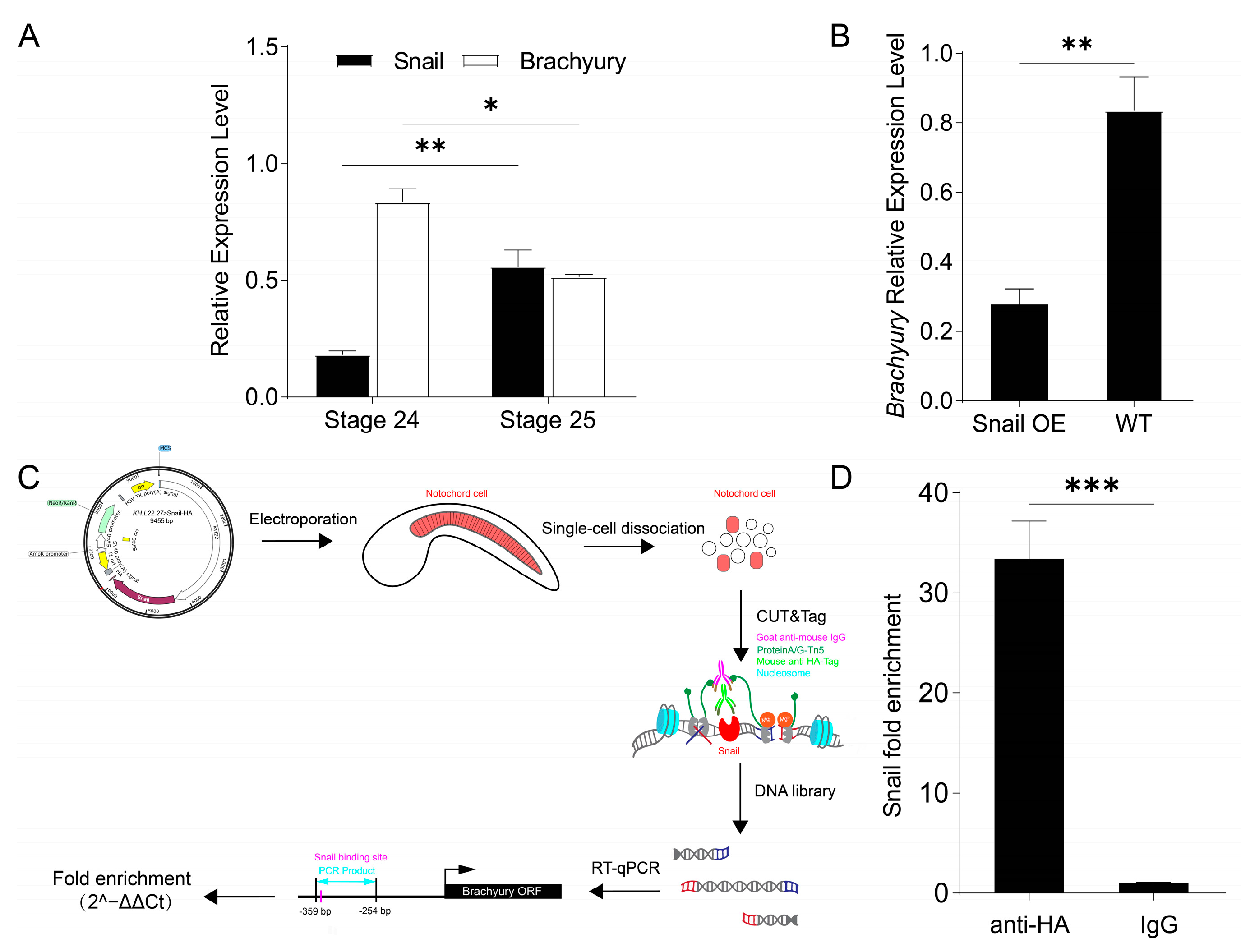
2.4. Ci-Snail Transcriptionally Represses Brachyury Expression to Regulate Notochord MET
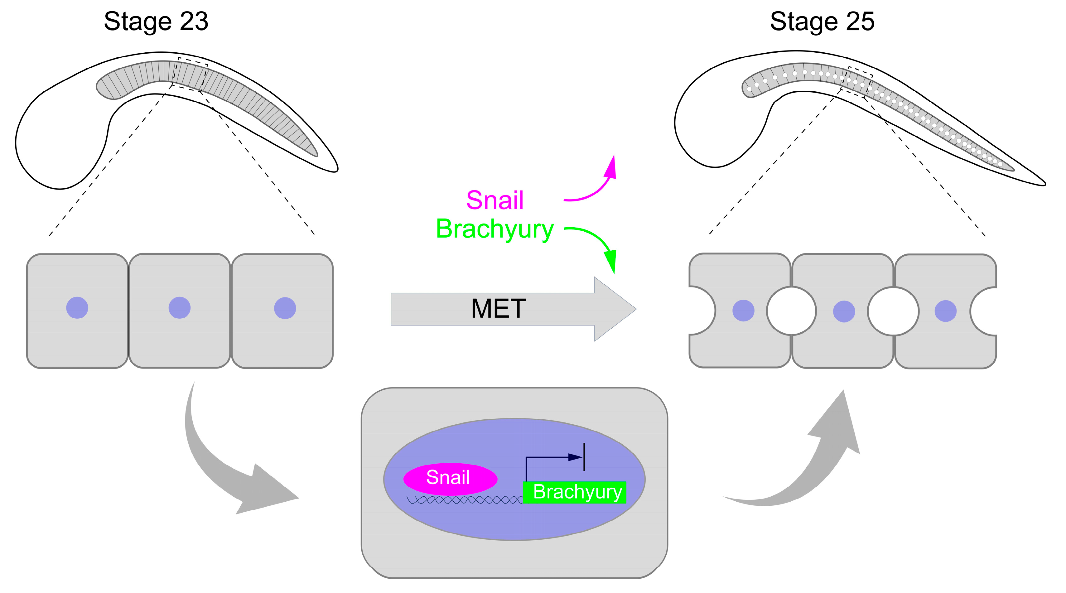
Ci-Snail OE could rescue the lumen formation defect caused by Brachyury OE, suggesting that Ci-Snail functions as a transcriptional repressor. To uncover the regulation of Ci-Snail on Brachyury expression, we first characterized the temporal expression of Ci-Snail and Brachyury of wild-type embryos at Stages 24 and 25 by RT-qPCR. Results showed that their expressions were inversely correlated (Figure 4A), which was in accordance with our Smart-Seq of notochord cells at the above stages (Figure S1). In Ciona embryos at the early tailbud stage, Ci-Snail directly binds the upstream regulatory region of Brachyury for transcriptional repression to promote the embryonic mesoderm development into tail muscle [12]. To reveal whether Ci-Snail transcriptionally represses Brachyury in the notochord cell MET, the expression of Brachyury was detected before and after Ci-Snail was overexpressed at Stage 24. It was found that the Ci-Snail OE down-regulated Brachyury significantly (Figure 4B). To validate the direct binding of Ci-Snail to the upstream region of Brachyury in notochord cells, we performed CUT&Tag-qPCR experiments (Figure 4C). Fold enrichment of the target sequence containing the predicted Ci-Snail binding site was significantly greater in the HA antibody incubated group compared to the IgG control group (Figure 4D). Taken together, our results indicated that Ci-Snail directly binds to the Brachyury upstream region to repress its transcription.

3. Discussion

We have presented evidence that Ci-Snail plays a crucial role in Ciona notochord cell MET process. MET occurrence require the presence of Ci-Snail but not overexpressed Brachyury. The elevated Ci-Snail proteins transcriptionally repress Brachyury expression to trigger MET occurrence at late tailbud stage.
Snail, as an EMT-related transcription repressor, is required for mesoderm and neural crest formation during early embryonic development [7,8]. Our previous work detecting temporal expression of Ci-Snail by RT-qPCR showed that it presented high expression during gastrulation (16 °C, 10 hpf) but decreased expression during the early tailbud stage (16 °C, 14 hpf); however, elevated expression again at late tailbud stage, accordingly the MET initiation stage (16 °C, 18 hpf) (Figure S1). The expression pattern indicated the transcriptional reactivation of Ci-Snail during the notochord MET process, which was also verified by ATAC-seq analysis (Figure S1). Immunofluorescence analysis of Ci-Snail revealed that it could be detected both in notochord cells and tail muscle cells at the late-tailbud stage (Figure S1). The temporal and spatial expression of Ci-Snail presented evidence for Ci-Snail function in notochord MET event.
Down-regulation of Snail was usually induced during the MET process [23], and even Snail silencing reversed EMT in prostate cancer cells [24]. Here, we demonstrated that Ci-Snail expression was required for MET occurrence, and knockout of Ci-Snail resulted in MET defect in Ciona notochord cells. The indispensability of Ci-Snail in two reverse biological processes may be realized through transcriptionally repressing different genes, which is speculated based on the evidence that Snail targets varied genes to regulate broad spectrum of biological processes, including cell proliferation, immune regulation, and stem cell biology [25,26,27].
The T-box transcription factor Brachyury has been proven to be an essential core gene for posterior mesoderm formation and notochord genesis and differentiation [11], and Brachyury-knockdown notochord progenitors survive but adopt neural and mesenchymal fates [28]. But when embryos developed into the late tailbud stage, Brachyury expression needed to be repressed by Ci-Snail to facilitate the MET process, just like the transcriptional regulation in Ciona tail muscle cells, though it resulted in different biological events [12]. Otherwise, Brachyury overexpression leads to failure of notochord cell MET. The result that Ci-Snail OE could only partially rescue MET failure caused by Brachyury OE ascribed to the fact that overexpressed Ci-Snail could transcriptionally represses the expression of an endogenous gene, which down-regulated the total level of Brachyury in the notochord cells. Thus, we conclude that the expression of Brachyury might be delicately regulated during the notochord cell MET process. The inhibition of Brachyury expression was reported to facilitate the MET process through the down-regulation of mesenchymal markers (Fibronectin, Vimentin) and up-regulation of cell junction-associated proteins (Plakoglobin) [10], which are conserved regulatory mechanisms in the MET process and might lead to the abnormal localization of caveolin in Ciona notochord cells.
Vertebrate Snail family members all share a similar organization, being composed of highly conserved four to six zinc fingers for E-box binding, and the repressor activity depends on the so-called SNAG domain [7]. It has been shown that some Snail family member does not seem to heterochromatinize the target region to ensure long-range silencing although it can silence neighboring genes efficiently [29,30,31]. Ci-Snail lost the SNAG domain just like some Drosophila family members and Caenorhabditis elegans, whose transcriptional repression activity is mediated through an interaction with co-repressor such as CtBP (Carboxy-terminal Binding Protein) [32]. It is speculated that Ci-Snail also conjuncts with CtBP to play the repressor activity because of the CtBP consensus site. The repression mechanism of Ci-Snail combined with CtBP or other different co-repressors remains elusive.
MET plays an essential role in the establishment of epithelial polarity in organogenesis during development [33], as well as in the malignant process of tumors [2]. The present study has verified the crucial role of Ci-Snail proteins in triggering MET occurrence, which may provide a new thought for deeply understanding the metamorphosis regulation and cancer treatment.
In summary, Ci-Snail proteins are required for notochord cell MET at late-tailbud embryo of Ciona. This process is regulated by elevating Ci-Snail expression to transcriptionally repress Brachyury (Figure 5).

4. Materials and Methods

4.1. Animal Breeding and Electroporation Experiments

200–300 Ciona adults were harvested from Sangou Bay, Rongcheng City, Shandong Province, China. After 24 h culture, 2–3 adult Ciona with stout white sperm glands and gray eggs in the oviduct were selected. Mature eggs and sperm were taken and artificially inseminated at room temperature in the laboratory. To obtain transgenic Ciona embryos, fertilized eggs were dechorionated as previously described [34]. The working solution for electroporation was prepared by mixing 60 μg plasmid, 420 μL of 0.77 M D-mannitol, and 300 μL of dechorionated eggs to a final volume of 800 μL. Electroporation was performed using the Gene Pulser Xcell system (BIO-RAD, Hercules, CA, USA) with parameters of voltage 50 V and capacitance 1500 μF. After electroporation, the eggs were transferred to plastic petri dishes coated with 1% agarose and incubated at 16 °C.

4.2. Phylogenetic Analysis and Protein Domain Analysis

The sequence of Ci-Snail was downloaded from Aniseed (https://aniseed.fr/) (accessed on 15 September 2021), and other Snail protein sequences including vertebrates, protochordates, and invertebrates were downloaded from NCBI (https://www.ncbi.nlm.nih.gov/) (accessed on 2 September 2021). Phylogenetic analysis was constructed using MEGA-X software (version 10.0, Mega Limited, Auckland, New Zealand) with maximum likelihood method and modeled as JTT + G + I, Sequence comparison of proteins using ClustalW method. CD-Search was used for conserved domain analysis.
Snail protein ID and protein sequence length information for all species are shown in Supplementary Table S2.

4.3. Immunofluorescence

Embryos were collected and fixed with 4% paraformaldehyde (PFA) in seawater for 2 h at room temperature, then washed with 0.1% PBST 3 times in 8 h. After being blocked with 10% goat serum (Solarbio, Beijing, China) for 1 h at room temperature, the embryos were incubated with Ci-Snail antibodies (made by our lab) diluted in 10% goat serum (Snail, 1:100) for 16 h at 4 °C, then followed a washing procedure as above. Another incubation with the secondary antibodies (Alexa Fluor ™ 555 anti-mouse IgG, 1:300, Thermo Fisher, Waltham, MA, USA) and the following washing was performed the same way as the Ci-Snail antibodies. Eventually, embryos are mounted with DAPI and imaged by Zeiss LSM900 or LSM980 confocal microscope.

4.4. Plasmid Construction

PCR amplification was performed using Phanta Max Super-Fidelity DNA polymerase (Vazyme, Nanjing, China). DNA was extracted with FastPure Gel DNA Extraction Mini Kit (Vazyme, Nanjing, China). DNA fragments were ligated by ClonExpress II One Step Cloning Kit (Vazyme, Nanjing, China). DNA sequencing was synthesized at Sangon Biotech (Shanghai, China).
The 4 kb promoter sequence of notochord-specific gene KH.L22.27 was amplified by PCR and ligated into the vector pEGFP-N1 (Clontech, Beijing, China) to generate KH.L22.27>eGFP. KH.L22.27>Snail-eGFP and KH.L22.27>Brachyury-eGFP were constructed by subcloning the coding sequence of Ci-Snail and Brachyury into vector KH.L22.27>eGFP at BamH1 site, respectively. KH.L22.27 was ligated into the vector ptdTomato-N1 (Clontech, Beijing, China) to generate KH.L22.27>tdTomato. KH.L22.27>Snail-tdTomato was constructed by subcloning Ci-Snail into KH.L22.27>tdTomato between EcoR1 and BamH1 sites.
The 4 kb promoter sequence of notochord-specific gene KH.L96.34 was amplified and ligated into the vector pEGFP-N1 to generate KH.L96.34>eGFP. KH.L96.34>Brachyury-eGFP was constructed in the same way as KH.L22.27>Brachyury-eGFP.
PCR programs during plasmid construction were as follows: pre-denaturation at 95 °C for 3 min; 35 cycles of denaturation at 95 °C for 15 s, annealing at 58 °C for 15 s and extension at 72 °C (1 kb/30 s); then extension at 72 °C for 5 min.
Primers for plasmid construction are shown in Supplementary Table S1.

4.5. Gene Knockout by CRISPR/Cas9

The single guide RNA (sgRNA) sequences targeted Ci-Snail were designed using the online website CRISPRdirect (http://crispr.dbcls.jp/) (accessed on 18 May 2022). The sgRNA was synthesized and cloned into the vector Ci-U6>sgRNA (F+E) (Addgene, Watertown, MA, USA 59986) for expression, and the sequence of control sgRNA was designed as previously described [35]. Two plasmids, Brachyury (1 kb)>NLS::Cas9::NLS::P2A-mCherry and EF1α>NLS::Cas9::NLS::P2A-mCherry, were employed for Cas9 expression.
The red fluorescence-emitting embryos were collected, and genomic DNA was extracted for PCR. The PCR products were purified by the Gene JET Gel Extraction Kit (Thermo Fisher, Waltham, MA, USA). A total of 200 ng PCR products were diluted to 17 μL by ddH2O and mixed with 2 μL T7 reaction buffer (Vazyme, Nanjing, China) and incubated with 1 μL T7 endonuclease (Vazyme, Nanjing, China) at 37 °C for 30 min. The fragments were analyzed with agarose gel electrophoresis. The PCR products were sequenced and analyzed by synthego (https://ice.synthego.com/) (accessed on 14 November 2023).
Primers for sgRNA are shown in Supplementary Table S1.

4.6. RNA Extraction and RT-qPCR

The total RNA was extracted by RNAiso plus reagent (Takara, Beijing, China). The integrity and quality of total RNA was evaluated by agarose gel electrophoresis and Nanodrop one (Thermo Fisher, Waltham, MA, USA). The first-strand cDNA was synthesized using 1 μg total RNA per 20 μL reaction system with HiScript II QRT SuperMix for qPCR Reverse transcription Kit (Vazyme, Nanjing, China).
The primers for real-time quantitative PCR (RT-qPCR) of the Ci-Snail and Brachyury were designed with Beacon Designer (Version 7.91, Premier Biosoft, San Diego, CA, USA). RT-qPCR was performed using the SYBR Green PCR Master Mix (Vazyme, Nanjing, China) on Light Cycler 480 (Roche, Basel, Switzerland). The qPCR procedures were as follows: 95 °C for 30 s (permutability); 45 cycles at 95 °C for 10 s, 58 °C for 30 s (cycle reaction); 95 °C for 15 s, 60 °C for 60 s, and 95 °C for 1 s (dissolution curves). The relative expression of Ci-Snail and Brachyury was normalized with U6 as a reference and calculated using the 2−ΔΔCt method.
Primers for RT-qPCR are shown in Supplementary Table S1.

4.7. CUT&Tag-qPCR

Fluorescent embryos expressing recombinant protein Ci-Snail-HA were digested into single-cell suspension with 1% trypsin. In the CUT&Tag procedure, cells were bound and held on Concanavalin A-coated magnetic beads. After permeabilization by the Digitonin (nonionic decontaminating agent), cells were incubated sequentially with the primary antibody (Mouse anti-HA-Tag mAb, AE008, Abclonal, Wuhan, China) against the Ci-Snail-HA protein and the corresponding secondary antibody (goat anti-mouse IgG H&L, ab6708, abcam, Cambridge, UK), and fusion protein of Protein-A/G and Tn5 transposase for genome localization, cleavage and adapter sequences insertion nearby the target protein. The fragmented DNA was retrieved as a template for subsequent RT-qPCR to amplify products containing Ci-Snail binding sequences. The fold enrichment of the target sequence was calculated through the 2−ΔΔCt method.

4.8. Statistical Analysis

All graphs in this study were produced using GraphPad Prism (Version 8.0.2, GraphPad, San Diego, CA, USA). Statistical analysis was performed using a t-test, and a p-value less than 0.05 was considered statistically significant, less than 0.01 was a highly significant difference, and less than 0.001 was an extremely significant difference.

Supplementary Materials

The following supporting information can be downloaded at: https://www.mdpi.com/article/10.3390/ijms25063413/s1.

Author Contributions

Conceptualization: B.D.; Investigation: B.W. and X.O.; Data analysis: B.W., X.O. and X.Y.; Writing—original draft: B.W.; Writing—review and editing: B.W., X.Y. and B.D.; Supervision: B.D.; Project administration: B.D.; Funding acquisition: B.D. All authors have read and agreed to the published version of the manuscript.

Funding

This research was funded by the Science and Technology Innovation Project of Laoshan Laboratory (No. LSKJ202203204).

Institutional Review Board Statement

Not applicable.

Informed Consent Statement

Not applicable.

Data Availability Statement

Data are contained within the article and Supplementary Materials.

Conflicts of Interest

The authors declare no conflict of interest.

References

  1. Pei, D.; Shu, X.; Gassama-Diagne, A.; Thiery, J.P. Mesenchymal–epithelial transition in development and reprogramming. Nat. Cell Biol. 2019, 21, 44–53. [Google Scholar] [CrossRef]
  2. Kalluri, R.; Weinberg, R.A. The basics of epithelial-mesenchymal transition. J. Clin. Investig. 2009, 119, 1420–1428. [Google Scholar] [CrossRef] [PubMed]
  3. Overeem, A.W.; Bryant, D.M.; van Ijzendoorn, S.C.D. Mechanisms of apical–basal axis orientation and epithelial lumen positioning. Trends Cell Biol. 2015, 25, 476–485. [Google Scholar] [CrossRef] [PubMed]
  4. Wu, J.; Ocampo, A.; Belmonte, J.C.I. Cellular Metabolism and Induced Pluripotency. Cell 2016, 166, 1371–1385. [Google Scholar] [CrossRef]
  5. Grau, Y.; Carteret, C.; Simpson, P. Mutations and Chromosomal Rearrangements Affecting the Expression of Snail, a Gene Involved in Embryonic Patterning in Drosophila Melanogaster. Genetics 1984, 108, 347–360. [Google Scholar] [CrossRef] [PubMed]
  6. Kosman, D.; Ip, Y.T.; Levine, M.; Arora, K. Establishment of the mesoderm-neuroectoderm boundary in the Drosophila embryo. Science 1991, 254, 118–122. [Google Scholar] [CrossRef]
  7. Nieto, M.A. The Snail superfamily of zinc-finger transcription factors. Nat. Rev. Mol. Cell Biol. 2002, 3, 155–166. [Google Scholar] [CrossRef]
  8. Corbo, C.J.; Albert, E.; Di, G.A. Dorsoventral patterning of the vertebrate neural tube is conserved in a protochordate. Development 1997, 124, 2335–2344. [Google Scholar] [CrossRef] [PubMed]
  9. Peinado, H.; Ballestar, E.; Esteller, M.; Cano, A. Snail Mediates E-Cadherin Repression by the Recruitment of the Sin3A/Histone Deacetylase 1 (HDAC1)/HDAC2 Complex. Mol. Cell. Biol. 2004, 24, 306–319. [Google Scholar] [CrossRef]
  10. Fernando, R.I.; Litzinger, M.; Trono, P.; Hamilton, D.H.; Schlom, J.; Palena, C. The T-box transcription factor Brachyury promotes epithelial-mesenchymal transition in human tumor cells. J. Clin. Investig. 2010, 120, 533–544. [Google Scholar] [CrossRef]
  11. Showell, C.; Binder, O.; Conlon, F.L. T-box Genes in Early Embryogenesis. Dev. Dyn. 2004, 229, 201–218. [Google Scholar] [CrossRef]
  12. Fujiwara, S.; Corbo, J.C.; Levine, M. The Snail repressor establishes a muscle/notochord boundary in the Ciona embryo. Development 1998, 125, 2511–2520. [Google Scholar] [CrossRef]
  13. Lu, Q.; Bhattachan, P.; Dong, B. Ascidian notochord elongation. Dev. Biol. 2019, 448, 147–153. [Google Scholar] [CrossRef]
  14. Peng, H.; Qiao, R.; Dong, B. Polarity Establishment and Maintenance in Ascidian Notochord. Front. Cell Dev. Biol. 2020, 8, 597446. [Google Scholar] [CrossRef]
  15. Denker, E.; Jiang, D. Ciona intestinalis notochord as a new model to investigate the cellular and molecular mechanisms of tubulogenesis. Semin. Cell Dev. Biol. 2012, 23, 308–319. [Google Scholar] [CrossRef]
  16. Dong, B.; Horie, T.; Denker, E.; Kusakabe, T.; Tsuda, M.; Smith, W.C.; Jiang, D. Tube formation by complex cellular processes in Ciona intestinalis notochord. Dev. Biol. 2009, 330, 237–249. [Google Scholar] [CrossRef] [PubMed]
  17. He, M.; Li, Y.; Li, Y.; Dong, B.; Yu, H. Dynamics of Chromatin Opening across Larval Development in the Urochordate Ascidian Ciona savignyi. Int. J. Mol. Sci. 2024, 25, 2793. [Google Scholar] [CrossRef] [PubMed]
  18. Chiang, C.; Ayyanathan, K. Snail/Gfi-1 (SNAG) family zinc finger proteins in transcription regulation, chromatin dynamics, cell signaling, development, and disease. Cytokine Growth Factor Rev. 2013, 24, 123–131. [Google Scholar] [CrossRef] [PubMed]
  19. Bhattachan, P.; Rae, J.; Yu, H.; Jung, W.; Wei, J.; Parton, R.G.; Dong, B. Ascidian caveolin induces membrane curvature and protects tissue integrity and morphology during embryogenesis. FASEB J. 2020, 34, 1345–1361. [Google Scholar] [CrossRef] [PubMed]
  20. Chiba, S.; Jiang, D.; Satoh, N.; Smith, W.C. Brachyury null mutant-induced defects in juvenile ascidian endodermal organs. Development 2009, 136, 35–39. [Google Scholar] [CrossRef] [PubMed]
  21. Hotta, K.; Mitsuhara, K.; Takahashi, H.; Inaba, K.; Oka, K.; Gojobori, T.; Ikeo, K. A web-based interactive developmental table for the ascidian Ciona intestinalis, including 3D real-image embryo reconstructions: I. From fertilized egg to hatching larva. Dev. Dyn. 2007, 236, 1790–1805. [Google Scholar] [CrossRef]
  22. Cao, C.; Lemaire, L.A.; Wang, W.; Yoon, P.H.; Choi, Y.A.; Parsons, L.R.; Matese, J.C.; Wang, W.; Levine, M.; Chen, K. Comprehensive single-cell transcriptome lineages of a proto-vertebrate. Nature 2019, 571, 349–354. [Google Scholar] [CrossRef] [PubMed]
  23. Yao, D.; Dai, C.; Peng, S. Mechanism of the Mesenchymal–Epithelial Transition and Its Relationship with Metastatic Tumor Formation. Mol. Cancer Res. 2011, 9, 1608–1620. [Google Scholar] [CrossRef] [PubMed]
  24. Wang, H.; Wang, Z.; Li, Y.; Lu, T.; Hu, G. Silencing Snail Reverses Epithelial-Mesenchymal Transition and Increases Radiosensitivity in Hypopharyngeal Carcinoma. OncoTargets Ther. 2020, 13, 497–511. [Google Scholar] [CrossRef] [PubMed]
  25. Wu, Y.; Zhou, B.P. Snail: More than EMT. Cell Adh. Migr. 2010, 4, 199–203. [Google Scholar] [CrossRef] [PubMed]
  26. Gingold, J.A.; Fidalgo, M.; Guallar, D.; Lau, Z.; Sun, Z.; Zhou, H.; Faiola, F.; Huang, X.; Lee, D.F.; Waghray, A.; et al. A Genome-wide RNAi Screen Identifies Opposing Functions of Snai1 and Snai2 on the Nanog Dependency in Reprogramming. Mol. Cell 2014, 56, 140–152. [Google Scholar] [CrossRef] [PubMed]
  27. Kudo-Saito, C.; Shirako, H.; Takeuchi, T.; Kawakami, Y. Cancer Metastasis Is Accelerated through Immunosuppression during Snail-Induced EMT of Cancer Cells. Cancer Cell 2009, 15, 195–206. [Google Scholar] [CrossRef]
  28. Zhu, J.; Kwan, K.M.; Mackem, S. Putative oncogene Brachyury (T) is essential to specify cell fate but dispensable for notochord progenitor proliferation and EMT. Proc. Natl. Acad. Sci. USA 2016, 113, 3820–3825. [Google Scholar] [CrossRef]
  29. Hemavathy, K.; Guru, S.C.; Harris, J.; Chen, J.D.; Ip, Y.T. Human Slug Is a Repressor That Localizes to Sites of Active Transcription. Mol. Cell. Biol. 2000, 55, 5087–5095. [Google Scholar] [CrossRef]
  30. Mezzasalma, M.; Andreone, F.; Glaw, F.; Guarino, F.M.; Odierna, G.; Petraccioli, A.; Picariello, O. Changes in heterochromatin content and ancient chromosome fusion. Salamandra 2019, 55, 140–144. [Google Scholar]
  31. Andrés, P.-P.; Mayra, F.-M. Heterochromatin as an Important Driver of Genome Organization. Front. Cell Dev. Biol. 2020, 8, 579137. [Google Scholar]
  32. Nibu, Y.; Zhang, H.; Bajor, E.; Barolo, S.; Small, S.; Levine, M. dCtBP mediates transcriptional repression by Knirps, Krüppel and Snail in the Drosophila embryo. EMBO J. 1998, 17, 7009–7020. [Google Scholar] [CrossRef] [PubMed]
  33. Lechner, M.S.; Dressler, G.R. The molecular basis of embryonic kidney development. Mech. Dev. 1997, 62, 105–120. [Google Scholar] [CrossRef] [PubMed]
  34. Corbo, J.C.; Levine, M.; Zeller, R.W. Characterization of a notochord-specific enhancer from the Brachyury promoter region of the ascidian, Ciona intestinalis. Development 1997, 124, 589–602. [Google Scholar] [CrossRef]
  35. Stolfi, A.; Gandhi, S.; Salek, F.; Christiaen, L. Tissue-specific genome editing in Ciona embryos by CRISPR/Cas9. Development 2014, 141, 4115–4120. [Google Scholar] [CrossRef]
Figure 1. Snail KO in the notochord leads to abnormal MET in Ciona. (A) Ci-Snail KO sgRNA was designed to target the second exon. (B) Ci-Snail KO in notochord cells resulted in failure of lumen formation. The yellow asterisk “*” indicates Ci-Snail KO cells. The green represents the cell membrane boundary, and the dark purple and purple represent nuclei/cytoplasm with Cas9 expression, respectively. (C) Ci-Snail KO in notochord cells resulted in a dispersed distribution of caveolin. The yellow asterisk “*” indicates Ci-Snail KO cells and the yellow arrowhead indicates caveolin localization. The orange, green, and purple represent the cell membrane boundary, caveolin localization, and the nuclei/cytoplasm with Cas9 expression, respectively. The schematic images on the right side illustrate the phenotypes of the transgenic notochord cells; the numbers at the bottom left of the images indicate the percentage of notochord cells with the illustrated phenotype. (D) Comparison of the tail length of the Ci-Snail KO group with the control group. The measurement is illustrated with a cartoon embryo. The tail length is illustrated with a green line, and the junction between the head and tail is a red line. The numbers in the upper right of the DIC images indicate the quantity of the embryos counted. Statistical analysis was performed using t-tests. “***” means the difference between the two groups is extremely significant (p < 0.001). All of the scale bars represent 10 μm.
Ijms 25 03413 g001
Figure 2. Brachyury OE leads to abnormal MET in Ciona notochord. (A) Ciona single-cell sequencing data shows that KH.L96.34 and KH.L22.27 are sequentially expressed in notochord cells at the initial tailbud and late tailbud stages. (B) Brachyury OE driven by promoters of KH.L96.34 and KH.L22.27 causes no lumen formation. The yellow asterisks “*” indicate Brahyury OE cells. The purple represents the cell membrane boundary, and green represents the nuclei with Brachyury OE expression and cytoplasm with GFP expression. (C) Caveolin dispersed in the cytoplasm of Brachyury OE cells while accumulating at the apical membrane in the control group. The yellow asterisk “*” indicates Brahyury OE cells, yellow arrowheads indicate caveolin localization. The orange, purple, and green represent the cell membrane boundary, caveolin localization, and nuclei with Brachyury OE expression, respectively. The schematic images on the right side illustrate the phenotypes of the transgenic notochord cells; the numbers at the bottom left of the images indicate the percentage of notochord cells with the illustrated phenotype. All of the scale bars represent 10 μm.
Ijms 25 03413 g002
Figure 3. Ci-Snail rescues MET failure caused by Brachyury OE. (A) No visible lumen between adjacent notochord cells was present in embryo with only Brachyury OE at stage 23, small lumen in embryo with strong Brachyury OE but weak Ci-Snail OE at stage 24, enlarged lumen in embryo with both strong Brachyury OE and Ci-Snail OE at stage 25. The numbers at the bottom left of the images indicate the percentage of notochord cells with illustrated phenotype. The yellow asterisks “*” indicate rescued cells, yellow arrowheads and dashed lines indicate the location of the rescued lumen and the cyan asterisks “*” indicate the wild-type lumen. (B) Images of embryos and the tail length comparison of Brachyury OE, Brachyury OE+Snail OE, and control group. The numbers in the upper right of the DIC images indicate the quantity of the embryos counted. Statistical analysis was performed using t-tests. “***” means the difference between the two groups is extremely significant (p < 0.001). All of the scale bars represent 10 μm.
Ijms 25 03413 g003
Figure 4. Ci-Snail represses Brachyury expression through direct binding. (A) Relative expression levels of Ci-Snail and Brachyury in wild-type embryos at Stages 24 and 25. (B) Relative expression levels of Brachyury were detected in Ci-Snail OE and wild-type (WT) groups. (C) Flow diagram of CUT&Tag-qPCR method. (D) Fold enrichment of Ci-Snail-HA incubated group and control group. Statistical analysis was performed using t-tests. “*” indicates significant difference (p < 0.05), “**” indicates highly significant difference (p < 0.01), “***” indicates extremely significant difference (p < 0.001).
Ijms 25 03413 g004
Figure 5. Working model of MET regulation in notochord cells. As development proceeds, Ci-Snail expression is up-regulated to repress Brachyury expression, which leads to MET. The purple represents the Snail protein, green represents the Brachyury gene, and light blue represents the nuclei.
Ijms 25 03413 g005
Disclaimer/Publisher’s Note: The statements, opinions and data contained in all publications are solely those of the individual author(s) and contributor(s) and not of MDPI and/or the editor(s). MDPI and/or the editor(s) disclaim responsibility for any injury to people or property resulting from any ideas, methods, instructions or products referred to in the content.

