Review

BCL-xL, a Mitochondrial Protein Involved in Successful Aging: From C. elegans to Human Centenarians

by
Consuelo Borrás
1,*,†,
Cristina Mas-Bargues
1,†,
Aurora Román-Domínguez
1,
Jorge Sanz-Ros
1,
Lucia Gimeno-Mallench
1,
Marta Inglés
2,
Juan Gambini
1 and
José Viña
1
1
Freshage Research Group, Department of Physiology, Faculty of Medicine, University of Valencia, CIBERFES, INCLIVA, Avenida Blasco Ibañez, 15 46010 Valencia, Spain
2
Freshage Research Group, Department of Physiotherapy, Faculty of Physiotherapy, University of Valencia, CIBERFES, INCLIVA, Avenida Blasco Ibañez, 15 46010 Valencia, Spain
*
Author to whom correspondence should be addressed.
These authors have contributed equally to the manuscript.
Int. J. Mol. Sci. 2020, 21(2), 418; https://doi.org/10.3390/ijms21020418
Submission received: 4 December 2019 / Revised: 1 January 2020 / Accepted: 4 January 2020 / Published: 9 January 2020
(This article belongs to the Special Issue Advances in Bcl-xL Research)

Abstract

:
B-Cell Lymphoma-extra-large (BCL-xL) is involved in longevity and successful aging, which indicates a role for BCL-xL in cell survival pathway regulation. Beyond its well described role as an inhibitor of apoptosis by preventing cytochrome c release, BCL-xL has also been related, indirectly, to autophagy and senescence pathways. Although in these latter cases, BCL-xL has dual roles, either activating or inhibiting, depending on the cell type and the specific conditions. Taken together, all these findings suggest a precise mechanism of action for BCL-xL, able to regulate the crosstalk between apoptosis, autophagy, and senescence, thus promoting cell survival or cell death. All three pathways can be both beneficial or detrimental depending on the circumstances. Thus, targeting BCL-xL would in turn be a “double-edge sword” and therefore, additional studies are needed to better comprehend this dual and apparently contradictory role of BCL-XL in longevity.

1. Introduction to BCL-xL

1.1. The BCL-2 Protein Family

BCL-2, the founding member of the BCL-2 family of proteins, was first identified while studying the chromosomal translocation t(14;18) which is a hallmark of several human follicular lymphomas [1,2]. Nevertheless, BCL-2 function was not elucidated until several years later, when Interleukin 3 (IL-3) dependent mouse cell lines were infected with a retroviral vector to express human BCL-2 protein. In the absence of IL-3, BCL-2 promoted cell survival of the infected cells, although they stopped proliferating [3]. Eventually, more proteins having homology sequences with BCL-2 with the capacity to regulate cell death were found. This set of proteins were labeled as the BCL-2 family of proteins.
Currently, it is known that the BCL-2 gene encodes a 26 kD protein consisting of 329 amino acids with a single highly hydrophobic domain at its C-terminus, which enables it to localize mainly in the outer mitochondrial membrane, and, to a lesser extent, in the nuclear envelope and the endoplasmic reticulum. This BCL-2 family of proteins contains all four BCL-2 Homology (BH) domains (BH1, BH2, BH3, and BH4) and a transmembrane (TM) domain. These BH domains facilitate the family members’ interaction with each other, and can result in a pro- or anti-apoptotic function [4,5,6]. More than 600 BCL-2 homologous proteins have been described in metazoan (animal) genomes and are currently available in the BCL2DB database [7]. In order to simplify the study of this large family, members of the BCL-2 family have been classified into three main subgroups by reason of their sequence homology regions [8,9,10]. These subfamilies are (i) Anti-apoptotic proteins, (ii) Pro-apoptotic proteins and (iii) BH3-only proteins.
  • The anti-apoptotic proteins possess the four BH regions (BH1–4), and include BCL-2, BCL-xL (BCL2-L1), BCL-W (BCL2-L2), A1 (also known as BFL-1 or BCL-2A1), and MCL-1 (Myeloid cell leukemia 1).
  • The pro-apoptotic proteins, which include BAX (BCL-2 associated X protein), BAK (BCL-2 antagonist killer), and BOK/MTD (BCL-2-related ovarian killer/Matador), have three BH domains (BH1–3) and are considered the pore-forming executioners.
  • BH3-only proteins generally possess one single BH region, the BH3 motif, and are also sometimes considered as a subcategory of pro-apoptotic proteins owing to their pro-cell death nature. Proteins like BID (BH3-interacting domain death antagonist), BIM/BOD (BCL2L11; BCL2-interacting mediator of cell death), BAD (BCL2 antagonist of cell death), PUMA/BBC3 (p53 up-regulated modulator of apoptosis), NOXA (phorbol-12-myristate-13-acetate-induced protein 1), BIK/BLK/NBK (BCL-2 interacting killer), HRK/DP5 and BMF (BCL-2 modifying factor) are included in this subgroup [11].
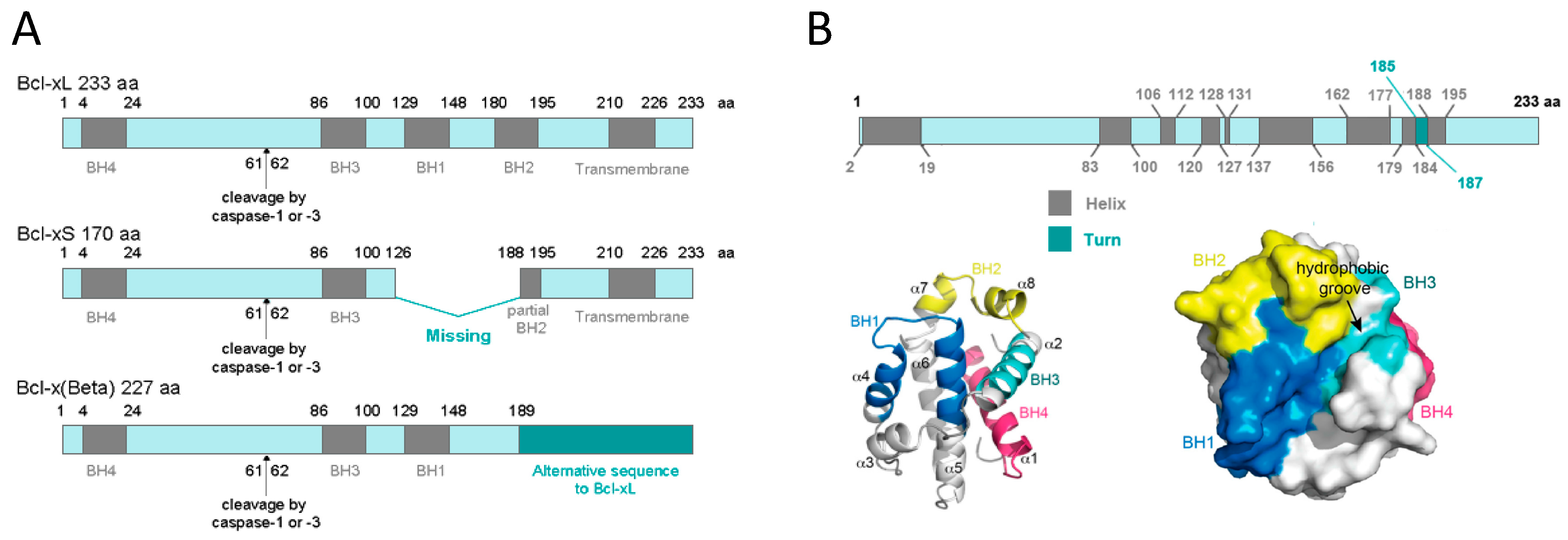
The role of the BCL-2 family in the regulation of apoptosis is typically described as the anti-apoptotic and pro-apoptotic BH3-only members existing in a state of competitive flux to influence the activation of the pore forming executioners [12,13]. Once the executioners are activated, they form pores in the outer mitochondrial membrane (MOM), triggering outer mitochondrial membrane permeability (MOMP), and subsequently the cell undergoes apoptosis [14,15,16].

1.2. BCL-xL

The human BCL2L1 or BCL-X gene is located in the chromosome 20 (20q11.21). Its DNA contains 58,394 bps (genomic size) and its RNA 2575 bps. The BCL-X gene promoter contains consensus motifs for three transcription factor families, STATs (Signal Transducer and Activator of Transcription), Rel/NF-kB (Rel/Nuclear Factor-κB) and Ets (E26 transformation specific sequence), that have been demonstrated to play an important role in the regulation of the BCL2L1 gene expression [17].
Alternative splicing of the BCL-X gene in human cells results in two major mRNA isoforms: the short isoform BCL-xS (591 bp) that has three exons, and the large isoform BCL-xL (780 bp) that has four exons. Many cis-regulatory elements and trans-acting factors exert combinatorial control of BCL-X splicing. Most known regulators, including Sam68, ASF/SF2 (Alternative Splicing Factor 1 / pre-mRNA-Splicing Factor 2, hnRNPA1 (Heterogenous Nuclear Rinonucleoprotein A1), SRp30c (splicing factor arginine/serine-rich 9 protein), and RBM25 (RNA Binding Motif Protein 25), are able to alter BCL-X alternative splicing in vitro or when they are overexpressed in cell cultures [18].

1.2.1. BCL-xL Protein Structure

At the protein level, three different transcript variants, which encode distinct isoforms, have been reported. The longer isoform BCL-xL (233 aa) acts as an apoptosis inhibitor and the shorter isoform, BCL-xS (170 aa) acts as an apoptosis activator. The third one, BCL-xβ (227 amino acids) differs from the longer and the shorter isoforms by a modification of the last 45 amino acids, however no specific function has yet been related to this isoform [19]. Figure 1A depicts all three BCL-X isoforms.
Human BCL-xL protein structure is formed by a total of eight α-helices, two of which (α5 and α6) have a central location and are disposed in a parallel fashion [20,21]. These central helices contain predominantly hydrophobic residues and are flanked by α3 and α4 on one side and by α1, α2, and α8 on the other side. Helices α1 and α2 are connected by a flexible 60-residue loop, which is characteristic of the BCL-xL proteins. It is indispensable for translocation to the nucleus [22] and is the main site for post-translational modifications (phosphorylation, deamidation, and cleavage), which have been shown to be efficient ways to regulate the anti-apoptotic function of BCL-xL [23]. In the context of the three-dimensional structure of BCL-xL, the BH domains make essential contributions to its tertiary structure: the BH1 and BH2 domains encompass turn regions linking two helices, α4 to α5 (in the case of BH1) and α7 to α8 (in the case of BH2). The BH3 domain is located entirely on α2 whilst the BH4 domain is located on α1 and makes a number of stabilizing hydrophobic contacts with α2, α5, and α6. Recent findings have proven a major structural feature on BCL-xL protein that is a large hydrophobic groove involving the BH1–BH3 domains. This hydrophobic groove represents the region of greatest difference between the pro-survival proteins. In BCL-xL, α3 and α4 are almost parallel and are relatively tightly packed resulting in a more closed groove. Mutagenesis studies confirm that this cleft could be the site of interaction with pro-apoptotic proteins [24]. Figure 1B shows BCL-xL primary, secondary, and tertiary structures.
BCL-xL has dual mechanisms to regulate apoptosis. First, the hydrophobic groove binds to the α-helical BH3 domain of the pro-apoptotic regulators, inhibiting apoptosis. Second, a site distal to the hydrophobic groove binds to cytosolic p53, inhibiting p53-dependent activation of BAX/BAK, and thus, apoptosis [25,26,27]. Consistent with the postulated multiple modes of action [28,29,30], BCL-xL exists in several conformations, both soluble and in the membrane. Generally, the transition between soluble and membrane conformations can follow two scenarios: anchoring of a relatively unperturbed protein to the lipid bilayer or a complete refolding and bilayer insertion of the protein (e.g., in bacterial toxins). A recent study conducted by Vasquez-Montes et al. [31], demonstrated that the core hydrophobic helix α6 of BCL-xL inserts into the mitochondrial bilayer without adopting a transmembrane orientation. Instead, this insertion disrupts the packing of BCL-xL and releases the regulatory N-terminal BH4 domain (α1) from the rest of the protein structure [31].
At the post-translational level, BCL-xL can be phosphorylated, deamidated, and ubiquitinated. Phosphorylation of BCL-xL at Ser-62 and Ser-49, have been well characterized and can alter BCL-xL intracellular localization and its loop conformation [32], which regulates the molecular association with other proteins such as BH3-only proteins and p53 [23]. Interestingly, BCL-xL proteins undergo dynamic phosphorylation/dephosphorylation on Ser-49 and Ser-62 residues during mitosis are important in the maintenance of chromosome integrity in normal cells [33]. The function of BCL-xL is regulated by several kinases, including PLK3 [34], JNK [35], CDK2 [36], and MST1 [37]. Deamidation [38] or ubiquitination [39] of BCL-xL are regulated processes that target it for degradation, and can determine susceptibility to DNA-damaging agents and other death stimuli.

1.2.2. BCL-xL Protein Localization

According to The Human Protein Atlas (https://www.proteinatlas.org), at the cellular level, BCL-xL has low RNA cell specificity, as it can be detected in several cell lines such as Caco-2 (colon cancer), MCF7 (mammary gland cancer), HeLa (breast cancer), A549 (lung cancer), etc. In all these cell lines, intracellular localization of BCL-xL protein is mainly in the mitochondria. At the tissue level, BCL-xL has low RNA tissue specificity but the consensus Normalized eXpression (NX) dataset, created as a combination from the three transcriptomics datasets (HPA, GTEx, and FANTOM5), depicts a slight overexpression in bone marrow and thymus. BCL-xL protein levels can be detected in low-medium score in a few tissues, including bone marrow, lymph node, brain, and cells in tubules (Figure 2).

2. BCL-xL Role in Mitochondrial Apoptosis

Several models have been proposed to explain how BCL-2 family proteins regulate outer mitochondrial membrane integrity through BAX and BAK:
  • The “neutralization model” stipulates that BAX and BAK activation is spontaneous and consequently, neutralization of all anti-apoptotic BCL-2 proteins by BH3-only proteins is sufficient to induce BAX and BAK oligomerization and outer mitochondrial membrane permeability (MOMP) [40,41,42].
  • The “direct activation model” posits that BH3-only proteins are divided into “de-repressors/sensitizers” that bind and inhibit anti-apoptotic proteins (e.g., BAD), and “direct-activators” (e.g., BID, BIM) that can also transiently interact with and activate the effectors BAX and BAK [43,44,45].
  • The “embedded together” model proposes that anti-apoptotic proteins act as dominant negative regulators of BAX and BAK by binding to direct activators and effectors in membranes, inhibiting both the activation and the oligomerization step [46,47].
  • Finally, a more recent study proposed a combination of the previous models and defined two “modes” in which anti-apoptotic proteins prevent apoptosis: by sequestering BH3-only direct activator proteins (MODE 1) or the active effectors BAX and BAK themselves (MODE 2) [48].
All models may be correct and are certainly non-exclusive, and BCL-xL has been reported to play a role in all proposed models. Thus, BCL-xL prevents BAX/BAK oligomerization in the MOM to form a pore for cytochrome c release from mitochondria. In the absence of BCL-xL, cytochrome c exits to the cytosol, and together with Apaf-1 and caspase-9, they form the apoptosome for the dimerization and activation of caspase-9, which, in turn, proteolytically cleaves downstream effector caspases, such as caspase-3, resulting in the activation of these caspases and leading to cell death. Therefore, BCL-xL, preventing cytochrome c release from the mitochondria, prevents apoptosis-mediated cell death.

BCL-xL Role in Mitochondrial Bioenergetics

The morphology of the mitochondrial population is the result of several interacting dynamical phenomena, including fission, fusion, movement, elimination, and biogenesis. Each of these phenomena is finely controlled by the underlying molecular machinery. Studies in mammals and model organisms have revealed that mitochondrial morphology, dynamics, and function appear to be subjected to regulation by the same proteins that regulate apoptotic cell death [49,50]. Although the biochemical mechanisms are still being investigated, several studies have provided hints regarding the BCL-xL pro-survival function, independently of its canonical role of inhibiting BAX and BAK. Cortical neurons lacking BCL-xL display fragmented mitochondria and restoring BCL-xL levels leads to elongated mitochondria, an enhanced mitochondrial fusion rate, and surprisingly, an even greater increase in the rate of fission. In addition, BCL-xL also increased total mitochondrial biomass (length) in cultured hippocampal neurons and cortical neurons [51,52]. These findings are consistent with the observation that BCL-xL can interact with the mitochondrial fission–fusion machinery [53].
Alavian and colleagues found that, in addition to its outer mitochondrial membrane localization, BCL-xL also localizes in the mitochondrial matrix where it co-immunoprecipitates with the beta subunit of F1FO ATP synthase. Exogenously applied, BCL-xL increases F1FO ATPase activity, while BCL-xL inhibition reduces the capacity of F1FO ATPase-containing sub-mitochondrial vesicles to sequester H+ ions. Thus, BCL-xL increases the efficiency of ATP synthesis by decreasing a proton leak within the F1FO ATPase, thus improving cellular metabolism and preventing oxidative stress [54].
Moreover, phosphorylation of BCL-xL at Ser-62 by the cyclin B1-CDK1 complex in the mitochondria leads to its dissociation from the beta subunit of the F1FO ATP synthase. The subsequent inhibition of ATP synthase activity causes complex I oxidative damage, inner mitochondrial membrane depolarization, and apoptotic death [55]. Supporting this fact, a study performed in yeast suggested that, ectopic expression of mammalian BCL-xL allowed cells to survive longer, and this protective effect of BCL-xL was more prominent in respiratory-competent cells that contained defects in mitochondrial ADP/ATP translocation [56].

3. BCL-xL Role in Autophagy

Autophagy is an evolutionarily conserved lysosomal pathway for degrading cytoplasmic proteins, macromolecules, and organelles. Three well defined types of autophagy have been established: macroautophagy, endosomal microautophagy, and chaperone-mediated autophagy. However, the current autophagy literature is often viewed as confusing, due to the association with apparently contradictory roles regarding cell survival and cell death [57].
Apoptosis and autophagy are both tightly regulated biological processes that play a central role in tissue homeostasis, development, and disease. The antiapoptotic protein BCL-xL, interacts with the evolutionarily conserved autophagy protein, Beclin-1 [58]. BCL-xL binds and inhibits Beclin-1 and this interaction involves the BH3 domain within Beclin-1 (residues 114–123) [59]. To directly test the impact of the endogenous BCL-xL on autophagy in the absence of apoptosis, Lindqvist and colleagues inhibited their activity in cells lacking the essential cell death mediators BAX and BAK.
In the absence of BAX and BAK; BCL-xL had no detectable effect on autophagy or cell death in myeloid or fibroblast cell lines. On the other hand, when BAX and BAK were present, inhibition of BCL-xL stimulated autophagy, but this correlated with increased cell death. These results demonstrate that BCL-xL does not directly inhibit components of the autophagic pathway but instead affects autophagy indirectly, owing to the inhibition of Bax and Bak [60,61]. For instance, BCL-xL inhibits both autophagy and apoptosis through interacting respectively with Beclin-1 and BAX/BAK using their BH3-binding pockets [62]. BCL-xL binds to Beclin-1 and thus prevents the interaction between Beclin-1 and the class III PI3K complex (PI3KC3) to inhibit autophagy [63]. Previous studies showed that BCL-xL phosphorylation may be a regulatory switch between autophagy and apoptosis [64,65]. In fact, JNK-mediated phosphorylation mediates the disruption of Beclin-1/BCL-xL and BAX-BAK/BCL-xL interaction, thus triggering autophagy [66,67,68,69].

4. BCL-xL Dual Role in Senescence

Like apoptosis, senescence is a cellular response to different stresses characterized by a permanent and irreversible state of cell cycle arrest. Although the determinants by which a cell undergoes senescence or apoptosis are still unclear, both processes seem to be exclusive in physiological conditions. Despite that, a large amount of evidence point to a crosstalk between apoptosis and cellular senescence, in addition to autophagy [70].
One of the first studies involving BCL-xL in senescent cells was performed in megakaryocytes derived from CD34+ progenitor cells and UT4 cells, with the aim of clarifying BCL-xL function in the maturation process of different hematopoietic cell lineages. BCL-xL expression was up-regulated during megakaryocyte differentiation [71], and subsequently, senescence was drastically reduced. We also reported that both MEFs and primary cultured human lymphocytes, transduced with a plasmid encoding BCL-xL, downregulated INK/ARF senescent pathway markers, such as p16INK4a, p14Arf, p21Cip, and senescence-associated β-galactosidase (SA-β-GAL) [72]. Taken together, these results suggest a negative correlation between BCL-xL expression and senescence.
In contrast with this data, there is also evidence suggesting that BCL-xL has a positive correlation with senescence. Likewise, BCL-2, BCL-xL is considered as an oncogene due to its ability to promote cell survival. Given that oncogene overexpression can trigger DNA response to damage in order to induce senescence, we could expect that BCL-xL overexpression could also induce this response [73]. In fact, it has been demonstrated that several cell types, when senescent, exhibit high levels of BCL-xL protein. As an example, several triple-negative breast cancer cell lines that survive and undergo cellular senescence after exposure to oncogenic suppressors showed increased basal levels of BCL-xL. Inhibition of BCL-xL in these cells was shown to change cell fates in response to oncogenic suppressors from senescence to apoptosis [74]. In fact, following DNA damage, BCL-xL has the ability to translocate to the nucleus where it binds to CDK1 during the G2/M cell-cycle checkpoint in human lymphoma cell lines. These arrested cells exhibited typical SA-β-GAL activity and were unable to synthesize new DNA when overexpressing BCL-xL, suggesting its capacity to stabilize senescence [22].
Overall, it is likely that BCL-xL could both favor and suppress cellular senescence. Presumably, the decision would be dependent on several factors such as the cell type and status (non-tumor or tumor cells), the type and level of stress to which the cell is exposed, and the influence of the surrounding cells and tissues. Nonetheless, further studies are needed towards the understanding of BCL-xL role in cellular senescence in order to elucidate this dual behavior. Summarizing, the main role of BCL-xL is to promote cell survival, either by inhibiting apoptosis or autophagy (or both). Furthermore, it has been described that a certain level of senescence is necessary for successful aging, such as in reprogramming [75], thus high levels of BCL-xL can be beneficial to maintain senescent cells during aging (Figure 3).

4.1. BCL-xL Role in Successful Aging

We have studied centenarians, as an example of successful aging, and have found that they overexpress BCL-xL [72]. By performing functional transcriptomic analysis of peripheral blood mononuclear cells (PMBC), we compared the expression patterns of 28.869 human genes in centenarians, septuagenarians, and young people. Results showed that the mRNA expression pattern of centenarians was similar to the one of young people, and completely different from that of septuagenarians. In particular, sub-network analysis of the 1.721 mRNAs that were found to be statistically different between the three populations, converged on the following six genes: interferon-γ (IFNG), T-cell receptor (TCR), tumor necrosis factor (TNF), SP1 transcription factor, transforming growth factor-β1 (TGFβ1), and cytokine IL-32.
Likewise, those six genes were related to BCL-xL, Fas, and Fas ligand (FasL), all of them known to be involved in the control of cell death regulation [72]. However, where BCL-xL is an antiapoptotic protein, Fas and FasL are proapoptotic. This could be considered a paradox, as centenarians overexpress anti and proapoptotic proteins at the same time, but there is an explanation: BCL-xL is involved in the inhibition of the intrinsic pathway of apoptosis, which is mainly mediated by mitochondria and activated after self-cell stress; however, Fas and Fas-L induce the extrinsic pathway of apoptosis, which means that they force the cell to die after external stress signals [76]. This suggests that centenarians have a better way to control apoptosis when cells are aging (intrinsic apoptosis), but at the same time, damaged cells by external signals are removed more efficiently (extrinsic apoptosis).
In order to further demonstrate the role of BCL-xL in longevity, we performed longevity curves using C. elegans with a gain function of Ced-9, the ortholog for human BCL-xL. Interestingly, animals overexpressing Ced-9 showed a significant increase in both the mean and the maximum survival time [72]. Although aging is a multifactorial process, these studies suggest that BCL-xL function is relevant in aging and may be one of the factors that contributes to exceptional longevity.
Moreover, it has been shown that BCL-xL regulates presynaptic plasticity and confers neuroprotection. While other members of the BCL-2 family of proteins substantially decline during development [77], BCL-xL expression is maintained throughout life in the adult brain [78]. This is consistent with the fact that BCL-xL exerts a critical function in mature, long lived neurons. For example, in hippocampal neurons, overexpression of BCL-xL markedly changed synaptic morphology [53]. In the squid giant presynaptic terminal, recombinant BCL-xL protein enhances the rate of recovery of neurotransmission following intense synaptic activity. BCL-xL-regulated recovery depends on stimulation, calmodulin, and BCL-xL translocation to clatherin-coated pits.
At synaptic vesicles, BCL-xL forms protein–protein interactions with the dynamin-like GTPase Drp1. Depletion of Drp1 or disruption of its interaction with BCL-xL slows down endocytosis and produces aberrantly shaped vesicle membranes [79]. Thus, the underlying molecular details of how BCL-xL alters neuronal synapses may stem from its direct effects on mitochondrial protein complexes as well as on lipids of the inner mitochondrial membrane. These findings could potentially explain the ability of BCL-xL to increase synaptic transmission within minutes when acutely injected into the presynaptic terminus, therefore acting independently of longer-term cell remodeling effects [80].

4.2. BCL-xL, a Senolytic Paradox?

It is well known that senescent cells tend to accumulate with age in multiple organisms provoking deleterious effects in neighboring cells and tissues, mainly due to senescence-associated secretory phenotype (SASP) [81]. In fact, cell senescence is considered one of the hallmarks of human aging [82]. In correlation with this, growing experimental evidence has recently demonstrated that the removal of senescent cells prolongs life span and enhances health [83]. Senescent cell clearance was first induced by a drug-activable “suicide” gene in transgenic mice with accelerated aging, resulting in an improvement in age-related phenotypes [84]. Since then, many drugs targeting anti-apoptotic and pro-survival factors have been identified to induce apoptosis in senescent cells in vitro and in vivo [85]. The molecules capable of selectively inducing death in senescent cells are named senolytics [86].
In order to identify potential senolytic targets, several anti-apoptotic and pro-survival factors that are upregulated in senescent cells were selected, with BCL-xL among them. Interfering expression of BCL-xL with siRNAs resulted in a reduced viability and survival of senescent but not proliferating human abdominal subcutaneous preadipocytes [87]. Moreover, when administered to old mice, the senolytic drugs Dastainib (D) and Quercetin (Q) were able to delay age-related symptoms and pathologies accompanied with reduced levels of BCL-xL [87]. In the same direction, it has been identified a new senolytic agent, Navitoclax (ABT-263), which induced apoptosis of senescent cells by inhibition of BCL-W and BCL-xL [88,89,90].
Similar results were obtained when using the small molecule ABT-737 in human and mice fibroblasts with induced senescence by inhibition of BCL-W and BCL-xL [91]. However, we must not forget that cellular senescence is a response to damage that involves inflammation and extracellular matrix remodeling and that resolves with the phagocytic elimination of the senescent cells. Cellular senescence plays an active and positive role during tissue regeneration, and the full benefits of senescence are achieved when the process includes the clearance of the senescent cells, thereby restoring the pre-damage status of the tissue [92]. Centenarians have presumably higher numbers of senescent cells as well as increased levels of BCL-xL, thus their clearance systems are working properly, offering the utmost benefits of cellular senescence for tissue damage repair.
Contrarily, we previously described that overexpression of BCL-xL decreases premature senescence in mouse embryo fibroblasts and in human isolated lymphocytes [72]. After four passages in culture, BCL-xL overexpressing cells exhibited lower levels of p16Ink4a, p14Arf, and p21Cip, all known to inhibit the cell cycle. Moreover, as discussed before, BCL-xL overexpression is found in individuals with extreme longevity.
In contrast, nematodes bearing a gain of function mutation in the BCL-xL ortholog ced-9 showed increased longevity, suggesting that BL-xL is involved in healthy aging [72]. Thus, we get to the following paradox by which BCL-xL is elevated in some senescent cells and its inhibition results in several beneficial effects for the individual, whereas BCL-xL overexpression reduces senescence markers in other cells. Similarly, there is also the contradiction that not only does overexpression of BCL-xL seem to play a role in longevity and life span, but also the interference of its expression in senescent cells has been linked to a wide range of beneficial effects.

5. Conclusions

In conclusion, BCL-xL is a complex protein involved in the three main defense pathways of cells: apoptosis, autophagy, and senescence. All three are interconnected and can be both beneficial and detrimental depending on the circumstances. BCL-xL localizes in the mitochondria, where is able to protect them by promoting efficient ATP synthesis and regulating Ca2+ flux from the endoplasmic reticulum, contributing to the energy balance, which is very important for healthy aging. Under healthy conditions, BCL-xL levels maintain cell survival directly by inhibiting apoptosis, or indirectly by repressing autophagy. Successful aging, such as the case of centenarians, is associated with high levels of BCL-xL. However, in ordinary aging, senescent cells may accumulate due to an inefficient immune system, and in this case, BCL-xL should be well monitored. Thus, targeting BCL-xL would, in turn, be a “double-edge sword” and therefore, additional studies are needed to better comprehend these dual and apparently contradictory roles of BCL-XL in longevity.

Funding

This work was supported by the following grants: Instituto de Salud Carlos III, FEDER [grant number PIE15/00013], SAF2016-75508-R from the Spanish Ministry of Education and Science (MEC), CB16/10/ 00435 (CIBERFES-ISCIII), PROMETEOII2014/056 de “Conselleria de Sanitat de la Generalitat Valenciana, GV/2018//067 from "Conselleria d’Educació, Cultura i Esport de la Generalitat Valenciana" to M.I., EU Funded FRAILOMIC Initiative (FP7-HEALTH-2012-Proposal no. 305483-2), ADVANTAGE-724099 (HP-JA) –DIALBFRAIL-LATAM (825546 H2020-SC1-BHC) to J.V., PCIN-2017-117 of the Ministry of Economy and Competitiveness, the EU Joint Programming Initiative ‘A Healthy Diet for a Healthy Life’ (JPI HDHL INTIMIC-085) to C.B. Dr. C. M.-B. is recipient of a postdoctoral grant financed by Generalitat Valenciana (APOSTD/2018/230) and FSE (European Social Fund).

Conflicts of Interest

The authors declare no conflict of interest.

References

  1. Pegoraro, L.; Palumbo, A.; Erikson, J.; Falda, M.; Giovanazzo, B.; Emanuel, B.S.; Rovera, G.; Nowell, P.C.; Croce, C.M. A 14;18 and an 8;14 chromosome translocation in a cell line derived from an acute B-cell leukemia. Proc. Natl. Acad. Sci. USA 1984, 81, 7166–7170. [Google Scholar] [CrossRef] [Green Version]
  2. Tsujimoto, Y.; Finger, L.R.; Yunis, J.; Nowell, P.C.; Croce, C.M. Cloning of the chromosome breakpoint of neoplastic B cells with the t(14;18) chromosome translocation. Science 1984, 226, 1097–1099. [Google Scholar] [CrossRef]
  3. Vaux, D.L.; Cory, S.; Adams, J.M. Bcl-2 gene promotes haemopoietic cell survival and cooperates with c-myc to immortalize pre-B cells. Nature 1988, 335, 440–442. [Google Scholar] [CrossRef]
  4. Warren, C.F.A.; Wong-Brown, M.W.; Bowden, N.A. BCL-2 family isoforms in apoptosis and cancer. Cell Death Dis. 2019, 10, 177. [Google Scholar] [CrossRef] [Green Version]
  5. Petros, A.M.; Medek, A.; Nettesheim, D.G.; Kim, D.H.; Yoon, H.S.; Swift, K.; Matayoshi, E.D.; Oltersdorf, T.; Fesik, S.W. Solution structure of the antiapoptotic protein bcl-2. Proc. Natl. Acad. Sci. USA 2001, 98, 3012–3017. [Google Scholar] [CrossRef] [Green Version]
  6. Yin, X.M.; Oltvai, Z.N.; Korsmeyer, S.J. BH1 and BH2 domains of Bcl-2 are required for inhibition of apoptosis and heterodimerization with Bax. Nature 1994, 369, 321–323. [Google Scholar] [CrossRef]
  7. Rech de Laval, V.; Deleage, G.; Aouacheria, A.; Combet, C. BCL2DB: Database of BCL-2 family members and BH3-only proteins. Database 2014, 2014. [Google Scholar] [CrossRef] [Green Version]
  8. Huska, J.D.; Lamb, H.M.; Hardwick, J.M. Overview of BCL-2 Family Proteins and Therapeutic Potentials. Methods Mol. Biol. 2019, 1877, 1–21. [Google Scholar]
  9. Moldoveanu, T.; Czabotar, P.E. BAX, BAK, and BOK: A Coming of Age for the BCL-2 Family Effector Proteins. Cold Spring Harb. Perspect. Biol. 2019. [Google Scholar] [CrossRef]
  10. Youle, R.J.; Strasser, A. The BCL-2 protein family: Opposing activities that mediate cell death. Nat. Rev. Mol. Cell Biol. 2008, 9, 47–59. [Google Scholar] [CrossRef]
  11. Knight, T.; Luedtke, D.; Edwards, H.; Taub, J.W.; Ge, Y. A delicate balance—The BCL-2 family and its role in apoptosis, oncogenesis, and cancer therapeutics. Biochem. Pharmacol. 2019, 162, 250–261. [Google Scholar] [CrossRef]
  12. Certo, M.; Del Gaizo Moore, V.; Nishino, M.; Wei, G.; Korsmeyer, S.; Armstrong, S.A.; Letai, A. Mitochondria primed by death signals determine cellular addiction to antiapoptotic BCL-2 family members. Cancer Cell 2006, 9, 351–365. [Google Scholar] [CrossRef] [Green Version]
  13. van Delft, M.F.; Huang, D.C. How the Bcl-2 family of proteins interact to regulate apoptosis. Cell Res. 2006, 16, 203–213. [Google Scholar] [CrossRef]
  14. Nechushtan, A.; Smith, C.L.; Lamensdorf, I.; Yoon, S.H.; Youle, R.J. Bax and Bak coalesce into novel mitochondria-associated clusters during apoptosis. J. Cell Biol. 2001, 153, 1265–1276. [Google Scholar] [CrossRef]
  15. Chipuk, J.E.; Kuwana, T.; Bouchier-Hayes, L.; Droin, N.M.; Newmeyer, D.D.; Schuler, M.; Green, D.R. Direct activation of Bax by p53 mediates mitochondrial membrane permeabilization and apoptosis. Science 2004, 303, 1010–1014. [Google Scholar] [CrossRef] [Green Version]
  16. Martinou, J.C.; Green, D.R. Breaking the mitochondrial barrier. Nat. Rev. Mol. Cell Biol. 2001, 2, 63–67. [Google Scholar] [CrossRef]
  17. Grillot, D.A.; Gonzalez-Garcia, M.; Ekhterae, D.; Duan, L.; Inohara, N.; Ohta, S.; Seldin, M.F.; Nunez, G. Genomic organization, promoter region analysis, and chromosome localization of the mouse bcl-x gene. J. Immunol. 1997, 158, 4750–4757. [Google Scholar]
  18. Moore, M.J.; Wang, Q.; Kennedy, C.J.; Silver, P.A. An alternative splicing network links cell-cycle control to apoptosis. Cell 2010, 142, 625–636. [Google Scholar] [CrossRef] [Green Version]
  19. Ban, J.; Eckhart, L.; Weninger, W.; Mildner, M.; Tschachler, E. Identification of a human cDNA encoding a novel Bcl-x isoform. Biochem. Biophys. Res. Commun. 1998, 248, 147–152. [Google Scholar] [CrossRef]
  20. Muchmore, S.W.; Sattler, M.; Liang, H.; Meadows, R.P.; Harlan, J.E.; Yoon, H.S.; Nettesheim, D.; Chang, B.S.; Thompson, C.B.; Wong, S.L.; et al. X-ray and NMR structure of human Bcl-xL, an inhibitor of programmed cell death. Nature 1996, 381, 335–341. [Google Scholar] [CrossRef]
  21. Petros, A.M.; Olejniczak, E.T.; Fesik, S.W. Structural biology of the Bcl-2 family of proteins. Biochim. Biophys. Acta 2004, 1644, 83–94. [Google Scholar] [CrossRef]
  22. Schmitt, E.; Beauchemin, M.; Bertrand, R. Nuclear colocalization and interaction between bcl-xL and cdk1(cdc2) during G2/M cell-cycle checkpoint. Oncogene 2007, 26, 5851–5865. [Google Scholar] [CrossRef] [Green Version]
  23. Follis, A.V.; Llambi, F.; Kalkavan, H.; Yao, Y.; Phillips, A.H.; Park, C.G.; Marassi, F.M.; Green, D.R.; Kriwacki, R.W. Regulation of apoptosis by an intrinsically disordered region of Bcl-xL. Nat. Chem. Biol. 2018, 14, 458–465. [Google Scholar] [CrossRef]
  24. Lee, E.F.; Fairlie, W.D. The Structural Biology of Bcl-xL. Int. J. Mol. Sci. 2019, 20, 2234. [Google Scholar] [CrossRef] [Green Version]
  25. Todt, F.; Cakir, Z.; Reichenbach, F.; Youle, R.J.; Edlich, F. The C-terminal helix of Bcl-xL mediates Bax retrotranslocation from the mitochondria. Cell Death Differ. 2013, 20, 333–342. [Google Scholar] [CrossRef]
  26. Edlich, F.; Banerjee, S.; Suzuki, M.; Cleland, M.M.; Arnoult, D.; Wang, C.; Neutzner, A.; Tjandra, N.; Youle, R.J. Bcl-xL Retrotranslocates Bax from the Mitochondria into the Cytosol. Cell 2011, 145, 104–116. [Google Scholar] [CrossRef] [Green Version]
  27. Follis, A.V.; Chipuk, J.E.; Fisher, J.C.; Yun, M.K.; Grace, C.R.; Nourse, A.; Baran, K.; Ou, L.; Min, L.; White, S.W.; et al. PUMA binding induces partial unfolding within BCL-xL to disrupt p53 binding and promote apoptosis. Nat. Chem. Biol. 2013, 9, 163. [Google Scholar] [CrossRef] [Green Version]
  28. Billen, L.P.; Kokoski, C.L.; Lovell, J.F.; Leber, B.; Andrews, D.W. Bcl-XL inhibits membrane permeabilization by competing with Bax. PLoS Biol. 2008, 6, e147. [Google Scholar] [CrossRef]
  29. Chi, X.; Kale, J.; Leber, B.; Andrews, D.W. Regulating cell death at, on, and in membranes. Biochim. Biophys. Acta BBA Mol. Cell Res. 2014, 1843, 2100–2113. [Google Scholar] [CrossRef] [Green Version]
  30. Moldoveanu, T.; Follis, A.V.; Kriwacki, R.W.; Green, D.R. Many players in BCL-2 family affairs. Trends Biochem. Sci. 2014, 39, 101–111. [Google Scholar] [CrossRef] [Green Version]
  31. Vasquez-Montes, V.; Vargas-Uribe, M.; Pandey, N.K.; Rodnin, M.V.; Langen, R.; Ladokhin, A.S. Lipid-modulation of membrane insertion and refolding of the apoptotic inhibitor Bcl-xL. Biochim. Biophys. Acta. Proteins Proteom. 2019, 1867, 691–700. [Google Scholar] [CrossRef]
  32. Priya, P.; Maity, A.; Ghosh Dastidar, S. The long unstructured region of Bcl-xl modulates its structural dynamics. Proteins 2017, 85, 1567–1579. [Google Scholar] [CrossRef]
  33. Baruah, P.S.; Beauchemin, M.; Hebert, J.; Bertrand, R. Dynamic Bcl-xL (S49) and (S62) Phosphorylation/Dephosphorylation during Mitosis Prevents Chromosome Instability and Aneuploidy in Normal Human Diploid Fibroblasts. PLoS ONE 2016, 11, e0159091. [Google Scholar] [CrossRef]
  34. Wang, J.; Beauchemin, M.; Bertrand, R. Bcl-xL phosphorylation at Ser49 by polo kinase 3 during cell cycle progression and checkpoints. Cell. Signal. 2011, 23, 2030–2038. [Google Scholar] [CrossRef] [Green Version]
  35. Kharbanda, S.; Saxena, S.; Yoshida, K.; Pandey, P.; Kaneki, M.; Wang, Q.; Cheng, K.; Chen, Y.N.; Campbell, A.; Sudha, T.; et al. Translocation of SAPK/JNK to mitochondria and interaction with Bcl-x(L) in response to DNA damage. J. Biol. Chem. 2000, 275, 322–327. [Google Scholar] [CrossRef] [Green Version]
  36. Megyesi, J.; Tarcsafalvi, A.; Seng, N.; Hodeify, R.; Price, P.M. Cdk2 phosphorylation of Bcl-xL after stress converts it to a pro-apoptotic protein mimicking Bax/Bak. Cell Death Discov. 2016, 2, 15066. [Google Scholar] [CrossRef] [Green Version]
  37. Del Re, D.P.; Matsuda, T.; Zhai, P.; Maejima, Y.; Jain, M.R.; Liu, T.; Li, H.; Hsu, C.P.; Sadoshima, J. Mst1 promotes cardiac myocyte apoptosis through phosphorylation and inhibition of Bcl-xL. Mol. Cell 2014, 54, 639–650. [Google Scholar] [CrossRef] [Green Version]
  38. Dho, S.H.; Deverman, B.E.; Lapid, C.; Manson, S.R.; Gan, L.; Riehm, J.J.; Aurora, R.; Kwon, K.S.; Weintraub, S.J. Control of cellular Bcl-xL levels by deamidation-regulated degradation. PLoS Biol. 2013, 11, e1001588. [Google Scholar] [CrossRef] [Green Version]
  39. Ni, F.; Yan, C.Y.; Zhou, S.; Hui, P.Y.; Du, Y.H.; Zheng, L.; Yu, J.; Hu, X.J.; Zhang, Z.G. Repression of GRIM19 expression potentiates cisplatin chemoresistance in advanced bladder cancer cells via disrupting ubiquitination-mediated Bcl-xL degradation. Cancer Chemother Pharm. 2018, 82, 593–605. [Google Scholar] [CrossRef]
  40. Uren, R.T.; Dewson, G.; Chen, L.; Coyne, S.C.; Huang, D.C.; Adams, J.M.; Kluck, R.M. Mitochondrial permeabilization relies on BH3 ligands engaging multiple prosurvival Bcl-2 relatives, not Bak. J. Cell Biol. 2007, 177, 277–287. [Google Scholar] [CrossRef] [Green Version]
  41. Willis, S.N.; Fletcher, J.I.; Kaufmann, T.; van Delft, M.F.; Chen, L.; Czabotar, P.E.; Ierino, H.; Lee, E.F.; Fairlie, W.D.; Bouillet, P.; et al. Apoptosis initiated when BH3 ligands engage multiple Bcl-2 homologs, not Bax or Bak. Science 2007, 315, 856–859. [Google Scholar] [CrossRef] [Green Version]
  42. Willis, S.N.; Chen, L.; Dewson, G.; Wei, A.; Naik, E.; Fletcher, J.I.; Adams, J.M.; Huang, D.C. Proapoptotic Bak is sequestered by Mcl-1 and Bcl-xL, but not Bcl-2, until displaced by BH3-only proteins. Genes Dev. 2005, 19, 1294–1305. [Google Scholar] [CrossRef] [Green Version]
  43. Kuwana, T.; Mackey, M.R.; Perkins, G.; Ellisman, M.H.; Latterich, M.; Schneiter, R.; Green, D.R.; Newmeyer, D.D. Bid, Bax, and lipids cooperate to form supramolecular openings in the outer mitochondrial membrane. Cell 2002, 111, 331–342. [Google Scholar] [CrossRef] [Green Version]
  44. Kuwana, T.; Bouchier-Hayes, L.; Chipuk, J.E.; Bonzon, C.; Sullivan, B.A.; Green, D.R.; Newmeyer, D.D. BH3 domains of BH3-only proteins differentially regulate Bax-mediated mitochondrial membrane permeabilization both directly and indirectly. Mol. Cell 2005, 17, 525–535. [Google Scholar] [CrossRef]
  45. Wei, M.C.; Lindsten, T.; Mootha, V.K.; Weiler, S.; Gross, A.; Ashiya, M.; Thompson, C.B.; Korsmeyer, S.J. tBID, a membrane-targeted death ligand, oligomerizes BAK to release cytochrome c. Genes Dev. 2000, 14, 2060–2071. [Google Scholar]
  46. Leber, B.; Lin, J.; Andrews, D.W. Embedded together: The life and death consequences of interaction of the Bcl-2 family with membranes. Apoptosis Int. J. Program. Cell Death 2007, 12, 897–911. [Google Scholar] [CrossRef] [Green Version]
  47. Leber, B.; Lin, J.; Andrews, D.W. Still embedded together binding to membranes regulates Bcl-2 protein interactions. Oncogene 2010, 29, 5221–5230. [Google Scholar] [CrossRef] [Green Version]
  48. Llambi, F.; Moldoveanu, T.; Tait, S.W.; Bouchier-Hayes, L.; Temirov, J.; McCormick, L.L.; Dillon, C.P.; Green, D.R. A unified model of mammalian BCL-2 protein family interactions at the mitochondria. Mol. Cell 2011, 44, 517–531. [Google Scholar] [CrossRef] [Green Version]
  49. Aouacheria, A.; Baghdiguian, S.; Lamb, H.M.; Huska, J.D.; Pineda, F.J.; Hardwick, J.M. Connecting mitochondrial dynamics and life-or-death events via Bcl-2 family proteins. Neurochem. Int. 2017, 109, 141–161. [Google Scholar] [CrossRef]
  50. Martinou, J.C.; Youle, R.J. Mitochondria in apoptosis: Bcl-2 family members and mitochondrial dynamics. Dev. Cell 2011, 21, 92–101. [Google Scholar] [CrossRef] [Green Version]
  51. Berman, S.B.; Pineda, F.J.; Hardwick, J.M. Mitochondrial fission and fusion dynamics: The long and short of it. Cell Death Differ. 2008, 15, 1147–1152. [Google Scholar] [CrossRef] [Green Version]
  52. Berman, S.B.; Chen, Y.B.; Qi, B.; McCaffery, J.M.; Rucker, E.B.; Goebbels, S.; Nave, K.A.; Arnold, B.A.; Jonas, E.A.; Pineda, F.J.; et al. Bcl-xL increases mitochondrial fission, fusion, and biomass in neurons. J. Cell Biol. 2009, 184, 707–719. [Google Scholar] [CrossRef] [Green Version]
  53. Li, H.; Chen, Y.; Jones, A.F.; Sanger, R.H.; Collis, L.P.; Flannery, R.; McNay, E.C.; Yu, T.; Schwarzenbacher, R.; Bossy, B.; et al. Bcl-xL induces Drp1-dependent synapse formation in cultured hippocampal neurons. Proc. Natl. Acad. Sci. USA 2008, 105, 2169–2174. [Google Scholar] [CrossRef] [Green Version]
  54. Alavian, K.N.; Li, H.; Collis, L.; Bonanni, L.; Zeng, L.; Sacchetti, S.; Lazrove, E.; Nabili, P.; Flaherty, B.; Graham, M.; et al. Bcl-xL regulates metabolic efficiency of neurons through interaction with the mitochondrial F1FO ATP synthase. Nat. Cell Biol. 2011, 13, 1224–1233. [Google Scholar] [CrossRef]
  55. Veas-Perez de Tudela, M.; Delgado-Esteban, M.; Maestre, C.; Bobo-Jimenez, V.; Jimenez-Blasco, D.; Vecino, R.; Bolanos, J.P.; Almeida, A. Regulation of Bcl-xL-ATP Synthase Interaction by Mitochondrial Cyclin B1-Cyclin-Dependent Kinase-1 Determines Neuronal Survival. J. Neurosci. 2015, 35, 9287–9301. [Google Scholar] [CrossRef] [Green Version]
  56. Trancikova, A.; Weisova, P.; Kissova, I.; Zeman, I.; Kolarov, J. Production of reactive oxygen species and loss of viability in yeast mitochondrial mutants: Protective effect of Bcl-xL. FEMS Yeast Res. 2004, 5, 149–156. [Google Scholar] [CrossRef] [Green Version]
  57. Dalby, K.N.; Tekedereli, I.; Lopez-Berestein, G.; Ozpolat, B. Targeting the prodeath and prosurvival functions of autophagy as novel therapeutic strategies in cancer. Autophagy 2010, 6, 322–329. [Google Scholar] [CrossRef] [Green Version]
  58. Pattingre, S.; Tassa, A.; Qu, X.; Garuti, R.; Liang, X.H.; Mizushima, N.; Packer, M.; Schneider, M.D.; Levine, B. Bcl-2 antiapoptotic proteins inhibit Beclin 1-dependent autophagy. Cell 2005, 122, 927–939. [Google Scholar] [CrossRef] [Green Version]
  59. Maiuri, M.C.; Le Toumelin, G.; Criollo, A.; Rain, J.C.; Gautier, F.; Juin, P.; Tasdemir, E.; Pierron, G.; Troulinaki, K.; Tavernarakis, N.; et al. Functional and physical interaction between Bcl-X(L) and a BH3-like domain in Beclin-1. EMBO J. 2007, 26, 2527–2539. [Google Scholar] [CrossRef]
  60. Lindqvist, L.M.; Heinlein, M.; Huang, D.C.; Vaux, D.L. Prosurvival Bcl-2 family members affect autophagy only indirectly, by inhibiting Bax and Bak. Proc. Natl. Acad. Sci. USA 2014, 111, 8512–8517. [Google Scholar] [CrossRef] [Green Version]
  61. Lindqvist, L.M.; Vaux, D.L. BCL2 and related prosurvival proteins require BAK1 and BAX to affect autophagy. Autophagy 2014, 10, 1474–1475. [Google Scholar] [CrossRef] [Green Version]
  62. Zhou, F.; Yang, Y.; Xing, D. Bcl-2 and Bcl-xL play important roles in the crosstalk between autophagy and apoptosis. FEBS J. 2011, 278, 403–413. [Google Scholar] [CrossRef] [PubMed]
  63. Noble, C.G.; Dong, J.M.; Manser, E.; Song, H. Bcl-xL and UVRAG cause a monomer-dimer switch in Beclin1. J. Biol. Chem. 2008, 283, 26274–26282. [Google Scholar] [CrossRef] [Green Version]
  64. Booth, L.A.; Tavallai, S.; Hamed, H.A.; Cruickshanks, N.; Dent, P. The role of cell signalling in the crosstalk between autophagy and apoptosis. Cell Signal 2014, 26, 549–555. [Google Scholar] [CrossRef] [Green Version]
  65. Levine, B.; Sinha, S.; Kroemer, G. Bcl-2 family members: Dual regulators of apoptosis and autophagy. Autophagy 2008, 4, 600–606. [Google Scholar] [CrossRef] [Green Version]
  66. Wei, Y.; Sinha, S.; Levine, B. Dual role of JNK1-mediated phosphorylation of Bcl-2 in autophagy and apoptosis regulation. Autophagy 2008, 4, 949–951. [Google Scholar] [CrossRef] [Green Version]
  67. Wei, Y.; Pattingre, S.; Sinha, S.; Bassik, M.; Levine, B. JNK1-mediated phosphorylation of Bcl-2 regulates starvation-induced autophagy. Mol. Cell 2008, 30, 678–688. [Google Scholar] [CrossRef] [Green Version]
  68. Maiuri, M.C.; Criollo, A.; Kroemer, G. Crosstalk between apoptosis and autophagy within the Beclin 1 interactome. EMBO J. 2010, 29, 515–516. [Google Scholar] [CrossRef] [Green Version]
  69. Yang, J.; Yao, S. JNK-Bcl-2/Bcl-xL-Bax/Bak Pathway Mediates the Crosstalk between Matrine-Induced Autophagy and Apoptosis via Interplay with Beclin 1. Int. J. Mol. Sci. 2015, 16, 25744–25758. [Google Scholar] [CrossRef] [Green Version]
  70. Vicencio, J.M.; Galluzzi, L.; Tajeddine, N.; Ortiz, C.; Criollo, A.; Tasdemir, E.; Morselli, E.; Ben Younes, A.; Maiuri, M.C.; Lavandero, S.; et al. Senescence, apoptosis or autophagy? When a damaged cell must decide its path—A mini-review. Gerontology 2008, 54, 92–99. [Google Scholar] [CrossRef]
  71. Sanz, C.; Benet, I.; Richard, C.; Badia, B.; Andreu, E.J.; Prosper, F.; Fernandez-Luna, J.L. Antiapoptotic protein Bcl-x(L) is up-regulated during megakaryocytic differentiation of CD34+ progenitors but is absent from senescent megakaryocytes. Exp. Hematol. 2001, 29, 728–735. [Google Scholar] [CrossRef]
  72. Borras, C.; Abdelaziz, K.M.; Gambini, J.; Serna, E.; Ingles, M.; de la Fuente, M.; Garcia, I.; Matheu, A.; Sanchis, P.; Belenguer, A.; et al. Human exceptional longevity: Transcriptome from centenarians is distinct from septuagenarians and reveals a role of Bcl-xL in successful aging. Aging 2016, 8, 3185–3208. [Google Scholar] [CrossRef] [Green Version]
  73. Jung, M.S.; Jin, D.H.; Chae, H.D.; Kang, S.; Kim, S.C.; Bang, Y.J.; Choi, T.S.; Choi, K.S.; Shin, D.Y. Bcl-xL and E1B-19K proteins inhibit p53-induced irreversible growth arrest and senescence by preventing reactive oxygen species-dependent p38 activation. J. Biol. Chem. 2004, 279, 17765–17771. [Google Scholar] [CrossRef] [Green Version]
  74. Gayle, S.S.; Sahni, J.M.; Webb, B.M.; Weber-Bonk, K.L.; Shively, M.S.; Spina, R.; Bar, E.E.; Summers, M.K.; Keri, R.A. Targeting BCL-xL improves the efficacy of bromodomain and extra-terminal protein inhibitors in triple-negative breast cancer by eliciting the death of senescent cells. J. Biol. Chem. 2019, 294, 875–886. [Google Scholar] [CrossRef] [Green Version]
  75. Mosteiro, L.; Pantoja, C.; Alcazar, N.; Marion, R.M.; Chondronasiou, D.; Rovira, M.; Fernandez-Marcos, P.J.; Munoz-Martin, M.; Blanco-Aparicio, C.; Pastor, J.; et al. Tissue damage and senescence provide critical signals for cellular reprogramming in vivo. Science 2016, 354. [Google Scholar] [CrossRef]
  76. Peter, M.E.; Krammer, P.H. The CD95(APO-1/Fas) DISC and beyond. Cell Death Differ. 2003, 10, 26–35. [Google Scholar] [CrossRef] [Green Version]
  77. Krajewska, M.; Mai, J.K.; Zapata, J.M.; Ashwell, K.W.; Schendel, S.L.; Reed, J.C.; Krajewski, S. Dynamics of expression of apoptosis-regulatory proteins Bid, Bcl-2, Bcl-X, Bax and Bak during development of murine nervous system. Cell Death Differ. 2002, 9, 145–157. [Google Scholar] [CrossRef]
  78. Gonzalez-Garcia, M.; Garcia, I.; Ding, L.; O’Shea, S.; Boise, L.H.; Thompson, C.B.; Nunez, G. Bcl-x is expressed in embryonic and postnatal neural tissues and functions to prevent neuronal cell death. Proc. Natl. Acad. Sci. USA 1995, 92, 4304–4308. [Google Scholar] [CrossRef] [Green Version]
  79. Li, H.; Alavian, K.N.; Lazrove, E.; Mehta, N.; Jones, A.; Zhang, P.; Licznerski, P.; Graham, M.; Uo, T.; Guo, J.; et al. A Bcl-xL-Drp1 complex regulates synaptic vesicle membrane dynamics during endocytosis. Nat. Cell Biol. 2013, 15, 773–785. [Google Scholar] [CrossRef]
  80. Hickman, J.A.; Hardwick, J.M.; Kaczmarek, L.K.; Jonas, E.A. Bcl-xL inhibitor ABT-737 reveals a dual role for Bcl-xL in synaptic transmission. J. Neurophysiol. 2008, 99, 1515–1522. [Google Scholar] [CrossRef]
  81. Coppe, J.P.; Patil, C.K.; Rodier, F.; Sun, Y.; Munoz, D.P.; Goldstein, J.; Nelson, P.S.; Desprez, P.Y.; Campisi, J. Senescence-associated secretory phenotypes reveal cell-nonautonomous functions of oncogenic RAS and the p53 tumor suppressor. PLoS Biol. 2008, 6, 2853–2868. [Google Scholar] [CrossRef]
  82. Lopez-Otin, C.; Blasco, M.A.; Partridge, L.; Serrano, M.; Kroemer, G. The hallmarks of aging. Cell 2013, 153, 1194–1217. [Google Scholar] [CrossRef] [Green Version]
  83. McHugh, D.; Gil, J. Senescence and aging: Causes, consequences, and therapeutic avenues. J. Cell Biol. 2017, 217, 65–77. [Google Scholar] [CrossRef]
  84. Baker, D.J.; Wijshake, T.; Tchkonia, T.; LeBrasseur, N.K.; Childs, B.G.; van de Sluis, B.; Kirkland, J.L.; van Deursen, J.M. Clearance of p16Ink4a-positive senescent cells delays ageing-associated disorders. Nature 2011, 479, 232–236. [Google Scholar] [CrossRef]
  85. Xu, M.; Palmer, A.K.; Ding, H.; Weivoda, M.M.; Pirtskhalava, T.; White, T.A.; Sepe, A.; Johnson, K.O.; Stout, M.B.; Giorgadze, N.; et al. Targeting senescent cells enhances adipogenesis and metabolic function in old age. Elife 2015, 4, e12997. [Google Scholar] [CrossRef]
  86. Soto-Gamez, A.; Demaria, M. Therapeutic interventions for aging: The case of cellular senescence. Drug Discov. Today 2017, 22, 786–795. [Google Scholar] [CrossRef]
  87. Zhu, Y.; Tchkonia, T.; Pirtskhalava, T.; Gower, A.C.; Ding, H.; Giorgadze, N.; Palmer, A.K.; Ikeno, Y.; Hubbard, G.B.; Lenburg, M.; et al. The Achilles’ heel of senescent cells: From transcriptome to senolytic drugs. Aging Cell 2015, 14, 644–658. [Google Scholar] [CrossRef]
  88. Zhu, Y.; Tchkonia, T.; Fuhrmann-Stroissnigg, H.; Dai, H.M.; Ling, Y.Y.; Stout, M.B.; Pirtskhalava, T.; Giorgadze, N.; Johnson, K.O.; Giles, C.B.; et al. Identification of a novel senolytic agent, navitoclax, targeting the Bcl-2 family of anti-apoptotic factors. Aging Cell 2016, 15, 428–435. [Google Scholar] [CrossRef]
  89. Chang, J.; Wang, Y.; Shao, L.; Laberge, R.M.; Demaria, M.; Campisi, J.; Janakiraman, K.; Sharpless, N.E.; Ding, S.; Feng, W.; et al. Clearance of senescent cells by ABT263 rejuvenates aged hematopoietic stem cells in mice. Nat. Med. 2016, 22, 78–83. [Google Scholar] [CrossRef] [Green Version]
  90. Zhu, Y.; Doornebal, E.J.; Pirtskhalava, T.; Giorgadze, N.; Wentworth, M.; Fuhrmann-Stroissnigg, H.; Niedernhofer, L.J.; Robbins, P.D.; Tchkonia, T.; Kirkland, J.L. New agents that target senescent cells: The flavone, fisetin, and the BCL-XL inhibitors, A1331852 and A1155463. Aging 2017, 9, 955–963. [Google Scholar] [CrossRef] [Green Version]
  91. Yosef, R.; Pilpel, N.; Tokarsky-Amiel, R.; Biran, A.; Ovadya, Y.; Cohen, S.; Vadai, E.; Dassa, L.; Shahar, E.; Condiotti, R.; et al. Directed elimination of senescent cells by inhibition of BCL-W and BCL-XL. Nat. Commun. 2016, 7, 11190. [Google Scholar] [CrossRef] [PubMed]
  92. Serrano, M. Understanding Aging. N. Engl. J. Med. 2017, 376, 1083–1085. [Google Scholar] [CrossRef] [PubMed]
Figure 1. BCL-xL protein structure. (A) BCL-xL, BCL-xS and BCL-xβ isoforms. This representation shows the presence of the BH domains as well as the cleavage site by caspase-1 or -3. (B) BCL-xL primary, secondary, and tertiary structures. The primary structure shows the linear position of helices and turns; the secondary structure represents the 3D position of the eight helices and the four BH domains; and the tertiary structure reveals the hydrophobic groove (adapted from Lee and Fairlie, IJMS 2019 [24]).
Ijms 21 00418 g001
Figure 2. BCL-xL localization. BCL-xL mRNA expression levels can be detected in different cell lines and cell types; BCL-xL protein subcellular location is mainly restricted to mitochondria (Image: MCF7 cells stained with CPTP-BCL2L1-2 antibody obtained from NCI-CPTAC). Consensus data for BCL-xL RNA expression in 55 human tissue types and six blood cell types reveals low tissue specificity. BCL-xL immunohistochemistry in four different tissues stained with HPA035734 antibody (obtained from Atlas Antibodies Sigma-Aldrich). All figures have been obtained from The Human Protein Atlas.
Ijms 21 00418 g002
Figure 3. BCL-xL promotes cell survival through apoptosis, autophagy, and senescence regulation.
Ijms 21 00418 g003

