Article

Evolution of the Proto Sex-Chromosome in Solea senegalensis

by
María Esther Rodríguez
1,
Belén Molina
1,
Manuel Alejandro Merlo
1,
Alberto Arias-Pérez
1,
Silvia Portela-Bens
1,
Aglaya García-Angulo
1,
Ismael Cross
1,
Thomas Liehr
2 and
Laureana Rebordinos
1,*
1
Área de Genética, Facultad de Ciencias del Mar y Ambientales, INMAR, Universidad de Cádiz, 11510 Cádiz, Spain
2
Molcular Cytogenetics, University Clinic Jena Institute of Human Genetics, 07747 Jena, Germany
*
Author to whom correspondence should be addressed.
Int. J. Mol. Sci. 2019, 20(20), 5111; https://doi.org/10.3390/ijms20205111
Submission received: 26 September 2019 / Revised: 11 October 2019 / Accepted: 13 October 2019 / Published: 15 October 2019
(This article belongs to the Special Issue Chromosome and Karyotype Variation)

Abstract

:
Solea senegalensis is a flatfish belonging to the Soleidae family within the Pleuronectiformes order. It has a karyotype of 2n = 42 (FN = 60; 6M + 4 SM + 8 St + 24 T) and a XX/XY system. The first pair of metacentric chromosomes has been proposed as a proto sex-chromosome originated by a Robertsonian fusion between acrocentric chromosomes. In order to elucidate a possible evolutionary origin of this chromosome 1, studies of genomic synteny were carried out with eight fish species. A total of 88 genes annotated within of 14 BACs located in the chromosome 1 of S. senegalensis were used to elaborate syntenic maps. Six BACs (BAC5K5, BAC52C17, BAC53B20, BAC84K7, BAC56H24, and BAC48P7) were distributed in, at least, 5 chromosomes in the species studied, and a group of four genes from BAC53B20 (grsf1, rufy3, slc4a4 and npffr2) and genes from BAC48K7 (dmrt2, dmrt3, dmrt1, c9orf117, kank1 and fbp1) formed a conserved cluster in all species. The analysis of repetitive sequences showed that the number of retroelements and simple repeat per BAC showed its highest value in the subcentromeric region where 53B20, 16E16 and 48K7 BACs were localized. This region contains all the dmrt genes, which are associated with sex determination in some species. In addition, the presence of a satellite “chromosome Y” (motif length: 860 bp) was detected in this region. These findings allowed to trace an evolutionary trend for the large metacentric chromosome of S. senegalensis, throughout different rearrangements, which could be at an initial phase of differentiation as sex chromosome.

1. Introduction

Teleosts are the most diversified group among vertebrates [1], colonizing aquatic environments with a wide range of salinity, temperature or photoperiod conditions and displaying a high variety of adaptations. This variability also reaches sex determination systems, of which a great variety of them have been described in fish that can be (i) simple system with heterogametic male as XX/XY, heterogametic female as ZZ/ZW, and homogametic female and male XX/X0 and ZZ/Z0 respectively, and (ii) multiple system with standard as XX/XY1Y2 which was observed in neo tropical fish Hoplias malabaricus in which XY1Y2 is a neo-sex chromosome, or constitution or numerical variations (revised in [2]). Pleuronectiformes are a diverse order composed of more than 600 species grouped into 11 families, they have a small genome [3] when compared to other teleost fish, making them an interesting group to study mechanisms of organization and gene expression. Within this order, both ZZ/ZW (e.g., Cynoglossus semilaevis, [4] and XX/XY (e.g., Solea senegalensis, [5]) sex chromosome determination systems have been described.
Sex determination is a process with many consequences at biological and evolutionary levels, and therefore, a high level of conservation would be expected as it happens with other important biological processes [6]. In fish, specifically, sex determination mechanisms involve, in many cases, more than one gene, as well as environmental factors and epigenetic mechanisms [7].
Sex determination in fish shows considerable variation among species, and six different master genes have described as responsible, dmy, gsdf, sox3, amhy, amhr2 and sdY [7,8]. Three of these have been described in different species of Oryzias. The first gene described in fish, dmy (DM-domain gene on the Y chromosome) has been reported in two species of Oryzias, O. latipes and O. curvinotus, and originated from a duplication of the autosomal gene dmrt1. The gsdf gene (gonadal soma derived factor on the Y chromosome), was reported in O. luzonensi [8]; and sox3, which was described on the Y chromosome of O. dancena, appeared independently, given that this species belongs to a different lineage [9]. Other genes are amhy (Y chromosome-specific anti-müllerian hormone) and amhr2 (anti-müllerian hormone receptor type II). The former gene is a male-specific duplicate gene that emerged from a duplication event of the autosomal amh gene [10]. It is located on a single metacentric/submetacentric chromosome and is expressed in presumptive Sertoli cells of XY individuals of Odonthestes hatcheri (Paragonian pejerrey). The amhr2 gene is expressed in somatic cells surrounding germ cells and it determines sex in Takifugu rubripes, likely due to a single-nucleotide difference between the Y and X chromosome that changes an amionacid (His/Asp384) in the kinase domain [11].
The last master sex-determining gene is sdY (sexually dimorphic on the Y-chromosome), an immune-related gene identified in the rainbow trout Oncorhynchus mykiss [12]. It has also been suggested that dmrt1 (doublesex and mab-3 related transcription factor 1) could be a sex-determining candidate of in C. semilaevis, which has a ZZ/ZW system [4].
The evolution of sex chromosomes has been often been mediated by fusions between sex chromosomes and autosomes of the type Y-A. This type of fusions has been observed at higher rates in fish and squamates reptiles than other sex chromosomes-autosomes fusions, being the XY lineages more frequently fused than ZW [13]. Hence, in two species of fishes of the Oplegnathus genus it was observed that multiple X1X2Y sex chromosomes were formed through a centric fusion of ancestral Y chromosome with an autosome, creating a large neo-Y chromosome [14]. The Neotropical fish H. malabaricus (Erythrinidae family) was described as having five karyomorphs, one of these, the (XX/XY1Y2) multiple systems G, derived by tandem fusion events between the acrocentric proto-sex chromosome pair and the submetacentric autosome pair from ancestral karyotype, [2].
In S. senegalensis, which has a XX/XY system, the chromosome 1, has been described as a proto-sex chromosome [15] originated by Robertsonian fusions between two acrocentric chromosomes [16].
It has been postulated that heteromorphic sex chromosomes evolved from an ordinary autosomal pair after acquisition of a sex determination (SD) locus. One of the first events in the evolution of sex chromosomes and what makes the W and Y different from X and Z chromosomes, is the suppression of the meiotic recombination between X and Y as well as Z and W chromosomes due to accumulation of heterochromatin and deletion of heterochromatin [17]. Moreover, other events associated with the evolution of sex chromosomes implied accumulation of TEs and simple repeats around young sex determination locus [17,18,19].
In heteromorphic sex chromosomes the SD system might involve (i) the whole chromosome and be detectable with cytogenetic techniques as described for Neotropical fish [2]; (ii) could be a cryptic region with cytogenetic level as observed in three-spined stickleback [20]; (iii) regions with a few kilobases as reported in O. latipes [21] and (iv) be a very tiny differentiation as observed in T. rubripes in which the only differentiation between X and Y is the single SNP in the amhr2 receptor [11].
The karyotype, of S. senegalensis has been described as 2n = 42 [22], its Fundamental Number (FN) is 60 and is composed by 6 M + 4 SM + 8 ST + 24 T. The largest metacentric chromosome is proposed to have evolved through the Robertsonian fusion of two acrocentric ones [23]. The Zoo-FISH technique (FISH: Fluorescence In Situ Hybridization) in two related flatfish species of the Soleidae family, Dicologlossa cuneata (2n = 50, and FN = 54) and Dagetichthys lusitanica (2n = 42, and FN = 50) confirmed this prediction [16].
Thus, with this antecedents/precedents, the aim of this work is deepen into the evolution of the metacentric chromosome 1 of S. senegalensis through studies of genomic synteny in closely-related flatfish species, Scophthalmus maximus and C. semilaevis, and also distant/faraway species not belonging to the Pleuronectiformes order (Tetraodon nigroviridis, Gasterosteus aculeatus, Xiphophorus maculatus, Oryzias latipes, Danio rerio and Lepisosteus oculatus).

2. Results

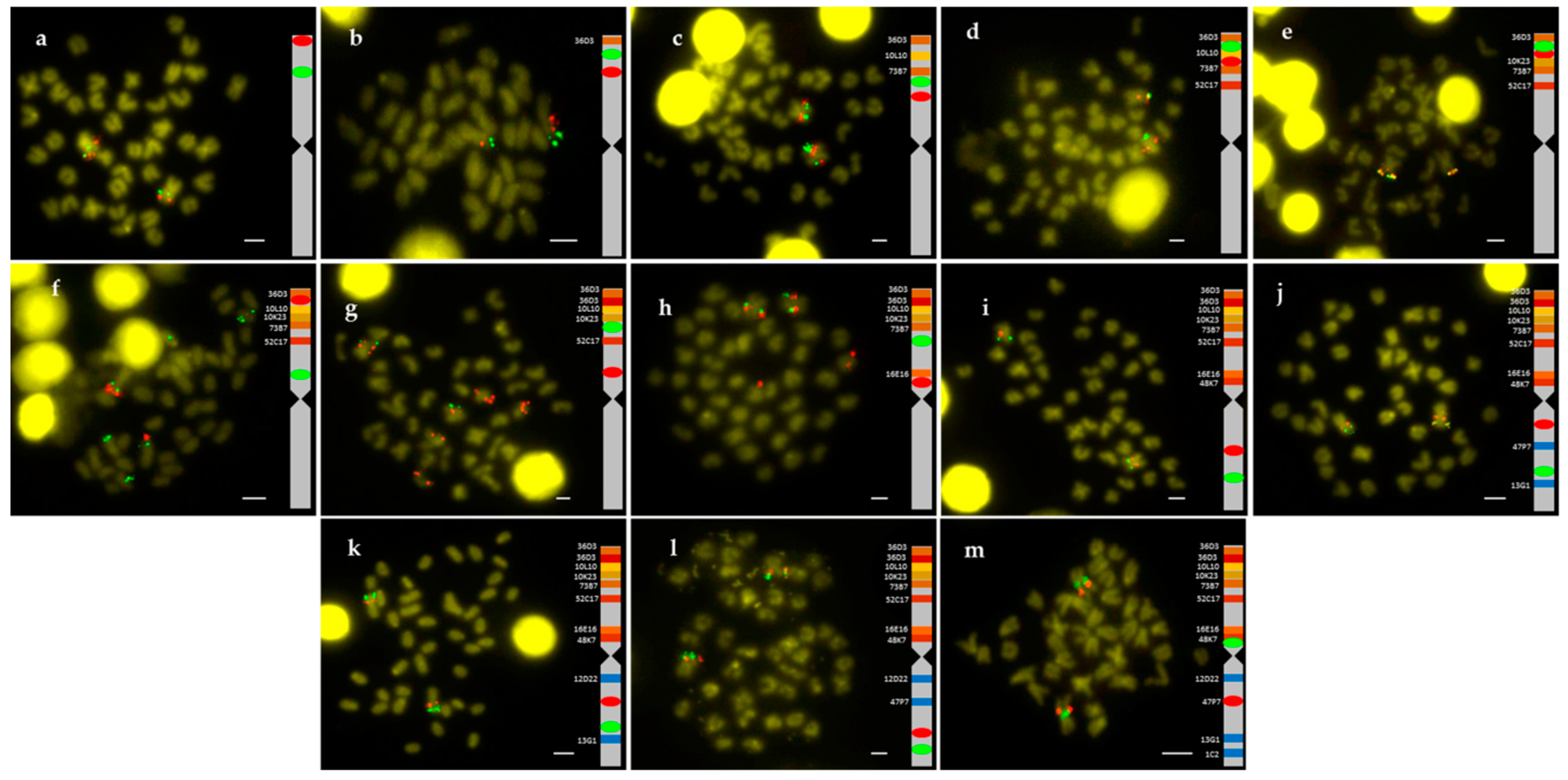
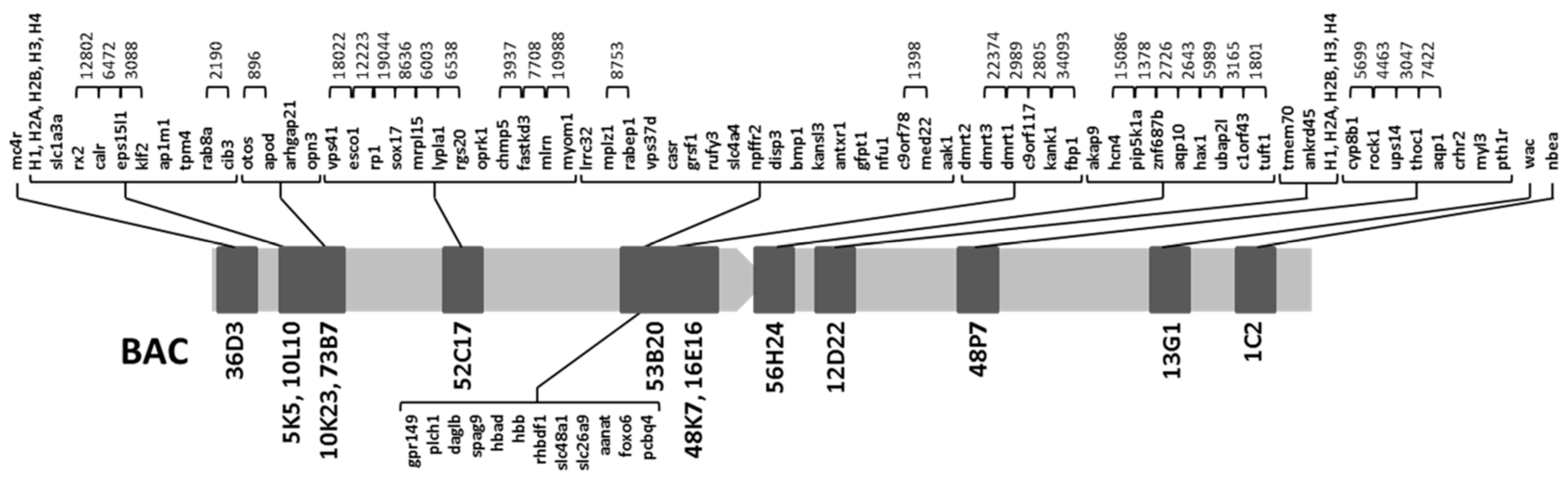
2.1. Obtention of the Integrated Map of Large Metacentric Chromosome 1 of S. senegalensis

A total of 14 BACs with 88 genes annotated were studied (Table 1). The relative BAC positions on the chromosome were obtained by double FISH-BAC (Figure 1).
All chromosome preparations showed 2n = 42. Nine out of the 14 BAC were found in arm 1 (BAC36D3, BAC5K5, BAC10L10, BAC10K23, BAC73B7, BAC52C17, BAC16E16, BAC48K7 and BAC53B20, telomere to centromere direction). The last three clones (BAC16E16, BAC48K7 and BAC53B20) were very close to the centromere and next to them is BAC56H24, located at centromeric position in a previous study [16]. The remaining 4 BAC were located in arm 2: 12D22, 47P7, 13G1 and 1C2 (centromere to telomere direction). The BLAST analysis demonstrated that BAC16E16 overlaps with BAC48K7 and the annotation analysis corroborated that these two BAC clones share the dmrt2 and dmrt3 genes. Similarly, the BLAST analysis showed overlap between BAC48K7 and BAC53B20, but not between BAC16E16 and BAC53B20.
To obtain the gene order within each BAC, C. semilaevis was used as reference species in the microsynteny study. Due to the high number of BAC located in the arm 1, most genes (67 of 88) belonged to such arm, 9 genes belonged to the centromeric region, and 17 genes were in arm 2 (keeping in mind that the five histone cluster genes were only counted in arm1). Distances between genes were obtained only for genes found in the same contig. Figure 2 shows the integrated genetic map for metacentric chromosome 1.

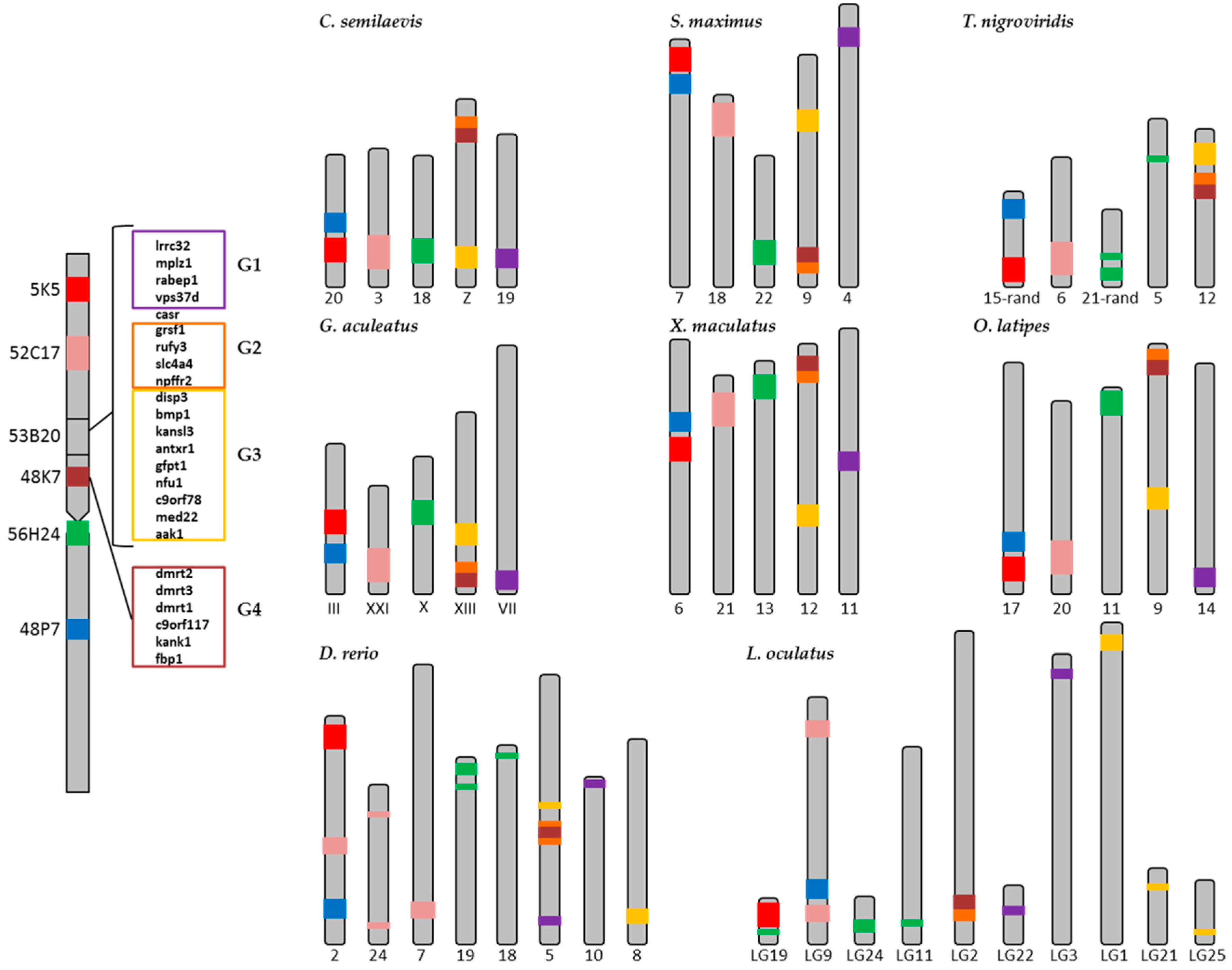
2.2. Synteny Analysis

In order to elucidate the possible evolutionary origin of the metacentric chromosome 1 of S. senegalensis, synteny studies were carried out with eight fish species. The integrated map obtained above served as starting point for the synteny analysis. The comparative study showed a high degree of conservation between the genomic regions within the BACs of S. senegalensis and genomic regions of close (C. semilaevis and S. maximus) and more distant species S. senegalensis genomic regions (T. nigroviridis, G. aculeatus, X. maculatus and O. latipes) (Figures S1–S8).
This high conservation was not so evident in D. rerio and L. oculatus. Eighteen of the 30 genes annotated in BAC53B20 were included in the synteny figures. The other 12 genes were scattered among different chromosomes (Table 2). The distribution of all S. senegalensis genes across the chromosomes of the other species is shown in Tables S1–S8.
One of the most important findings was that six BACs (BAC5K5, BAC52C17, BAC53B20, BAC84K7, BAC56H24, and BAC48P7) were distributed among at least 5 different chromosomes in the reference species (Figure 3), thus allowing us to trace an evolutionary trend for the appearance of the large metacentric chromosome of S. senegalensis, throughout different rearrangement events.
BAC5K5 and BAC48P7 BACs, which are located in different arms in S. senegalensis, were found near each other in the same chromosome in all species except in T. nigroviridis and D. rerio. In these two species, BAC5K5 and BAC48P7 were located far away from each other near the two telomeric regions. Moreover, in L. oculatus, these two BACs were found in different chromosomes linked to genes representative of other BACs (Figure 3). All genes found within each BAC were conserved among the studied species.
The gene organization of BAC52C17 was conserved among all species, except in D. rerio and L. oculatus. Genes in this BAC were distributed among three chromosomes in D. rerio and in one linkage group in L. oculatus, but scattered and located in the extremes. The same situation was observed for BAC56H24, except for D. rerio, L. oculatus and T. nigroviridis.
The most interesting finding in the synteny analysis was obtained in the BAC53B20 and BAC48K7. In all species a group of four genes of BAC53B20 (grsf1, rufy3, slc4a4 and npffr2) (henceforth/from now on named G2) and the genes of BAC48K7 (dmrt2, dmrt3, dmrt1, c9orf117, kank1 and fbp1) (henceforth/from now on named G4) are present in the same cluster. However, in S. senegalensis, between G2 and G4 clusters, a group of genes (disp3, bmp1, kansl3, antxr1, gfpt1, nfu1, c9orf78, med22 and aak1; from now on named as G3) was observed. These genes are positioned in the same chromosome in six species, but in a different position to the G2-G4 cluster.
The exceptions were D. rerio, in which only two genes of G3 (c9orf78 and med22) were in the same chromosome (Chr. 5), and L. oculatus, in which all G3 genes were in three different linkage groups (LG1, LG21 and LG25). The position of G2 and G4 was inverted (taking as reference G3) for T. nigroviridis, G. aculeatus and X. maculatus with respect to C. semilaevis, S. maximus and O. latipes. Other genes of BAC53B20 (lrrc32, mplz1, rabep1 and vps37d) (from now on named G1) were conserved and positioned in different chromosomes in five species, C. semilaevis, S. maximus, G. aculeatus, X maculatus and O. latipes. In D. rerio and L. oculatus, these genes were placed in two different chromosomes. For T. nigroviridis it was impossible to find the G1 orthologous genes. The remaining genes of BAC53B20 (Table 2) were also located in other different chromosomes.

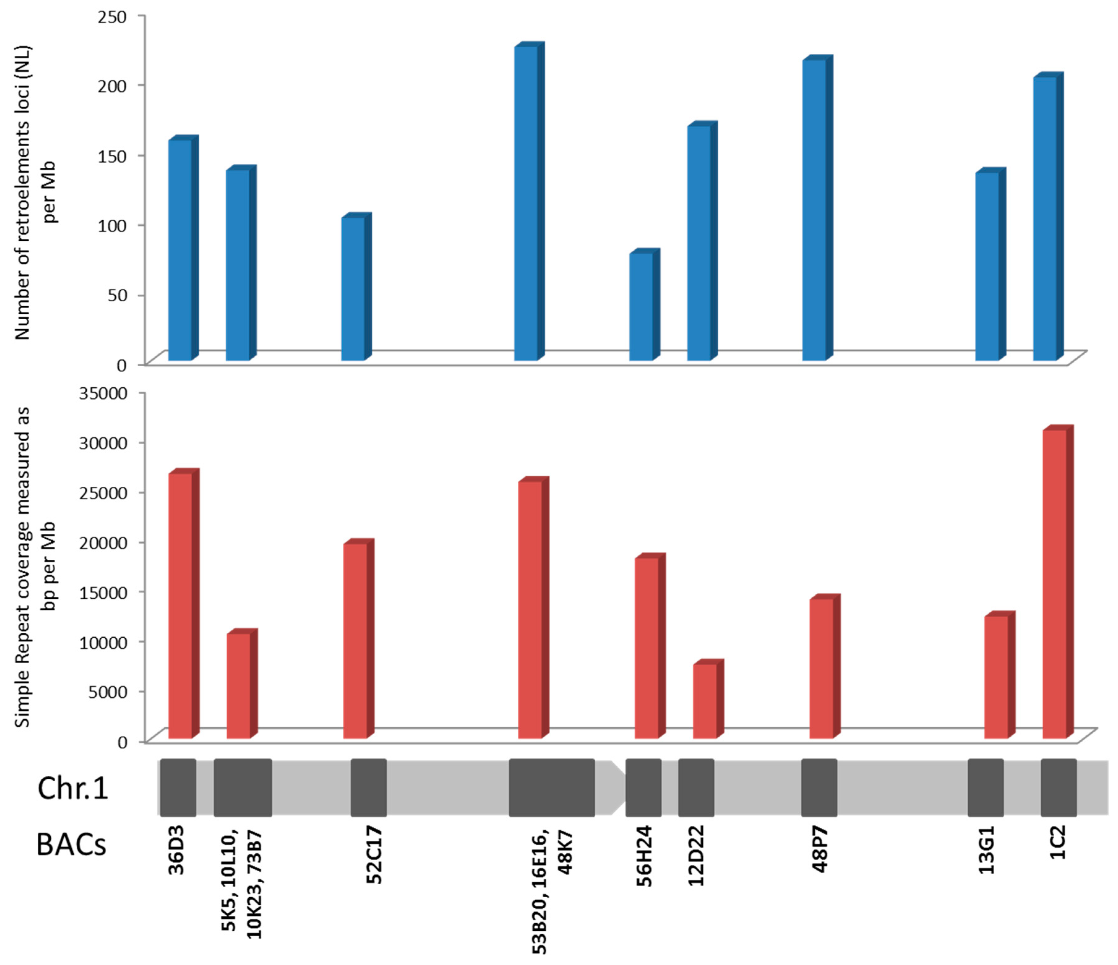
2.3. Repetitive Sequences

The 14 BACs listed in Table 1 represent a total sequence length of 2.79 Mb. The number of transposable elements (TEs) and small non-coding RNAs (sRNA elements) was 1297, showing 464.4 loci per BAC and Mb. The number of satellite DNA and low complexity loci was 1449, with an average of 518.8 loci per Mb. The length of these sequences was 58 kb, representing 2.1% of the genome analyzed (Tables S9 and S10). In relation to the repetitive elements distribution along the chromosomes, and taking into account that some BACs overlapped both in FISH and bioinformatics analysis, BACs 5K5, 10L10, 10K23, 73B7 on one hand, and 53B20, 16E16, 48K7 on the other, were grouped for analysis (Figure 4, Table S11). The distribution of DNA transposons, sRNA, satellites and low complexity sequences was homogeneous along the chromosomes and the differences among then were not related to their location in the arms (Figure S9, Table S11). However, the number of loci of retroelements per BAC and Mb (Figure 4) showed the highest value in the subcentromeric region where 53B20, 16E16 and 48K7 BACs were localized (224.34 loci/Mb). This subcentromeric region contains all the dmrt genes associated with sex determining in some species. On the other side, the coverage of simple repeats showed three regions with higher values than the rest of the chromosome: telomeric BACs 36D3 and 1C2 (26460.44 and 30811.13 bp/Mb respectively) and again the subcentromeric region containing dmrt genes (53B20, 16E16 and 48K7 BACs) (25649.10 bp/Mb) (Figure 4).
It is worth mentioning the presence of a “chromosome Y” satellite (motif length: 860 bp) in the subcentromeric region that contains dmrt genes. The BLAST analysis of this satellite sequence showed the existence of this sequence in centromeric position of several vertebrate chromosomes and in the long arm of the human Y chromosome, indicating the presence of this satellite in a sex determining region.

3. Discussion

The comparative genomic study of 14 BAC with 88 genes located in the large metacentric chromosome 1 of S. senegalensis was carried out in eight fish species, including the spotted gar, which belongs to a non-teleost fish lineage that did not suffer the teleost genome duplication (TGD) [24]. The taxonomic relation among all species included in the present work is shown in Figure 5. Three out of the nine species were flatfish belonging to the Pleuronectiformes order, thus in the taxonomic tree three families of Pleuronectiformes: Soleidae (S. senegalensis), Cynoglossidae (C. semilaevis) and Scophthalmidae (S. maximus) are/were represented. The Pleuronectiformes have been the subject of controversy regarding the monophyletic or polyphyletic origin of the group. Recently, dates generated by analysis from more than 1000 UCE (Ultraconserved DNA Elements) loci provided strong molecular support for the monophyletic origin of flatfishes and for the single origin of cranial asymmetry [25,26]. Despite the taxonomic proximity of S. maximus and C. semilaevis, the analysis of the gene groups carried out in the Figure 3, they were more similar to other distant species that to S. senegalensis. García-Angulo et al. [16] showed that the metacentric chromosome 1 of S. senegalensis hybridized to two acrocentric chromosome pairs of two species of the Soleidae family, thus demonstrating the origin of the metacentric chromosome 1 by a Robertsonian fusion. In addition, more than 80% of the metacentric chromosome 1 genes are distributed in four chromosomes of both C. semilaevis and S. maximus. Moreover, the conservation of groups of genes in all species studied, which are also conserved in L. oculatus, demonstrates a common origin in the evolution of Teleostei and Holostei [27]. Although two acrocentric pairs of both Dagetichthys and Dicologlossa show hybridization to each arm of chromosome 1 of S. senegalensis [16], there are no data about the distribution of BACs in these acrocentric chromosomes. On the other hand Merlo et al. [23] reported pericentric inversions in the chromosome 1 of S. senegalensis, which would support that the acrocentric chromosomes of Dagetichthys and Dicologlossa do not have the same distribution that the BACs in the arms of the metacentric chromosome 1 of S. senegalensis have. If this were the case, more differences will be observed between S. senegalensis and Dagetichthys and Dicologlossa, despite all of them belonging to the Soleidae family (taking into account the distribution of BACs on Figure 3), than between the other families of Pleuronectiformes or even among other Euteleosteomorpha families included in this work. Further FISH studies are necessary to assess the distribution of BACs in other Soleidae species.
In relation to the localization of genes from groups G2-G4, the G4 group (Figure 3) contains the dmrt1-dmrt2-dmrt3 gene cluster and this cluster was found in the Z sex-chromosome of C. semilaevis; only the Z chromosome contains a functional copy of dmrt1 in this species, and it was proposed as an important sex-determining gene in C. semilaevis [4]. Moreover, the same cluster, G4, was positioned in the dmY region of medaka (except dmrt1 and c9orf117 genes), which was described as sex determination (SD) gene that emerged from a segment duplication of a small autosomal region containing the precursor dmrt1, followed by an insertion into the proto-Y chromosome [21]. The same dmY gene was described in two different species of medaka, O. latipes and Oryzias curvinotus [8,21]. These findings suggest that the pericentromeric region of the metacentric chromosome 1 of S. senegalensis might be a sex-determining zone, but future studies are necessary.
Sex chromosomes are good model systems for the study of evolutionary processes that occur in the absence of recombination. The non-recombining parts of the Y chromosome are unique genomic regions in which the processes of gene degeneration and accumulation of repetitive elements, including microsatellites, are active [28,29,30,31]. Microsatellites are sometimes co-localized with other genomic repeats, especially TE. In humans, microsatellites are associated with other repetitive DNA, especially non- LTR retrotransposons [32,33]. In plants as Arabidopsis, rice, soybean, and wheat, however, microsatellites are preferentially associated to non-repetitive DNA regions, indicating that they reside in regions pre-dating genome expansion [34,35]. In fish as X. maculatus it has been found a high content of active TEs and satellite sequences on the SD segments of the X and Y chromosomes, and the distribution of long repeats is different between X and Y chromosomes [18,19]. In O. latipes, it has also been observed an accumulation of TEs after the formation of the SD region containing the dmrt1Y gene [19]. Moreover, the existence of a conserved genetic cascade of SD common to all vertebrates governed by different lineages or species-specific genes such as dmrt1 has been hypothesized [36]. In this study, a high number of retrotransposons loci and a high abundance of simple repeats elements were found in a region containing dmrt genes in the proto-sexual chromosome 1 of S. senegalensis. Besides, in this region, a satellite element related with human Y chromosome was observed too. In Rumex acetosa (Y1Y2X system, 12–13 mya), microsatellites expanded on the relatively young and large Y chromosomes [31] but not on the older and small Y chromosome of human (XY system, 150 mya) [37] or Marchantia polymorpha (XY system) [38]. In this way, R. acetosa data indicates that microsatellite arrays are targets for TE insertions. It has been suggested that early Y chromosome evolution is accompanied by microsatellite expansion and that expansion ability is an intrinsic property of some microsatellites, and the non-recombining regions of the young Y chromosome provide the opportunity for expansion [39], while the revitalizing process of recombination opposes this expansion on other chromosomes [31]. It has also been reported that the accumulation of DNA repetitive sequences increased the size of X and Y chromosomes in the initial phase of differentiation of sex chromosomes [17]. This model can explain sequence patterns on older Y chromosomes, which is characterized by short microsatellite arrays and higher number of TE and it has been proposed that microsatellites served as targets for TE insertions [31]. Support for this is provided by the colocalization of microsatellites with transposable elements in a partial human genome sequence [32].
In this study, simple repeats elements and retroelements were observed in a pericentromeric location in the chromosome 1 of S. senegalensis where dmrt genes, usually associated to sex determining, are present. This could indicate a non- (or low-rate) recombining region in the genome of S. senegalensis, and the accumulation of these repetitive sequences could account for the evolution of the putative sex-determining chromosome of this species.
The synteny data and repetitive sequences analysis could give insights about the evolution of this chromosome which is proposed as neo sex proto-chromosome. The synteny analysis (Figure 3) and the distribution of BACs 5K5, 52C17, 53B20, 48K7, 56H24 and 48P7 in five chromosomes seems to indicate that this distribution is conserved from the ancestral Eutelosteomorpha to the actual teleosts as it was observed in the reference species. This distribution, however, is not present in S. senegalensis. It was reported in studies of vertebrate evolution that the genome of the bony vertebrate ancestor contained both macro and micro-chromosomes, some of which remain conserved in L. oculatus. These chromosomes suffered fusions in the lineage leading to teleosts after divergence from L. oculatus, followed by the TGD of the fused chromosomes and subsequent interchromosomal rearrangements and rediploidization, which led to the formation of macrochromosomes in teleosts, as for example O. latipes [27]. These events could lead to establishing the current situation observed in Figure 3, just like for all other species, as included in this work.
To explain the evolutionary origin of the proto sex- chromosome 1 of S. senegalensis from a common ancestor of Eutelosteomorpha three scenarios (Figure 6a), starting with five ancestral chromosomes, are proposed. The situations described in Figure 6b,c show how translocations, fusions and rearrangements events among “A”, “B”, and “C” chromosomes could have originated a new chromosome called “F”. In the formation of sex chromosomes events such as fusions between sex chromosomes and autosome of the type Y-A has been described [13], or in the formation of (XX/XY1Y2) multiple system in neo tropical fish [2], or in the X1X2Y sex system, which was formed through a centric fusion of ancestral Y chromosome with an autosome, creating a large neo-Y chromosome in the Oplegnathus genus [14]. In the situation described in Figure 6d only translocations and rearrangements are proposed to originate the “F” chromosome. The next step, as described by Merlo et al. [23], a Robertsonain fusion must have happened between “F” chromosome and another one called “G” (Figure 6e), which is unknown with respect to its gene content, followed by a pericentromeric inversion (Figure 6f). Hence, this type of rearrangement could be related with the evolution of the proto sex chromosome in S. senegalensis. For example, a large inversion has been described in the zone corresponding to the sex-determining region of the XY system in the nine-spined stickcleback Pungitius pungitius [6], and both, an inversion and a deletion in G. aculeatus Y chromosomes, suggesting that the process of sex chromosome evolution can occur rapidly in sex chromosomes evolution and after the acquisition of the sex determining region (SD) [20]. At the same time, translocations and rearrangements with the “C” and also with “E” chromosomes and accumulation of repetitive sequences could have occurred in the pericentromeric region (Figure 6g). This is suggested in our data by the presence of repetitive sequences in the zone of the BAC53B20 and 48K7 (see above). This could lead to the actual proto-sex chromosome of S. senegalensis (Figure 6h).

4. Materials and Methods

4.1. BAC Clones

BACs used in this work were isolated from a BAC library of S. senegalensis, composed of 29,184 positive clones distributed in 384-well plates (76 plates in total). The 4D-PCR method [40] was used to isolate BACs. Details of the construction of the BAC library and the strategy used to find and isolate BAC clones are described by García-Cegarra et al. [41]. The name of each BAC indicates the library plate number (first set of digits) and well coordinates (A- and 1- ).

4.2. Chromosome Preparation and Double BAC-FISH

Chromosome preparations were carried out using larvae (1–3 days after hatching) of S. senegalensis. Larvae were pre-treated with 0.02% colchicine for 3 h to obtain a large number of metaphasic cells. After that, they were treated with KCl (0.4%), and finally they were fixed in a freshly solution of Carnoy (absolute ethanol:acetic acid, 3:1). Prior to hybridization and to isolate the BAC, 50 mL of LB supplemented with chloramphenicol were inoculated from a pre-culture (8 h growth at 37 °C in a shaker). DNA-BAC were purified using Plasmid Midi Kit (Qiagen, Hilden, Germany), following the manufacturer’s instructions. This DNA was labeled by Nick Translation using the DIG or BIO -Nik Translation Mix (Roche Molecular Biochemical) and used as probe as described in the manufacturer’s instructions.
Pre-treatment of chromosome preparations and hybridization was carried out as described by García-Cegarra et al. [41]. Then, 2-colour FISH was used. For digoxigenin-labeled probes, hybridization sites were detected using anti-Digoxigenin/Digoxin (Vector Labs, Burlingame, CA, USA) at 1:200 and anti-Goat IgG (vector Labs) at 1:100. Avidin FITC (Vector Labs) at 1:200 and biotinylated anti- avidin (Vector labs) at 1:100 were used for biotin-labeled probes. These antibodies were prepared in TNFM (4x SSC, 0.05% Tween 20, 5% skim milk). Slides were then washed in TNFM, dehydrated with ethanol (70%, 90% and 100%) and counterstained with 0.4 mg/mL DAPI-Vectashield (ATOM). Images were recorded using a Zeiss PALM MicroBeam microdissector and fluorescence microscopes equipped with an AxioCam MRm digital camera.

4.3. Sequencing and Bioinformatic Analysis

The DNA-BAC extracted with the Large Construct kit (Qiagen, Hilden, Germany) was sequenced using the Illumina sequencing platform (Illumina, San Diego, CA, USA). The BAC clones were deposited in the GenBank database under the accession numbers AC278047 to AC278120. The functional and structural annotation of the genes identified in each BAC was carried out as described by García-Angulo et al. [41].
For the analysis of micro-synteny, C. semilaevis was selected as the genome reference and the ENSEMBL database was used to obtain the order of the genes. The order of the contigs within each BAC and the distance between the genes within each contig of S. senegalensis were estimated using the information provided by the Geneious R11 [42]. The “nucleotide BLAST” tool was used to found overlapping among contiguous BACs and the study of the repetitive sequences.

4.4. Repetitive Elements Analysis

After determining the chromosome location of BAC clones, using the FISH technique, a homology-based approach using the Repbase database (https://www.girinst.org/repbase/, release 23.07) with RepeatMasker software (http://www.repeatmasker.org/) [43] was applied. Distribution analysis of repetitive elements as TEs (Retroelements and DNA transposons), sRNA, DNA satellite (measured as satellites and simple repeats) and low complexity sequences, in S. senegalensis metacentric chromosome, was possible by using both the information of BAC clone position, obtained afterthe double BACFISH technique, and the coordinates of the elements from the RepeatMasker software. The average number of TEs and sRNA was calculated as the total number of identified loci in relation to the BAC sequences length analyzed and normalized by Mb. The coverage of DNA satellite and low complexity elements was calculated as the quantity of sequences (bp) in relation to the BAC sequences length analyzed, and again normalized by Mb.

4.5. Synteny Analyses

For comparative genomic analysis eight species of fish were used, Cynoglossus semilaevis (2n = 42) or smooth tongue sole, Scophthalmus maximus (2n = 44) or turbot, Tetraodon nigroviridis (2n = 42) or smooth pufferfish, Gasterosteus aculeatus (2n = 42) or three-spined stickleback, Xiphophorus maculatus (2n = 48) or platyfish, Oryzias latipes (2n = 48) or medaka, Danio rerio (2n = 50) or zebrafish and Lepisosteus oculatus (2n = 58, [27]) or spotted gar. The lineages of these references species and S. senegalensis were obtained from the National Center for Biotechnology Information (NCBI) database to obtain the taxonomic tree (Figure 5).
The ENSEMBL database and Genome Data Viewer (NCBI) were applied for comparative genomic analysis. The data were used to identify reorganizations within the chromosomes of reference species.

5. Conclusions

The evolution of the sex-proto chromosome of S. senegalensis, which belong to the Soleidae family, is very different from the evolution of the karyotype of the other families of Pleuronectiformes.
Cytogenetic events, fusions and pericentric inversions reported previously, the analysis synteny carried out (Figure 3) and the detection of simple repeat elements and retroelements in a pericentromeric location in the chromosome 1 of S. senegalensis, in the region where dmrt genes have been located, indicates that this chromosome is in an initial phase of differentiation as sex chromosome.

Supplementary Materials

Supplementary materials can be found at https://www.mdpi.com/1422-0067/20/20/5111/s1.

Author Contributions

Conceptualization, L.R.; Formal analysis, M.E.R, M.A.M., A.A.-P., and I.C.; Funding acquisition, L.R.; Investigation, M.E.R., I.C., B.M., T.L., and A.G.-A.; Methodology, S.P.-B., and M.E.R.; Project administration, L.R.; Resources: L.R., I.C., and M.A.M.; Supervision, L.R.; Visualization, M.E.R., L.R., A.A.-P., and M.A.M; Writing—original, M.E.R., I.C., and S.P.-B.; Writing—review & editing, L.R, M.A.M., and A.A.-P.

Funding

This research was funded by the Spanish Ministerio de Ciencia e Innovación MICINN-FEDER (projects AGL2014- 51860-C2-1-P and RTI2018-096847-B-C21).

Acknowledgments

The authors acknowledge to Manuel Manchado from El Toruño IFAPA Center for providing the larvae of S. senegalensis.

Conflicts of Interest

The authors declare no conflict of interest.

References

  1. Nelson, J.S. Fishes of the World, 4th ed.; John Willey & Sons, Inc.: Hoboken, NJ, USA, 2006; p. 624. [Google Scholar]
  2. De Oliveira, E.A.; Sember, A.; Bertollo, L.A.C.; Yano, C.F.; Ezaz, T.; Moreira-Filho, O.; Hatanaka, T.; Trifonov, V.; Liehr, T.; Al-Rikabi, A.B.H.; et al. Tracking the evolutionary pathway of sex chromosomes among fishes: Characterizing the unique XX/XY1Y2 system in Hoplias malabaricus (Teleostei, Characiformes). Chromosoma 2018, 127, 115–128. [Google Scholar] [CrossRef] [PubMed]
  3. Robles, F.; De La Herrán, R.; Navajas-Pérez, R.; Cano-Roldán, B.; Sola-Campoy, P.J.; García-Zea, J.A.; Ruiz-Rejón, C. Centromeric satellite DNA in flatfish (order Pleuronectiformes) and its relation to speciation processes. J. Hered. 2017, 108, 217–222. [Google Scholar]
  4. Chen, S.; Zhang, G.; Shao, C.; Huang, Q.; Liu, G.; Zhang, P.; Song, W.; An, N.; Chalopin, D.; Volff, J.N.; et al. Whole-genome sequenced of a flatfish provides insights into ZW sex chromosome evolution and adaptation to a benthic lifestyle. Nat. Genet. 2014, 46, 253–260. [Google Scholar] [CrossRef] [PubMed]
  5. Molina-Luzón, M.J.; Hermida, M.; Navajas-Pérez, R.; Robles, F.; Navas, J.I.; Ruiz-Rejón, C.; Martínez, P.; De la Herrán, R. First haploid genetic map base don microsatellite markers in Senegalense sole (Solea senegalensis, Kaup, 1858). Mar. Biotecnol. 2015, 17, 8–12. [Google Scholar] [CrossRef] [PubMed]
  6. Natri, H.M.; Merilä, J.; Shikano, T. The evolution of sex determination associated with a chromosomal inversion. Nat. Commun. 2019, 10, 145. [Google Scholar] [CrossRef]
  7. Martínez, P.; Viñas, A.M.; Sánchez, L.; Díaz, N.; Ribas, L.; Piferrer, F. Genetic architecture of sex determination in fish: Applications to sex ratio control in aquaculture. Front. Genet. 2014, 5, 340. [Google Scholar] [CrossRef]
  8. Matsuda, M.; Sakaizumi, M. Evolution of the sex-determining gene in teleostean genus Oryzias. Gen. Comp. Endocrinol. 2016, 239, 80–88. [Google Scholar] [CrossRef]
  9. Takehana, Y.; Matsuda, M.; Myosho, T.; Suster, M.L.; Kawakami, K.; Shin-I, T.; Kohara, Y.; Kuroki, Y.; Toyoda, A.; Fujiyama, A.; et al. Co-option of sox3 as the male-determining factor on the Y chromosome in fish Oryzias dancena. Nat. Commun. 2014, 5, 4157. [Google Scholar] [CrossRef]
  10. Hattori, R.S.; Murai, Y.; Oura, M.; Masuda, S.; Majhi, S.K.; Sakamoto, T.; Fernandino, J.I.; Somoza, G.M.; Yokota, M.; Strüssmann, C.A. A Y-linked anti-Müllerian hormone duplication takes over a critical role in sex determination. Proc. Nat. Acad. Sci. USA 2012, 109, 2955–2959. [Google Scholar] [CrossRef]
  11. Kamiya, T.; Kai, W.; Tasumi, S.; Oka, A.; Matsunaga, T.; Mizuno, N.; Fujita, M.; Suetake, H.; Suzuki, S.; Hosoya, S.; et al. A trans-species missense SNP in Amhr2 is associated with sex determination in the tiger pufferfish, Takifugu rubripes (fugu). PLoS Genet. 2012, 8, e1002798. [Google Scholar] [CrossRef]
  12. Yano, A.; Nicol, B.; Jouanno, E.; Quillet, E.; Fostier, A.; Guyomard, R.; Guguen, Y. The sexually dimorphic on the Y-chromosome gene (sdY) is a conserved male-specific Y-chromosome sequence in many salmonids. Evol. Appl. 2013, 6, 486–496. [Google Scholar] [CrossRef] [PubMed]
  13. Pennell, M.W.; Kirkpatrick, M.; Otto, S.P.; Vamosi, J.C.; Peichel, C.L.; Valenzuela, N.; Kitano, J. Y fuse? Sex chromosome fusions in fishes and reptiles. PLoS Genet. 2015, 11, e1005237. [Google Scholar] [CrossRef] [PubMed]
  14. Xu, D.; Sember, A.; Zhu, Q.; De Oliveira, E.A.; Liehr, T.; Al-Rikabi, A.B.H.; Xiao, Z.; Song, H.; Cioffi, M.B. Deciphering the origin and evolution of the X1X2Y system in two closely-related Oplegnathus species (Oplegnathidae and Centrarchiformes). Int. J. Mol Sci. 2019, 20, 3517. [Google Scholar] [CrossRef] [PubMed]
  15. Portela-Bens, S.; Merlo, M.A.; Rodríguez, M.E.; Cross, I.; Manchado, M.; Kosyakova, N.; Liehr, T.; Rebordinos, L. Integrated gene mapping and synteny studies give insights into the evolution of a sex proto-chromosome in Solea senegalensis. Chromosoma 2017, 126, 261–277. [Google Scholar] [CrossRef] [PubMed]
  16. García-Angulo, A.; Merlo, M.A.; Portela-Bens, S.; Rodríguez, M.E.; García, E.; Al-Rikabi, A.; Liehr, T.; Rebordinos, L. Evidence for a Robertsonian fusion in Solea senegalensis (Kaup, 1858) revealed by zoo-FISH and comparative genome analysis. BMC Genomics 2018, 19, 818. [Google Scholar] [CrossRef]
  17. Schartl, M.; Schmid, M.; Nanda, I. Dynamics of vertebrate sex chromosome evolution: From equal size to giants and dwarfs. Chromosoma 2016, 125, 553–571. [Google Scholar] [CrossRef]
  18. Zhou, Q.A. Swimy locus on Y chromosome of the platyfish (Xiphophorus maculatus) is derived from a novel DNA transposon Zisupton. Gene 2012, 503, 254–259. [Google Scholar] [CrossRef]
  19. Chalopin, D.; Volff, J.N.; Galiana, D.; Anderson, J.L.; Schartl, M. Transposable elements and early evolution of sex chromosome in fish. Chromosome Res. 2015, 23, 545–560. [Google Scholar] [CrossRef]
  20. Ross, J.A.; Peichel, C.L. Molecular cytogenetic evidence of rearrangements on the Y chromosome of the threespine stickleback fish. Genetics 2008, 179, 2173–2182. [Google Scholar] [CrossRef]
  21. Matsuda, M.; Nagahama, Y.; Shinomiya, A.; Sato, T.; Matsuda, C.; Kobayashi, T.; Morrey, C.E.; Shibata, N.; Asakawa, S.; Shimizu, N.; et al. DMY is a Y-specific DM-domain gene required for male development in the medaka fish. Nature 2002, 417, 559–563. [Google Scholar] [CrossRef]
  22. Vega, L.; Díaz, E.; Cross, I.; Rebordinos, L. Caracterizaciones citogenética e isoenzimática del lengauado Solea senegalensis Kaup, 1858. Bol. Inst. Esp. Oceanogr. 2002, 18, 245–250. [Google Scholar]
  23. Merlo, M.A.; Iziga, R.; Portela-Bens, S.; Cross, I.; Kosyakova, N.; Liehr, T.; Manchado, M.; Rebordinos, L. Analysis of the histone cluster in Senegalese sole (Solea senegalensis): Evidence for a divergent evolution of two canonical histone cluster. Genome 2017, 60, 441–453. [Google Scholar] [CrossRef] [PubMed]
  24. Amores, A.; Catchen, J.; Ferrara, A.; Fontenot, Q.; Postlethwait, J.H. Genome evolution and meiotic maps by massively parallel DNA sequencing: Spotted gar, an outgroup for the teleost genome duplication. Genetics 2011, 188, 799–808. [Google Scholar] [CrossRef] [PubMed]
  25. Harrington, R.C.; Faircloth, B.C.; Eytan, R.I.; Smith, W.L.; Near, T.J.; Alfaro, M.E.; Friedman, M. Phylogenomic analysis of carangimorph fishes reveals flatfish asymmetry arose in blink of evolutionary eye. BMC Evol. Biol. 2016, 16, 224. [Google Scholar] [CrossRef] [PubMed]
  26. Shi, W.; Chen, S.; Kong, X.; Si, L.; Gong, L.; Zhang, Y.; Yu, H. Flatfish monophyly refereed by the relationship of Psettodes in Carangimorphariae. BMC Genom. 2018, 19, 400. [Google Scholar] [CrossRef] [PubMed]
  27. Braasch, I.; Gehrke, A.R.; Smith, J.J.; Kawasaki, K.; Manousaki, T.; Pasquier, J.; Amores, A.; Desvignes, T.; Batzel, P.; Catchen, L.; et al. The spotted gar genome illuminates vertebrate evolution and facilitates human-teleost comparisons. Nat. Genet. 2016, 48, 427–437. [Google Scholar] [CrossRef] [Green Version]
  28. Bachtrog, D.; Weiss, S.; Zangerl, B.; Brem, G.; Schlotterer, C. Distribution of dinucleotide microsatellites in the Drosophila melanogaster genome. Mol. Biol. Evol. 1999, 16, 602–610. [Google Scholar] [CrossRef]
  29. Kubat, Z.; Hobza, R.; Vyskot, B.; Kejnovsky, E. Microsatellite accumulation on the Y chromosome in Silene latifolia. Genome 2008, 51, 350–356. [Google Scholar] [CrossRef]
  30. Kejnovsky, E.; Hobza, R.; Cermak, T.; Kubat, Z.; Vyskot, B. The role of repetitive DNA in structure and evolution of sex chromosomes in plants. Heredity 2009, 103, 533–541. [Google Scholar] [CrossRef]
  31. Kejnovský, E.; Michalovova, M.; Steflova, P.; Kejnovska, I.; Manzano, S.; Hobza, R.; Kubat, Z.; Kovarik, J.; Jamilena, M.; Vyskot, B. Expansion of microsatellites on evolutionary young Y chromosome. PLoS ONE 2013, 8, e45519. [Google Scholar] [CrossRef]
  32. Nadir, E.; Margalit, H.; Gallily, T.; Ben-Sasson, S. Microsatellite spreading in the human genome: Evolutionary mechanisms and structural implications. Proc. Natl. Acad. Sci. USA 1996, 93, 6470–6475. [Google Scholar] [CrossRef] [PubMed]
  33. Kelkar, Y.D.; Eckert, K.A.; Chiaromonte, F.; Makova, K.D. A matter of life or death: How microsatellites emerge in and vanish from the human genome. Genome Res. 2011, 21, 2038–2048. [Google Scholar] [CrossRef] [PubMed] [Green Version]
  34. Temnykh, S.; DeClerk, G.; Lukashova, A.; Lipovich, L.; Cartinhour, S.; McCouh, S. Computational and experimental analysis of microsatellites in rice (Oryza sativa L.): Frequency, length variation, transposon association, and genetic marker potential. Genome Res. 2001, 11, 1441–1452. [Google Scholar] [CrossRef] [PubMed]
  35. Morgante, M.; Hanafey, M.; Powell, W. Microsatellites are preferentially associated with nonrepetitive DNA in plant genomes. Nat. Genet. 2002, 30, 194–200. [Google Scholar] [CrossRef] [PubMed]
  36. Heule, C.; Salzburger, W.; Böhne, A. Genetics of sexual development: An evolutionary playground for fish. Genetics 2014, 196, 579–591. [Google Scholar] [CrossRef]
  37. Graves, J.A.M. The rise and fall of SRY. Trends Genet. 2002, 18, 259–264. [Google Scholar] [CrossRef]
  38. Yamato, K.T.; Ishizaki, K.; Fujisawa, M.; Okada, S.; Nakayama, S.; Fujishita, M.; Bando, H.; Yodoya, K.; Hayashi, K.; Bando, T.; et al. Gene organization of the liverworth Y chromosome reveals distinct sex chromosom evolution in a haploid system. Proc. Natl Acad Sci USA 2007, 104, 6472–6477. [Google Scholar] [CrossRef]
  39. Charlesworth, B.; Sniegowski, P.; Stephan, W. The evolutionary dynamics of repetitive DNA in eukaryotes. Nature 1994, 371, 215–220. [Google Scholar] [CrossRef]
  40. Asakawa, S.; Abe, I.; Kudoh, Y.; Kishi, N.; Wang, Y.; Kubota, R.; Kudoh, J.; Kawasaki, K.; Minoshima, S.; Shimizu, N. Human BAC library: Construction and rapid screening. Gene 1997, 191, 69–79. [Google Scholar] [CrossRef]
  41. García-Cegarra, A.; Merlo, M.A.; Ponce, M.; Portela-Bens, S.; Cross, I.; Manchado, M.; Rebordinos, L. A preliminary genetic map in Solea senegalensis (Pleuronectiformes, Soleidae) using BAC-FISH and next-generation sequencing. Cytogenet. Genome Res. 2013, 141, 227–240. [Google Scholar] [CrossRef]
  42. Kearse, M.; Moir, R.; Wilson, A.; Stones-Havas, S.; Cheung, M.; Sturrock, S.; Buxton, S.; Cooper, A.; Markowitz, S.; Duran, C.; et al. Geneious Basic: An integrated and extendable desktop software platform for the organization and analysis of sequence data. Bioinformatics 2012, 28, 1647–1649. [Google Scholar] [CrossRef] [PubMed]
  43. Smit, A.F.A.; Hubley, R.; Green, P. RepeatMasker Open-4.0. (2013–2015). Available online: http://www.repeatmasker.org (accessed on 24 May 2018).
Figure 1. Double FISH-BAC of (a) 73B7 (green)/36D3 (red), (b) 10L10 (green)/73B7 (red), (c) 10K23 (green)/52C17 (red), (d) 5K5 (green)/10K23 (red), (e) 5K5 (green)/10L10 (red), (f) 16E16 (green)/5K5 (red), (g) 73B7 (green)/16E16 (red), (h) 52C17 (green)/48K7 (red), (i) 13G1 (green)/47P7 (red), (j) 1C2 (green)/12D22 (red), (k) 1C2 (green/47P7 (red), (l) 1C2 (green)/13G1 (red), and (m) 53B20 (green)/47P7 (red). Bar = 2 µm.
Ijms 20 05111 g001
Figure 2. Integrated genetic map of metacentric chromosome 1 of S. senegalensis.
Ijms 20 05111 g002
Figure 3. Organization of the BACs and specific regions of the metacentric chromosome 1 of S. senegalensis in the species.
Ijms 20 05111 g003
Figure 4. Repetitive elements distribution along S. senegalensis chromosome 1.
Ijms 20 05111 g004
Figure 5. Taxonomic tree showing the species of fishes included in this work.
Ijms 20 05111 g005
Figure 6. Evolution of the proto sex- chromosome 1 of S. senegalensis from ancestral chromosomes after evolution of Teleost fish and Euteleosteomprpha. (a) A, B, C, D and E represent ancestral chromosomes. (bd) are the proposed paths for the evolution of the chromosome of S. senegalensis. (e) Robertsonian fusions. (f) Pericentric inversions and rearrangements. The black square represents the region in which pericentric inversion occurred. (g) Translocation and rearrangements events. (h) Current metacentric proto sex-chromosome of S. senegalensis.
Ijms 20 05111 g006
Table 1. BAC clones located on the metacentric chromosome 1.
Name of BACGene AnnotationReferences
36D3mcr4[16]
5K5H1, H2a, H2b, H3, H4, slc1a3a, rx2, calr, eps15l1, klf2, ap1m1, tpm4, rab8a, cib3[16,23]
10L10rx2, calr, eps15l1, klf2[16]
10K23calr, eps15l1, otos, apod, arhgap21, opn3[15]
73B7otos, apod, arhgap21[16]
52C17vps41, esco1, rp1, sox17, mrpl15, lypla1, rgs20, oprk1, chmp5, fastkd3, mlrn, myom1[16]
53B20lrrc32, mplz1, rabep1, vps37d, casr, gpr149, plch1, grsf1, rufy3, slc4a4, npffr2, daglb, spag9, hbad, hbb, rhbdf1, slc48a1, slc26a9, aanat, foxo6, pcbp4, disp3, bmp1, kansl3, antxr1, gfpt1, nfu1, c9orf78, med22, aak1This work
16E16dmrt2, dmrt3[15,16]
48K7dmrt2, dmrt3, dmrt1, c9orf117, kank1, fbp1[16]
56H24akap9, hcn4, pip5k1a, znf687b, aqp10, hax1, ubap2l, c1orf43, tuft1[16]
12D22H1, H2a, H2b, H3, H4, tmem70, ankrd45[23]
48P7cyp8b1, rock1, ups14, thoc1, aqp1, crhr2, myl3, pth1r[16]
13G1wac[16]
1C2nbea[16]
Table 2. Distribution of 12 genes out of 30 annotated in BAC53B20 on the chromosomes of the reference species.
GenesC. semilaevisS. maximusT. nigroviridisG. aculeatusX. maculatusO. latipesD. rerioL. oculatus
gpr1494316I18131LG14
plch14316I181318LG14
daglb8182V1019121
spag98182V101912LG10
hbad8183XI1083LG13
hbb8183XI10193LG13
rhbdf117183XI1083LG13
slc48a1101111XII1723LG4
slc26a91119XII1723LG3
aanat983XI1683LG13
foxo61371X131119LG6
pcbp4169XII1722LG5
1 This gene was not found.

