
Liquid Chromatography-Mass Spectrometry (LC-MS) Derivatization-Based Methods for the Determination of Fatty Acids in Biological Samples


Abstract
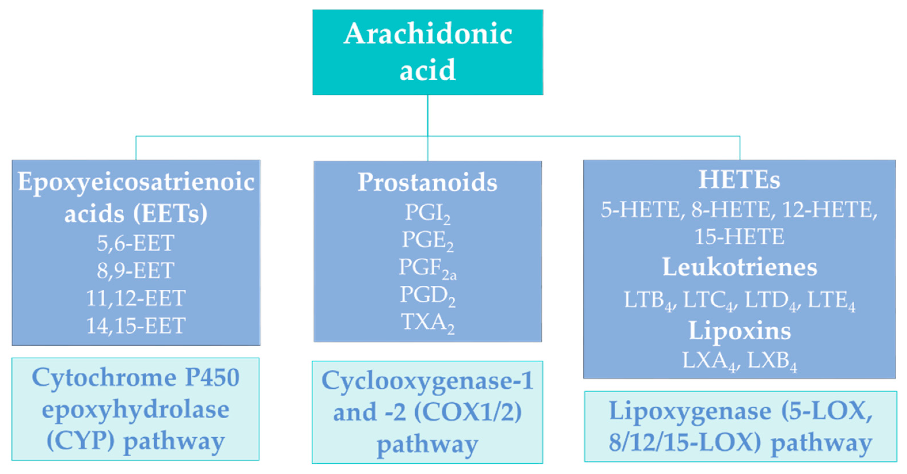
1. Introduction
2. Derivatization of Carboxyl Group—Classes of Derivatizing Agents
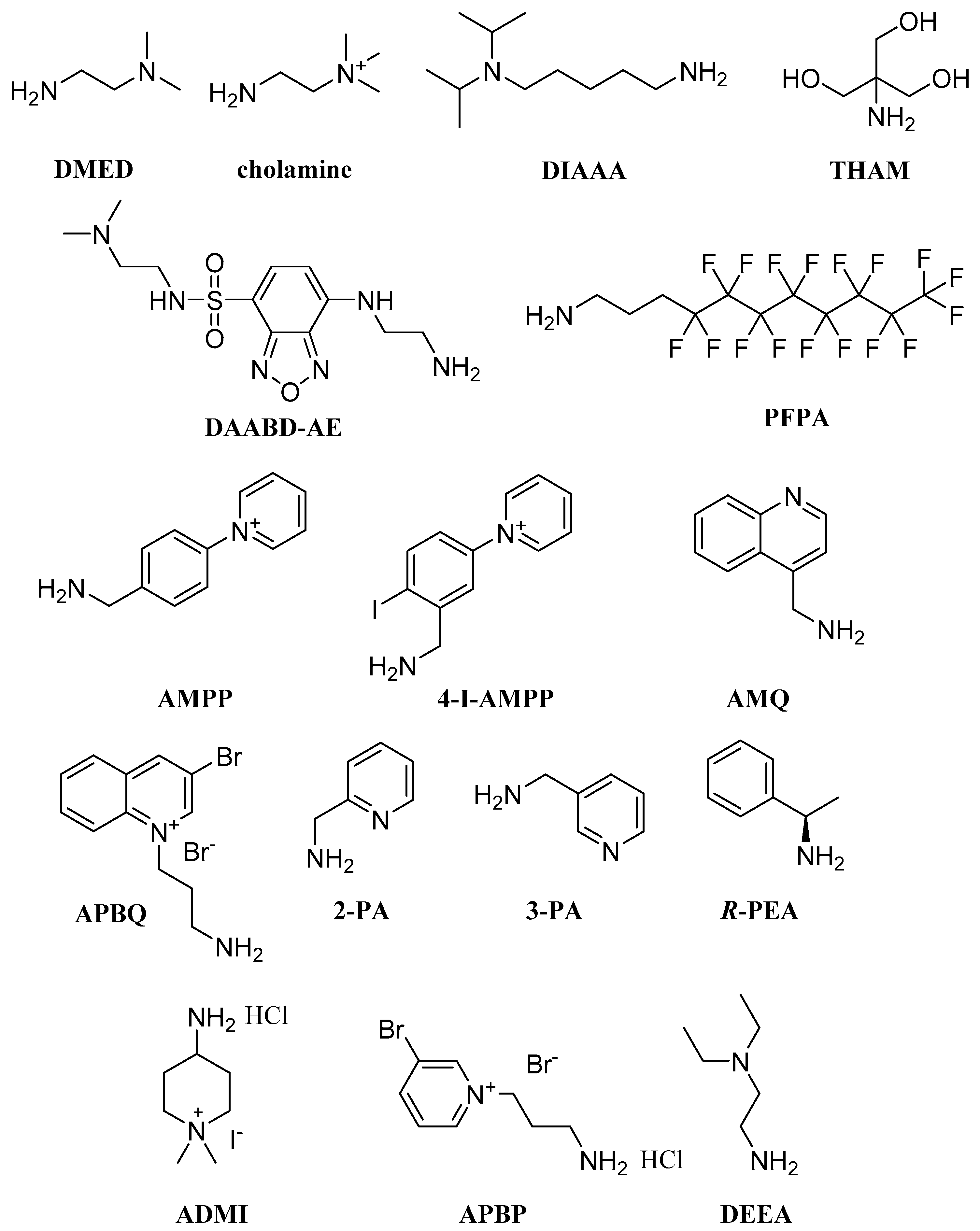
2.1. Primary Amines
2.2. Secondary Amines
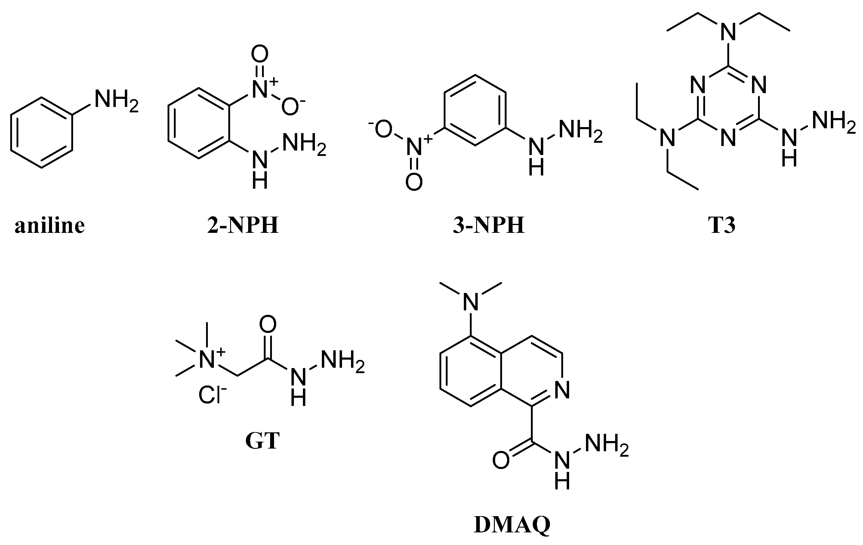
2.3. Aromatic Amines
2.4. Hydrazines and Hydrazides
2.5. Bromides
2.6. Hydroxylamines
2.7. Other Derivatization Reagents
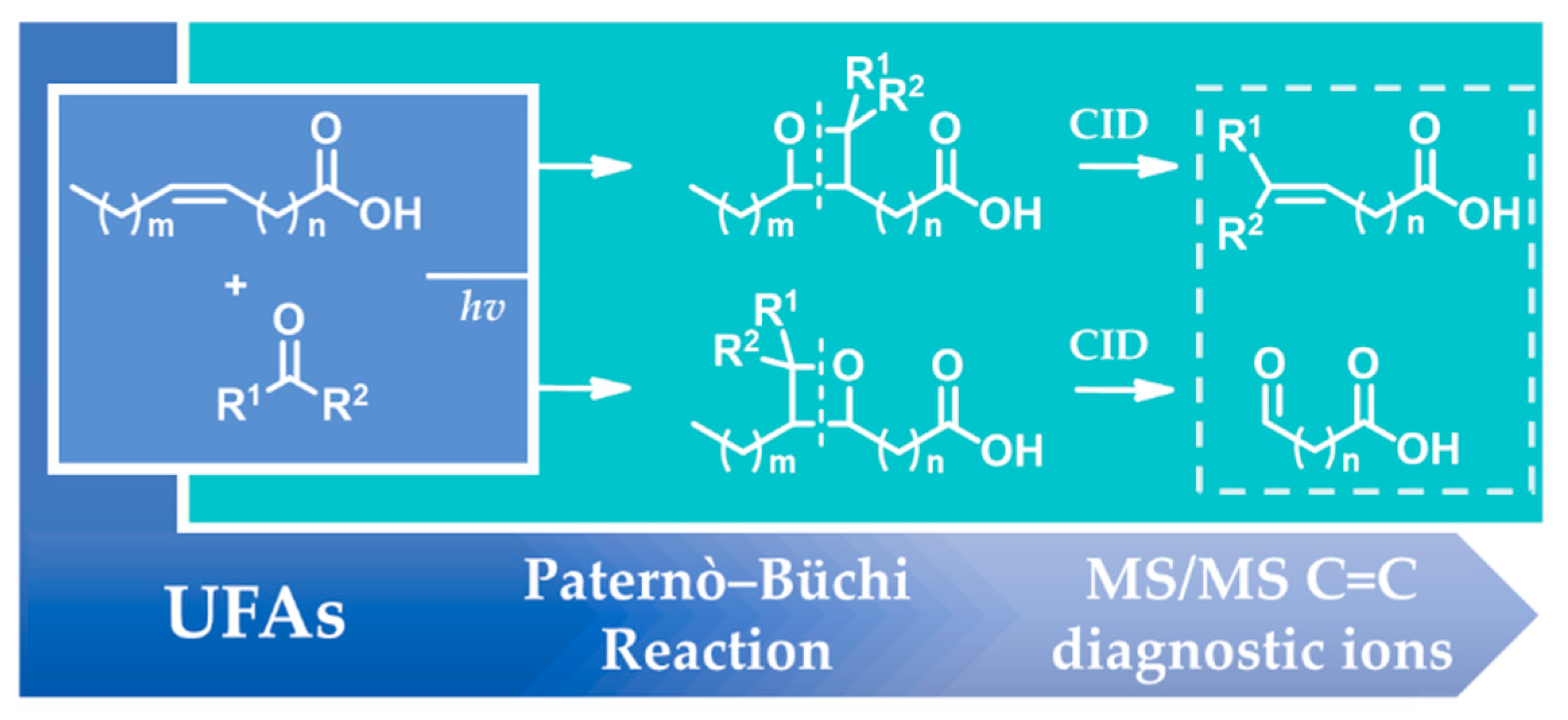
3. Derivatization of Unsaturated Fatty Acids (UFAs)
4. Conclusions
Author Contributions
Funding
Institutional Review Board Statement
Informed Consent Statement
Data Availability Statement
Conflicts of Interest
References
- Calder, P.C.; Burdge, G.C. Fatty Acids in Bioactive Lipids; The Oily Press: Bridgewater, UK, 2004; pp. 1–36. [Google Scholar]
- Georgiadi, A.; Kersten, S. Mechanisms of gene regulation by fatty acids. Adv. Nutr. 2012, 3, 127–134. [Google Scholar] [CrossRef] [PubMed]
- Michalik, L.; Auwerx, J.; Berger, J.P.; Chatterjee, V.K.; Glass, C.K.; Gonzalez, F.J.; Grimaldi, P.A.; Kadowaki, T.; Lazar, M.A.; O’Rahilly, S.; et al. International Union of Pharmacology. LXI. Peroxisome proliferator-activated receptors. Pharmacol. Rev. 2006, 58, 726–741. [Google Scholar] [CrossRef] [PubMed]
- Kimura, I.; Ichimura, A.; Ohue-Kitano, R.; Igarashi, M. Free fatty acid receptors in health and disease. Physiol. Rev. 2020, 100, 171–210. [Google Scholar] [CrossRef]
- Li, Z.; Xu, X.; Huang, W.; Qian, H. Free fatty acid receptor 1 (FFAR1) as an emerging therapeutic target for type 2 diabetes mellitus: Recent progress and prevailing challenges. Med. Res. Rev. 2018, 38, 381–425. [Google Scholar] [CrossRef]
- Ulven, T.; Christiansen, E. Dietary fatty acids and their potential for controlling metabolic diseases through activation of FFA4/GPR120. Annu. Rev. Nutr. 2015, 35, 239–263. [Google Scholar] [CrossRef] [PubMed]
- Bolognini, D.; Dedeo, D.; Milligan, G. Metabolic and inflammatory functions of short-chain fatty acid receptors. Curr. Opin. Endocr. Metab. Res. 2021, 16, 1–9. [Google Scholar] [CrossRef]
- Dąbrowski, G.; Konopka, I. Update on food sources and biological activity of odd-chain, branched and cyclic fatty acids—A review. Trends Food Sci. Technol. 2022, 119, 514–529. [Google Scholar] [CrossRef]
- Christie, W.W.; Harwood, J.L. Oxidation of polyunsaturated fatty acids to produce lipid mediators. Essays Biochem. 2020, 64, 401–421. [Google Scholar] [CrossRef]
- Kokotou, M.G.; Kokotos, A.C.; Gkikas, D.; Mountanea, O.G.; Mantzourani, C.; Almutairi, A.; Lei, X.; Ramanadham, S.; Politis, P.K.; Kokotos, G. Saturated hydroxy fatty acids exhibit a cell growth inhibitory activity and suppress the cytokine-induced β-cell apoptosis. J. Med. Chem. 2020, 63, 12666–12681. [Google Scholar] [CrossRef]
- Kokotou, M.G.; Mantzourani, C.; Bourboula, A.; Mountanea, O.G.; Kokotos, G. A Liquid chromatography-high resolution mass spectrometry (LC-HRMS) method for the determination of free hydroxy fatty acids in cow and goat milk. Molecules 2020, 25, 3947. [Google Scholar] [CrossRef]
- Liu, J.; Sahin, C.; Ahmad, S.; Magomedova, L.; Zhang, M.; Jia, Z.; Metherel, A.H.; Orellana, A.; Poda, G.; Bazinet, R.P.; et al. The omega-3 hydroxy fatty acid 7(S)-HDHA is a high-affinity PPARα ligand that regulates brain neuronal morphology. Sci. Signal. 2022, 15, eabo1857. [Google Scholar] [CrossRef] [PubMed]
- Batsika, C.S.; Mantzourani, C.; Gkikas, D.; Kokotou, M.G.; Mountanea, O.G.; Kokotos, C.G.; Politis, P.K.; Kokotos, G. Saturated oxo fatty acids (SOFAs): A previously unrecognized class of endogenous bioactive lipids exhibiting a cell growth inhibitory activity. J. Med. Chem. 2021, 64, 5654–5666. [Google Scholar] [CrossRef] [PubMed]
- Kokotou, M.G.; Batsika, C.S.; Mantzourani, C.; Kokotos, G. Free saturated oxo fatty acids (SOFAs) and ricinoleic acid in milk determined by a liquid chromatography-high-resolution mass spectrometry (LC-HRMS) method. Metabolites 2021, 11, 46. [Google Scholar] [CrossRef] [PubMed]
- Avato, P.; Tava, A. Rare fatty acids and lipids in plant oilseeds: Occurrence and bioactivity. Phytochem. Rev. 2022, 21, 401–428. [Google Scholar] [CrossRef]
- Imig, J.D.; Jankiewicz, W.K.; Khan, A.H. Epoxy fatty acids: From salt regulation to kidney and cardiovascular therapeutics. Hypertension 2020, 76, 3–15. [Google Scholar] [CrossRef]
- Koutoulogenis, G.S.; Kokotos, G. Nitro fatty acids (NO2-FAs): An emerging class of bioactive fatty acids. Molecules 2021, 26, 7536. [Google Scholar] [CrossRef]
- Neves, B.; Pérez-Sala, D.; Ferreira, H.B.; Guerra, I.M.S.; Moreira, A.S.P.; Domingues, P.; Domingues, M.R.; Melo, T. Understanding the nitrolipidome: From chemistry to mass spectrometry and biological significance of modified complex lipids. Prog. Lipid Res. 2022, 87, 101176. [Google Scholar] [CrossRef]
- Dennis, E.A.; Cao, J.; Hsu, Y.-H.; Magrioti, V.; Kokotos, G. Phospholipase A2 enzymes: Physical structure, biological function, disease implication, chemical inhibition, and therapeutic intervention. Chem. Rev. 2011, 111, 6130–6185. [Google Scholar] [CrossRef]
- Funk, C.D. Prostaglandins and leukotrienes: Advances in eicosanoid biology. Science 2001, 294, 1871–1875. [Google Scholar] [CrossRef]
- Dennis, E.A.; Norris, P.C. Eicosanoid storm in infection and inflammation. Nat. Rev. Immunol. 2015, 15, 511–523. [Google Scholar] [CrossRef]
- Yore, M.M.; Syed, I.; Moraes-Vieira, P.M.; Zhang, T.; Herman, M.A.; Homan, E.A.; Patel, R.T.; Lee, J.; Chen, S.; Peroni, O.D.; et al. Discovery of a class of endogenous mammalian lipids with anti-diabetic and anti-inflammatory effects. Cell 2014, 159, 318–332. [Google Scholar] [CrossRef] [PubMed]
- Kokotou, M.G. Analytical methods for the determination of fatty acid esters of hydroxy fatty acids (FAHFAs) in biological samples, plants and foods. Biomolecules 2020, 10, 1092. [Google Scholar] [CrossRef] [PubMed]
- Riecan, M.; Paluchova, V.; Lopes, M.; Brejchova, K.; Kuda, O. Branched and linear fatty acid esters of hydroxy fatty acids (FAHFA) relevant to human health. Pharmacol. Ther. 2022, 231, 107972. [Google Scholar] [CrossRef]
- Christie, W.W. Gas chromatography–mass spectrometry methods for structural analysis of fatty acids. Lipids 1998, 33, 343–353. [Google Scholar] [CrossRef]
- Ichihara, K.; Kohsaka, C.; Tomari, N.; Kiyono, T.; Wada, J.; Hirooka, K.; Yamamoto, Y. Fatty acid analysis of triacylglycerols: Preparation of fatty acid methyl esters for gas chromatography. Anal. Biochem. 2016, 495, 6e8. [Google Scholar] [CrossRef]
- Wang, Z.; Wang, D.H.; Park, H.G.; Tobias, H.J.; Kothapalli, K.S.D.; Brenna, J.T. Structural identification of monounsaturated branched chain fatty acid methyl esters by combination of electron ionization and covalent adduct chemical ionization tandem mass spectrometry. Anal. Chem. 2019, 91, 15147–15154. [Google Scholar] [CrossRef] [PubMed]
- Quehenberger, O.; Armando, A.M.; Dennis, E.A. High sensitivity quantitative lipidomics analysis of fatty acids in biological samples by gas chromatography-mass spectrometry. Biochim. Biophys. Acta 2011, 1811, 648–656. [Google Scholar] [CrossRef] [PubMed]
- Quehenberger, O.; Armando, A.M.; Brown, A.H.; Milne, S.B.; Myers, D.S.; Merrill, A.H.; Bandyopadhyay, S.; Jones, K.N.; Kelly, S.; Shaner, R.L.; et al. Lipidomics reveals a remarkable diversity of lipids in human plasma. J. Lipid Res. 2010, 51, 3299–3305. [Google Scholar] [CrossRef]
- Hellmuth, C.; Weber, M.; Koletzko, B.; Peissner, W. Nonesterified fatty acid determination for functional lipidomics: Comprehensive ultrahigh performance liquid chromatography−tandem mass spectrometry quantitation, qualification, and parameter prediction. Anal. Chem. 2012, 84, 1483–1490. [Google Scholar] [CrossRef]
- Christinat, N.; Morin-Rivron, D.; Masoodi, M. High-Throughput quantitative lipidomics analysis of nonesterified fatty acids in plasma by LC-MS. Methods Mol. Biol. 2017, 1619, 183–191. [Google Scholar]
- Kokotou, M.G.; Mantzourani, C.; Kokotos, G. Development of a liquid chromatography-high resolution mass spectrometry method for the determination of free fatty acids in milk. Molecules 2020, 25, 1548. [Google Scholar] [CrossRef] [PubMed]
- Kokotou, M.G.; Mantzourani, C.; Batsika, C.S.; Mountanea, O.G.; Eleftheriadou, I.; Kosta, O.; Tentolouris, N.; Kokotos, G. Lipidomics Analysis of free fatty acids in human plasma of healthy and diabetic subjects by liquid chromatography-high resolution mass spectrometry (LC-HRMS). Biomedicines 2022, 10, 1189. [Google Scholar] [CrossRef] [PubMed]
- Zhu, Y.; Deng, P.; Zhong, D. Derivatization methods for LC–MS analysis of endogenous compounds. Bioanalysis 2015, 7, 2557–2581. [Google Scholar] [CrossRef] [PubMed]
- Xiang, L.; Zhu, L.; Huang, Y.; Cai, Z. Application of derivatization in fatty acids and fatty acyls detection: Mass spectrometry-based targeted lipidomics. Small Methods 2020, 4, 2000160. [Google Scholar] [CrossRef]
- Zhu, Q.-F.; Hao, Y.-H.; Liu, M.-Z.; Yue, J.; Ni, J.; Yuan, B.-F.; Feng, Y.-Q. Analysis of cytochrome P450 metabolites of arachidonic acid by stable isotope probe labeling coupled with ultra high-performance liquid chromatography/mass spectrometry. J. Chromatogr. A 2015, 1410, 154–163. [Google Scholar] [CrossRef]
- Zhu, Q.-F.; Yan, J.-W.; Gao, Y.; Zhang, J.-W.; Yuan, B.-F.; Feng, Y.-Q. Highly sensitive determination of fatty acid esters of hydroxyl fatty acids by liquid chromatography-mass spectrometry. J. Chromatogr. B 2017, 1061–1062, 34–40. [Google Scholar] [CrossRef]
- Ding, J.; Kind, T.; Zhu, Q.-F.; Wang, Y.; Yan, J.-W.; Fiehn, O.; Feng, Y.-Q. In-silico-generated library for sensitive detection of 2-dimethylaminoethylamine derivatized FAHFA lipids using high-resolution tandem mass spectrometry. Anal. Chem. 2020, 92, 5960–5968. [Google Scholar] [CrossRef]
- An, N.; Zhu, Q.-F.; Wang, Y.-Z.; Xiong, C.-F.; Hu, Y.-N.; Feng, Y.-Q. Integration of chemical derivatization and in-source fragmentation mass spectrometry for high-coverage profiling of submetabolomes. Anal. Chem. 2021, 93, 11321–11328. [Google Scholar] [CrossRef]
- Gowda, S.G.B.; Gowda, D.; Liang, C.; Li, Y.; Kawakami, K.; Fukiya, S.; Yokota, A.; Chiba, H.; Hui, S.-P. Chemical labeling assisted detection and identification of short chain fatty acid esters of hydroxy fatty acid in rat colon and cecum contents. Metabolites 2020, 10, 398. [Google Scholar] [CrossRef]
- Gowda, D.; Li, Y.; Gowda, S.G.B.; Ohno, M.; Chiba, H.; Hui, S.-P. Determination of short-chain fatty acids by N,N-dimethylethylenediamine derivatization combined with liquid chromatography/mass spectrometry and their implication in influenza virus infection. Anal. Bioanal. Chem. 2022, 414, 6419–6430. [Google Scholar] [CrossRef]
- Bian, X.; Sun, B.; Zheng, P.; Li, N.; Wu, J.-L. Derivatization enhanced separation and sensitivity of long chain-free fatty acids: Application to asthma using targeted and non-targeted liquid chromatography-mass spectrometry approach. Anal. Chim. Acta 2017, 989, 59–70. [Google Scholar] [CrossRef] [PubMed]
- Bian, X.; Li, N.; Tan, B.; Sun, B.; Guo, M.-Q.; Huang, G.; Fu, L.; Hsiao, W.L.W.; Liu, L.; Wu, J.-L. Polarity-tuning derivatization-LC-MS approach for probing global carboxyl-containing metabolites in colorectal cancer. Anal. Chem. 2018, 90, 11210–11215. [Google Scholar] [CrossRef] [PubMed]
- Yang, R.; Xia, F.; Su, H.; Wan, J.-B. Quantitative analysis of n-3 polyunsaturated fatty acids and their metabolites by chemical isotope labeling coupled with liquid chromatography—Mass spectrometry. J. Chromatogr. B 2021, 1172, 122666. [Google Scholar] [CrossRef] [PubMed]
- Williams, J.; Pandarinathan, L.; Wood, J.A.; Vouros, P.; Makriyannis, A. Endocannabinoid metabolomics: A novel liquid chromatography-mass spectrometry reagent for fatty acid analysis. AAPS J. 2006, 8, E655–E660. [Google Scholar] [CrossRef] [PubMed]
- Tsukamoto, Y.; Santa, T.; Saimaru, H.; Imai, K.; Funatsu, T. Synthesis of benzofurazan derivatization reagents for carboxylic acids and its application to analysis of fatty acids in rat plasma by high-performance liquid chromatography-electrospray ionization mass spectrometry. Biomed. Chromatogr. 2005, 19, 802–808. [Google Scholar] [CrossRef]
- Zheng, J.-Y.; Jin, Y.-Y.; Shi, Z.-Q.; Zhou, J.-L.; Liu, L.-F.; Xin, G.-Z. Fluorous-paired derivatization approach towards highly sensitive and accurate determination of long chain unsaturated fatty acids by liquid chromatography-tandem mass spectrometry. Anal. Chim. Acta 2020, 1136, 187–195. [Google Scholar] [CrossRef]
- Bollinger, J.G.; Thompson, W.; Lai, Y.; Oslund, R.C.; Hallstrand, T.S.; Sadilek, M.; Turecek, F.; Gelb, M.H. Improved sensitivity mass spectrometric detection of eicosanoids by charge reversal derivatization. Anal. Chem. 2010, 82, 6790–6796. [Google Scholar] [CrossRef]
- Bollinger, J.G.; Rohan, G.; Sadilek, M.; Gelb, M.H. LC/ESI-MS/MS Detection of FAs by charge reversal derivatization with more than four orders of magnitude improvement in sensitivity. J. Lipid Res. 2013, 54, 3523–3530. [Google Scholar] [CrossRef]
- Liu, X.; Moon, S.H.; Mancuso, D.J.; Jenkins, C.M.; Guan, S.; Sims, H.F.; Gross, R.W. Oxidized fatty acid analysis by charge-switch derivatization, selected reaction monitoring, and accurate mass quantitation. Anal. Biochem. 2013, 442, 40–50. [Google Scholar] [CrossRef][Green Version]
- Narreddula, V.R.; Boase, N.R.; Ailuri, R.; Marshall, D.L.; Poad, B.L.J.; Kelso, M.J.; Trevitt, A.J.; Mitchell, T.W.; Blanksby, S.J. Introduction of a fixed-charge, photolabile derivative for enhanced structural elucidation of fatty acids. Anal. Chem. 2019, 91, 9901–9909. [Google Scholar] [CrossRef]
- Fu, H.; Zhang, Q.; Huang, X.; Ma, Z.; Zheng, X.; Li, S.; Duan, H.; Sun, X.; Lin, F.; Zhao, L.; et al. A rapid and convenient derivatization method for quantitation of short-chain fatty acids in human feces by ultra-performance liquid chromatography/tandem mass spectrometry. Rapid Commun. Mass Spectrom. 2020, 34, e8730. [Google Scholar] [CrossRef] [PubMed]
- Mochizuki, Y.; Inagaki, S.; Suzuki, M.; Min, J.Z.; Inoue, K.; Todoroki, K.; Toyo’oka, T. A novel derivatization reagent possessing a bromoquinolinium structure for biological carboxylic acids in HPLC-ESI-MS/MS: Liquid Chromatography. J. Sep. Sci. 2013, 36, 1883–1889. [Google Scholar] [CrossRef] [PubMed]
- Li, X.; Franke, A.A. Improved LC−MS method for the determination of fatty acids in red blood cells by LC−orbitrap MS. Anal. Chem. 2011, 83, 3192–3198. [Google Scholar] [CrossRef]
- Nagatomo, R.; Okada, Y.; Ichimura, M.; Tsuneyama, K.; Inoue, K. Application of 2-picolylamine derivatized ultra-high performance liquid chromatography tandem mass spectrometry for the determination of short-chain fatty acids in feces samples. Anal. Sci. 2018, 34, 1031–1036. [Google Scholar] [CrossRef] [PubMed]
- Wu, Q.; Xu, Y.; Ji, H.; Wang, Y.; Zhang, Z.; Lu, H. Enhancing coverage in LC–MS-based untargeted metabolomics by a new sample preparation procedure using mixed-mode solid-phase extraction and two derivatizations. Anal. Bioanal. Chem. 2019, 411, 6189–6202. [Google Scholar] [PubMed]
- Algarra, M.; Rodríguez-Borges, J.E.; Esteves da Silva, J.C.G. LC-MS Identification of derivatized free fatty acids from adipocere in soil samples: Liquid chromatography. J. Sep. Sci. 2010, 33, 143–154. [Google Scholar] [CrossRef]
- Zhu, Z.; Li, X.; Tang, C.; Shen, J.; Liu, J.; Ye, Y. A Derivatization strategy for comprehensive identification of 2- and 3-hydroxyl fatty acids by LC-MS. Anal. Chim. Acta 2022, 1216, 339981. [Google Scholar] [CrossRef] [PubMed]
- Barry, S.J.; Carr, R.M.; Lane, S.J.; Leavens, W.J.; Monté, S.; Waterhouse, I. Derivatisation for liquid chromatography/electrospray mass spectrometry: Synthesis of pyridinium compounds and their amine and carboxylic acid derivatives: Derivatisation for LC/ESI-MS. Rapid Commun. Mass Spectrom. 2003, 17, 603a–620. [Google Scholar]
- Zhang, J.; Yang, S.; Wang, J.; Xu, Y.; Zhao, H.; Lei, J.; Zhou, Y.; Chen, Y.; Wu, L.; Zhou, M.; et al. Integrated LC-MS metabolomics with dual derivatization for quantification of FFAs in fecal samples of hepatocellular carcinoma patients. J. Lipid Res. 2021, 62, 100143. [Google Scholar] [CrossRef]
- Leng, J.; Wang, H.; Zhang, L.; Zhang, J.; Wang, H.; Guo, Y. A highly sensitive isotope-coded derivatization method and its application for the mass spectrometric analysis of analytes containing the carboxyl group. Anal. Chim. Acta 2013, 758, 114–121. [Google Scholar] [CrossRef]
- Leng, J.; Guan, Q.; Sun, T.; Wu, Y.; Cao, Y.; Guo, Y. Application of isotope-based carboxy group derivatization in LC–MS/MS analysis of tissue free-fatty acids for thyroid carcinoma. J. Pharm. Biomed. Anal. 2013, 84, 256–262. [Google Scholar] [CrossRef] [PubMed]
- Jiang, R.; Jiao, Y.; Zhang, P.; Liu, Y.; Wang, X.; Huang, Y.; Zhang, Z.; Xu, F. Twin Derivatization strategy for high-coverage quantification of free fatty acids by liquid chromatography–tandem mass spectrometry. Anal. Chem. 2017, 89, 12223–12230. [Google Scholar] [CrossRef] [PubMed]
- Xia, F.; He, C.; Ren, M.; Xu, F.-G.; Wan, J.-B. Quantitative profiling of eicosanoids derived from n-6 and n-3 polyunsaturated fatty acids by twin derivatization strategy combined with LC-MS/MS in patients with type 2 diabetes mellitus. Anal. Chim. Acta 2020, 1120, 24–35. [Google Scholar] [CrossRef] [PubMed]
- Wei, J.; Xiang, L.; Li, X.; Song, Y.; Yang, C.; Ji, F.; Chung, A.C.K.; Li, K.; Lin, Z.; Cai, Z. Derivatization strategy combined with parallel reaction monitoring for the characterization of short-chain fatty acids and their hydroxylated derivatives in mouse. Anal. Chim. Acta 2020, 1100, 66–74. [Google Scholar] [CrossRef] [PubMed]
- Chan, J.C.Y.; Kioh, D.Y.Q.; Yap, G.C.; Lee, B.W.; Chan, E.C.Y. A novel LCMSMS method for quantitative measurement of short-chain fatty acids in human stool derivatized with 12C- and 13C-labelled aniline. J Pharm. Biomed. Anal. 2017, 138, 43–53. [Google Scholar] [CrossRef] [PubMed]
- Han, J.; Lin, K.; Sequeira, C.; Borchers, C.H. An isotope-labeled chemical derivatization method for the quantitation of short-chain fatty acids in human feces by liquid chromatography–tandem mass spectrometry. Anal. Chim. Acta 2015, 854, 86–94. [Google Scholar] [CrossRef]
- Dei Cas, M.; Paroni, R.; Saccardo, A.; Casagni, E.; Arnoldi, S.; Gambaro, V.; Saresella, M.; Mario, C.; La Rosa, F.; Marventano, I.; et al. A straightforward LC-MS/MS analysis to study serum profile of short and medium chain fatty acids. J. Chromatogr. B 2020, 1154, 121982. [Google Scholar] [CrossRef]
- Chen, Z.; Wu, Y.; Shrestha, R.; Gao, Z.; Zhao, Y.; Miura, Y.; Tamakoshi, A.; Chiba, H.; Hui, S.-P. Determination of total, free and esterified short-chain fatty acid in human serum by liquid chromatography-mass spectrometry. Ann. Clin. Biochem. 2019, 56, 190–197. [Google Scholar] [CrossRef]
- Chen, Z.; Gao, Z.; Wu, Y.; Shrestha, R.; Imai, H.; Uemura, N.; Hirano, K.; Chiba, H.; Hui, S.-P. Development of a simultaneous quantitation for short-, medium-, long-, and very long-chain fatty acids in human plasma by 2-nitrophenylhydrazine-derivatization and liquid chromatography–tandem mass spectrometry. J. Chromatogr. B 2019, 1126–1127, 121771. [Google Scholar] [CrossRef]
- Hu, T.; Tie, C.; Wang, Z.; Zhang, J.-L. Highly sensitive and specific derivatization strategy to profile and quantitate eicosanoids by UPLC-MS/MS. Anal. Chim. Acta 2017, 950, 108–118. [Google Scholar] [CrossRef]
- Song, W.-S.; Park, H.-G.; Kim, S.-M.; Jo, S.-H.; Kim, B.-G.; Theberge, A.B.; Kim, Y.-G. Chemical derivatization-based LC–MS/MS method for quantitation of gut microbial short-chain fatty acids. J. Ind. Eng. Chem. 2020, 83, 297–302. [Google Scholar] [CrossRef]
- Feng, X.; Liu, N.; Yang, Y.; Feng, S.; Wang, J.; Meng, Q. Isotope-coded chemical derivatization method for highly accurately and sensitively quantifying short-chain fatty acids. J. Agric. Food Chem. 2022, 70, 6253–6263. [Google Scholar] [CrossRef] [PubMed]
- Uji, Y.; Noma, A.; Shiraki, M.; Maeda, M.; Tsuji, A.; Okabc, H. Separation and quantitation of plasma free fatty acids as phenacyl esters by HPLC. Biomed. Chromatogr. 1987, 2, 110–114. [Google Scholar] [CrossRef] [PubMed]
- L’emeillat, Y.; Ménez, J.F.; Berthou, F.; Bardou, L. Quantitative gas chromatographic determination of low-molecular-weight straight-chain carboxylic acids as their p-bromophenacyl esters after extractive alkylation in acidic Medium. J. Chromatogr. A 1981, 206, 89–100. [Google Scholar] [CrossRef]
- Ma, S.-R.; Tong, Q.; Zhao, Z.-X.; Cong, L.; Yu, J.-B.; Fu, J.; Han, P.; Pan, L.-B.; Gu, R.; Peng, R.; et al. Determination of berberine-upregulated endogenous short-chain fatty acids through derivatization by 2-bromoacetophenone. Anal. Bioanal. Chem. 2019, 411, 3191–3207. [Google Scholar] [CrossRef]
- Willacey, C.C.W.; Karu, N.; Harms, A.C.; Hankemeier, T. Metabolic profiling of material-limited cell samples by dimethylaminophenacyl bromide derivatization with UPLC-MS/MS analysis. Microchem. J. 2020, 159, 105445. [Google Scholar] [CrossRef]
- Zeng, M.; Cao, H. Fast Quantification of short chain fatty acids and ketone bodies by liquid chromatography-tandem mass spectrometry after facile derivatization coupled with liquid-liquid extraction. J. Chromatogr. B 2018, 1083, 137–145. [Google Scholar] [CrossRef] [PubMed]
- Jaochico, A.; Sangaraju, D.; Shahidi-Latham, S.K. A rapid derivatization based LC–MS/MS method for quantitation of short chain fatty acids in human plasma and urine. Bioanalysis 2019, 11, 741–753. [Google Scholar] [CrossRef]
- Song, H.E.; Lee, H.Y.; Kim, S.J.; Back, S.H.; Yoo, H.J. A facile profiling method of short chain fatty acids using liquid chromatography-mass spectrometry. Metabolites 2019, 9, 173. [Google Scholar] [CrossRef]
- Pettinella, C.; Lee, S.H.; Cipollone, F.; Blair, I.A. Targeted quantitative analysis of fatty acids in atherosclerotic plaques by high sensitivity liquid chromatography/tandem mass spectrometry. J. Chromatogr. B 2007, 850, 168–176. [Google Scholar] [CrossRef]
- Ma, X.; Xia, Y. Pinpointing double bonds in lipids by Paternò-Büchi reactions and mass spectrometry. Angew. Chem. Int. Ed. 2014, 53, 2592–2596. [Google Scholar] [CrossRef] [PubMed]
- Ma, X.; Chong, L.; Tian, R.; Shi, R.; Hu, T.Y.; Ouyang, Z.; Xia, Y. Identification and quantitation of lipid C=C location isomers: A shotgun lipidomics approach enabled by photochemical reaction. Proc. Natl. Acad. Sci. USA 2016, 113, 2573–2578. [Google Scholar] [CrossRef] [PubMed]
- Murphy, R.C.; Okuno, T.; Johnson, C.A.; Barkley, R.M. Determination of double bond positions in polyunsaturated fatty acids using the photochemical Paternò-Büchi reaction with acetone and tandem mass spectrometry. Anal. Chem. 2017, 89, 8545–8553. [Google Scholar] [CrossRef] [PubMed]
- Esch, P.; Heiles, S. Charging and charge switching of unsaturated lipids and apolar compounds using Paternò-Büchi reactions. J. Am. Soc. Mass Spectrom. 2018, 29, 1971–1980. [Google Scholar] [CrossRef] [PubMed]
- Esch, P.; Heiles, S. Investigating C=C positions and hydroxylation sites in lipids using Paternò–Büchi functionalization mass spectrometry. Analyst 2020, 145, 2256–2266. [Google Scholar] [CrossRef]
- Zhao, J.; Xie, X.; Lin, Q.; Ma, X.; Su, P.; Xia, Y. Next-generation Paternò–Büchi reagents for lipid analysis by mass spectrometry. Anal. Chem. 2020, 92, 13470–13477. [Google Scholar] [CrossRef]
- Zhao, J.; Fang, M.; Xia, Y. A liquid chromatography-mass spectrometry workflow for in-depth quantitation of fatty acid double bond location isomers. J. Lipid Res. 2021, 62, 100110. [Google Scholar] [CrossRef]
- Wang, D.H.; Park, H.G.; Wang, Z.; Lacombe, R.J.S.; Shmanai, V.V.; Bekish, A.V.; Schmidt, K.; Shchepinov, M.S.; Brenna, J.T. Toward quantitative sequencing of deuteration of unsaturated hydrocarbon chains in fatty acids. Anal. Chem. 2021, 93, 8238–8247. [Google Scholar] [CrossRef]
- Mao, R.; Li, W.; Jia, P.; Ding, H.; Teka, T.; Zhang, L.; Fu, Z.; Fu, X.; Kaushal, S.; Dou, Z.; et al. An efficient and sensitive method on the identification of unsaturated fatty acids in biosamples: Total lipid extract from bovine liver as a case study. J. Chromatogr. A 2022, 1675, 463176. [Google Scholar] [CrossRef]
- Han, Y.; Chen, P.; Li, Z.; Wang, X.; Sun, C. Multi-wavelength visible-light induced [2+2] cycloaddition for identification of lipid isomers in biological samples. J. Chromatogr. A 2022, 1662, 462742. [Google Scholar] [CrossRef]


| Derivatization Reagent | Analytical Technique | Instrumental Analysis | Column/Mobile Phase | Sample Preparation— Solvent Extraction/ Cartridge-Column | Sample | Analyte | Ref. |
|---|---|---|---|---|---|---|---|
| DMED | UPLC–ESI–MS/MS (+) ESI mode | ABI/SCIEX 4500 Triple QuadTM coupled to Shimadzu LC-30AD UPLC. | Acquity UPLC BEH phenyl column (2.1 mm × 50 mm, 1.7 μm, Waters) / (Solvent A) HCOOH in H2O (0.1%, v/v) and (Solvent B) ACN/MeOH (7/3, v/v); flow rate 0.4 mL/min; temperature 40 °C. | Extraction with EtOAc containing 10 μL of BHT (0.10 mM) and 10 μL of 0.5% HCOOH | Serum | Cytochrome P450 metabolites of arachidonic acid | [36] |
| DMED | UHPLC-ESI–MS/MS (+) ESI mode | Shimadzu MS-8040 mass spectrometer (Tokyo, Japan) with an electrospray ionization source (Turbo Ionspray) coupled to a Shimadzu LC-30AD UPLC system (Tokyo, Japan) | Acquity UPLC BEH C18 column (2.1 mm × 50 mm, 1.7 μm, Waters). The mobile phase consisted of (A) HCOOH in ACN/H2O (0.1%, 6/4, v/v) and (B) HCOOH in IPA/ACN (0.1%, 9/1, v/v); flow rate 0.4 mL/min; temperature 40 °C | Extraction with cold saline solution and ACN containing 0.1% NH3/ SAX SPE-cartridge (1 mL, 50 mg) Weltech Co. (Wuhan, China) | Rat tissue Human serum | FAHFAs | [37] |
| DMED | UHPLC/LTQ-Orbitrap MS (+) ESI mode | LTQ-Orbitrap-MS coupled to Shimadzu UHPLC. | Atlantic T3 C18 reverse-phase column (2.1 mm × 150 mm, 3 µm, Waters, Milford, MA, USA) / (Solvent A) 10 mM of aqueous CH3COONH4 with 0.1% CH3COOH, (Solvent B) IPA, (Solvent C) MeOH; flow rate 0.2 mL/min; temperature 40 °C. | Folch method | Colon contents | Short chain FAHFAs | [40] |
| DMED | LC-MS/MS (+) ESI mode | Prominence UFLC (Shimadzu, Kyoto, Japan) coupled to TSQ Quantum Mass Spectrometer System (Thermo Fisher Scientific, San Jose, CA, USA) | Hypersil gold C8 column (50 mm × 2.1 mm, 5 μm; Thermo Fisher Scientific) / Mobile phase A: 20 mM CH3COONH4, B: MeOH and ACN (1:1); flow rate 0.4 mL/min; temperature 40 °C. | Extraction with ACN | Intestinal contents | SCFAs and hydroxy SCFAs | [41] |
| Cholamine | UHPLC-Q-TOF/MS (+) ESI mode | Agilent 6550 UHD accurate-mass Q-TOF/MS system coupled to Agilent 1290 Infinity LC system (UHPLC, Santa Clara, CA, USA). | Agilent Eclipse XDB-C18 column (2.1 mm × 100 mm, 1.8 μm) / Mobile phase A and B were 0.1% HCOOH-containing H2O and 0.1% HCOOH-containing ACN; temperature 40 °C. | Extraction with EtOAc | Serum | Long chain FFAs | [42] |
| Cholamine | LC-MS/MS (+) ESI mode | Xevo TQD triple-quadrupole tandem mass spectrometry coupled to an ACQUITY UPLC system (UPLC-QQQ-MS/MS, Waters Corp., Manchester, UK). | ACQUITY BEH C18 column (150 mm × 2.1 mm i.d., 1.7 μm) / ACN containing 0.1% HCOOH (A, v/v) and 0.1% aqueous HCOOH solution (B, v/v); flow rate 0.3 mL/min; temperature 45 °C. | Extraction with cold EtOAc containing 0.5% HCOOH | Serum | n-3 PUFAs and their metabolites | [44] |
| DIAAA | UHPLC-Q-TOF/MS (+) ESI mode | Agilent 6550 UHD accurate-mass Q-TOF/MS coupled to an Agilent 1290 Infinity LC system (UHPLC, Santa Clara, CA, USA) | Waters ACQUITY UPLC HSS T3 column (2.1 mm × 100 mm, 1.8 μm) / Mobile phase A and B were 0.1% HCOOH containing H2O and 0.1% HCOOH containing ACN; flow rate 0.3 mL/min; temperature 40 °C. | Extraction with cold MeOH | Serum | SCFAs and LCFAs | [43] |
| PFPA | UPLC-MS/MS (+) ESI mode | 5500 QTRAP mass spectrometer (AB Sciex, Foster City, CA, USA) coupled to a Shimadzu LC-30AD UHPLC system (Tokyo, Japan). | Thermo Scientific Accucore pentafluorophenyl (PFP) column (2.1 mm × 15 mm, 2.6 μm) / 0.1% HCOOH in H2O/MeOH (3:7, v/v) as mobile phase A and 0.1% HCOOH in MeOH/IPA (6:4, v/v) as mobile phase B; flow rate 0.3 mL/min; temperature 40 °C. | Extraction with cold CH2Cl2/MeOH (2:1, v/v) | Serum, lung tissue | LCUFAs | [46] |
| DAABD-AE | LC–ESI-MS (+) ESI mode | HP 1090 series II system (Hewlett-Packard GmbH) coupled to an ESI ion trap spectrometer (Esquire 3000+, Brucker Daltonics, Billeria, MA, USA). | Capcellpak C18 (35 mm × 2.0 mm, i.d., 5 µm; Shiseido, Tokyo, Japan) / Mobile phase A: ACN–H2O (10:90, v/v) containing 0.1% HCOOH, and mobile phase B: ACN–H2O (90:10, v/v); flow rate 0.2 mL/min. | Bligh and Dyer method | Rat plasma | FAs | [47] |
| AMPP | LC/ESI-MS/MS (+) ESI mode | Waters Xevo TQ triple quadrupole mass spectrometer coupled to an Acquity UPLC. | Waters Acquity UPLC BEH Shield RP18 (2.1 mm × 100 mm, 1.7 μm) / Solvent A: 100% H2O/0.1% HCOOH, solvent B: ACN/0.1% HCOOH; flow rate 0.4 mL/min; temperature 45 °C. | Extraction with MeOH/1N HCl and isooctane | Serum | FAs | [48] |
| AMPP | LC/ESI-MS/MS (+) ESI mode | LTQ Orbitrap mass spectrometer (Thermo Scientific, San Jose, CA, USA) coupled to a Surveyor HPLC system (Thermo Scientific). | C18 reverse phase column (Ascentis Express, 2.7 μm particles, 150 mm × 2.1 mm) / Solvent A: 0.1% glacial CH3COOH in H2O) and solvent B: 0.1% glacial CH3COOH in ACN; flow rate 0.2 mL/min; temperature 23 °C. | Bligh and Dyer procedure/ Strata-X SPE cartridge (30 mg/mL) Phenomenex (Torrance, CA, USA). | Hepatic tissue | Linoleic acid, arachidonic acid, docosahexaenoic acid metabolites | [49] |
| AMQ | UPLC/MS/MS (+) ESI mode | Triple quadrupole 5500 mass spectrometer (AB SCIEX, Redwood City, CA, USA) coupled to a Waters ACQUITY UPLC I-Class system (Waters, Milford, MA, USA). | Waters BEH C18 (2.1 mm × 100 mm I.D., 1.7 μm) UPLC column / H2O:HCOOH (1000:1, v/v; solvent A) and MeOH:HCOOH (1000:1, v/v; solvent B); flow rate 0.4 mL/min; temperature 50 °C. | Extraction with ethanol | Feces | SCFAs | [51] |
| APBQ | HPLC-ESI-MS/MS (+) ESI mode | API 3000 triple quadrupole-mass spectrometer (Applied Biosystems, Foster City, CA, USA) coupled to an Agilent 1100 HPLC (Agilent Technologies, Santa Clara, CA, USA). | XbridgeTM C18 column (3.5 μm, 150 mm × 2.1 mm id; Waters, Milford, MA, USA) / H2O / ACN containing 0.1% v/v HCOOH; flow rate 0.2 mL/min; temperature 40 °C. | EtOH/SPE cartridge (SOLA-C18, 10 mg/mL, Thermo Scientific, Bellefonte, PA, USA). | Plasma, saliva | FAs | [52] |
| 2-PA | UPLC-ESI/MS/MS (+) ESI mode | Waters Xevo TQD triple quadrupole mass spectrometer coupled to a Waters Acquity H Class UPLC system (Waters Co., Milford, MA, USA). | Acquity BEH C18 column (1.7 μm, 2.1 mm × 100 mm) / Mobile phase A and B were 0.1% HCOOH in H2O and 0.1% HCOOH in MeOH; flow rate 0.3 mL/min; temperature 40 °C. | Extraction with MeOH | Feces | SCFAs | [56] |
| (R)-PEA | LC-DAD-MS (+) API mode | Finnigan LCQ DECA XP MAX (Finnigan, San Jose, CA, USA) quadrupole IT equipped with an API source coupled to a Finnigan Surveyor series liquid chromatograph. | Chromolith RP-18 column (125 4.6 mm) from Merck KGaA / MeOH / H2O (90:10); flow rate 0.5 mL/min; temperature 25 °C. | Soxhlet extraction with 1:1 CH2Cl2/ether solution | Soil samples | FFAs | [57] |
| ADMI | LC–ESI–MS/MS (+) ESI mode | (UPLC-Q/TOF) MS System (H-class UPLC and Synapt G2-Si MS, Waters, Milford, MA, USA) | ACQUITY UPLC BEH C8 column (2.1 mm × 100 mm, 1.8 μm) / Mobile phase A and B were 0.1% HCOOH containing H2O and 0.1% HCOOH containing ACN; flow rate 0.4 mL/min; temperature 45 °C. | Addition of H2O and extraction with EtOAc | Mouse melanoma samples | 2/3 OHUFAS 2/3 OHFAS | [58] |
| DMPP | LC–ESI–MS/MS (+) ESI mode | Triple-quadrupole time-of flight (Q-TOF) mass spectrometer (G6500, Agilent) coupled to an HPLC system (1260 Series LC, Agilent) and an ESI source. | Agilent Zorbax SB-C8, 2.1 mm × 100 mm, 1.8 μm, Santa Clara, CA, USA) / deionized H2O (solvent A) and ACN (solvent B); flow rate 0.3 mL/min. | Oasis HLB cartridge (1 cc, Waters Corporation) | Urine | FFAs | [61] |
| DMPP | LC–ESI–MS/MS (+) ESI mode | Triple-quadrupole mass spectrometer (G6410A, Agilent, Santa Clara, CA, USA). | Agilent Zorbax SB-C8, 2.1 mm × 100 mm, 1.8 μm, Santa Clara, CA, USA) / deionized H2O (solvent A) and ACN (solvent B); flow rate 0.3 mL/min. | Extraction with CHCl3–MeOH (4:1, v/v) | Human thyroid | FFAs | [62] |
| Dns-PP/Dens-PP | LC-MS/MS (+) ESI mode | MS-8040 triple-quadrupole mass spectrometer (Shimadzu Co., Tokyo, Japan) equipped with an ESI source coupled to a Shimadzu Nexera UPLC system. | Agilent Zorbax Eclipse XDB-C18 column (2.1 mm × 100 mm, 1.8 μm, Agilent Technologies, Santa Clara, CA, USA)/Mobile phase A: 0.1% HCOOH in H2O, mobile phase B: MeOH; flow rate 0.4 mL/min; temperature 50 °C. | Extraction with 0.5% HCOOH in H2O and EtOAc | Serum | FFAs | [63] |
| Dns-PP/Dens-PP | LC-MS/MS (+) ESI mode | ACQUITY™ ultra performance liquid chromatography (UPLC) coupled to Xevo TQD triple-quadrupole tandem mass spectrometry (UPLC-QqQ-MS/MS, Waters, Manchester, UK). | Hypersil GOLD™ C18 column (150 mm × 2.1 mm i.d., 1.9 μm) / Mobile phase A (0.1% aqueous HCOOH solution) and mobile phase B (ACN/0.1% HCOOH); flow rate 0.4 mL/min; temperature 40 °C. | Oasis HLB cartridge (30 mg, 1 cc, Waters, Manchester, UK) | Plasma | Eicosanoids | [64] |
| DHPP | LC-MS/MS (+) ESI mode | UHPLC coupled to high-resolution orbitrap fusion mass spectrometer (Ultimate 3000 RSLC-Orbitrap Fusion, Thermofisher scientific, Waltham, MA, USA). | Phenomenex polar C18 column (1.6 μm, 2.1 mm × 150 mm) / Mobile phase A: H2O/ 0.1% HCOOH and mobile phase B: ACN; flow rate 0.3 mL/min; temperature 30 °C. | Extraction with precooled MeOH | Feces, serum, liver tissue | SCFAs and OH-SCFAs | [65] |
| Aniline | LC-MS/MS (+) ESI mode | AB Sciex QTRAP 5500 hybrid linear ion-trap quadrupole mass spectrometer equipped with a TurboIonSpray source (Applied Biosystems, Foster City, CA) coupled to an Agilent 1290 Infinity LC system (Agilent Technologies, Santa Clara, CA, USA). | Acquity UPLC HSS T3 (1.8 μm, 2.1 mm × 100 mm) / H2O and HPLC-grade IPA, both acidified with 0.1% HCOOH; flow rate 0.35 mL/min; temperature 50 °C. | Extraction with 1:1 v/v ACN/H2O | Human stool | SCFAs | [66] |
| 3-NPH | UPLC-MS/MS (-) ESI mode | 4000 QTRAP triple-quadrupole mass spectrometer (AB Sciex, Concord, ON, Canada) with an ESI source coupled to Ultimate 3000 RSLC system (Dionex Inc., Amsterdam, The Netherlands). | Waters BEH C18 (2.1 mm × 100 mm, 1.7 μm) / H2O:HCOOH (100:0.01, v/v; solvent A) and ACN:HCOOH (100:0.01, v/v; solvent B); flow rate 0.35 mL/min; temperature 40 °C. | Extraction with 50% aqueous ACN | Feces | SCFAs | [67] |
| 3-NPH | LC-MS/MS (-) ESI mode | AB Sciex 3200 QTRAP (Sciex, Milan, Italy) coupled to an HPLC Dionex 3000 UltiMate system (Thermo Fisher Scientific, MA, USA). | Restek Raptor C18 (2.7 μm, 2.1 mm × 100 mm, Bellefonte, PA, USA) / Mobile phase A: H2O +0.1% HCOOH and mobile phase B: ACN; flow rate 0.4 mL/min; temperature 35 °C. | Extraction with MeOH/0.05% HCOOH, diluted with deionized H2O | Serum | SCFAs and MCFAs | [68] |
| 2-NPH | LC-MS/MS (-) ESI mode | Thermo Finnigan Surveyor HPLC-TSQ Quantum Quadrupole mass spectrometer system (Thermo Fisher Scientific Inc., Waltham, MA, USA). | Ascentis® Express Phenyl-Hexyl column (5 cm × 2.1 mm I.D., 2.7 μm, Supelco, Inc., Bellefonte, PA) / 5 mM aqueous CH3COONH4 (A), IPA (B), and MeOH(C); flow rate 0.2 mL/min; temperature 40 °C. | Saponification with 0.3 M KOH-EtOH | Plasma | SCFAs, MCFAs, LCFAs, VLCFAs | [70] |
| T3 | UPLC-MS/MS (+) ESI mode | Agilent 1290 series UPLC system coupled to an Agilent 6490 triple-quadrupole mass spectrometer (Agilent Technologies, Inc. Santa Clara, CA, USA) with an AJS electrospray ionization (AJS-ESI) device. | UPLC BEH C18 column (1.7 μm, 100 mm × 2.1 mm i.d.) / Solvent A: H2O (0.2% HCOOH) and solvent B: ACN (0.2% HCOOH); flow rate 0.5 mL/min; temperature 45 °C. | Extraction with MeOH (containing 0.001 M of BHT)/ HLB (30 mg, 1 cc) SPE cartridge | Plasma, heart tissue | Eicosanoids | [71] |
| GT | LC-MS/MS (+) ESI mode | Agilent 6420 triple quadrupole LC/MS (Santa Clara, CA, USA) coupled to an Agilent 1260 Infinity Binary LC (Santa Clara, CA, USA). | Agilent Zorbax HILIC plus column (4.6 mm × 100 mm, 3.5 μm) / Solvent A: H2O containing 20 mM of CH3COONH4 and 20 mM of CH3COOH, solvent B: 100% ACN; flow rate 0.5 mL/min. | Dilution with distilled H2O | Gut bacteria E. rectale culture medium | SCFAs | [72] |
| PABr | LC-MS/MS (+) ESI mode | LC-MS/MS 8050, Shimadzu Corporation, (Kyoto, Japan). | xBridge C18 column (100 mm × 2.1 mm × 3.5 μm, Waters, Milford, MA, USA) / HCOOH:H2O (0.1:100, v/v) (phase A) and MeOH (phase B); flow rate 0.4 mL/min; temperature 30 °C. | Dilution in H2O and saturated sodium carbonate solution | Plasma, feces | SCFAs | [76] |
| DmPABr | UPLC-MS/MS (+) ESI mode | AB Sciex QTrap 6500 mass spectrometer (Framingham, MA, USA) coupled to a Waters Acquity UPLC Class II (Milford, MA, USA) | AccQ-tag C18 column (2.1 mm × 100 mm, 1.4 μm, Milford, MA, USA) / Mobile phase A: H2O containing 10 mM of NH4COOH and 0.1% HCOOH, mobile phase B: 100% ACN; flow rate 0.7 mL/min; temperature 60 °C. | Extraction with H2O/MeOH (1:4 v/v) | HepG2 cells | FFAs | [77] |
| O-BHA | LC-MS/MS (+) ESI mode | 5500 triple-quad mass spectrometer (Sciex, Concord, ON, Canada) equipped with Turbospray ESI source coupled to a Shimadzu Nexera X2 UHPLC system (Shimadzu, Kyoto, Japan). | Kinetex C18 (100 mm × 2.1 mm 2.6 μm, Phenomenex, Torrance, CA, USA) / 0.1% HCOOH in H2O with 10 mM of NH4COOH (solvent A) and 0.1% HCOOH in MeOH: IPA (9:1 v/v) (solvent B); flow rate 0.4 mL/min; temperature 45 °C. | Extraction with MeOH | Plasma | SCFAs | [78] |
| AABD-SH | LC-MS/MS (+) ESI mode | QTRAP 5500 (ABSciex, Framingham, MA, USA) coupled to a 1290 HPLC instrument (Agilent Technologies, Glostrup, Denmark). | Pursuit 5 C18 (150 × 2.0 mm; Agilent Technologies, Santa Clara, CA, USA) / Mobile phase A (0.1 % HCOOH in H2O) and mobile phase B (0.1% HCOOH in ACN); flow rate 0.5 µL/min; temperature 40 °C. | Dilution with H2O | Feces, plasma | SCFAs | [80] |
| TMAE | LC-MS/MS (+) ESI mode | Finnigan TSQ Quantum Ultra AM mass spectrometer (Thermo Electron Corporation, San Jose, CA, USA) coupled to a Hitachi L-2130 pump equipped with Hitachi Autosampler L-2200 (Hitachi, San Jose, CA, USA) | Varian Pursuit Diphenyl 3μm column (150 mm × 2 mm i.d., 3μm) / Solvent A: 5 mM of CH3COONH4 in H2O, solvent B: 5mM of CH3COONH4 in ACN; flow rate 0.5 mL/min; temperature 25 °C. | Hydrolysis with 40% aqueous KOH and extraction with diethyl ether/hexane | Atherosclerotic plaques from carotid arteries | FAs | [81] |
| Derivatization Reagent | Analytical Technique | Instrumental Analysis | Column/Mobile Phase | Sample Preparation— Solvent Extraction/ Cartridge-Column | Sample | Analyte | Ref. |
|---|---|---|---|---|---|---|---|
| Acetone | MS/MS (-) ESI mode | 4000 QTRAP triple quadrupole/linear ion trap (LIT) hybrid mass spectrometer (Sciex, Toronto, Canada) | - | Extraction with MeOH/iso-octane | Rat brain tissue | UFAs | [83] |
| Acetone | MS/MS (-) ESI mode | 4000 QTRAP (Applied Biosystems, Thornhill, Ontario, Canada) | - | Bligh−Dyer method | RAW 264.7 cells | PUFAs | [84] |
| Acpy | MS/MS (+) ESI mode | Orbitrap Q Exactive HF instrument (Thermo Fisher Scientific GmbH, Germany) | - | Extraction with MTBE | Mouse brain tissue | MUFAs and PUFAs | [85] |
| TriFAP | MS/MS (+) ESI mode | QTRAP 4500 mass spectrometer (SCIEX, Toronto, Canada) | HILIC column Sigma-Aldrich, MO, USA (150 mm × 2.1 mm, 2.7 μm) / 10 mM of aqueous CH3COONH4 (A), ACN (B); flow rate 0.2 mL/min; temperature 30 °C. | Extract from Avanti Polar Lipids, Inc. (Alabaster, AL, USA) | Lipid extract from bovine liver | UFAs | [87] |
| 2-Acpy | MS/MS (+) ESI mode | X500R QTOF mass spectrometer (Sciex, Toronto, Canada) coupled to a Shimadzu LC-20AD system (Kyoto, Japan) | C18 column Sigma-Aldrich, MO, USA (150 mm × 3.0 mm, 2.7 μm) / H2O:ACN (40:60, v/v with 20 mM ofHCOONH4) (A), IPA:ACN (40:60, v/v with 0.2% FA).(B); flow rate 0.45 mL/min. | Folch method | Human plasma | FAs and UFAs | [88] |
| 3-PYA | UHPLC–MS (+) ESI mode | QTRAP 6500 + (AB SCIEX, Framingham, MA, USA) coupled to a Shimadzu LC-30AD system (Shimadzu, Kyoto, Japan) | ACQUITY UPLC BEH C 18 column (Waters, 1.7 μm, 2.1 mm × 100 mm) / H2O (A), ACN (B). both mobile phases contained 5 mM of CH3COONH4; flow rate 0.3 mL/min. | Extract from Aladdin Trading Co., Ltd. (Shanghai, China) | Lipid extract from bovine liver | UFAs | [90] |
| BIQD DF-BIQD | LC–MS/MS and MALDI–MS/MS (+) ESI mode | ESI-Q-TOF mass spectrometer (Bruker Daltonics, Billerica, MA, USA) and MALDI-TOF/TOF mass spectrometer (rapiflex TM, Bruker Daltonics, Billerica, MA, USA) | ACQUITY UPLC HSS T3 column (Waters, 3 mm ×100 mm, 1.8 μm) / H2O, 0.1% FA (A), ACN/IPA, 9:1, v/v, 0.1% FA, (B); flow rate 0.25 mL/min; temperature 35 °C. | Extraction with saline solution and ACN | Rat heart, brain, lung, spleen, thymus, kidney, liver and plasma samples | Unsaturated lipids | [91] |
| Type of derivatization | Sensitivity | Fragmentation |
|---|---|---|
| Carboxylic group derivatization | Increase | Increase structural information |
| Double bond derivatization | Increase | Identification of the double bond location |
Publisher’s Note: MDPI stays neutral with regard to jurisdictional claims in published maps and institutional affiliations. |
© 2022 by the authors. Licensee MDPI, Basel, Switzerland. This article is an open access article distributed under the terms and conditions of the Creative Commons Attribution (CC BY) license (https://creativecommons.org/licenses/by/4.0/).
Share and Cite
Mantzourani, C.; Kokotou, M.G. Liquid Chromatography-Mass Spectrometry (LC-MS) Derivatization-Based Methods for the Determination of Fatty Acids in Biological Samples. Molecules 2022, 27, 5717. https://doi.org/10.3390/molecules27175717
Mantzourani C, Kokotou MG. Liquid Chromatography-Mass Spectrometry (LC-MS) Derivatization-Based Methods for the Determination of Fatty Acids in Biological Samples. Molecules. 2022; 27(17):5717. https://doi.org/10.3390/molecules27175717
Chicago/Turabian StyleMantzourani, Christiana, and Maroula G. Kokotou. 2022. "Liquid Chromatography-Mass Spectrometry (LC-MS) Derivatization-Based Methods for the Determination of Fatty Acids in Biological Samples" Molecules 27, no. 17: 5717. https://doi.org/10.3390/molecules27175717
APA StyleMantzourani, C., & Kokotou, M. G. (2022). Liquid Chromatography-Mass Spectrometry (LC-MS) Derivatization-Based Methods for the Determination of Fatty Acids in Biological Samples. Molecules, 27(17), 5717. https://doi.org/10.3390/molecules27175717