Review

Polysaccharide Hydrogels as Delivery Platforms for Natural Bioactive Molecules: From Tissue Regeneration to Infection Control

1
Research Institute on Terrestrial Ecosystems (IRET), National Research Council of Italy (CNR), Via Pietro Castellino 111, 80131 Naples, Italy
2
National Biodiversity Future Center (NBFC), 90133 Palermo, Italy
3
Faculty of Medicine and Surgery, Saint Camillus International University of Health Sciences, Via di Sant’Alessandro 8, 00131 Rome, Italy
*
Author to whom correspondence should be addressed.
These authors contributed equally to this work.
Gels 2025, 11(3), 198; https://doi.org/10.3390/gels11030198
Submission received: 10 February 2025 / Revised: 10 March 2025 / Accepted: 11 March 2025 / Published: 12 March 2025

Abstract

:
Polysaccharide-based hydrogels have emerged as indispensable materials in tissue engineering and wound healing, offering a unique combination of biocompatibility, biodegradability, and structural versatility. Indeed, their three-dimensional polymeric network and high water content closely resemble the natural extracellular matrix, creating a microenvironment for cell growth, differentiation, and tissue regeneration. Moreover, their intrinsic biodegradability, tunable chemical structure, non-toxicity, and minimal immunogenicity make them optimal candidates for prolonged drug delivery systems. Notwithstanding numerous advantages, these polysaccharide-based hydrogels are confronted with setbacks such as variability in material qualities depending on their source, susceptibility to microbial contamination, unregulated water absorption, inadequate mechanical strength, and unpredictable degradation patterns which limit their efficacy in real-world applications. This review summarizes recent advancements in the application of polysaccharide-based hydrogels, including cellulose, starch, pectin, zein, dextran, pullulan and hyaluronic acid as innovative solutions in wound healing, drug delivery, tissue engineering, and regenerative medicine. Future research should concentrate on optimizing hydrogel formulations to enhance their effectiveness in regenerative medicine and antimicrobial therapy.

1. Introduction

With the rising demand for biodegradable and non-toxic drug delivery systems, polysaccharide-based hydrogels have emerged as a leading alternative to synthetic polymer carriers, offering enhanced biocompatibility and controlled release mechanisms. These hydrogels play a crucial role in biomedical applications, particularly in drug delivery, wound healing, and antimicrobial therapies, where their ability to encapsulate and regulate the release of bioactive compounds improves therapeutic outcomes while minimizing systemic side effects [1,2]. Additionally, their renewable and biodegradable nature aligns with the growing emphasis to waste management and demonstrating the potential for enhanced and enduring sustainability [3,4]. Polysaccharide-based hydrogels are derived from natural sources, including plant-based polysaccharides such as starch, pectin, cellulose or plant proteins such as and zein, as well as those produced through enzymatic activity, such as dextran, pullulan, and hyaluronic acid. These materials exhibit unique physicochemical properties—high water absorption, biocompatibility, and tunable mechanical strength—that make them ideal candidates for biomedical applications (Figure 1) [5,6]. Their structural and mechanical properties can be tailored through cross-linking strategies, swelling behavior modulation, and responsiveness to external stimuli, enabling precise control over drug release kinetics and therapeutic effectiveness [7,8]. Beyond drug delivery, polysaccharide-based hydrogels have demonstrated significant potential in tissue engineering and regenerative medicine. Their ability to retain moisture, encapsulate bioactive agents, and provide a favorable microenvironment for cell growth enhances wound healing and infection control while effectively modulating inflammatory responses [9]. Comparative studies indicate that these hydrogels exhibit lower environmental impact than conventional synthetic polymers, making them promising candidates for eco-friendly biomedical solutions [10]. This review explores the biomedical applications of various polysaccharide-based hydrogels, with a particular focus on their advantages in drug delivery, wound healing, and infection control. Additionally, we discuss current challenges, including scalability and mechanical limitations, and highlight future research directions aimed at optimizing their performance in biomedical and regenerative medicine applications.

Outline of the Review

This review explores the biomedical applications of plant- and microbial-derived polysaccharide-based hydrogels, focusing on drug delivery, wound healing, and regenerative medicine. Section 2 outlines the key properties that enhance their suitability for bioactive release. Section 3 and Section 4 examine hydrogels from plant- and microbial-derived polysaccharides and proteins, respectively. Section 5 discusses emerging trends, including nanocomposites and smart hydrogels, and future directions for clinical and industrial development.

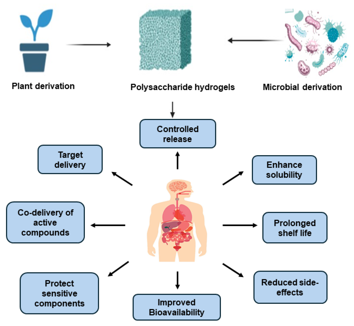
2. Polysaccharide Hydrogel System Characteristics

Polysaccharide-based hydrogels obtained from natural sources have gained significant interest due to their inherent biocompatibility, biodegradability, and versatility across various fields. Composed of long-chain carbohydrate polymers such as cellulose, hyaluronic acid, and starch derivatives, these hydrogels exhibit a remarkable capacity to retain water while maintaining their structural framework. Their natural origin provides key advantages, including reduced toxicity, minimal ecological footprint, and favorable interactions with biological environments [11]. Consequently, these hydrogels have been extensively explored in biomedical applications such as drug delivery, wound care, and tissue regeneration [12]. Their ability to form hydrogels via physical or chemical cross-linking enables broad adaptability, ranging from scaffolds supporting cellular growth to carriers for controlled therapeutic delivery. However, the successful incorporation of bioactive molecules requires mild cross-linking strategies to preserve molecular integrity and functionality. The cross-linking approaches for polysaccharide-based hydrogels are primarily categorized into physical and chemical methods [13]. Physically cross-linked hydrogels are assembled through non-covalent interactions, forming reversible networks that do not require chemical cross-linking agents. This characteristic allows for mild gelation conditions, beneficial for preserving biomolecular structure and activity [14]. Several mechanisms contribute to physical cross-linking, including electrostatic interactions, hydrophobic associations, ionic binding with multivalent ions, van der Waals forces, and host-guest inclusion complexes [15]. In contrast, chemically cross-linked hydrogels rely on irreversible covalent bonds, often facilitated by reactive small molecules such as monomers, photo-initiated groups, or oligomers [16]. Functional groups like carboxyl and amine moieties present in polysaccharide chains are instrumental in covalent polymerization. By incorporating reactive groups such as thiols, acrylates, or alkenes, polysaccharides can undergo targeted chemical modifications to enable stable cross-linking [17]. Both physically and chemically cross-linked hydrogels incorporate bioactive agents through weak interactions, entrapment, or cleavable linkages. These weak forces allow for a tunable approach to loading and releasing bioactive compounds, making polysaccharide-based hydrogels highly adaptable for diverse therapeutic applications. The ability to facilitate controlled release through reversible adsorption or stimulus-induced cleavage ensures precise regulation over biomolecule delivery kinetics [18]. Notably, the integration of hydrogels into biomolecule delivery represents a major advancement, as their intrinsic properties significantly influence drug release dynamics. Due to their biocompatible nature, these hydrogels seamlessly interact with biological systems without inducing adverse effects. Their high water retention capacity mimics the extracellular matrix, promoting biomolecule diffusion and facilitating cellular functions [19]. Furthermore, hydrogel properties can be tailored to optimize specific parameters such as mechanical strength, swelling characteristics, and degradation rates, thereby ensuring efficiency in controlled drug delivery [20]. Their capability to encapsulate various molecules, including proteins, peptides, and nucleic acids, further enhances their utility [21]. Additionally, hydrogels offer protection against enzymatic degradation and immune clearance, prolonging the circulation of therapeutic agents within the body [22]. Some hydrogels also exhibit stimuli-responsive behaviors, enabling the on-demand release of bioactive agents under specific environmental triggers. This feature allows for highly regulated biomolecule delivery, responding to changes in external stimuli such as ionic strength, light exposure, pH variation, magnetic or electric fields, temperature fluctuations, redox potential, or the presence of specific biomolecules [23]. Different strategies are employed for biomolecule release, including diffusion-based delivery, where hydrogel porosity dictates release kinetics, and swelling-controlled release, where hydrogel restructuring modulates molecule dispersion [24]. Biodegradation-mediated delivery leverages the natural breakdown of hydrogels, often via enzymatic activity, to gradually release therapeutic agents [25]. Affinity-based mechanisms further enable modulated release by leveraging interactions between hydrogel components and encapsulated biomolecules, ensuring sustained therapeutic effects based on molecular affinity parameters [26]. Lastly, link-breaking strategies involve the covalent attachment of biomolecules, with controlled cleavage under physiological conditions to achieve precise, time-dependent release [27].

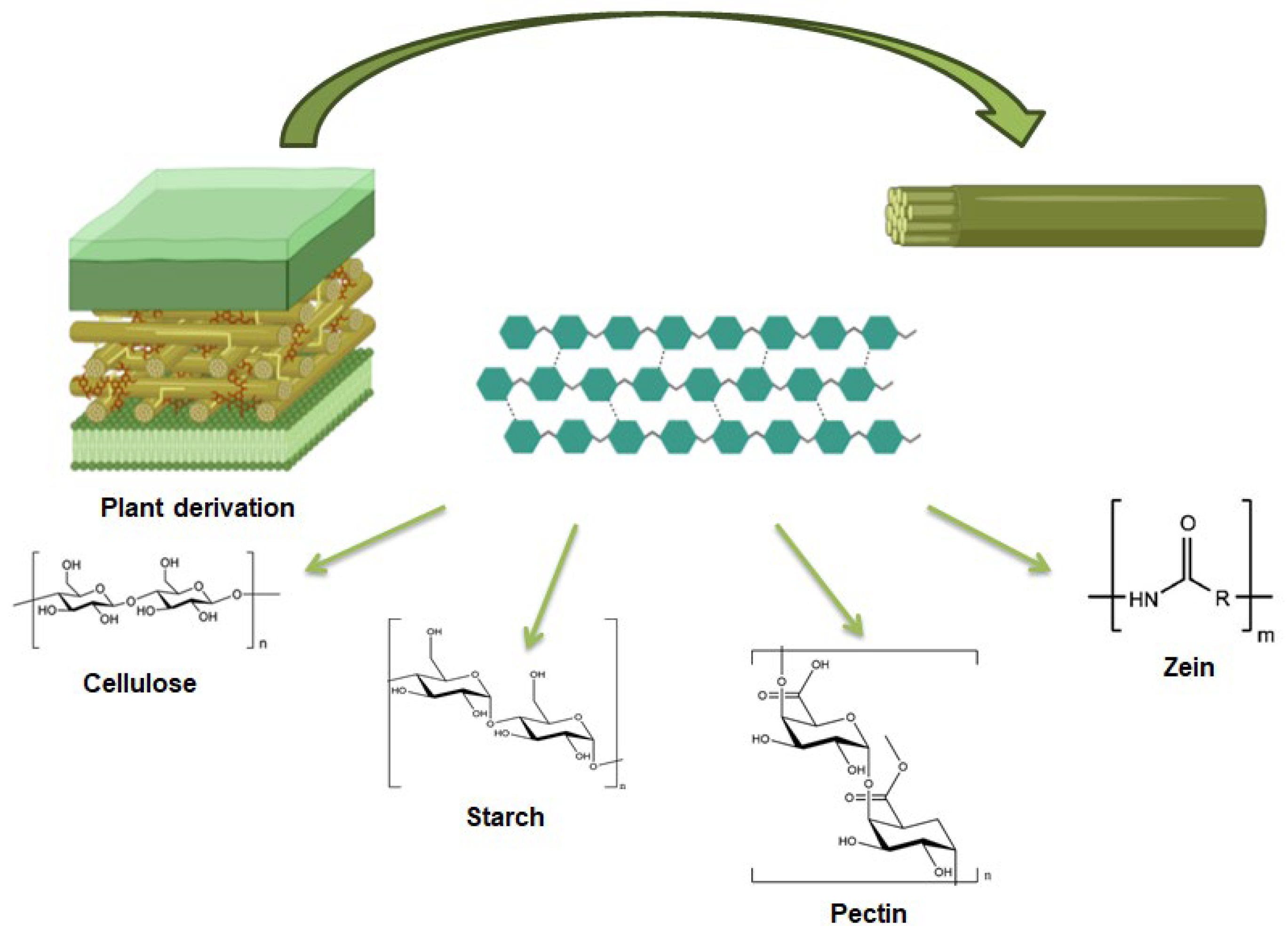
3. Plant Derived Polysaccharides

Plant-derived polysaccharides and proteins represent a significant source for developing innovative biomaterials with diverse medical applications, offering both practical and economic advantages [28], as already discussed for polysaccharides of marine origin in Sepe et al. [23]. Numerous studies have highlighted the potential of these polysaccharides for enhancing the delivery of therapeutic agents, such as antioxidants, anti-inflammatory compounds, and anticancer drugs, by improving their stability and bioavailability within biological systems [28]. Their unique properties allow for the formulation of controlled-release drug delivery systems, which can protect active ingredients during transit through the body and enable their gradual and targeted release at specific sites, making them valuable for applications including immunotherapy. Additionally, plant-derived polysaccharides are employed in the creation of biomaterials such as hydrogels, scaffolds, and membranes, which are utilized in tissue engineering, wound care, and regenerative medicine [29]. These polysaccharides are also used in the food industry as natural thickeners, stabilizers, and biodegradable packaging materials [30], as well as in environmental applications for water purification and soil conditioning [31]. Figure 2 displays the structures of plant derived polysaccharides and proteins.

3.1. Starch

3.1.1. Starch: Origin and Property

Starch is a natural polysaccharide that serves as an energy reserve in both plants and animals. It is predominantly stored in seeds, tubers, roots, and stem pith of plants (e.g., maize, wheat, potatoes, and tapioca). Industrially, starch is primarily extracted from cereal grains, tubers or the pith of plants [32]. Starch possesses a range of properties that make it valuable across various industries. Its strong hydrophilicity gives it a high affinity for water, making it easily soluble in aqueous solutions. This is crucial for its use as a thickening agent in food products and as a binder in pharmaceutical formulations. The viscosity of starch solutions can vary depending on concentration, temperature, and type, allowing for versatility in producing gels, pastes, and other formulations where viscosity control is essential [33]. When heated in water, starch granules undergo gelatinization, resulting in gel formation that provides texture, stability, and consistency in food products [34]. After gelatinization, starch gels can undergo retrogradation upon cooling, leading to a firmer gel or crystallization, which helps control food texture and prevent staling. Starch also can form flexible, transparent films when dispersed in water and dried. This property is used to produce biodegradable packaging and coatings for food and pharmaceutical products [35]. The size of starch granules is key to their function. Rice starch with small granules is highly valued in medical and cosmetic powders for topical use, effectively absorbing skin oils and acting as an excipient in dry shampoos and as a lubricant in surgical materials. Starch-based excipients enhance drug formulation and production by improving safety, cost-effectiveness, and product quality, serving as binders, diluents, disintegrants, lubricants, and glidants in granules, capsules, and tablets [35].

3.1.2. Starch-Based Hydrogels Applications

Starch-based hydrogels form three-dimensional networks created by crosslinking of starch linear molecules [36]. In their manufacture, starch molecules are cross-linked by physical, chemical or enzymatic processes to form a network capable of absorbing and retaining large amounts of water. Starch-based hydrogels have diverse applications across various fields, including biomedicine, agriculture, food, and environmental engineering. In particular, such hydrogels have been investigated as carriers for encapsulating bioactive natural compounds such as plant extracts, essential oils and phytochemicals with therapeutic properties. These compounds find application in wound healing, anti-inflammatory processes and as antimicrobial therapies. The hydrogel support facilitates the controlled release of the molecules, extending their benefits in therapeutic treatments. For example, Ghaffar et al. [37] developed a hydrogel consisting of poly(amide/acrylic acid) to promote the release of rutin, a flavonoid glycoside found in citrus fruits with anti-inflammatory and anti-allergic properties. In vitro studies revealed a pH-dependent release behavior of rutin from the hydrogel. Moreover, in vivo experiments showed that the hydrogel effectively inhibited inflammation of colon by reducing toxicity. This suggests that such pH-sensitive hydrogels could serve as an efficient anti-inflammatory approach to enhance the therapeutic efficacy of rutin in treating inflammatory conditions. Stable hydrogels were successfully formed by D’Aniello et al. [38] in the presence of the green tea extract and glycerol in the starch suspension. The controlled and steady release of bioactive compounds from the hydrogel matrix over time indicated that small molecules, such as polyphenols, interacted effectively with the rice starch hydrogel network [38]. Phosphorylated starch, using sodium tripolyphosphate as a crosslinking agent, has demonstrated a high encapsulation efficiency for anthocyanins from purple maize, achieving a productivity rate of 87.63% [39]. Additionally, starch modified with octenyl succinic anhydride (OSA) in its amorphous form is currently being utilized for the encapsulation of hydrophobic polyphenols [40]. The OSA modified waxy maize starch was able to interact with tea polyphenols leading to the gradual release of catechins during in vivo digestion [40]. All these described examples are summarized in Table 1.

3.2. Pectin

3.2.1. Pectin: Origin and Property

Pectin is a polysaccharide found in the cell walls of plants, particularly in fruits. Extracted commercially from various citrus sources such as apple, pomace, and oranges, pectin undergoes extraction under mildly acidic conditions. It is primarily categorized into two major groups: high methoxyl pectin and low methoxyl pectin [41]. As a high-value functional food ingredient, pectin finds widespread application as a gelling agent and stabilizer in the food industry. Furthermore, its potential extends to biomedical realms, notably in targeted drug delivery and various biomedical applications. Rapid degradation by colonic microorganisms makes pectin a promising candidate for colon-targeted drug delivery systems [42].

3.2.2. Pectin-Based Hydrogels Applications

Pectin-based formulations exhibit significant promises as innovative biomaterials for the development of implantable devices and prosthetics. In fact, it is biodegradable and suitable for use in implants, packaging materials and for the control of the viscosity of liquid products. Pectin biocompatibility is useful in biomedical applications, such as drug delivery systems and tissue engineering [43]. Pectin-based hydrogels have a remarkable capacity to absorb and retain significant amounts of water. Their soft and rubbery consistency minimizes irritation to surrounding tissues, rendering them exceptional candidates for applications in artificial skin and tissue engineering [44]. Additionally, pectin-based hydrogels find diverse utility as matrices for controlled drug delivery, soft contact lenses, protein separation, and cell encapsulation [45]. Nevertheless, pectin formulations suffer from drawbacks such as premature drug release, low mechanical strength, suboptimal drug loading efficiency, and limited shear stability. Consequently, complexes with other polysaccharides were developed. For example, Mishra et al. produced pH sensitive polyacrylamide grafted pectin by simple graft copolymerization. This method showed a better ability to gel and form a film than the pectin [46]. These complexes are particularly adapt to deliver natural substances [46]. A hydrogel mixture of pectin/polyethylene glycol and calcium carbonate microparticles was synthesized for the targeted release of bovine serum albumin (BSA) into the colon. The encapsulation efficiency of BSA in the hydrogel blend was about 98%. In vitro studies demonstrated that the hydrogels released the protein for approximately 9 h in the colon, highlighting their ability to transport drugs [47]. Yang et al. [48] developed caseinate-reinforced pectin hydrogel beads (PCHG-CAS) with the aim of improving the stability of (-)-epigallocatechin (EGC) and preventing its degradation. PCHG-CAS formed a compact structure due to the hydrogen bond between caseinate and pectin, achieving a high encapsulation efficiency of EGC of 93.39%. EGC showed a rapid release from PCHG-CAS in the first 30 min, and between 30 and 120 min showed a stabilization phase, reaching 76% of release. The addition of protein demonstrated their effect on release kinetics, significantly slowing the release of EGC from PCHG-CAS into water. This condition simultaneously ensured a controlled release of the hydrogel under simulated gastrointestinal conditions. Notably, EGC in PCHG-CAS remained chemically stable for a storage period of 6 days at 37 °C, preventing epimerization, oxidation, dimerization and trimerization [48]. Bostancı et al. [49] studied photo-cross-linked pectin/gelatin hydrogels for curcumin release. Hydrogels of different compositions were successfully synthesized from methacrylate derivatives of gelatin and pectin (PCMA). The hydrogel consisting of the highest PCMA content had pores with an average diameter of 44 μm, demonstrating remarkable stability, retaining 38% of its initial weight after 21 days in phosphate-buffered saline (PBS). In PBS (10 mM) at pH 7.4, curcumin was released four times faster than at pH 5.0. Furthermore, disk diffusion assays demonstrated the efficacy of the hydrogels against S. aureus and E. coli bacteria. In addition, tests of the hydrogels with L929 fibroblasts using Alamar Blue assays showed their cytocompatibility [49]. Hydrogel beads with varying ratios of alginate and pectin were investigated by Wu et al. [50] to encapsulate Pickering emulsions containing resveratrol. Cross-linking the beads with Ca2⁺ allowed better flexibility in drug delivery. Alginate/pectin hydrogel beads exhibited pH reactivity when tested under simulated gastrointestinal conditions. This limited the release of resveratrol into the simulated stomach-like gastric fluid and promoted greater release into the simulated intestinal fluid (SIF), thereby improving the bioaccessibility of resveratrol [50]. Mala and Anal [51] explored the release and protection capacity of bromelain encapsulated within hydrogel beads made of pectin and maize starch acid-resistant. The use of the 4.5:1.5 (w/w) pectin/starch ratio allowed to reach 81.25% of the encapsulation efficiency. In fact, the addition of a starch resistant to the acid improves the entrapment of bromelain, with a better swelling, a greater stability at gastric level and the prolongation of the release compared to the hydrogels constituted only by pectin. Bromelain encapsulated in hydrogels showed faster release in simulating intestinal fluid (pH 7.4) than gastric fluid (pH 1.2) [51]. Alsakhawy et al. [52] developed a hydrogel using Arabic gum (AG) and pectin to encapsulate naringenin (NAR). The developed NAR-loaded hydrogel achieved an impressive encapsulation efficiency of approximately 99.79% with a load of drug of about 16%. The NAR-loaded AG-pectin hydrogel accelerated wound healing by promoting angiogenesis and collagen deposition. Furthermore, the mRNA expression of inflammatory (TNF-α) and apoptosis-related (BAX) markers was significantly reduced [52]. The water solubility was improved by making a combination of quaternized chitosan (QCS) and pectin which also improved the antibacterial properties, the tensile strength as well as stability of the hydrogel. Incorporation of propolis into the film improved the wound healing property. The propolis-loaded hydrogel films exhibited antioxidant activity ranging from 22% to 37%. Release percentages of propolis were approximately 22% for QCS-loaded films, 78% for pectin films, and 64% for QCS–pectin hydrogel films. Wound closure with propolis-loaded QCS hydrogel films was around 3.25%, but pectin addition improved wound closures up to 53.13%, also showed substantial antibacterial activity against S. aureus and S. pyogenes. Additionally, the hydrogel films were non-toxic to NCTC clone 929 mouse fibroblast cells [53]. The cited examples are summarized in Table 2.

3.3. Cellulose

3.3.1. Cellulose: Origin and Property

Cellulose is a polysaccharide consisting of linear chains of glucose molecules linked together through β(1→4) glycosidic bonds. It is the most abundant organic compound on Earth and serves as a fundamental structural component in the cell walls of plants, providing rigidity and strength. It is synthesized by plants through the process of photosynthesis, utilizing carbon dioxide and water to produce glucose, which is then polymerized into cellulose [54]. Cellulose has strong and rigid fibers; it is insoluble in water due to the extensive hydrogen bonding between its glucose chains but is able to absorb and retain moisture. This property is beneficial in various applications, including as a thickening agent in food products and as a component in wound dressings. Moreover, cellulose is chemically stable under normal conditions, resisting degradation from acids, alkalis, and most organic solvents [55]. The production of cellulose-based hydrogels typically involves the crosslinking of cellulose molecules using various methods such as physical, chemical or enzymatic crosslinking. These methods result in the formation of a network structure capable of absorbing and retaining large amounts of water [56].

3.3.2. Cellulose-Based Hydrogels Applications

Cellulose-based hydrogels have numerous applications in a wide range of fields, including biomedical, agricultural, food and environmental engineering. In biomedicine, they can be used as wound dressings, scaffolds for tissue engineering, and drug delivery systems. They are also employed as components in diagnostic tools due to their soft and flexible nature, ability to mimic the extracellular matrix and to absorb water up to several thousand times their dry weight [57]. Cellulose-based hydrogels resulted particularly attractive for biomedical thanks to their biocompatibility as they offer a protective, aqueous environment that can shield sensitive cells and delicate drugs, including peptides and proteins. Hydrogels also facilitate efficient nutrient transport to cells and the removal of metabolic waste, can be easily modified with cell adhesion ligands, and may be injected in vivo as a liquid that solidifies at body temperature [58]. Phan et al. prepared cellulose hydrogel by physical cross-linking in a NaOH/urea medium with α-mangostin employed as an active pharmaceutical ingredient. The hydrogel’s inhibitory activity against the growth of MC3T3-E1 cells identifies it as a potential drug carrier for α-mangostin in ankylosing spondylitis treatment [59]. Gami et al. synthesized a xylan-β-cyclodextrin cellulose hydrogel through a chemical crosslinking method and investigated its absorption and release properties for curcumin. In phosphate-buffered saline (PBS), the drug on the surface of the hydrogel was rapidly released, resulting in a faster initial release rate. Curcumin formed inclusion complexes with β-cyclodextrin within the gel, inhibiting its release [60]. A cellulose hydrogel matrix with pectin and mucin was synthesized using a urea/NaOH solvent system and cross-linked with epichlorohydrin to develop superabsorbent cellulose-based hydrogels [61]. The hydrogels were evaluated for their pH-responsive swelling behavior and in vitro drug release characteristics. Results indicated a significant increase in swelling capacity for the Pec/Muc-modified hydrogels compared to the unmodified cellulose hydrogel, with the highest swelling ratio reaching 2781% at pH 10 for the Pec-modified sample. Additionally, variations in drug release profiles were observed across different pH conditions. Hydrogels exhibiting greater swelling and reduced matrix erosion demonstrated a slower drug release due to an extended diffusion pathway [61]. Regenerated cellulose derived from sugarcane bagasse was utilized in the fabrication of hydrogels, employing epichlorohydrin as a crosslinking agent and green zinc oxide nanoparticles (ZNPs), synthesized using muskmelon seed extract. The ZNPs were characterized using field emission scanning electron microscopy and energy-dispersive X-ray analysis. The swelling behavior of the hydrogel was evaluated through swelling ratio measurements. For drug delivery applications, curcumin was selected as a model drug due to its notable antimicrobial and anticancer properties. Investigations such as Fourier transform infrared spectroscopy, X-ray diffraction, thermogravimetric analysis and scanning electron microscopy confirmed the incorporation of ZNPs and curcumin into the hybrid hydrogel. Optimization of drug loading efficiency was conducted using the Taguchi method. Furthermore, the antimicrobial efficacy of the curcumin-loaded hybrid hydrogel and pure cellulose hydrogel was assessed against Staphylococcus aureus and Trichophyton rubrum. These findings suggest that the developed biomass-derived hybrid hydrogel, loaded with curcumin, holds significant potential for applications in the treatment of skin infections [62]. Table 3 recaps the described applications.

3.4. Zein

3.4.1. Zein: Origin and Property

Zein is a naturally occurring protein found in corn (maize) kernels. It is the major storage protein in corn and accounts for approximately 50% of the total protein content in maize. Zein is biocompatible, biodegradable and has excellent film-forming properties, allowing it to form thin, transparent films when dissolved in alcohol or other organic solvents. These films have barrier properties against moisture, oxygen, and other gases, making them useful for coating food and pharmaceutical products. Zein is insoluble in water, but soluble in alcohol and other organic solvents. This property allows for the production of water-resistant coatings and films with good thermal stability and suitable for applications that require heat processing, such as extrusion coating and hot-melt adhesives [63]. Finally, Zein is gluten-free and suitable for individuals with gluten sensitivities or celiac disease. This property has led to its use in gluten-free food products as a substitute for wheat-based ingredients [63].

3.4.2. Zein-Based Hydrogels Applications

The adaptability of zein-based hydrogels allows the integration of a wide range of drugs, peptides and small molecules. In tissue engineering, plant-based hydrogels play a critical role as a supportive scaffold to promote cell growth and tissue regeneration. Their porous architecture facilitates the diffusion of nutrients and oxygen, thereby promoting vital cellular activities. In addition, these hydrogels can be customized to reproduce the mechanical characteristics of tissues, creating the conditions for cells to attach, proliferate and differentiate. Zein-based hydrogels are largely diffused also for drug delivery application. For example, Ji et al. [64] encapsulated cinnamaldehyde (CA) in a composite carriers made of zein and chitosan, taking advantage of its antimicrobial properties. The interaction between CA and the zein–chitosan composite significantly reduced the volatilization rate of CA. The mass ratio of zein to CS resulted in varying structural configurations, affecting encapsulation efficiency, release behavior, and antimicrobial efficacy. Interestingly, the co-carrier’s presence slowed the release of CA [64]. Interestingly, systems made of zein loaded with hydrophobic substances were also studied. For instance, zein devices loaded with maytansine not only improved tumor targeting in non-small-cell lung cancer (A549 cells), but also reduced possible side effects due to maytansine [65]. Zein hydrogels with curcumin demonstrated significant antibacterial activity, with increasing inhibition observed as curcumin content rose [66]. Encapsulation of curcumin in whey protein isolate (WPI)-zein composite nanogels significantly improved its dispersibility [67]. Additionally, zein-hyaluronic acid composite nanogels, prepared using a layer-by-layer assembly technique, effectively enhanced the light, thermal, and storage stability of both curcumin and quercetagetin [68]. Furthermore, numerous studies have reported the encapsulation quercetin [69], resveratrol [70], rutin [71], β-carotene [72], vitamin D3 [73] and retinol [74] as hydrophobic agents in zein vectors. Finally, curcumin-loaded zein-hyaluronic acid gels demonstrated strong anti-cancer activity against CT26 colorectal cancer cells, probably due to the hyaluronic acid enhancing CD44 receptor targeting [75]. The cited applications were summarized in Table 4.

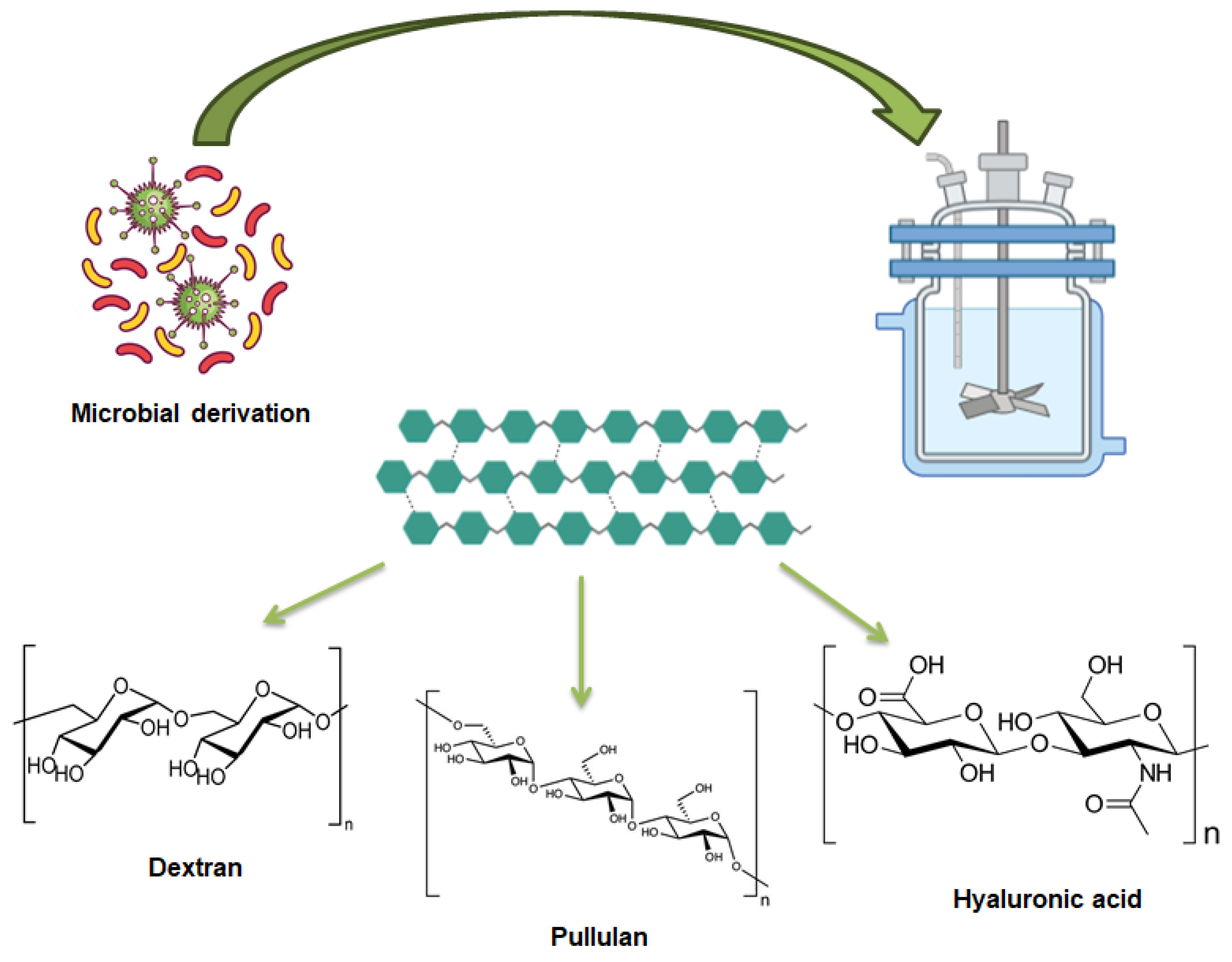
4. Microbial-Derived Polysaccharide

Microbial derived polysaccharides represent a promising and versatile category of natural materials with substantial potential for various biomedical and industrial applications. Through targeted microbial growth, these polysaccharides can be tailored to possess specific properties, enhancing their effectiveness as carriers for therapeutic agents [76,77]. Microbial (bacterial and fungal) sources are increasingly favored for their ability to produce high-yield polysaccharides. Different studies have demonstrated that polysaccharides from microbial sources can improve the stability, bioavailability, and targeted delivery of bioactive compounds, such as antioxidants, anti-inflammatory agents, and anticancer drugs [78,79,80]. Their ability to form hydrogels, scaffolds, and membranes makes them invaluable in tissue engineering, wound care, and regenerative medicine, where they offer tailored support and controlled release of therapeutic agents. Beyond biomedicine, enzymatically derived polysaccharides are utilized in the food industry as natural additives and stabilizers, and in environmental applications for water treatment and pollutant removal [81]. Figure 3 displays the structures of microbial derived polysaccharides.

4.1. Dextran

4.1.1. Dextran: Origin and Property

Dextran is nontoxic water-soluble branched homo polysaccharide of glucose linked via α-1, 6 glycosidic linkages with various branches at α (1, 2) or (1–4) and (1–6) positions [82] and with variable length. This polysaccharide is of microbial origin; in particular, it has been produced by lactic acid bacteria such as Lactococcus, Carnobacterium, Weissella and Leuconostoc, nonpathogenic species generally regarded as safe [83]. Dextran characteristic of water solubility, biodegradability, and biocompatibility make it a good candidate for the structuring of hydrogel-based platforms for drug delivery. Adequate chemical modifications allow obtaining dextran derivatives (dextran sulphate, thiolated dextran, phosphorylated dextran) improving drug administration. Different substituents can be linked to dextran to improve the performance of the material with high drug loading efficiency and targeting of specific cells [84]. In addition, dextran is a neutral polysaccharide and hydrogels, when used as a delivery vehicle, have the ability to cross the mucus layer due to their hydrophilicity [85]. Dextran hydrogels have the property of protecting drugs from enzymatic and chemical degradation prior to release. Due to the action of dextranase, the polysaccharides they contain can swell and dissolve rapidly [86].

4.1.2. Dextran-Based Hydrogels Applications

The preparation process is critical to achieving important hydrogel properties such as mechanical strength, self-healing and swelling capacity [87]. There are several crosslinking methods used to prepare dextran-based hydrogels, which can be summarized as physical and chemical crosslinking or a combination of the two [85]. The ability of dextran to implement the hydrogel formulation compared to conventional dressings has been highlighted by several works. A hydrogel of halloysite nanotubes mixed with chitosan/oxidized dextran, showed strong antibacterial capacity, improving Staphylococcus aureus-infected resistant wounds [88]. Antimicrobial properties have been demonstrated in dextran/chitosan hydrogels loaded with polydopamine nanoparticles [89], and in an oxidizing dextran and gelatin hydrogel loaded with nanoparticles of cerium oxide and curcumin as bioactive compounds [90]. Amphiphilic alkylated dextran nanoparticles served to encapsulate curcumin, improving bioavailability and delivery to the desired site. The development of combined hydrogels based on dextran-hyaluronic acid loaded with Sanguinarine, derived from the roots of Sanguinaria canadensis L. and Chelidonium majus L., showed antibacterial activity against S. aureus and Escherichia coli. Long-lasting drug release and wound regeneration capacity were also demonstrated in a rat burn infection model [91]. In the field of skin tissue regeneration an injectable hydrogel composed of gelatin, oxidized dextran loaded with apocynin, a naturally occurring methoxy-substituted catechol isolated from the roots of Apocynum cannabinum, was stabilized by the dynamic Schiff base. The hydrogel had the characteristics of good injectability, self-healing and hemostatic properties. Moreover, studies on murine and human cell evidenced an acceleration in angiogenesis with wound inflammation reduction [92]. The specificity of rosmarinic acid, a natural polyphenol with a structure similar to dopamine, allowed the construction of a rosmarinic acid-grafted dextran/gelatin hydrogel [93]. The hydrogel loaded with the natural bioactive compound showed very rapid gelation (61.6 ± 2.8 s) and good mechanical properties. In addition to highlighting antibacterial properties, tests on rat models demonstrated the effectiveness of the gel in healing skin wounds. Dextran based cyclodextrins (DC) are often used for complexation [94]. These cyclodextrins consist of glucopyranoside units linked via α-1,4 glycosidic bonds, synthesized by bacterial enzymes (cyclodextrin glycosyltransferases). Due to their hydrophilic surface and hydrophobic cavity, they can form inclusion complexes with various lipophilic bioactive increasing their solubility, stability and bioavailability [95]. For example, hypromellose hydrogels with different DC was used to incorporate 3-O-Methylquercetin, a flavonoid from Achyrocline satureioides [95]. The formulation of the gel allowed the skin release of the bioactive molecule (100%), directing it to the site of replication of melanoma cells. DC-based hydrogels have numerous uses in the biomedical field. An example was the hydrogel composed of β-CD, polyethyleneimine (PEI) and silk fibroin cross-linked with epichlorohydrin and loaded with Centella asiatica extract and hydrocortisone acetate. The bioactive molecules were released effectively, even if the released C. asiatica extract was pH dependent, healing of bedsores was observed (in 6 days), which was not found in the negative control [96]. A cyclodextrin-based hydrogel loaded with gallic acid allowed a slow release of the phenolic acid (in 48 h) showing antibacterial activity against S. epidermidis, S. aureus and K. pneumoniae without causing any damage to the surrounding tissue [97]. Cui et al. [98] produced an hydrogel based on methyl-β-cyclodextrin and soluble soy polysaccharide loaded with Satureja montana L. essential oil. Compared with the soluble soybean hydrogel alone, the cyclodextrins gave a more compact gel structure. Furthermore, a good antibacterial activity of the hydrogel against S. aureus was verified in meat samples [98]. The described examples are summarized in Table 5.

4.2. Pullulan

4.2.1. Pullulan: Origin and Property

Pullulan is a homopolysaccharide of natural origin, synthesized during the fermentation of the fungus Aureobasidium pullulans [99]. The polymer chemical structure is linear, consisting of maltotriose units interconnected by α-1,6 and α-1,4 glycosidic bonds that make the polysaccharides neutral and non-ionic [99]. Thanks to the regular alternation of glycosidic bonds, pullulan exhibits structural flexibility, good elastic properties, solubility in aqueous solvents and high thermal stability (melting temperature range 250–300 °C) [100]. Pullulan has the ability to form films and is also non-carcinogenic, non-ionic, non-irritant, non-hygroscopic, non-toxic, blood-compatible, biodegradable, hydrophilic, odorless and tasteless [101]. Its important properties make the polysaccharide applicable in a wide range of fields, from textiles to food packaging, cosmetics and pharmaceutical formulation, the limitation to the widespread use of pullulan concerns its production costs [100].

4.2.2. Pullulan Based Hydrogels Applications

Pullulan has antibacterial, anticoagulant and anti-inflammatory properties and is therefore used as a carrier for trans-mucosal drug delivery systems and wound dressings, in capsule and tablet coatings or for topical formulation in the form of gels and oral films [102]. These unique characteristics are also exploited for the preparation of hydrogels for various applications [103]. Hydrogels characterized by a three-dimensional cross-linked structure are able to retain large volumes of water and swell without dissolving, trapping small biomolecules [104]. Bioactive compounds of natural origin, frequently used for the treatment of numerous pathologies, can be promisingly incorporated into pullulan hydrogels. In fact, the characteristics of the polymer allow significant quenching of free radicals, a useful function in skin wound treatment. An Asiatic traditional medicine for the treatment of inflammation and skin wounds, Ulmus davidiana var. japonica root bark, has been used for the development of pullulan hydrogel. Hydrogels containing U. davidiana had good thermal stability and mechanically improved properties compared to the pullulan-only gel film, with swelling capacity and skin adherence. Hydrogels containing up to 50% extract showed a cell viability of 90%. Furthermore, an improvement in wound healing was observed, collagen deposition with dermal regeneration was achieved in mouse wound model [105]. A pullulan-based hydrogel was formulated to load an aqueous extract of Rhus verniciflua a tree used in traditional Asiatic herbal medicine as immunostimulant and endowed with antioxidant, anticancer, anti-inflammatory and antimicrobial activities [106]. The pullulan hydrogel was able to release approximately 87% of the bioactive compound in 12 h of application, stabilizing the release in 24–48 h. The application of this hydrogel, capable of creating an anti-scratch film on the skin, in the treatment of atopic dermatitis highlighted a better infiltration of mast cells, thus confirming the therapeutic efficacy [106]. Curcumin is known for its antioxidant, antibacterial, and anti-inflammatory properties, but its low bioavailability and poor solubility in water limit its use as a drug. An injectable hydrogel platform of hyaluronic acid-pullulan and pluronic was designed for the slow release of curcumin to repair diabetic wounds [107]. In the hydrogel with optimized composition, 50% release of the bioactive was observed after 8 h, continuing the release over 24 h. Interestingly, using the curcumin-loaded hydrogel, a wound healing rate of 13 days was observed compared to the 35 days required without the bioactive compound [107]. Antibacterial, antifungal activities and bacteriostatic effect after 48 and 72 h against S. aureus and E. coli, respectively, was also found in a clove oil-loaded hydrogel synthesized by combining covalent and physical cross-linking methods [108]. The hydrogels loaded with optimized amounts of clove oil exhibited excellent mechanical properties, good swelling ability combined with rapid recovery and sustained release of oil, essential properties for wound dressing applications [108]. The potential of plant extracts released from pullulan hydrogels for wound dressings was investigated also by Pelin et al. [109]. Pullulan/poly(vinyl alcohol) hydrogels, prepared by an environmentally friendly method by covalent and physical cross-linking, were loaded with the hydroalcoholic extract of Calendula officinalis by dipping after loading. The formation of hydrogen bonds between the polymer and the bioactive allowed high loading efficiency. It was also possible to give the gel good mechanical properties, with bio adhesiveness improving as the amount of bioactive loaded increased. The pullulan-based hydrogel loaded with extracts of C. officinalis were also characterized by a high antioxidant and antibacterial activity, both against Gram-positive and Gram-negative bacteria, and were not cytotoxic against Human Dermal Fibroblasts cells [109]. The cited applications are summarized in Table 6.

4.3. Hyaluronic Acid

4.3.1. Hyaluronic Acid: Origin and Property

Hyaluronic acid (HA) is a linear anionic polysaccharide that belongs to the glycosaminoglycan family, consisting of repeating units of N-acetyl-D-glucosamine and glucuronic acid [110]. Historically, HA has been obtained from rooster combs and bovine vitreous humor. However, microbial fermentation using streptococcus and bacillus species has become an alternative and widely adopted method for HA production due to its efficiency and reduced risk of contamination from animal-derived sources [111]. In the human body, HA is abundant in synovial fluid and vitreous humor, where it plays an important structural role in articular cartilage and skin [112]. HA possesses excellent water solubility and forms highly viscous solutions, contributing to its free radical scavenging activity, bacteriostatic properties, and facilitation of tissue repair [113]. However, the homopolymeric form of HA lacks sufficient mechanical strength and stability for use as a structural scaffold. To overcome this limitation, HA is chemically modified by cross-linking with ethyl esters, benzyl esters, or other biodegradable polymers, enhancing its mechanical properties while preserving biocompatibility [114]. These cross-linked HA hydrogels are highly adaptable and can be fabricated into various structures, including membranes, sponges, fibers, and scaffolds. They have been extensively explored for applications in wound healing, tracheal regeneration, and the repair of cartilage, vasculature, and neural tissues [114].

4.3.2. Hyaluronan-Based Hydrogel Applications

HA-based hydrogels exhibit characteristics of biodegradability, great adaptability to environmental stimuli and the ability to stimulate the immune system. These characteristics make such hydrogels of great interest in biomedicine, in particular in tissue and regenerative engineering and drug administration [23]. For instance, Kwon et al. developed pH-responsive hydrogels incorporating the phenol isoliquiritigenin from licorice, employing hydroxyethylcellulose and HA for transcutaneous delivery. These hydrogels were obtained by conjugate Michael addition and allowed for improved drug release at pH above 7, indicating a pH dependence [115]. Furthermore, Xin Jin et al. designed a composite hydrogel consisting of tannic acid and carbon in dopamine-coated particles enriched with phenolic compounds. This hydrogel via oxidative coupling improved cross-linking and adhesion by establishing covalent and hydrogen bonds with wound tissues [116]. The hydrogel exhibited fast gelation speed (<6 s) and remarkable adhesiveness (>8.1 kPa). In vitro evaluations indicated minimal hemolysis activity, low cytotoxicity, and improved fibroblast proliferation and migration. An in vivo model of skin defect to which the hydrogel was applied demonstrated accelerated wound healing through photothermal treatment. It also reduced inflammation, alleviated tissue hypoxia and promoted angiogenesis and epithelialization [116]. In another study, Gallelli et al. [117] evaluated the therapeutic potential of HA nanohydrogels incorporated with quercetin and oleic acid. This formulation significantly reduced the healing time of skin wounds on the lower limbs of diabetic patients compared to free hyaluronic acid (0.2%) and exhibited a favorable safety profile with no adverse drug reactions [117]. Similarly, Conte et al. [118] formulated a novel periodontal drug delivery system by encapsulating Opuntia ficus-indica extract, recognized as anti-inflammatory, antioxidant and antibacterial, inside chitosan nanoparticles immersed in pluronic-hyaluronic hydrogels temperature responsive. This system demonstrated effective eradication of biofilms formed by Streptococcus mutans, Pseudomonas aeruginosa, and Porphyromonas gingivalis while disrupting extracellular polymeric substance formation. Additionally, these hydrogels exhibited immune-modulating properties [118]. In further research, the same authors developed HA-based hydrogels containing resveratrol-loaded chitosan nanoparticles for atopic dermatitis treatment.
The encapsulation in HA of the nanoparticles delayed their hydrolysis, allowing a controlled and sustained release of resveratrol. The formulation mitigated oxidative stress in human keratinocytes with TNF-α/INF-γ (HaCaT) and reduced the expression and secretion of pro-inflammatory cytokines [119]. The studies discussed in this section are summarized in Table 7.

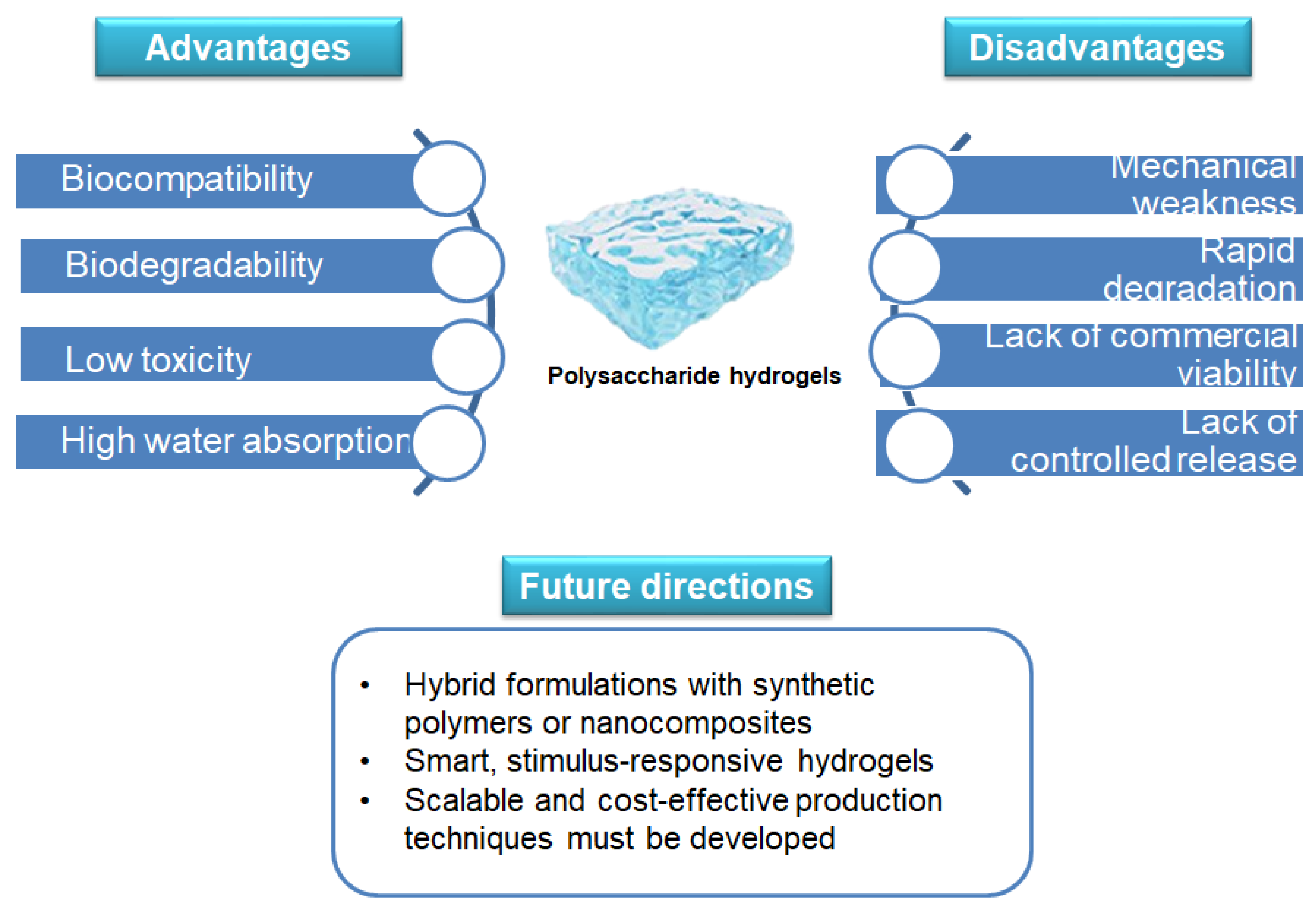
5. Challenges and Future Directions

Despite their numerous advantages, polysaccharide-based hydrogels also present significant limitations that must be addressed to facilitate their widespread clinical and commercial adoption. One of the major challenges is their mechanical weakness, which can limit their use in load-bearing applications such as cartilage repair [120]. Many natural polysaccharides lack the necessary structural integrity and durability, making them prone to rapid degradation or mechanical failure under physiological conditions [121]. Furthermore, their high swelling capacity, while beneficial for bioactive release, can lead to instability in certain biomedical applications, reducing their functional lifespan [121]. Another limitation is the complexity of achieving precise control over degradation rates and drug release kinetics [122]. Polysaccharide-based hydrogels often exhibit variability in their degradation profiles due to differences in cross-linking methods, environmental conditions, and interactions with biological fluids. This inconsistency poses challenges for developing reproducible and standardized formulations suitable for regulatory approval [122]. Additionally, the potential for immunogenic responses or contamination from plant-derived or microbial sources needs careful evaluation to ensure patient safety [123]. To overcome these challenges, future research should focus on developing reinforced protein and polysaccharide-based hydrogels through hybrid formulations with synthetic polymers or nanocomposites. The incorporation of nanoparticles, graphene derivatives, or bioceramic fillers can significantly enhance mechanical properties, providing better structural stability for applications in tissue engineering and regenerative medicine [124]. Smart, stimulus-responsive hydrogels that react to changes in pH, temperature, or enzymatic activity can also improve control over bioactive release, ensuring more predictable therapeutic outcomes [125]. Issues related to large-scale production, cost-effectiveness, and reproducibility remain major hurdles in industrial manufacturing. Then, scalable and cost-effective production techniques must be developed to facilitate commercial viability [126]. Moreover, regulatory frameworks should also be adapted to accommodate the complexities of these materials, with standardized protocols for biocompatibility assessment, long-term stability testing, and large-scale production [127] (Figure 4).

6. Conclusions

Polysaccharide-based hydrogels represent a powerful and sustainable platform for the controlled release of natural bioactive compounds, with wide-ranging applications in drug delivery, wound healing, and antimicrobial therapies. Their biocompatibility, biodegradability, and tunable physicochemical properties make them ideal candidates for biomedical applications, allowing for precise modulation of drug release profiles and enhanced therapeutic outcomes. This review has explored hydrogels derived from plant-based polysaccharides and proteins, including starch, pectin, cellulose, and zein, as well as those obtained through microbial action, such as dextran, pullulan, and hyaluronic acid. The incorporation of these materials into hydrogel-based devices has demonstrated significant advantages, including improved stability, prolonged bioactive release, and enhanced interaction with biological tissues. The optimization of cross-linking mechanisms, swelling behavior, and responsiveness to external stimuli, permitted to tailor hydrogels for specific therapeutic needs, reducing side effects and increasing treatment efficacy. Moreover, the use of naturally derived bioactives within these hydrogels has shown promising results in accelerating wound healing, preventing infections, and modulating inflammatory responses, reinforcing their role in regenerative medicine and targeted drug delivery. As research continues to advance, further innovations in hydrogel formulation and functionalization are expected to drive the development of next-generation biomaterials that combine efficacy, sustainability, and biocompatibility.

Author Contributions

Conceptualization, A.V., R.C., O.P., L.M. and A.C.; writing—original draft preparation and figure drawing, F.S., A.V. and R.C.; writing—review and editing, F.S., A.V., O.P., R.C., L.M., S.M. and A.C.; visualization, S.M. and G.P.; supervision, O.P., R.C., L.M., A.V. and A.C.; literature review, F.S., A.V. and R.C.; resources, O.P. and L.M.; funding acquisition, G.P. and A.C. All authors have read and agreed to the published version of the manuscript.

Funding

We acknowledge financial support under the National Recovery and Resilience Plan (NRRP), Mission 4 Component 2 Investment 1.4—Call for tender No. 3138 of 16 December 2021, rectified by Decree n. 3175 of 18 December 2021 of the Italian Ministry of University and Research funded by the European Union—NextGenerationEU, Award Number: Project code CN_00000033; Concession Decree No. 1034 of 17 June 2022 adopted by the Italian Ministry of University and Research, CUP B83C22002930006, Project title “National Biodiversity Future Center—NBFC”; and National Recovery and Resilience Plan (NRRP), Mission 4, Component 2, Investment 1.1, Call for tender No. 1409 published on 14 September 2022 by the Italian Ministry of University and Research (MUR), funded by the European Union—Next Generation EU, (PRIN 2022 PNRR) CUP B53D23031790001, Injectable Hydrogels for treatment of periodontal disease (HYPE)—P2022RZ8WM—Grant Assignment Decree No. n. 1369 adopted on 01/09/2023 1 September 2023 by the Italian Ministry of Ministry of University and Research (MUR).

Institutional Review Board Statement

Not applicable.

Informed Consent Statement

Not applicable.

Data Availability Statement

No new data were created or analyzed in this study.

Acknowledgments

We would like to acknowledge Annunziata Bilongi for the administrative support (IBBC-NA, CNR), Marina Russo for technical support to data collection process and Pasquale Lo Schiavo for assistance with graphic design.

Conflicts of Interest

The authors declare no conflicts of interest.

References

  1. Rumon, M.M.H.; Akib, A.A.; Sarkar, S.D.; Khan, M.A.R.; Uddin, M.M.; Nasrin, D.; Roy, C.K. Polysaccharide-Based Hydrogels for Advanced Biomedical Engineering Applications. ACS Polym. Au 2024, 4, 463–486. [Google Scholar] [CrossRef] [PubMed]
  2. Chai, Q.; Jiao, Y.; Yu, X. Hydrogels for Biomedical Applications: Their Characteristics and the Mechanisms behind Them. Gels 2017, 3, 6. [Google Scholar] [CrossRef] [PubMed]
  3. Asthana, N.; Pal, K.; Khan, A.A.; Malik, A. Novel biopolymeric materials potential utilization for environmental practices. J. Mol. Struct. 2024, 1311, 138390. [Google Scholar] [CrossRef]
  4. Beena Unni, A.; Muringayil Joseph, T. Enhancing Polymer Sustainability: Eco-Conscious Strategies. Polymers 2024, 16, 1769. [Google Scholar] [CrossRef]
  5. Salehi, M.; Rashidinejad, A. Multifaceted roles of plant-derived bioactive polysaccharides: A review of their biological functions, delivery, bioavailability, and applications within the food and pharmaceutical sectors. Int. J. Biol. Macromol. 2025, 290, 138855. [Google Scholar] [CrossRef]
  6. Rial-Hermida, M.I.; Rey-Rico, A.; Blanco-Fernandez, B.; Carballo-Pedrares, N.; Byrne, E.M.; Mano, J.F. Recent Progress on Polysaccharide-Based Hydrogels for Controlled Delivery of Therapeutic Biomolecules. ACS Biomater. Sci. Eng. 2021, 7, 4102–4127. [Google Scholar] [CrossRef]
  7. Valentino, A.; Yazdanpanah, S.; Conte, R.; Calarco, A.; Peluso, G. Smart Nanocomposite Hydrogels as Next-Generation Therapeutic and Diagnostic Solutions. Gels 2024, 10, 689. [Google Scholar] [CrossRef]
  8. Conte, R.; Valentino, A.; Romano, S.; Margarucci, S.; Petillo, O.; Calarco, A. Stimuli-Responsive Nanocomposite Hydrogels for Oral Diseases. Gels 2024, 10, 478. [Google Scholar] [CrossRef]
  9. Olteanu, G.; Neacșu, S.M.; Joița, F.A.; Musuc, A.M.; Lupu, E.C.; Ioniță-Mîndrican, C.B.; Lupuliasa, D.; Mititelu, M. Advancements in Regenerative Hydrogels in Skin Wound Treatment: A Comprehensive Review. Int. J. Mol. Sci. 2024, 25, 3849. [Google Scholar] [CrossRef]
  10. Zhao, L.; Zhou, Y.; Zhang, J.; Liang, H.; Chen, X.; Tan, H. Natural Polymer-Based Hydrogels: From Polymer to Biomedical Applications. Pharmaceutics 2023, 15, 2514. [Google Scholar] [CrossRef]
  11. Satchanska, G.; Davidova, S.; Petrov, P.D. Natural and Synthetic Polymers for Biomedical and Environmental Applications. Polymers 2024, 16, 1159. [Google Scholar] [CrossRef] [PubMed]
  12. Revete, A.; Aparicio, A.; Cisterna, B.A.; Revete, J.; Luis, L.; Ibarra, E.; Segura González, E.A.; Molino, J.; Reginensi, D. Advancements in the Use of Hydrogels for Regenerative Medicine: Properties and Biomedical Applications. Int. J. Biomater. 2022, 2022, 3606765. [Google Scholar] [CrossRef] [PubMed]
  13. Manzoor, A.; Dar, A.H.; Pandey, V.K.; Shams, R.; Khan, S.; Panesar, P.S.; Kennedy, J.F.; Fayaz, U.; Khan, S.A. Recent insights into polysaccharide-based hydrogels and their potential applications in food sector: A review. Int. J. Biol. Macromol 2022, 213, 987–1006. [Google Scholar] [CrossRef] [PubMed]
  14. Xue, X.; Hu, Y.; Wang, S.; Chen, X.; Jiang, Y.; Su, J. Fabrication of physical and chemical crosslinked hydrogels for bone tissue engineering. Bioact. Mater. 2022, 12, 327–339. [Google Scholar] [CrossRef]
  15. Kamel, S.; El-Sayed, N.S. Cross-linking Strategies for the Design of Smart Injectable Hydrogels. In Injectable Smart Hydrogels for Biomedical Applications; Dodda, J.M., Ashammakhi, N., Sadiku, E.R., Eds.; Royal Society of Chemistry: Edinburgh, UK, 2024; Volume 17, pp. 128–149. [Google Scholar]
  16. Bustamante-Torres, M.; Romero-Fierro, D.; Arcentales-Vera, B.; Palomino, K.; Magaña, H.; Bucio, E. Hydrogels Classification According to the Physical or Chemical Interactions and as Stimuli-Sensitive Materials. Gels 2021, 7, 182. [Google Scholar] [CrossRef]
  17. Alavarse, A.C.; Frachini, E.C.G.; da Silva, R.L.C.G.; Lima, V.H.; Shavandi, A.; Petri, D.F.S. Crosslinkers for polysaccharides and proteins: Synthesis conditions, mechanisms, and crosslinking efficiency, a review. Int. J. Biol. Macromol. 2022, 202, 558–596. [Google Scholar] [CrossRef]
  18. Liu, D.; Yang, F.; Xiong, F.; Gu, N. The Smart Drug Delivery System and Its Clinical Potential. Theranostics 2016, 6, 1306–1323. [Google Scholar] [CrossRef]
  19. Kaur, H.; Gogoi, B.; Sharma, I.; Das, D.K.; Azad, M.A.; Pramanik, D.D.; Pramanik, A. Hydrogels as a Potential Biomaterial for Multimodal Therapeutic Applications. Mol. Pharm. 2024, 21, 4827–4848. [Google Scholar] [CrossRef]
  20. Liu, B.; Chen, K. Advances in Hydrogel-Based Drug Delivery Systems. Gels 2024, 10, 262. [Google Scholar] [CrossRef]
  21. Correa, S.; Grosskopf, A.K.; Lopez Hernandez, H.; Chan, D.; Yu, A.C.; Stapleton, L.M.; Appel, E.A. Translational Applications of Hydrogels. Chem. Rev. 2021, 121, 11385–11457. [Google Scholar] [CrossRef]
  22. Kim, K.M.; D’Elia, A.M.; Rodell, C.B. Hydrogel-based approaches to target hypersensitivity mechanisms underlying autoimmune disease. Adv. Drug Deliv. Rev. 2024, 212, 115395. [Google Scholar] [CrossRef] [PubMed]
  23. Sepe, F.; Valentino, A.; Marcolongo, L.; Petillo, O.; Conte, R.; Margarucci, S.; Peluso, G.; Calarco, A. Marine-Derived Polysaccharide Hydrogels as Delivery Platforms for Natural Bioactive Compounds. Int. J. Mol. Sci. 2025, 26, 764. [Google Scholar] [CrossRef] [PubMed]
  24. Li, J.; Mooney, D.J. Designing hydrogels for controlled drug delivery. Nat. Rev. Mater. 2016, 1, 16071. [Google Scholar] [CrossRef]
  25. Knipe, J.; Chen, F.; Peppas, N. Enzymatic Biodegradation of Hydrogels for Protein Delivery Targeted to the Small Intestine. Biomacromolecules 2015, 16, 962–972. [Google Scholar] [CrossRef]
  26. Vulic, K.; Shoichet, M.S. Affinity-Based Drug Delivery Systems for Tissue Repair and Regeneration. Biomacromolecules 2014, 15, 3867–3880. [Google Scholar] [CrossRef]
  27. Ali, F.; Khan, I.; Chen, J.; Akhtar, K.; Bakhsh, E.M.; Khan, S.B. Emerging Fabrication Strategies of Hydrogels and Its Applications. Gels 2022, 8, 205. [Google Scholar] [CrossRef]
  28. Bahú, J.O.; de Andrade, L.R.M.; de Melo Barbosa, R.; Crivellin, S.; da Silva, A.P.; Souza, S.D.A.; Cárdenas Concha, V.O.; Severino, P.; Souto, E.B. Plant Polysaccharides in Engineered Pharmaceutical Gels. Bioengineering 2022, 9, 376. [Google Scholar] [CrossRef]
  29. Shang, L.; Wang, S.; Mao, Y. Recent advances in plant-derived polysaccharide scaffolds in tissue engineering: A review. Int. J. Biol. Macromol. 2024, 277, 133830. [Google Scholar] [CrossRef]
  30. Navasingh, R.J.H.; Gurunathan, M.K.; Nikolova, M.P.; Królczyk, J.B. Sustainable Bioplastics for Food Packaging Produced from Renewable Natural Sources. Polymers 2023, 15, 3760. [Google Scholar] [CrossRef]
  31. Fakhri, V.; Jafari, A.; Layaei Vahed, F.; Su, C.-H.; Pirouzfar, V. Polysaccharides as eco-friendly bio-adsorbents for wastewater remediation: Current state and future perspective. J. Water Process Eng. 2023, 54, 103980. [Google Scholar] [CrossRef]
  32. Apriyanto, A.; Compart, J.; Fettke, J. A review of starch, a unique biopolymer—Structure, metabolism and in planta modifications. Plant Sci. 2022, 318, 111223. [Google Scholar] [CrossRef] [PubMed]
  33. Wang, X.; Huang, L.; Zhang, C.; Deng, Y.; Xie, P.; Liu, L.; Cheng, J. Research advances in chemical modifications of starch for hydrophobicity and its applications: A review. Carbohydr. Polym. 2020, 240, 116292. [Google Scholar] [CrossRef] [PubMed]
  34. Gong, Y.; Xiao, S.; Yao, Z.; Deng, H.; Chen, X.; Yang, T. Factors and modification techniques enhancing starch gel structure and their applications in foods:A review. Food Chem. 2024, 24, 102045. [Google Scholar] [CrossRef] [PubMed]
  35. Dong, Y.; Ghasemzadeh, M.; Khorsandi, Z.; Sheibani, R.; Nasrollahzadeh, M. Starch-based hydrogels for environmental applications: A review. Int. J. Biol. Macromol. 2024, 269, 131956. [Google Scholar] [CrossRef]
  36. Koshenaj, K.; Ferrari, G. A Comprehensive Review on Starch-Based Hydrogels: From Tradition to Innovation, Opportunities, and Drawbacks. Polymers 2024, 16, 1991. [Google Scholar] [CrossRef]
  37. Abdel-Ghaffar, A.M. Radiation synthesis and modification of biopolymers and polymeric composites for biomedical applications. Poly. Compos. 2023, 31, 1–13. [Google Scholar] [CrossRef]
  38. D’Aniello, A.; Koshenaj, K.; Ferrari, G. A Preliminary Study on the Release of Bioactive Compounds from Rice Starch Hydrogels Produced by High-Pressure Processing (HPP). Gels 2023, 9, 521. [Google Scholar] [CrossRef]
  39. García Tejeda, Y.; Salinas-Moreno, Y.; Hernandez-Martinez, A.; MartÍNez-Bustos, F. Encapsulation of Purple Maize Anthocyanins in Phosphorylated Starch by Spray Drying. Cereal Chem. 2015, 93, 130–137. [Google Scholar] [CrossRef]
  40. Peng, S.; Xue, L.; Leng, X.; Yang, R.; Zhang, G.; Hamaker, B.R. Slow digestion property of octenyl succinic anhydride modified waxy maize starch in the presence of tea polyphenols. J. Agric. Food Chem. 2015, 63, 2820–2829. [Google Scholar] [CrossRef]
  41. Roman-Benn, A.; Contador, C.; Li, M.W.; Lam, H.-M.; Ah-Hen, K.; Ulloa, P.; Ravanal, M. Pectin: An overview of sources, extraction and applications in food products and health. Food Chem. Adv. 2023, 2, 100192. [Google Scholar] [CrossRef]
  42. Liu, L.; Fishman, M.L.; Kost, J.; Hicks, K.B. Pectin-based systems for colon-specific drug delivery via oral route. Biomaterials 2003, 24, 3333–3343. [Google Scholar] [CrossRef] [PubMed]
  43. Moutaharrik, S.; Palugan, L.; Cerea, M.; Meroni, G.; Casagni, E.; Roda, G.; Martino, P.A.; Gazzaniga, A.; Maroni, A.; Foppoli, A. Colon Drug Delivery Systems Based on Swellable and Microbially Degradable High-Methoxyl Pectin: Coating Process and In Vitro Performance. Pharmaceutics 2024, 16, 508. [Google Scholar] [CrossRef] [PubMed]
  44. Tortorella, S.; Inzalaco, G.; Dapporto, F.; Maturi, M.; Sambri, L.; Vetri Buratti, V.; Chiariello, M.; Comes Franchini, M.; Locatelli, E. Biocompatible pectin-based hybrid hydrogels for tissue engineering applications. New J. Chem. 2021, 45, 22386–22395. [Google Scholar] [CrossRef]
  45. Mishra, R.; Banthia, A.; Majeed, A. Pectin based formulations for biomedical applications: A review. Asian J. Pharm. Biol. Res. 2012, 5, 1–7. [Google Scholar] [CrossRef]
  46. Mishra, R.; Ray, A. Synthesis and Characterization of Poly{N-[3-(dimethylamino) propyl] methacrylamide-co-itaconic acid} Hydrogels for Drug Delivery. J. Appl. Polym. Sci. 2011, 119, 3199–3206. [Google Scholar] [CrossRef]
  47. Gautam, M.; Santhiya, D. In-situ mineralization of calcium carbonate in pectin based edible hydrogel for the delivery of protein at colon. J. Drug Deliv. Sci. Technol. 2019, 53, 101137. [Google Scholar] [CrossRef]
  48. Yang, M.; Zhao, S.; Zhao, C.; Cui, J.; Wang, Y.; Fang, X.; Zheng, J. Caseinate-reinforced pectin hydrogels: Efficient encapsulation, desirable release, and chemical stabilization of (−)-epigallocatechin. Int. J. Biol. Macromol. 2023, 230, 123298. [Google Scholar] [CrossRef]
  49. Bostancı, N.S.; Büyüksungur, S.; Hasirci, N.; Tezcaner, A. pH responsive release of curcumin from photocrosslinked pectin/gelatin hydrogel wound dressings. Biomater. Adv. 2022, 134, 112717. [Google Scholar] [CrossRef]
  50. Wu, B.; Li, Y.; Li, Y.; Li, H.; Li, L.; Xia, Q. Encapsulation of resveratrol-loaded Pickering emulsions in alginate/pectin hydrogel beads: Improved stability and modification of digestive behavior in the gastrointestinal tract. Int. J. Biol. Macromol. 2022, 222, 337–347. [Google Scholar] [CrossRef]
  51. Mala, T.; Anal, A.K. Protection and Controlled Gastrointestinal Release of Bromelain by Encapsulating in Pectin-Resistant Starch Based Hydrogel Beads. Front. Bioeng. Biotechnol. 2021, 9, 757176. [Google Scholar] [CrossRef]
  52. Alsakhawy, M.A.; Abdelmonsif, D.A.; Haroun, M.; Sabra, S.A. Naringin-loaded Arabic gum/pectin hydrogel as a potential wound healing material. Int. J. Biol. Macromol. 2022, 222, 701–714. [Google Scholar] [CrossRef] [PubMed]
  53. Phonrachom, O.; Charoensuk, P.; Kiti, K.; Saichana, N.; Kakumyan, P.; Suwantong, O. Potential use of propolis-loaded quaternized chitosan/pectin hydrogel films as wound dressings: Preparation, characterization, antibacterial evaluation, and in vitro healing assay. Int. J. Biol. Macromol. 2023, 241, 124633. [Google Scholar] [CrossRef] [PubMed]
  54. McNamara, J.T.; Morgan, J.L.; Zimmer, J. A molecular description of cellulose biosynthesis. Annu. Rev. Biochem. 2015, 84, 895–921. [Google Scholar] [CrossRef] [PubMed]
  55. Etale, A.; Onyianta, A.J.; Turner, S.R.; Eichhorn, S.J. Cellulose: A Review of Water Interactions, Applications in Composites, and Water Treatment. Chem. Rev. 2023, 123, 2016–2048. [Google Scholar] [CrossRef] [PubMed]
  56. Zainal, S.; Mohd, N.; Suhaili, N.; Anuar, F.; Lazim, A.M.; Othaman, R. Preparation of Cellulose-based Hydrogel: A Review. J. Mater. Res. Technol. 2020, 10, 935–952. [Google Scholar] [CrossRef]
  57. Chang, C.; Na, N. Cellulose-based hydrogels: Present status and application prospects. Carbohydr. Polym. 2011, 84, 40–53. [Google Scholar] [CrossRef]
  58. Silva, I.G.R.d.; Pantoja, B.T.d.S.; Almeida, G.H.D.R.; Carreira, A.C.O.; Miglino, M.A. Bacterial Cellulose and ECM Hydrogels: An Innovative Approach for Cardiovascular Regenerative Medicine. Int. J. Mol. Sci. 2022, 23, 3955. [Google Scholar] [CrossRef]
  59. Phan, H.L.; Tran, N.C.T.; Le, T.H.Y.; Le, Q.V.; Le, T.T.; Thach, U.D. Fabrication of polydopamine-modified cellulose hydrogel for controlled release of α-mangostin. Biopolymers 2024, 115, e23613. [Google Scholar] [CrossRef]
  60. Gami, P.; Kundu, D.; Seera, S.D.K.; Banerjee, T. Chemically crosslinked xylan-β-Cyclodextrin hydrogel for the in vitro delivery of curcumin and 5-Fluorouracil. Int. J. Biol. Macromol. 2020, 158, 18–31. [Google Scholar] [CrossRef]
  61. Islam, F.; Wong, S.Y.; Li, X.; Arafat, M.T. Pectin and mucin modified cellulose-based superabsorbent hydrogel for controlled curcumin release. Cellulose 2022, 29, 5207–5222. [Google Scholar] [CrossRef]
  62. Anagha, B.; George, D.; Maheswari, P.U.; Begum, K.M.M.S. Biomass Derived Antimicrobial Hybrid Cellulose Hydrogel with Green ZnO Nanoparticles for Curcumin Delivery and its Kinetic Modelling. J. Polym. Environ. 2019, 27, 2054–2067. [Google Scholar] [CrossRef]
  63. Tortorella, S.; Maturi, M.; Vetri Buratti, V.; Vozzolo, G.; Locatelli, E.; Sambri, L.; Comes Franchini, M. Zein as a versatile biopolymer: Different shapes for different biomedical applications. RSC Adv. 2021, 11, 39004–39026. [Google Scholar] [CrossRef] [PubMed]
  64. Ji, Z.; Yao, J.; Shao, Z.; Chen, X. Zein/chitosan composite nanospheres for controlled release of fragrances. Mater. Lett. 2023, 349, 134863. [Google Scholar] [CrossRef]
  65. Yu, X.; Wu, H.; Hu, H.; Dong, Z.; Dang, Y.; Qi, Q.; Wang, Y.; Du, S.; Lu, Y. Zein nanoparticles as nontoxic delivery system for maytansine in the treatment of non-small cell lung cancer. Drug Deliv. 2020, 27, 100–109. [Google Scholar] [CrossRef]
  66. Wang, H.; Hao, L.; Wang, P.; Chen, M.; Jiang, S.; Jiang, S. Release kinetics and antibacterial activity of curcumin loaded zein fibers. Food Hydrocoll. 2017, 63, 437–446. [Google Scholar] [CrossRef]
  67. Zhan, X.; Dai, L.; Zhang, L.; Gao, Y. Entrapment of curcumin in whey protein isolate and zein composite nanoparticles using pH-driven method. Food Hydrocoll. 2020, 106, 105839. [Google Scholar] [CrossRef]
  68. Chen, S.; Han, Y.; Huang, J.; Dai, L.; Du, J.; McClements, D.J.; Mao, L.; Liu, J.; Gao, Y. Fabrication and Characterization of Layer-by-Layer Composite Nanoparticles Based on Zein and Hyaluronic Acid for Codelivery of Curcumin and Quercetagetin. ACS Appl. Mater. Interfaces 2019, 11, 16922–16933. [Google Scholar] [CrossRef]
  69. Rodríguez-Félix, F.; Del-Toro-Sánchez, C.L.; Javier Cinco-Moroyoqui, F.; Juárez, J.; Ruiz-Cruz, S.; López-Ahumada, G.A.; Carvajal-Millan, E.; Castro-Enríquez, D.D.; Barreras-Urbina, C.G.; Tapia-Hernández, J.A. Preparation and Characterization of Quercetin-Loaded Zein Nanoparticles by Electrospraying and Study of In Vitro Bioavailability. J. Food Sci. 2019, 84, 2883–2897. [Google Scholar] [CrossRef]
  70. Ren, X.; Hou, T.; Liang, Q.; Zhang, X.; Hu, D.; Xu, B.; Chen, X.; Chalamaiah, M.; Ma, H. Effects of frequency ultrasound on the properties of zein-chitosan complex coacervation for resveratrol encapsulation. Food Chem. 2019, 279, 223–230. [Google Scholar] [CrossRef]
  71. Gali, L.; Bedjou, F.; Ferrari, G.; Donsì, F. Formulation and characterization of zein/gum arabic nanoparticles for the encapsulation of a rutin-rich extract from Ruta chalepensis L. Food Chem. 2022, 367, 129982. [Google Scholar] [CrossRef]
  72. Wang, M.; Fu, Y.; Chen, G.; Shi, Y.; Li, X.; Zhang, H.; Shen, Y. Fabrication and characterization of carboxymethyl chitosan and tea polyphenols coating on zein nanoparticles to encapsulate β-carotene by anti-solvent precipitation method. Food Hydrocoll. 2018, 77, 577–587. [Google Scholar] [CrossRef]
  73. Luo, Y.; Teng, Z.; Wang, Q. Development of zein nanoparticles coated with carboxymethyl chitosan for encapsulation and controlled release of vitamin D3. J. Agric. Food Chem. 2012, 60, 836–843. [Google Scholar] [CrossRef] [PubMed]
  74. Park, C.-E.; Park, D.-J.; Kim, B.-K. Effects of a chitosan coating on properties of retinol-encapsulated zein nanoparticles. Food Sci. Biotechnol. 2015, 24, 1725–1733. [Google Scholar] [CrossRef]
  75. Seok, H.Y.; Sanoj Rejinold, N.; Lekshmi, K.M.; Cherukula, K.; Park, I.K.; Kim, Y.C. CD44 targeting biocompatible and biodegradable hyaluronic acid cross-linked zein nanogels for curcumin delivery to cancer cells: In vitro and in vivo evaluation. J. Control. Release 2018, 280, 20–30. [Google Scholar] [CrossRef] [PubMed]
  76. Jabeen, N.; Atif, M. Polysaccharides based biopolymers for biomedical applications: A review. Polym. Adv. Technol. 2024, 35, e6203. [Google Scholar] [CrossRef]
  77. Petrovici, A.R.; Pinteala, M.; Simionescu, N. Dextran Formulations as Effective Delivery Systems of Therapeutic Agents. Molecules 2023, 28, 1086. [Google Scholar] [CrossRef]
  78. Karaki, N.; Aljawish, A.; Humeau, C.; Muniglia, L.; Jasniewski, J. Enzymatic Modification of Polysaccharides: Mechanisms, Properties, and Potential Applications: A Review. Enzyme Microb. Technol. 2016, 90, 1–18. [Google Scholar] [CrossRef]
  79. Yadav, N.; Francis, A.P.; Priya, V.V.; Patil, S.; Mustaq, S.; Khan, S.S.; Alzahrani, K.J.; Banjer, H.J.; Mohan, S.K.; Mony, U.; et al. Polysaccharide-Drug Conjugates: A Tool for Enhanced Cancer Therapy. Polymers 2022, 14, 950. [Google Scholar] [CrossRef]
  80. Ng, J.Y.; Obuobi, S.; Chua, M.L.; Zhang, C.; Hong, S.; Kumar, Y.; Gokhale, R.; Ee, P.L.R. Biomimicry of microbial polysaccharide hydrogels for tissue engineering and regenerative medicine—A review. Carbohydr. Polym. 2020, 241, 116345. [Google Scholar] [CrossRef]
  81. Al-Hazmi, H.E.; Łuczak, J.; Habibzadeh, S.; Hasanin, M.S.; Mohammadi, A.; Esmaeili, A.; Kim, S.-J.; Khodadadi Yazdi, M.; Rabiee, N.; Badawi, M.; et al. Polysaccharide nanocomposites in wastewater treatment: A review. Chemosphere 2024, 347, 140578. [Google Scholar] [CrossRef]
  82. Dangre, P.V.; Gurumukhi, V.C.; Meshram, S.S.; Zade, S.M. Chapter 8—Hydrogels Based on Dextran. In Polysaccharide Hydrogels for Drug Delivery and Regenerative Medicine; Giri, T.K., Ghosh, B., Badwaik, H., Eds.; Elsevier: Amsterdam, The Netherlands, 2024; pp. 129–137. ISBN 978-0-323-95351-1. [Google Scholar]
  83. Dhaneshwar, S.; Bhilare, N.; Roy, S. Dextran Pharmaceutical Applications. In Polysaccharides of Microbial Origin: Biomedical Applications; Oliveira, J., Radhouani, H., Reis, R.L., Eds.; Springer International Publishing: Cham, Switzerland, 2020; pp. 1–28. [Google Scholar]
  84. Díaz-Montes, E. Dextran: Sources, Structures, and Properties. Polysaccharides 2021, 2, 554–565. [Google Scholar] [CrossRef]
  85. Patenaude, M.; Smeets, N.M.; Hoare, T. Designing injectable, covalently cross-linked hydrogels for biomedical applications. Macromol. Rapid Commun. 2014, 35, 598–617. [Google Scholar] [CrossRef] [PubMed]
  86. Li, D.; Han, J.; Ding, J.; Chen, L.; Chen, X. Acid-sensitive dextran prodrug: A higher molecular weight makes a better efficacy. Carbohyd. Polym. 2017, 161, 33–41. [Google Scholar] [CrossRef]
  87. Akhtar, M.F.; Hanif, M.; Ranjha, N.M. Methods of synthesis of hydrogels A review. Saudi Pharm. J. 2016, 24, 554–559. [Google Scholar] [CrossRef] [PubMed]
  88. Li, Z.; Li, B.; Li, X.; Lin, Z.; Chen, L.; Chen, H.; Jin, Y.; Zhang, T.; Xia, H.; Lu, Y.; et al. Ultrafast In-Situ Forming Halloysite Nanotube-Doped Chitosan/Oxidized Dextran Hydrogels for Hemostasis and Wound Repair. Carbohyd. Polym. 2021, 267, 118155. [Google Scholar] [CrossRef]
  89. Fu, Y.; Zhang, J.; Wang, Y.; Li, J.; Bao, J.; Xu, X.; Zhang, C.; Li, Y.; Wu, H.; Gu, Z. Reduced polydopamine nanoparticles incorporated oxidized dextran/chitosan hybrid hydrogels with enhanced antioxidative and antibacterial properties for accelerated wound healing. Carbohydr. Polym. 2021, 257, 117598. [Google Scholar] [CrossRef]
  90. Andrabi, S.M.; Majumder, S.; Gupta, K.C.; Kumar, A. Dextran based amphiphilic nano-hybrid hydrogel system incorporated with curcumin and cerium oxide nanoparticles for wound healing. Colloids Surf. B Biointerfaces 2020, 195, 111263. [Google Scholar] [CrossRef]
  91. Zhu, Q.; Jiang, M.; Liu, Q.; Yan, S.; Feng, L.; Lan, Y.; Shan, G.; Xue, W.; Guo, R. Enhanced healing activity of burn wound infection by a dextran-HA hydrogel enriched with sanguinarine. Biomater. Sci. 2018, 6, 2472–2486. [Google Scholar] [CrossRef]
  92. Guan, S.; Zhang, K.; Cui, L.; Liang, J.; Li, J.; Guan, F. Injectable gelatin/oxidized dextran hydrogel loaded with apocynin for skin tissue regeneration. Biomater. Adv. 2022, 133, 112604. [Google Scholar] [CrossRef]
  93. Yin, Y.; Xu, Q.; Wei, X.; Ma, Q.; Li, D.; Zhao, J. Rosmarinic Acid-Grafted Dextran/Gelatin Hydrogel as a Wound Dressing with Improved Properties: Strong Tissue Adhesion, Antibacterial, Antioxidant and Anti-Inflammatory. Molecules 2023, 28, 4034. [Google Scholar] [CrossRef]
  94. Wüpper, S.; Lüersen, K.; Rimbach, G. Cyclodextrins, Natural Compounds, and Plant Bioactives—A Nutritional Perspective. Biomolecules 2021, 11, 401. [Google Scholar] [CrossRef] [PubMed]
  95. Liu, J.; Tian, B.; Liu, Y.; Wan, J.-B. Cyclodextrin-Containing Hydrogels: A Review of Preparation Method, Drug Delivery, and Degradation Behavior. Int. J. Mol. Sci. 2021, 22, 13516. [Google Scholar] [CrossRef] [PubMed]
  96. Lee, M.S.; Seo, S.R.; Kim, J.C. A β-cyclodextrin, polyethyleneimine and silk fibroin hydrogel containing Centella asiatica extract and hydrocortisone acetate: Releasing properties and in vivo efficacy for healing of pressure sores. Clin. Exp. Dermatol. 2012, 37, 762–771. [Google Scholar] [CrossRef]
  97. Pinho, E.; Henriques, M.; Soares, G. Cyclodextrin/cellulose hydrogel with gallic acid to prevent wound infection. Cellulose 2014, 21, 4519–4530. [Google Scholar] [CrossRef]
  98. Cui, H.; Wang, Y.; Li, C.; Chen, X.; Lin, L. Antibacterial efficacy of Satureja montana L. essential oil encapsulated in methyl-β-cyclodextrin/soy soluble polysaccharide hydrogel and its assessment as meat preservative. LWT 2021, 152, 112427. [Google Scholar] [CrossRef]
  99. Ponnusami, V.; Gunasekar, V. Production of Pullulan by Microbial Fermentation. In Polysaccharides: Bioactivity and Biotechnology; Ramawat, K.G., Mérillon, J.-M., Eds.; Springer International Publishing: Cham, Switzerland, 2015; pp. 581–596. [Google Scholar]
  100. Sugumaran, K.R.; Ponnusami, V. Review on production, downstream processing and characterization of microbial pullulan. Carbohydr. Polym. 2017, 173, 573–591. [Google Scholar] [CrossRef]
  101. Teixeira, M.O.; Marinho, E.; Silva, C.; Antunes, J.C.; Felgueiras, H.P. Pullulan hydrogels as drug release platforms in biomedicine. J. Drug Deliv. Sci. Technol. 2023, 89, 105066. [Google Scholar] [CrossRef]
  102. Rai, M.; Wypij, M.; Ingle, A.P.; Trzcińska-Wencel, J.; Golińska, P. Emerging Trends in Pullulan-Based Antimicrobial Systems for Various Applications. Int. J. Mol. Sci. 2021, 22, 13596. [Google Scholar] [CrossRef]
  103. Zhu, T.; Mao, J.; Cheng, Y.; Liu, H.; Lv, L.; Ge, M.; Li, S.; Huang, J.; Chen, Z.; Li, H.; et al. Recent Progress of Polysaccharide-Based Hydrogel Interfaces for Wound Healing and Tissue Engineering. Adv. Mater. Interfaces 2019, 6, 1900761. [Google Scholar] [CrossRef]
  104. Gupta, A.; Kowalczuk, M.; Heaselgrave, W.; Britland, S.; Martin, C.; Radecka, I. The production and application of hydrogels for Wound Management: A Review. Eur. Polym. J. 2018, 111, 134–151. [Google Scholar] [CrossRef]
  105. Park, T.H.; Lee, S.; Amatya, R.; Maharjan, P.; Kim, H.-J.; Park, W.S.; Ahn, M.-J.; Kim, S.Y.; Moon, C.; Cheong, H.; et al. Development and characterization of a superabsorbing hydrogel film containing Ulmus davidiana var. Japonica root bark and pullulan for skin wound healing. Saudi Pharm. J. 2020, 28, 791–802. [Google Scholar] [CrossRef] [PubMed]
  106. Jeong, J.; Back, S.; An, J.; Lee, N.-s.; Kim, D.K.; Na, C.; Jeong, Y.G.; Han, S. Topical film prepared with Rhus verniciflua extract-loaded pullulan hydrogel for atopic dermatitis treatment. J. Biomed. Mater. Res. B Appl. Biomater. 2019, 107, 2325–2334. [Google Scholar] [CrossRef]
  107. Shah, S.A.; Sohail, M.; Minhas, M.U.; Khan, S.; Hussain, Z.; Mahmood, A.; Kousar, M.; Thu, H.E.; Abbasi, M.; Kashif, M.U.R. Curcumin-laden hyaluronic acid-co-Pullulan-based biomaterials as a potential platform to synergistically enhance the diabetic wound repair. Int. J. Biol. Macromol. 2021, 185, 350–368. [Google Scholar] [CrossRef] [PubMed]
  108. Suflet, D.M.; Constantin, M.; Pelin, I.M.; Popescu, I.; Rimbu, C.M.; Horhogea, C.E.; Fundueanu, G. Chitosan-Oxidized Pullulan Hydrogels Loaded with Essential Clove Oil: Synthesis, Characterization, Antioxidant and Antimicrobial Properties. Gels 2024, 10, 227. [Google Scholar] [CrossRef] [PubMed]
  109. Pelin, I.M.; Silion, M.; Popescu, I.; Rîmbu, C.M.; Fundueanu, G.; Constantin, M. Pullulan/Poly(vinyl alcohol) Hydrogels Loaded with Calendula officinalis Extract: Design and In Vitro Evaluation for Wound Healing Applications. Pharmaceutics 2023, 15, 1674. [Google Scholar] [CrossRef]
  110. Lierova, A.; Kasparova, J.; Filipova, A.; Cizkova, J.; Pekarova, L.; Korecka, L.; Mannova, N.; Bilkova, Z.; Sinkorova, Z. Hyaluronic Acid: Known for Almost a Century, but Still in Vogue. Pharmaceutics 2022, 14, 838. [Google Scholar] [CrossRef]
  111. Serra, M.; Casas, A.; Toubarro, D.; Barros, A.N.; Teixeira, J.A. Microbial Hyaluronic Acid Production: A Review. Molecules 2023, 28, 2084. [Google Scholar] [CrossRef]
  112. Sprott, H.; Fleck, C. Hyaluronic Acid in Rheumatology. Pharmaceutics 2023, 15, 2247. [Google Scholar] [CrossRef]
  113. Valachová, K.; Šoltés, L. Hyaluronan as a Prominent Biomolecule with Numerous Applications in Medicine. Int. J. Mol. Sci. 2021, 22, 7077. [Google Scholar] [CrossRef]
  114. Fallacara, A.; Baldini, E.; Manfredini, S.; Vertuani, S. Hyaluronic Acid in the Third Millennium. Polymers 2018, 10, 701. [Google Scholar] [CrossRef]
  115. Kwon, S.S.; Kong, B.J.; Park, S.N. Physicochemical properties of pH-sensitive hydrogels based on hydroxyethyl cellulose-hyaluronic acid and for applications as transdermal delivery systems for skin lesions. Eur. J. Pharm. Biopharm. 2015, 92, 146–154. [Google Scholar] [CrossRef] [PubMed]
  116. Jin, X.; Wei, C.; Li, K.; Yin, P.; Wu, C.; Zhang, W. Polyphenol-mediated hyaluronic acid/tannic acid hydrogel with short gelation time and high adhesion strength for accelerating wound healing. Carbohydr. Polym. 2024, 342, 122372. [Google Scholar] [CrossRef] [PubMed]
  117. Gallelli, G.; Cione, E.; Serra, R.; Leo, A.; Citraro, R.; Matricardi, P.; Di Meo, C.; Bisceglia, F.; Caroleo, M.C.; Basile, S.; et al. Nano-hydrogel embedded with quercetin and oleic acid as a new formulation in the treatment of diabetic foot ulcer: A pilot study. Int. Wound J. 2020, 17, 485–490. [Google Scholar] [CrossRef] [PubMed]
  118. Conte, R.; Valentino, A.; De Luca, I.; Soares Pontes, G.; Calarco, A.; Cerruti, P. Thermo-Responsive Hydrogel Containing Microfluidic Chitosan Nanoparticles Loaded with Opuntia ficus-indica Extract for Periodontitis Treatment. Int. J. Mol. Sci. 2024, 25, 9374. [Google Scholar] [CrossRef]
  119. Conte, R.; De Luca, I.; Valentino, A.; Cerruti, P.; Pedram, P.; Cabrera-Barjas, G.; Moeini, A.; Calarco, A. Hyaluronic Acid Hydrogel Containing Resveratrol-Loaded Chitosan Nanoparticles as an Adjuvant in Atopic Dermatitis Treatment. J. Funct. Biomater. 2023, 14, 82. [Google Scholar] [CrossRef]
  120. Gao, Y.; Zhang, X.; Zhou, H. Biomimetic Hydrogel Applications and Challenges in Bone, Cartilage, and Nerve Repair. Pharmaceutics 2023, 15, 2405. [Google Scholar] [CrossRef]
  121. Fuchs, S.; Shariati, K.; Ma, M. Specialty Tough Hydrogels and Their Biomedical Applications. Adv. Healthc. Mater. 2020, 9, e1901396. [Google Scholar] [CrossRef]
  122. Lu, P.; Ruan, D.; Huang, M.; Tian, M.; Zhu, K.; Gan, Z.; Xiao, Z. Harnessing the potential of hydrogels for advanced therapeutic applications: Current achievements and future directions. Signal Transduct. Target Ther. 2024, 9, 166. [Google Scholar] [CrossRef]
  123. Pineda, C.; Castañeda Hernández, G.; Jacobs, I.A.; Alvarez, D.F.; Carini, C. Assessing the Immunogenicity of Biopharmaceuticals. BioDrugs 2016, 30, 195–206. [Google Scholar] [CrossRef]
  124. Delgado-Pujol, E.J.; Martínez, G.; Casado-Jurado, D.; Vázquez, J.; León-Barberena, J.; Rodríguez-Lucena, D.; Torres, Y.; Alcudia, A.; Begines, B. Hydrogels and Nanogels: Pioneering the Future of Advanced Drug Delivery Systems. Pharmaceutics 2025, 17, 215. [Google Scholar] [CrossRef]
  125. Kumi, M.; Ejeromedoghene, O.; Sudane, W.D.; Zhang, Z. Unlocking the biological response of smart Stimuli-Responsive hydrogels and their application in biological systems. Eur. Polym. J. 2024, 209, 112906. [Google Scholar] [CrossRef]
  126. Negrete-Bolagay, D.; Guerrero, V.H. Opportunities and Challenges in the Application of Bioplastics: Perspectives from Formulation, Processing, and Performance. Polymers 2024, 16, 2561. [Google Scholar] [CrossRef] [PubMed]
  127. Catoira, M.C.; González-Payo, J.; Fusaro, L.; Ramella, M.; Boccafoschi, F. Natural hydrogels R&D process: Technical and regulatory aspects for industrial implementation. J. Mater. Sci. Mater. Med. 2020, 31, 64. [Google Scholar] [CrossRef] [PubMed]
Figure 1. Advantages of plant and microbial derived polysaccharide hydrogels. Image created with BioRender.com.
Gels 11 00198 g001
Figure 2. Plant-derived polysaccharides and proteins. Image created with BioRender.com.
Gels 11 00198 g002
Figure 3. Microbial-derived polysaccharides. Image created with BioRender.com.
Gels 11 00198 g003
Figure 4. Advantages/disadvantages of natural polysaccharides. Image created with BioRender.com.
Gels 11 00198 g004
Table 1. Applications of starch-based hydrogels.
ApplicationAdministrationStarch-Based FormulationBenefitsReferences
Drug deliveryOral routePoly(starch/acrylic acid) (1:10 wt) hydrogel for rutin deliveryInhibition of colonic inflammation with reduced toxicity[37]
Drug deliveryOral routeGlycerol-starch suspension with green tea extractSteady and sustained release of these compounds over time[38]
Drug deliveryOral routePhosphorylated starch with sodium tripolyphosphate as a crosslinking agent containing anthocyanins from purple maizeHigh encapsulation efficiency[39]
Drug deliveryOral routeStarch modified with octenyl succinic anhydride (OSA) containing tea polyphenols and catechinsGradual release during digestion[40]
Table 2. Applications of pectin-based hydrogels.
ApplicationAdministrationPectin-Based FormulationBenefitsReferences
Drug deliveryOral routepH sensitive polyacrylamide grafted pectin to deliver natural substancesBetter gelling and film forming ability than pectin[36]
Colon localized releaseOral routeCalcium carbonate microparticles within a pectin/polyethylene glycol hydrogel blend for the targeted delivery of bovine serum albumin (BSA)High encapsulation efficiency[37]
Drug deliveryOral routePectin hydrogel beads reinforced with caseinate (PCHG-CAS) to improve the stability of (−)-epigallocatechin (EGC)Delayed EGC release in water and providing controlled release under simulated gastrointestinal conditions. EGC remained chemically stable over a 6-day storage period at 37 °C, preventing epimerization, oxidation, dimerization, and trimerization[38]
Drug deliveryOral routePhoto-crosslinked methacrylated derivatives of pectin/gelatin hydrogels containing curcuminStabilization of bioactive compounds[49]
Drug deliveryOral routeHydrogel beads with varying alginate and pectin ratios to encapsulate Pickering emulsions loaded with resveratrolpH-responsive behavior[40]
Gastric delivery deviceOral routeHydrogel beads made from pectin and acid-resistant maize starch containing bromelainBetter swelling properties, sustained release, and greater gastric stability compared to hydrogels made with pectin alone[51]
Wound healingTopicalHydrogel with Arabic gum and pectin encapsulating naringeninAcceleration of wound healing by promoting angiogenesis and collagen deposition and reduction in the mRNA expression of inflammatory markers and apoptosis-related factors[52]
Wound healingTopicalQuaternized chitosan and pectin hydrogel containing propolisStrong antioxidant and antibacterial activity[53]
Table 3. Applications of cellulose-based hydrogels.
ApplicationAdministrationCellulose-Based FormulationBenefitsReferences
Treatment of ankylosing spondylitisScaffoldCellulose hydrogel by physical cross-linking in a NaOH/urea medium with α-mangostin employed as an active pharmaceutical ingredientInhibitory activity against the growth of MC3T3-E1 cells[59]
Drug deliveryScaffoldXylan-β-cyclodextrin cellulose hydrogel with curcuminCurcumin forms inclusion complexes with β-cyclodextrin within the gel, prolonging the release[60]
Drug deliveryScaffoldCellulose hydrogel matrix with pectin and mucin cross-linked with epichlorohydrin to develop superabsorbent cellulose-based hydrogelsGreater swelling and reduced matrix erosion; Slower drug release due to an extended diffusion pathway[61]
Skin infectionTopicalHydrogel of regenerated cellulose derived from sugarcane bagasse with zinc oxide nanoparticles delivering musk melon seed extract and curcuminStrong antimicrobial efficacy[62]
Table 4. Applications of zein-based hydrogels.
ApplicationAdministrationZein-Based FormulationBenefitsReferences
Antimicrobial deviceOral routeComposite carrier made of zein and chitosan containing cinnamaldehydeInteraction of drug and zein-chitosan composite reduces volatilization rate with good encapsulation efficiency, release behavior and antimicrobial efficacy[64]
Anticancer delivery systemOral routeMaytansine-loaded zein devicesEnhanced tumor targeting for non-small cell lung cancer (A549 cells) and reduced toxic side effects of maytansine[55]
Drug deliveryOral routeCurcumin-loaded zein devicesEnhanced antibacterial activity[66]
Drug deliveryOral routeWhey protein isolate (WPI)-zein composite nanogels containing curcuminImproved dispersibility of Curcumin[67]
Drug deliveyOral routeZein-hyaluronic acid composite nanogels containing curcumin and quercetagetinPreparation using a layer-by-layer assembly technique; increase in the light, thermal, and storage stability of drugs[68]
CarriersOral routeZein hydrogel containing quercetinGood encapsulation efficiency[69]
CarriersOral routeZein hydrogel containing resveratrolGood encapsulation efficiency[70]
CarriersOral routeZein hydrogel containing rutinGood encapsulation efficiency[71]
CarriersOral routeZein hydrogel containing β-caroteneGood encapsulation efficiency[62]
CarriersOral routeZein hydrogel containing Vitamin D3Good encapsulation efficiency[73]
CarriersOral routeZein hydrogel containing retinolGood encapsulation efficiency[74]
Localized drug delivery to colorectal cellsOral routeCurcumin-loaded zein–hyaluronic acid gelStrong anticancer activity against CT26 colorectal cancer cells, attributed to the enhanced targeting of the CD44 receptor[75]
Table 5. Applications of dextran-based hydrogels.
ApplicationAdministrationDextran-Based FormulationBenefitsReferences
Antibacterial device for wound healingTopicalHydrogel of halloysite nanotubes mixed with chitosan/oxidized dextran.Strong antibacterial capacity; Improvement of S. aureus-infected resistant wounds[88]
Antimicrobial device for wound healingTopicalDextran/chitosan hydrogels loaded with polydopamine nanoparticlesStrong antimicrobial capacity[89]
Antimicrobial device for wound healingTopicalOxidizing dextran and gelatin hydrogel, loaded with nanoparticles of cerium oxide and curcuminStrong antimicrobial capacity[90]
Wound healingTopicalCombined hydrogels based on dextran-hyaluronic acid loaded with SanguinarineAntibacterial efficacy against S. aureus and E. coli; Long-duration drug release; Wound regeneration ability[91]
Wound healingTopicalInjectable hydrogel composed of gelatin and oxidized dextran loaded with apocyninGood injectability, self-healing and hemostatic properties[92]
Wound healingTopicalRosmarinic acid-grafted dextran/gelatin hydrogelHigh biocompatibility, very rapid gelation times and good mechanical properties[93]
Melanoma-directed delivery deviceTopicalHypromellose hydrogels with different dextran based cyclodextrins incorporating 3-O-Methylquercetin.Improved skin release[95]
Localized drug deliveryOral routeHydrogel composed of dextran β-CD, polyethylenimine and silk fibroin loaded with Centella asiatica extract and hydrocortisone acetatepH dependent release[96]
Localized drug deliveryTopicalDextran cyclodextrin-based hydrogel loaded with gallic acidSlow release; Antibacterial activity against S. epidermidis, S. aureus and K. pneumoniae without causing any damage to the surrounding tissue[97]
Antibacterial deviceOral route (in association with ingested food)Hydrogel based on dextran methyl-β-cyclodextrin and soluble soy polysaccharide loaded with Satureja montana L. essential oil.Strong antibacterial action[98]
Table 6. Applications of pullulan-based hydrogels.
ApplicationAdministrationPullulan-Based FormulationBenefitsReferences
Wound healingTopicalPullulan hydrogel with Ulmus davidiana var. japonica root barkImproved skin adherence[105]
Anti-scratch film on the skin for the treatment of atopic dermatitisTopicalPullulan-based hydrogel loaded with an aqueous extract of Rhus vernicifluaImmunostimulant, antioxidant, anticancer, anti-inflammatory and antimicrobial[106]
Repair diabetic woundsTopicalInjectable hydrogel platform of hyaluronic acid-pullulan and pluronic containing curcuminControlled release of curcumin[107]
Wound dressingTopicalClove oil-loaded pullulan hydrogelExcellent mechanical properties, good swelling ability combined with rapid shape recovery; Sustained release of the oil[108]
Wound healingTopicalPullulan/poly(vinyl alcohol) hydrogels loaded with the hydroalcoholic extract of Calendula officinalisHigh loading efficiency; Good mechanical properties; Increased bioadhesiveness[109]
Table 7. Applications of hyaluronic acid-based hydrogels.
ApplicationAdministrationHyaluronic Acid-Based FormulationBenefitsReferences
Transdermal drug deliveryTopicalpH-sensitive hydrogel formed of hydroxyethyl cellulose and hyaluronic acid, containing isoliquiritigeninpH-dependent drug release[115]
Wound healingTopicalHyaluronic acid based composite hydrogel incorporating tannic acid and dopamine-coated carbon particles rich in phenolsRapid gelation time; Good adhesive strength, Low hemolytic activity; Minimal cytotoxicity; Ability to promote fibroblast proliferation and migration[116]
Treatment of lower limb skin wound in patients with diabetes mellitusTopicalHyaluronic acid-based nanohydrogel embedded with quercetin and oleic acidReduction in wound healing time without developing adverse drug reactions[117]
Localized and sustained release in periodontal pocketsScaffoldOpuntia ficus-indica extract encapsulated in chitosan nanoparticles embedded in pluronic–hyaluronic thermo-responsive hydrogelSystem able to eradicate biofilms of S. mutans, P. aeruginosa and P. gingivalis and disrupt extracellular polymeric substance formation.[118]
Treatment of atopic dermatitisTopicalHyaluronic acid hydrogel containing resveratrol-loaded chitosan (CS) nanoparticlesDelayed hydrolytic degradation; Decreased oxidative damage; Reduced secretion and gene expression of proinflammatory cytokines[119]
Disclaimer/Publisher’s Note: The statements, opinions and data contained in all publications are solely those of the individual author(s) and contributor(s) and not of MDPI and/or the editor(s). MDPI and/or the editor(s) disclaim responsibility for any injury to people or property resulting from any ideas, methods, instructions or products referred to in the content.

