Advancing Precision Diagnosis and Therapy with Targeted Radiopharmaceuticals in Cancer and Systemic Diseases

A special issue of Pharmaceutics (ISSN 1999-4923). This special issue belongs to the section "Drug Targeting and Design".

Deadline for manuscript submissions: 31 October 2026 | Viewed by 1811

Special Issue Editors


E-Mail Website1 Website2
Guest Editor
1. Division of Radiotherapy and Imaging, The Institute of Cancer Research, London SW7 3RP, UK
2. Department of Radiopharmacy and Preclinical PET Imaging, Maria Skłodowska-Curie National Research Institute of Oncology, Gliwice Branch, Wybrzeze Armii Krajowej 15, 44-101 Gliwice, Poland
Interests: immuno-PET; molecular imaging; theranostics; translational research

E-Mail Website
Guest Editor
Crump Institute for Molecular Imaging, University of California Los Angeles (UCLA), Los Angeles, CA 90095, USA
Interests: PET; SPECT; radiotracer; radiochemistry; radioligand therapy; cGMP/GMP for radiopharmaceutical; microfluidic radiosynthesis

Special Issue Information

Dear Colleagues,

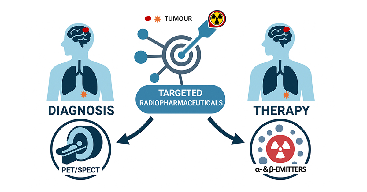
Recent advancements in radiopharmaceuticals have significantly advanced nuclear medicine, particularly in precision diagnostics and targeted therapy for cancer and neurological disorders. Among these innovations, targeted radioligand therapy (TRT) has emerged as a transformative approach that is already improving patient outcomes in prostate and neuroendocrine cancers.

TRT employs small carrier radioligands that function like precision-guided missiles. These carriers first home in on tumour-specific markers, delivering a cytotoxic payload of radioactivity directly to cancer cells while sparing most surrounding healthy tissue. Compared to external beam radiotherapy, TRTs often offer superior tumour targeting, allowing for higher radiation doses with fewer off-target effects.

Radiopharmaceuticals are now used globally on a daily basis, not only for cancer detection and staging but increasingly for therapy, particularly in oncology. They are also widely applied in the management of cardiovascular, endocrine, neurological, and psychiatric conditions. However, despite their growing clinical utility, key challenges persist in both their diagnostic and therapeutic applications.

Access to therapeutic radionuclides, particularly alpha- and beta-emitters, remains limited. The short physical half-lives of many isotopes necessitate their frequent production and rapid distribution, placing considerable logistical demands on infrastructure. The manufacturing process itself requires specialized facilities, trained personnel, and strict regulatory oversight, all of which vary widely between countries and often hinder clinical translation. In addition, radioligand therapies can be associated with off-target effects, especially in organs involved in metabolism and clearance. These challenges collectively restrict access to new agents and slow their translation from bench to bedside applications.

This Special Issue seeks to highlight recent developments in targeted radiopharmaceuticals for both imaging and therapy, with a focus on cancer and other systemic diseases. We welcome contributions on topics including novel radionuclide production and the design and preclinical/clinical evaluation of innovative radiotracers, cutting-edge TRT strategies, and technological advances that aim to streamline production and improve accessibility.

We welcome original research articles and comprehensive reviews.

Prof. Dr. Gabriela Kramer-Marek
Dr. Yingqing Lu
Guest Editors

Manuscript Submission Information

Manuscripts should be submitted online at www.mdpi.com by registering and logging in to this website. Once you are registered, click here to go to the submission form. Manuscripts can be submitted until the deadline. All submissions that pass pre-check are peer-reviewed. Accepted papers will be published continuously in the journal (as soon as accepted) and will be listed together on the special issue website. Research articles, review articles as well as short communications are invited. For planned papers, a title and short abstract (about 250 words) can be sent to the Editorial Office for assessment.

Submitted manuscripts should not have been published previously, nor be under consideration for publication elsewhere (except conference proceedings papers). All manuscripts are thoroughly refereed through a single-blind peer-review process. A guide for authors and other relevant information for submission of manuscripts is available on the Instructions for Authors page. Pharmaceutics is an international peer-reviewed open access monthly journal published by MDPI.

Please visit the Instructions for Authors page before submitting a manuscript. The Article Processing Charge (APC) for publication in this open access journal is 2900 CHF (Swiss Francs). Submitted papers should be well formatted and use good English. Authors may use MDPI's English editing service prior to publication or during author revisions.

Keywords

  • radiopharmaceuticals
  • diagnostics imaging
  • theranostics
  • radionuclides
  • radioligand therapy
  • cancer
  • neurological disorders
  • dosimetry
  • preclinical studies
  • clinical studies
  • clinical translation

Benefits of Publishing in a Special Issue

  • Ease of navigation: Grouping papers by topic helps scholars navigate broad scope journals more efficiently.
  • Greater discoverability: Special Issues support the reach and impact of scientific research. Articles in Special Issues are more discoverable and cited more frequently.
  • Expansion of research network: Special Issues facilitate connections among authors, fostering scientific collaborations.
  • External promotion: Articles in Special Issues are often promoted through the journal's social media, increasing their visibility.
  • Reprint: MDPI Books provides the opportunity to republish successful Special Issues in book format, both online and in print.

Further information on MDPI's Special Issue policies can be found here.

Published Papers (2 papers)

Order results
Result details
Select all
Export citation of selected articles as:

Research

15 pages, 1405 KB  
Article
[161Tb]Tb-BPAMD as a High-Affinity Agent for Skeletal Targeting: Radiochemical and Biodistribution Insights
by Magdalena Radović, Pavle Sitarica, Dragana Stanković, Marija Mirković, Drina Janković, Miloš Marić, Marko Perić, Sanja Vranješ-Đurić and Aleksandar Vukadinović
Pharmaceutics 2026, 18(3), 312; https://doi.org/10.3390/pharmaceutics18030312 - 28 Feb 2026
Viewed by 676
Abstract
Background: Bone-seeking radiopharmaceuticals based on bisphosphonates enable targeted therapy of skeletal metastases. They are suitable carriers for therapeutic radionuclides such as terbium-161 (161Tb), a β emitter that additionally releases short-range conversion and Auger electrons, which may enhance radiation dose [...] Read more.
Background: Bone-seeking radiopharmaceuticals based on bisphosphonates enable targeted therapy of skeletal metastases. They are suitable carriers for therapeutic radionuclides such as terbium-161 (161Tb), a β emitter that additionally releases short-range conversion and Auger electrons, which may enhance radiation dose delivery to small lesions. This study explored the potential of the well-established DOTA conjugated bisphosphonate BPAMD (4-{[(bis(phosphonomethyl))carbamoyl]methyl}-7,10-bis(carboxymethyl)-1,4,7,10 tetraazacyclododec-1-yl)acetic acid) radiolabeled with 161Tb as a bone-targeted radiopharmaceutical, focusing on the theranostic and radiophysical advantages conferred by the radionuclide. Methods: BPAMD was radiolabeled with 161Tb and 177Lu under mild conditions (pH 4.5, 95 °C, 30 min); subsequently, the radiochemical purity was assessed by radio-TLC. Physicochemical properties (charge, lipophilicity, protein binding), in vitro stability (saline and human serum, 48 h), and hydroxyapatite (HAP) binding were evaluated for [161Tb]Tb-BPAMD. Biodistribution was investigated in healthy Wistar rats (n = 3 per time point) at 2 h, 24 h, and 7 days post-injection. Computational density functional theory (DFT) analyses were performed to explore the coordination chemistry of Tb3+ and Lu3+ with BPAMD. Results: Both complexes achieved a radiochemical yield of greater than 98%. [161Tb]Tb-BPAMD exhibited negative charge, high hydrophilicity (logP = −3.92 ± 0.13), low protein binding (19.07 ± 1.01%), excellent radiochemical stability under simulated physiological conditions (>97% at 48 h), and strong hydroxyapatite affinity (>98% with ≥10 mg HAP). Biodistribution showed high, stable bone uptake (8.06% ID/g at 2 h; 6.70% ID/g at 24 h; 5.31% ID/g at 7 d) with rapid blood clearance (<0.001% ID/g at 24 h) and low non-target retention. To contextualize its performance, [161Tb]Tb-BPAMD was compared with [177Lu]Lu-BPAMD, which demonstrated similarly strong skeletal retention (8.74% ID/g at 2 h; 8.08% ID/g at 24 h; 5.25% ID/g at 7 d) but comparatively higher non-target organ uptake. DFT calculations indicate that both Tb3+ and Lu3+ favor octa-coordinated BPAMD complexes. Conclusions: [161Tb]Tb-BPAMD exhibits excellent radiochemical and pharmacokinetic properties, with enhanced biodistribution selectivity over [177Lu]Lu-BPAMD. Combined with the radiobiological advantages of 161Tb, it represents a promising theranostic candidate for targeted therapy of bone metastases. Full article
Show Figures

Graphical abstract

12 pages, 1359 KB  
Article
89Zr-girentuximab PET/CT Enables Noninvasive Assessment of Indeterminate Renal Masses and Metastatic Clear-Cell Renal Cell Carcinoma
by Yihan Cao, Jonathan Kim, Justin Talluto, Taylor McVeigh, Michael L. Blute, Douglas M. Dahl, Keyan Salari, Pedram Heidari and Shadi A. Esfahani
Pharmaceutics 2026, 18(2), 258; https://doi.org/10.3390/pharmaceutics18020258 - 19 Feb 2026
Viewed by 740
Abstract
Background: Indeterminate renal masses (IRMs) frequently require biopsy for characterization and often lead to unnecessary surgical interventions. 89Zr-girentuximab is a positron emission tomography (PET) radiopharmaceutical targeting carbonic anhydrase IX, a biomarker overexpressed in clear-cell renal cell carcinoma (ccRCC). This real-world experience demonstrates [...] Read more.
Background: Indeterminate renal masses (IRMs) frequently require biopsy for characterization and often lead to unnecessary surgical interventions. 89Zr-girentuximab is a positron emission tomography (PET) radiopharmaceutical targeting carbonic anhydrase IX, a biomarker overexpressed in clear-cell renal cell carcinoma (ccRCC). This real-world experience demonstrates the impact of 89Zr-girentuximab PET on the clinical management of patients with IRM and its role in differentiating primary and metastatic ccRCC from other etiologies. Methods: This prospective single-center study, part of an expanded access program (NCT06090331), investigated patients with IRM on conventional imaging who underwent 89Zr-girentuximab PET/computed tomography (PET/CT). Qualitative and quantitative PET/CT features of each lesion were assessed. Pathologic or clinical diagnosis was determined for all lesions. Referring physicians were surveyed to evaluate the impact of PET on patient management. Results: Seven male patients (age range, 57–78 years) were included; four had ccRCC (including two with metastatic disease) and three had oncocytoma (including one with Birt-Hogg-Dubé syndrome). Across all 32 lesions identified, 89Zr-girentuximab PET/CT accurately characterized each lesion based on pathologic or clinical diagnosis. 89Zr-girentuximab PET/CT identified ccRCC tumor thrombi in the inferior vena cava and renal vein branches (SUVmax 12.0–13.0), a perinephric deposit (SUVmax 36.4), and intramuscular (SUVmax 103.0), pulmonary (SUVmax 4.0–10.5), and osseous (SUVmax 10.2) metastases. 89Zr-girentuximab PET/CT enabled the diagnosis of oncocytomatosis in one patient and detected a renal lesion with positive uptake that was occult on MRI. According to referring physicians, 89Zr-girentuximab PET/CT changed clinical management in six of seven patients and improved patient care in all cases. Conclusions: 89Zr-girentuximab PET/CT provides a noninvasive tool for characterizing indeterminate renal masses and metastatic ccRCC and may improve clinical problem-solving in complex scenarios. Full article
Show Figures

Figure 1

Back to TopTop