Article

Analysis of the Roles of the ISLR2 Gene in Regulating the Toxicity of Zearalenone Exposure in Porcine Intestinal Epithelial Cells

1
Key Laboratory for Animal Genetics, Breeding, Reproduction and Molecular Design, College of Animal Science and Technology, Yangzhou University, Yangzhou 225009, China
2
Lvliang Central Animal Husbandry and Veterinary Station, Huaian 211600, China
3
Joint International Research Laboratory of Agriculture & Agri-Product Safety, Yangzhou University, Yangzhou 225009, China
*
Authors to whom correspondence should be addressed.
Toxins 2022, 14(9), 639; https://doi.org/10.3390/toxins14090639
Submission received: 7 August 2022 / Revised: 7 September 2022 / Accepted: 8 September 2022 / Published: 16 September 2022

Abstract

:
Zearalenone (ZEN) is one of the mycotoxins that pose high risks for human and animal health, as well as food safety. However, the regulators involved in ZEN cellular toxicity remain largely unknown. Herein, we showed that cell viability of porcine intestinal epithelial cells (IPEC-J2) tended to decrease with increasing doses of ZEN by the cell counting kit-8 assay. Expression of the ISLR2 (immunoglobulin superfamily containing leucine-rich repeat 2) gene in IPEC-J2 cells was significantly downregulated upon ZEN exposure. Furthermore, we found the dose–effect of ZEN on ISLR2 expression. We then overexpressed the ISLR2 gene and observed that overexpression of ISLR2 obviously reduced the effects of ZEN on cell viability, apoptosis rate and oxidative stress level. In addition, ISLR2 overexpression significantly decreased the expression of TNF-α and IFN-α induced by ZEN. Our findings revealed the effects of ZEN on the ISLR2 gene expression and indicated the ISLR2 gene as a novel regulator of ZEN-induced cytotoxicity, which provides potential molecular targets against ZEN toxicity.
Key Contribution: This study reveals the functional roles of the ISLR2 gene in alleviating zearalenone-induced toxic effects in porcine epithelial cells, shows the dose–effect of ZEN on the expression of the ISLR2 gene, and highlights the potentiality of ISLR2 as a molecular target against ZEN toxicity.

1. Introduction

Mycotoxins are highly toxic secondary products produced by fungi species such as Aspergillus, Penicillium, and Fusarium. The toxic syndrome caused by the ingestion of mycotoxins in humans and animals is called mycotoxicosis [1,2]. Mycotoxicity causes the development of cancer, mutations and malformations, as well as toxic reactions in cells, nerves and kidneys and other guts [1]. Several mycotoxins are particularly important for public health and agronomic development, including aflatoxin, ochratoxin A, fumonisin, deoxynivalenol and zearalenone (ZEN) [2]. ZEN is a non-steroidal estrogenic mycotoxin [3]. From a structural point of view, ZEN has a lactone ring, a feature that allows it to be heat resistant up to 150 °C [4], and thus it shows high stability during both post-processing and storage. This characteristic of ZEN stability at increased temperature and pressure makes it detectable in fresh plants and processed products [5,6]. The non-degradability of mycotoxins at high temperature and pressure results in great potential harm of mycotoxin contamination for food security.
Among the farm animals, pigs are the most significantly affected species and are much more sensitive to zearalenone than rodents and other farm animals. The intestine is the main organ that absorbs ZEN. Ingestion of zearalenone-contaminated feed by livestock can cause damage to their intestinal function and integrity, which causes the occurrence of diseases. ZEN induces apoptosis and oxidative stress in porcine intestinal epithelial cells, and these cellular damages induced by ZEN are closely linked to several key signaling pathways; it has been shown that glutamine and DL-selenomethionine play a mitigating role in ZEN-induced apoptosis and oxidative stress through PI3K/Akt and Nrf2/Keap1 signaling pathways, respectively [7,8]. According to previous studies, ZEN mainly causes structural damage in the porcine jejunum, inducing inflammation and apoptosis of jejunal epithelial cells [9,10,11]. However, the underlying mechanism of ZEN-induced jejunal epithelial injury is largely unknown.
ISLR2 (immunoglobulin superfamily containing leucine-rich repeat 2) is a member of the leucine-rich repeat (LRR) and immunoglobulin (LIG) family of membrane proteins, which is preferentially expressed in the central and peripheral nervous systems [12]. It is surprising that few molecules contain both sequence elements. In contrast, the nature of the LRR motif in the ISLR2 gene is important for generating various interactions with exogenous factors in the immune system and with a large number of different cell types in the developing nervous system [13]. Both intracellular and extracellular LRR proteins, are well characterized in the natural immune system, from plants to mammals [14]. Interactions of ISLR2 with ret proto-oncogene was involved in regulating the development of the gastrointestinal tract, and alterations of ISLR2 expression levels were found to be associated with pseudoexfoliation syndrome [12,15]. We previously found that the expression of ISLR2 was significantly changed in porcine intestinal epithelial cells upon ZEN exposure [16]. However, the regulatory mechanisms and functions of ISLR2 in response to ZEN exposure remain unknown.
In this study, we explored the effects of ZEN on the expression of the ISLR2 gene in porcine intestinal epithelial cells (IPEC-J2). In addition, we investigated the roles of the ISLR2 gene in regulating the cytotoxic effects induced by ZEN. Our findings will provide insight into the roles of the ISLR2 gene in cellular responses to ZEN exposure and enhance our understanding of the molecular mechanisms involved in the toxicological processes of ZEN.

2. Results

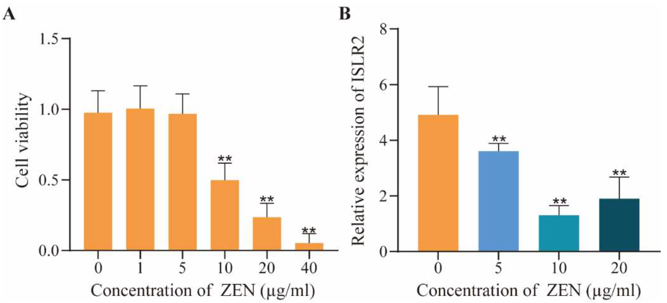
2.1. Dose-Effect of ZEN on Cell Viability and ISLR2 Expression

We first determined the optimal doses of ZEN on IPEC-J2 cells using cell viability analysis and found that the cell viability was significantly decreased (p < 0.01) and around 50% at a dose of 10 μg/mL compared with the control (Figure 1A). Meanwhile, we detected the expression of ISLR2 by qPCR at different ZEN doses and found that the expression of ISLR2 was significantly decreased (p < 0.01) with a higher dose of ZEN in comparison with the control (Figure 1B).

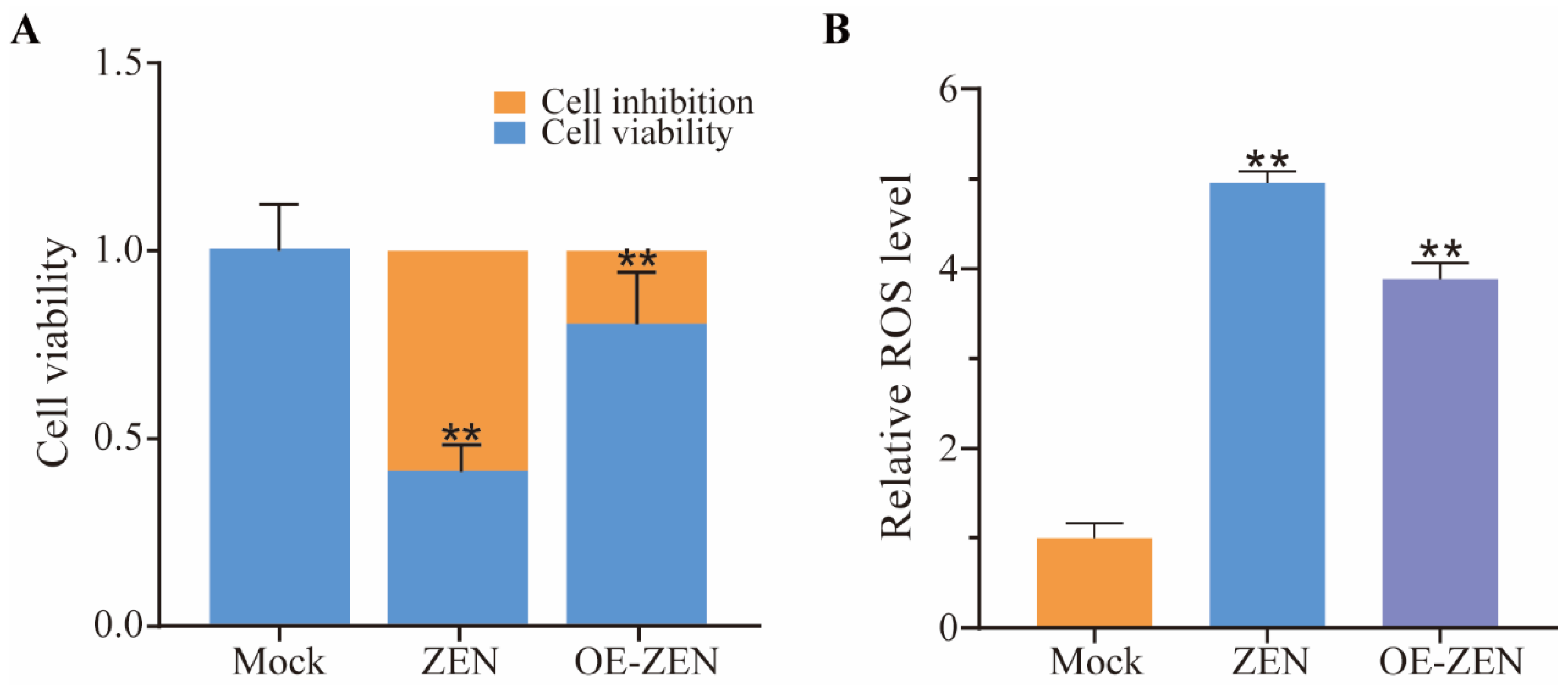
2.2. The Role of ISLR2 in ZEN Induced Cytotoxicity and Oxidative Stress

To investigate the role of ISLR2, we constructed the plasmids overexpressing ISLR2. The recombinant plasmid containing the ISLR2 coding sequence was confirmed by agarose gel analysis (Supplementary Materials, Figure S1A) and DNA sequencing, which shows consistent sequences with the known sequences of the ISLR2 coding region (Supplementary Materials, Figure S1B). Expression analysis indicated that ISLR2 expression in the overexpression group was more than 900-fold higher than that in the control group (p < 0.01) (Supplementary Materials, Figure S1C). To investigate the functions of ISLR2 in response to ZEN-induced toxicity, we analyzed cell viability and ROS level in cells overexpressing ISLR2. The results showed that the cell viability of ZEN-treated cells was significantly decreased compared with the control group, while the cell viability in cells overexpressing ISLR2 was significantly higher than that of the ZEN exposure group (Figure 2A). In addition, after ZEN treatment, ROS levels in cells overexpressing ISLR2 were significantly lower than that of control cells (p < 0.01) (Figure 2B), indicating the involvement of ISLR2 in mediating ZEN-induced ROS production.

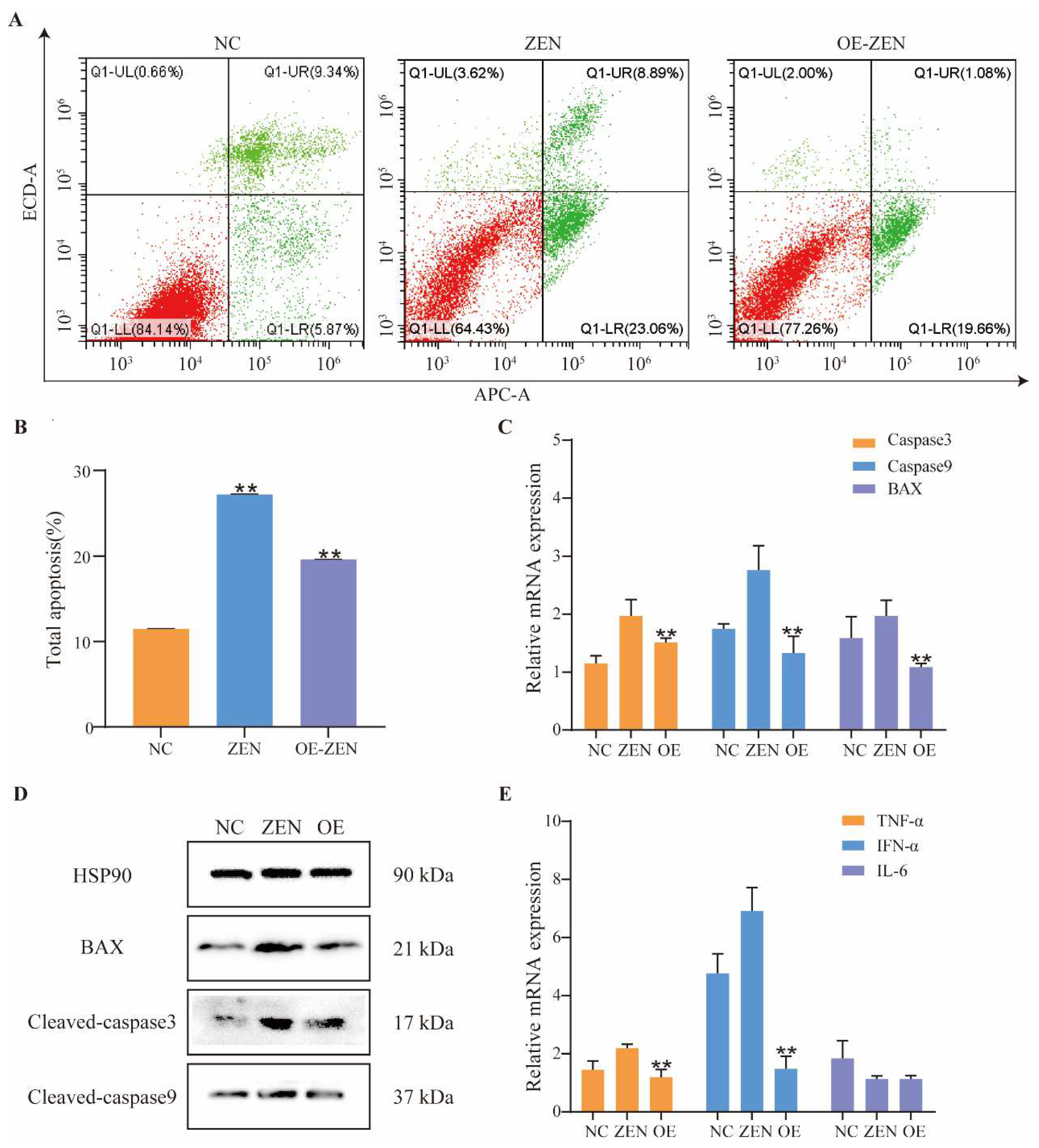
2.3. Role of ISLR2 in ZEN-Induced Cell Apoptosis and Inflammation

To further investigate the roles of ISLR2 in ZEN-induced toxicity, we detected the apoptotic level of cells exposed to ZEN by Annexin V-APC/PI. The results showed that the total apoptotic cells increased after ZEN exposure. The apoptosis rate in cells overexpressing ISLR2 was significantly lower than that of control cells (p < 0.01) (Figure 3A,B).
Expression analysis of apoptosis-related proteins showed that the expression of Caspase3, Caspase9, and BAX in cells overexpressing ISLR2 was significantly lower than that of the control cells (Figure 3C) (p < 0.01). Western blot assay further confirmed the expression of these proteins (Figure 3D). Furthermore, we detected the expression of proinflammatory cytokines (TNF-α, IL-6) and interferon IFN-α. The results showed that ISLR2 overexpression significantly decreased the expression of TNF-α and IFN-α induced by ZEN (p < 0.01), while the IL-6 expression level did not change significantly (Figure 3E).

3. Discussion

ZEN exerts different mechanisms of toxicity in different cell types at different doses, which leads to estrogen-like effects at low doses and cell death at high doses [17]. Previous studies showed that ZEN induces HSP70 expression in a time and dose-dependent manner in HEPG2 cells [18]. Under different doses of DON treatment, DON inhibited expression of the TEM8 gene that plays important roles in cell migration, and the inhibitory effects of DON on cell migration could be reduced by overexpression of TEM8 [19]. In this study, we revealed the dose–effect of ZEN on ISLR2 expression in IPEC-J2 cells. These findings indicated that the expression of some genes is responsive to mycotoxin doses, and these genes may be important potential regulators involved in mycotoxin toxicological processes.
ZEN exposure can result in an imbalance of oxidative and antioxidant effects, allowing the massive production of free radicals. The massive accumulation of free radicals leads to the destruction of DNA, proteins, and lipids [20]. The accumulation of excessive ROS is one of the causes of apoptosis. Previous studies have reported that SelS overexpression mitigates OTA-induced cytotoxicity and apoptosis [21]. In addition, overexpression of HO-1 reduces the DON-induced ROS and DNA damage by maintaining DNA repair, antioxidant activity, and autophagy [22]. In this study, we observed that ISLR2 overexpression significantly increased the cell viability and reduced the ROS levels of cells exposed to ZEN, indicating the potential of ISLR2 as a molecular target for controlling ZEN-induced toxicity.
After ISLR2 gene overexpression, the toxicity of ZEN in IPEC-J2 cells could be inhibited. It has been reported that ISLR2 is composed of two parts: leucine-rich repeat (LRR) and immunoglobulin (LIG) families, while LRR in them plays important roles in the innate immune system [13], indicating that the function of ISLR2 may be related to immunity. According to relevant reports, the loss of ISLR in stromal cells significantly impaired the regeneration of the intestine and inhibited the development of colon tumors [23]. In non-small cell lung cancer (NSCLC), silencing ISLR increased the apoptotic rate of cells [24]. In chicken myoblasts, silencing ISLR similarly caused significantly higher expression of apoptosis-related proteins such as Caspase3, Caspase8, and Caspase9 [25]. These results suggested that inhibition of ISLR expression may be detrimental to cell proliferation, and we therefore speculate that ISLR2 may play a similar role as a paralogous homolog of ISLR. Combined with the analysis of our results, we suggest that high expression of ISLR2 contributes to increased cellular resistance to ZEN through oxidative stress and apoptosis. Herein, our data showed that high expression of ISLR2 decreases the expression of these inflammatory factors, suggesting that ISLR2 may play a role in the inflammatory response. Taken together, our results suggest that ISLR2 alleviated the damage caused by ZEN by reducing apoptosis and alleviating inflammatory responses.

4. Conclusions

In conclusion, we found the dose–effect of ZEN on the expression of the ISLR2 gene and explored the functions of ISLR2 expression in mitigating the cytotoxicity induced by ZEN exposure. Findings of this study shed light on the roles of ISLR2 in IPEC-J2 cells upon ZEN exposure and provided potential molecular targets for protection against ZEN cytotoxicity.

5. Materials and Methods

5.1. Construction of ISLR2 Overexpression Vector

The CDS region of ISLR2 was amplified by PCR with the addition of restriction of endonucleases digestion sites. A 50 μL reaction system consisted of 200 ng cDNA, 1.5 µL each of forward and reverse primers (10 µmol/L), 25 µL 2×PCR Buffer, 10 µL 2 mM dNTPs, 1.0 µL KOD FX (1.0 U/µL) mixture, and ddH2O (50 µL total) (Toyobo, Osaka, Japan). PCR reactions were as follows: 94 °C for 2 min, 40 cycles of 98 °C for 10 s, 68 °C for 2 min. PCR products were double digested and then ligated into the linear pcDNA3.1 plasmid using T4 DNA ligase at 16 °C overnight (Vazyme, Nanjing, China). PCR products were confirmed by agarose gel and Sanger sequencing. The plasmids expressing ISLR2 were transfected into cells using jetPRIME (Polyplus, Illkirch, France) following the manufacturer’s protocols. The primer information is shown in Table 1.

5.2. Cell Culture

IPEC-J2 cells were plated into 6-well and 12-well plates and incubated in DMEM medium containing 1% penicillin streptomycin (1 mg/mL) and 10% FBS at 37 °C and 5% CO2 in an incubator.

5.3. Cell Viability Assay

Cells were plated into 96-well plates at a density of 2000 cells per well. After overnight incubation, cells were treated with different concentrations (0, 1, 5, 10, 20, 40 μg/mL) of ZEN and incubated for 48 h. Cell viability was measured using CCK8 reagents following the manufacturer’s protocols. The absorbance at 450 nm was quantified on a Tecan Infinite 200 microplate reader (Tecan, Männedorf, Switzerland).

5.4. Cell Apoptosis Detection

Cells were plated on six-well plates and exposed to 10 μg/mL of ZEN for 48 h. Cell samples were washed with PBS and sequentially stained with Annexin V-APC and (PI) using the Annexin V-APC/PI Apoptosis Kit (Elabscience, Wuhan, China). The stained samples were analyzed by flow cytometry and followed by CytExpert 2.3 (Beckman Coulter, Brea, CA, USA) to count cell apoptosis rate.

5.5. Determination of Oxidative Stress Index

Cells were plated on 6-well plates and exposed to 10 μg/mL of ZEN for 48 h. Cell samples were washed twice with pre-chilled PBS and then incubated with 10 µM DCHF-DA for 30 min at 37 °C. ROS levels in cells were measured with Reactive Oxygen Species Analysis Kit (Solarbio, Beijing, China) and analyzed by flow cytometry (Beckman Coulter, Brea, CA, USA).

5.6. qRT-PCR

Total RNA was isolated using the Trizol method. Reverse transcription was performed with the HiScript II Q Select RT SuperMix for qPCR kit (Vazyme, Nanjing, China). The cDNA was used as the template. The reactions were performed on a real-time PCR machine (ABI Step One Plus, Pleasanton, CA, USA). The GAPDH gene was used as the housekeeping gene. Gene relative expression was calculated by the 2−ΔΔCT method [26]. The primer sequences of each gene are shown in Table 1.

5.7. Western Blot Analysis

Cells were washed twice with pre-cooled PBS, mixed with RIPA lysate (Applygen, Beijing, China) and protease inhibitor (CWBIO, Beijing, China). Total proteins were isolated by utilizing a protein extraction reagent, denatured by boiling for 10 min, separated with SDS-PAGE, and transferred to PVDF membranes. Blocking of the membrane was performed with 5% non-fat dry milk and 0.2%Tween for 2 h at room temperature. Proteins were incubated with antibodies, including anti-Cleaved-Caspase3, anti-Cleaved-Caspase9, anti-BAX and anti-HSP90, overnight at 4 °C. Incubation with secondary antibody for 2 h was then conducted at room temperature. Finally, the bands were detected by ECL.

Supplementary Materials

The following supporting information can be downloaded at: https://www.mdpi.com/article/10.3390/toxins14090639/s1, Figure S1. Construction of ISLR2 overexpression cells. (A) Enzyme-digested products of the plasmids overexpressing ISLR2 by agarose gel. M: 1 kb ladder; S1: enzyme-digested products of the plasmids with XhoI; S2: products of undigested sample; (B) PCR sequencing of the plasmids overexpressing ISLR2. (C) Efficiency of ISLR2 overexpression by qRT-PCR.NC, cells transfected with an empty vector; OE-ISLR2, cells transfected with an ISLR2 overexpression vector. Data are presented as mean ± SD. Significance compared with control, ** p < 0.01.

Author Contributions

Conceptualization, H.W. and S.W.; methodology, Z.B. and Y.X.; validation, Z.B., X.G.; formal analysis, Z.B., X.G., and Y.Z.; resources, S.W. and W.B.; writing—original draft preparation, Z.B., X.G., and H.W.; writing—review and editing, H.W., W.B., and S.W. All authors have read and agreed to the published version of the manuscript.

Funding

This study was financially supported by Seed Industry Vitalization Research Projects of Jiangsu Province (JBGS [2021]024), Key Research and Development Project (Modern Agriculture) of Jiangsu Province (BE2019341), Qing Lan Project of Yangzhou University, and Priority Academic Program Development of Jiangsu Higher Education Institution.

Institutional Review Board Statement

Not applicable.

Informed Consent Statement

Not applicable.

Data Availability Statement

Not applicable.

Conflicts of Interest

The authors declare no conflict of interest.

References

  1. Abrunhosa, L.; Morales, H.; Soares, C.; Calado, T.; ila-Chã, A.S.; Pereira, M.; Venâncio, A. A Review of Mycotoxins in Food and Feed Products in Portugal and Estimation of Probable Daily Intakes. Crit. Rev. Food. Sci. Nutr. 2016, 56, 249–265. [Google Scholar] [CrossRef] [PubMed]
  2. Galvano, F.; Piva, A.; Ritieni, A. Dietary strategies to counteract the effects of mycotoxins: A review. J. Food. Prot. 2001, 64, 120–131. [Google Scholar] [CrossRef] [PubMed]
  3. Bennett, J.W.; Klich, M. Mycotoxins. Clin. Micro. Rev. 2003, 16, 497–516. [Google Scholar] [CrossRef]
  4. Yumbe-Guevara, B.E.; Imoto, T.; Yoshizawa, T. Effects of heating procedures on deoxynivalenol, nivalenol and zearalenone levels in naturally contaminated barley and wheat. Food. Addit. Contam. 2003, 20, 1132–1140. [Google Scholar] [CrossRef]
  5. Cheli, F.; Pinotti, L.; Rossi, L.; Dell’Orto, V. Effect of milling procedures on mycotoxin distribution in wheat fractions: A review. LWT-Food Sci. Technol. 2013, 54, 307–314. [Google Scholar] [CrossRef]
  6. Heidari, S.; Milani, J.; Nazari, S.S. Effect of the bread-making process on zearalenone levels. Food Addit. Contam. Part A 2014, 31, 2047–2054. [Google Scholar] [CrossRef] [PubMed]
  7. Wang, T.; Wang, J.; Zhang, T.; Gu, A.; Li, J.; Shan, A. The Antagonistic Effect of Glutamine on Zearalenone-Induced Apoptosis via PI3K/Akt Signaling Pathway in IPEC-J2 Cells. Toxins 2021, 13, 891. [Google Scholar] [CrossRef]
  8. Sun, H.; Zhang, M.; Li, J.; Shan, A. DL-Selenomethionine Alleviates Oxidative Stress Induced by Zearalenone via Nrf2/Keap1 Signaling Pathway in IPEC-J2 Cells. Toxins 2021, 13, 557. [Google Scholar] [CrossRef]
  9. Shen, T.; Miao, Y.; Ding, C.; Fan, W.; Liu, S.; Lv, Y.; Gao, X.; De Boevre, M.; Yan, L.; Okoth, S.; et al. Activation of the p38/MAPK pathway regulates autophagy in response to the CYPOR-dependent oxidative stress induced by zearalenone in porcine intestinal epithelial cells. Food Chem. Toxicol. 2019, 131, 110527. [Google Scholar] [CrossRef]
  10. Cao, L.; Zhao, J.; Ma, L.; Chen, J.; Xu, J.; Rahman, S.U.; Feng, S.; Li, Y.; Wu, J.; Wang, X. Lycopene attenuates zearalenone-induced oxidative damage of piglet sertoli cells through the nuclear factor erythroid-2 related factor 2 signaling pathway. Ecotoxicol. Environ. Saf. 2021, 225, 112737. [Google Scholar] [CrossRef]
  11. Fan, W.; Shen, T.; Ding, Q.; Lv, Y.; Li, L.; Huang, K.; Yan, L.; Song, S. Zearalenone induces ROS-mediated mitochondrial damage in porcine IPEC-J2 cells. J. Biochem. Mol. Toxicol. 2017, 31, e21944. [Google Scholar] [CrossRef]
  12. Mandai, K.; Guo, T.; St Hillaire, C.; Meabon, J.S.; Kanning, K.C.; Bothwell, M.; Ginty, D.D. LIG family receptor tyrosine kinase-associated proteins modulate growth factor signals during neural development. Neuron 2009, 63, 614–627. [Google Scholar] [CrossRef] [PubMed]
  13. Dolan, J.; Walshe, K.; Alsbury, S.; Hokamp, K.; O’Keeffe, S.; Okafuji, T.; Miller, S.F.; Tear, G.; Mitchell, K.J. The extracellular leucine-rich repeat superfamily; a comparative survey and analysis of evolutionary relationships and expression patterns. BMC Genom. 2007, 8, 320. [Google Scholar] [CrossRef] [PubMed]
  14. Nürnberger, T.; Brunner, F.; Kemmerling, B.; Piater, L. Innate immunity in plants and animals: Striking similarities and obvious differences. Immunol. Rev. 2004, 198, 249–266. [Google Scholar] [CrossRef] [PubMed]
  15. Berner, D.; Hoja, U.; Zenkel, M.; Ross, J.J.; Uebe, S.; Paoli, D.; Frezzotti, P.; Rautenbach, R.M.; Ziskind, A.; Williams, S.E.; et al. The protective variant rs7173049 at LOXL1 locus impacts on retinoic acid signaling pathway in pseudoexfoliation syndrome. Hum. Mol. Genet. 2019, 28, 2531–2548. [Google Scholar] [CrossRef]
  16. Wang, H.; Jin, J.; Wu, J.; Qu, H.; Wu, S.; Bao, W. Transcriptome and chromatin accessibility in porcine intestinal epithelial cells upon Zearalenone exposure. Sci. Data 2019, 6, 298. [Google Scholar] [CrossRef]
  17. Zheng, W.; Wang, B.; Li, X.; Wang, T.; Zou, H.; Gu, J.; Yuan, Y.; Liu, X.; Bai, J.; Bian, J.; et al. Zearalenone Promotes Cell Proliferation or Causes Cell Death? Toxins 2018, 10, 184. [Google Scholar] [CrossRef]
  18. Hassen, W.; El Golli, E.; Baudrimont, I.; Mobio, A.T.; Ladjimi, M.M.; Creppy, E.E.; Bacha, H. Cytotoxicity and Hsp70 induction in HepG2 cells in response to zearalenone and cytoprotection by sub-lethal heat shock. Toxicology 2005, 207, 293–301. [Google Scholar] [CrossRef]
  19. Mu, H.; Mu, P.; Zhu, W.; Huang, B.; Li, H.; Yuan, L.; Deng, Y. Low doses of deoxynivalenol inhibit the cell migration mediated by H3K27me3-targeted downregulation of TEM8 expression. Biochem. Pharmacol. 2020, 175, 113897. [Google Scholar] [CrossRef]
  20. Zinedine, A.; Soriano, J.M.; Molto, J.C.; Manes, J. Review on the toxicity, occurrence, metabolism, detoxification, regulations and intake of zearalenone: An oestrogenic mycotoxin. Food Chem. Toxicol. 2007, 45, 1–18. [Google Scholar] [CrossRef]
  21. Gan, F.; Hu, Z.; Zhou, Y.; Huang, K. Overexpression and Low Expression of Selenoprotein S Impact Ochratoxin A-Induced Porcine Cytotoxicity and Apoptosis In Vitro. J. Agric. Food Chem. 2017, 65, 6972–6981. [Google Scholar] [CrossRef] [PubMed]
  22. Meng, Z.; Wang, L.; Liao, Y.; Peng, Z.; Li, D.; Zhou, X.; Liu, S.; Li, Y.; Nüssler, A.K.; Liu, L.; et al. The Protective Effect of Heme Oxygenase-1 on Liver Injury Caused by DON-Induced Oxidative Stress and Cytotoxicity. Toxins 2021, 13, 732. [Google Scholar] [CrossRef] [PubMed]
  23. Xu, J.; Tang, Y.; Sheng, X.; Tian, Y.; Deng, M.; Du, S.; Lv, C.; Li, G.; Pan, Y.; Song, Y.; et al. Secreted stromal protein ISLR promotes intestinal regeneration by suppressing epithelial Hippo signaling. EMBO J. 2020, 39, e103255. [Google Scholar] [CrossRef] [PubMed]
  24. Zhang, P.; Li, Z.; Yang, G. Silencing of ISLR inhibits tumour progression and glycolysis by inactivating the IL6/JAK/STAT3 pathway in nonsmall cell lung cancer. Int. J. Mol. Med. 2021, 48, 222. [Google Scholar] [CrossRef]
  25. Cui, C.; Han, S.; Shen, X.; He, H.; Chen, Y.; Zhao, J.; Wei, Y.; Wang, Y.; Zhu, Q.; Li, D.; et al. ISLR regulates skeletal muscle atrophy via IGF1-PI3K/Akt-Foxo signaling pathway. Cell Tissue Res. 2020, 381, 479–492. [Google Scholar] [CrossRef]
  26. Livak, K.J.; Schmittgen, T.D. Analysis of relative gene expression data using real-time quantitative PCR and the 2(-Delta Delta C(T)) Method. Methods 2001, 25, 402–408. [Google Scholar] [CrossRef]
Figure 1. Effects of ZEN on cell viability and ISLR2 expression. (A) Detection of cell viability of cells exposed to different doses (0, 1, 5, 10, 20, 40 μg/mL) of ZEN. (B) ISLR2 expression changes in cells exposed to different doses (0, 5, 10, 20 μg/mL) of ZEN for 48 h. Different color of bars represents different concentration of ZEN. Data are presented as mean ± SD. Significance compared with control, ** p < 0.01.
Toxins 14 00639 g001
Figure 2. Effects of ISLR2 overexpression on ZEN-induced cytotoxicity and oxidative stress. (A) Detection of cell viability in the Mock, ZEN, and OE-ZEN groups. (B) Relative ROS levels in the Mock, ZEN, and OE-ZEN groups. Mock, cells without ZEN treatment; ZEN, cells exposed to 10 μg/mL ZEN; OE-ZEN, cells overexpressing ISLR2 and exposed to 10 μg/mL ZEN. Data are presented as mean ± SD. Significance compared with control, ** p < 0.01.
Toxins 14 00639 g002
Figure 3. ISLR2 overexpression alleviates ZEN-induced cell apoptosis and inflammation. (A) Cell apoptosis rate determined by annexin V-APC/PI staining using flow cytometry. Red and green color indicate the percentage of normal cells and apoptotic cells in the total cells, respectively. (B) Quantification of cell apoptosis rate in NC, ZEN, and OE-ZEN groups. (C) Relative expression of Caspase3, Caspase9 and BAX in the NC, ZEN, and OE groups. (D) Protein expression of Cleaved-Caspase3, Cleaved-Caspase9, and BAX in the NC, ZEN and OE groups. (E) Relative expression of TNF-α, IFN-α, and IL-6 in the NC, ZEN, and OE groups. NC, cells without ZEN treatment; ZEN, cells exposed to 10 μg/mL ZEN; OE-ZEN, cells overexpressing ISLR2 and exposed to 10 μg/mL ZEN. Data are presented as mean ± SD. Significance compared with control, ** p < 0.01.
Toxins 14 00639 g003
Table 1. Primer sequences used in qRT-PCR and PCR assays.
GeneSequence (5′–3′)Product Length
GAPDHF:GGTCGGAGTGAACGGATTT
R:ATTTGATGTTGGCGGGAT
245 bp
ISLR2F:GGTCCAAGGCGAGGTTGC
R:CGAACTGATGCGCGTACTTG
242 bp
Caspase3F:GGAATGGCATGTCGATCTGGT
R:ACTGTCCGTCTCAATCCCAC
351 bp
Caspase9F:TGGAACTCAAGCCAGAGGAG
R:CTGCATTCAGGACGTAAGCC
195 bp
BAXF:TGCCTCAGGATGCATCTACC
R:AAGTAGAAAAGCGCGACCAC
199 bp
TNF-aF:TTCCAGCTGGCCCCTTGAGC
R:GAGGGCATTGGCATACCCAC
146 bp
IL-6F:TTCACCTCTCCGGACAAAAC
R:TCTGCCAGTACCTCCTTGCT
122 bp
OE-ISLR2F:ttAAGCTTgccaccATGGGCTCCAGCCCAGAC
R:caGAATTCTCAGCCTGCTGTCTGCCTATAG
2431 bp
ISLR2-SNPF:CTGGGTTTAGGTATGTTAGA
R:ACACTGGCTCAGGACTC
419 bp
Note: For the primer of OE-ISLR2, the underlined letters indicate the restriction enzyme cutting sites, the italic is the Kozak sequence, and the lowercase is the protected base of the restriction enzyme cutting site.
Publisher’s Note: MDPI stays neutral with regard to jurisdictional claims in published maps and institutional affiliations.

