Review

Exploring the Antimicrobial Potential of Natural Substances and Their Applications in Cosmetic Formulations

by
Katarzyna Kulik-Siarek
1,*,
Marta Klimek-Szczykutowicz
1,
Ewelina Błońska-Sikora
1,
Emilia Zarembska
2 and
Małgorzata Wrzosek
1,3,*
1
Department of Pharmaceutical Sciences, Collegium Medicum, Jan Kochanowski University, IX Wieków Kielc19a, 25-516 Kielce, Poland
2
Student Scientific Association “Farmakon”, Medical University of Warsaw, 1 Banacha St., 02-097 Warsaw, Poland
3
Department of Biochemistry and Pharmacogenomics, Medical University of Warsaw, 1 Banacha St., 02-097 Warsaw, Poland
*
Authors to whom correspondence should be addressed.
Cosmetics 2025, 12(1), 1; https://doi.org/10.3390/cosmetics12010001
Submission received: 21 November 2024 / Revised: 13 December 2024 / Accepted: 25 December 2024 / Published: 29 December 2024

Abstract

:
The aim of this review is to analyze natural substances exhibiting antibacterial and antifungal activity against skin pathogens, along with their exemplary applications in cosmetic products. Growing concerns related to increasing infection rates and pathogen resistance have prompted the search for alternative therapeutic methods. This article discusses various natural products, derived from plants, animals, and minerals, with antimicrobial potential. Special attention is given to the antimicrobial efficacy of natural substances derived from Allium L., Salvia L., Lavandula L., Origanum L., Melaleuca alternifolia, Aloe vera, Black Cumin, and Trigonella L. in improving treatment outcomes, either alone or in combination with conventional medications. In addition, the presented natural products, such as propolis, honey, cosmetic mud, and clays, can serve as viable alternatives or complementary treatments for mild skin infections and may help prevent recurrence. The promising potential of these natural products encourages further research into discovering new antimicrobial agents. However, the lack of standardization of natural preparations can result in inconsistent therapeutic effects and unforeseen side effects. This review significantly contributes to the pharmaceutical and cosmetic industries by emphasizing the potential of natural products and highlighting the need for further research and regulatory measures to ensure their safe and effective integration with existing therapies.

1. Introduction

Skin infections represent a serious global health issue. The incidence of skin infections is influenced by various factors, including geographical region, socio-economic status, individual sensitivity, and overall health status [1]. Skin and soft tissue infections can manifest as standalone conditions or complications of chronic diseases such as atopic dermatitis, diabetes, or post-surgical wounds. The most common include acne vulgaris, folliculitis, furuncles (or carbuncles), impetigo, cellulitis, erysipelas, cutaneous mycobacterial infections, erythematosquamous dermatitis, pitted keratolysis, dermatophytosis (affecting glabrous skin, hairy skin, or nails), green nail syndrome, cutaneous candidiasis, pityriasis versicolor, and infections following aesthetic medical procedures. Table 1 presents the main bacterial and fungal skin diseases and the microorganisms that cause these conditions.
Data obtained from the Global Burden of Disease (GBD) study show an increase in skin and subcutaneous diseases worldwide, with fungal diseases accounting for 34% and bacterial infections constituting 23% of all dermatological conditions [17]. During systemic analysis of bacterial skin diseases, the highest incidence was observed for pyoderma, with an increase in its occurrence [18]. A study in Poland conducted between 2010 and 2014, involving 1765 patients, reported that 49% of infections were caused by dermatophytes, 39.8% by yeast-like fungi, 9.1% by mold fungi, and 2.1% by mixed infections. The most frequently isolated pathogens from affected areas, including glabrous skin, nails, hands, feet, and scalp, were Trichophyton rubrum, Trichophyton mentagrophytes, Microsporum canis, Epidermophyton floccosum, Candida albicans, Candida glabrata, Rhodotorula spp., Malassezia furfur, Scopulariopsis brevicaulis, Aspergillus spp., and Alternaria spp. [19].
Studies conducted in India on 250 patients confirmed that the majority of superficial fungal infections of the skin, hair, and nails were caused by Candida albicans, dermatophytes such as Trichophyton rubrum and Trichophyton mentagrophytes, and non-dermatophytic molds [1].
According to data from 2020–2021, collected from patients visiting outpatient clinics and hospital wards in Pakistan, the most frequently isolated pathogens were Escherichia coli, Klebsiella spp., Pseudomonas aeruginosa, and Staphylococcus aureus [20].
Other findings were corroborated in Nigeria, where pathogens were isolated from 80 samples taken from purulent skin lesions. The results showed that Staphylococcus aureus had the highest percentage occurrence, followed by Escherichia coli, Pseudomonas aeruginosa, and Proteus sp. [21].
Bacterial skin infections can range from mild to severe and may present with accompanying symptoms depending on the depth of the infection. They are classified as superficial or deep, with differing treatment approaches and prognoses. In addition, antimicrobial resistance among fungal and bacterial pathogens poses a growing challenge to public health. It represents a direct threat to patient life and health while generating high treatment costs. Among the bacterial pathogens exhibiting antimicrobial resistance, the most frequently cited are Staphylococcus aureus, Enterococcus spp., Escherichia coli, Proteus spp., Klebsiella spp., and Pseudomonas spp. [22,23]. Fungal pathogens showing resistance include, among others, Trichophyton spp., Malassezia spp., and Candida spp. [24].
Accurate diagnosis of skin and soft tissue infections is crucial for effective treatment [25] According to analyses conducted by Zilberberg et al., inadequate treatment of skin infections most frequently occurred in patients hospitalized due to device-related infections, pressure ulcers, cellulitis, postoperative wound infections, and abscesses. The pathogens that were inadequately treated mainly included Enterococcus faecalis Enterococcus faecium, vancomycin-resistant Enterococcus, P. aeruginosa, E. coli, Klebsiella sp., polymicrobial infections, and mixed infections [26].
In light of the increasing demand for antimicrobial agents, preparations based on natural substances may become adjuncts in treatment and preventive measures for various dermatological diseases [27,28]. In this review, substances with antimicrobial potential are classified into plant-derived natural substances, animal-derived substances, and mineral-derived natural substances, accompanied by a critical analysis of their advantages and disadvantages. Their effects against pathogenic bacteria and fungi are discussed through both in vitro and in vivo studies. Additionally, cosmetic formulations available on the market containing studied substances are presented.

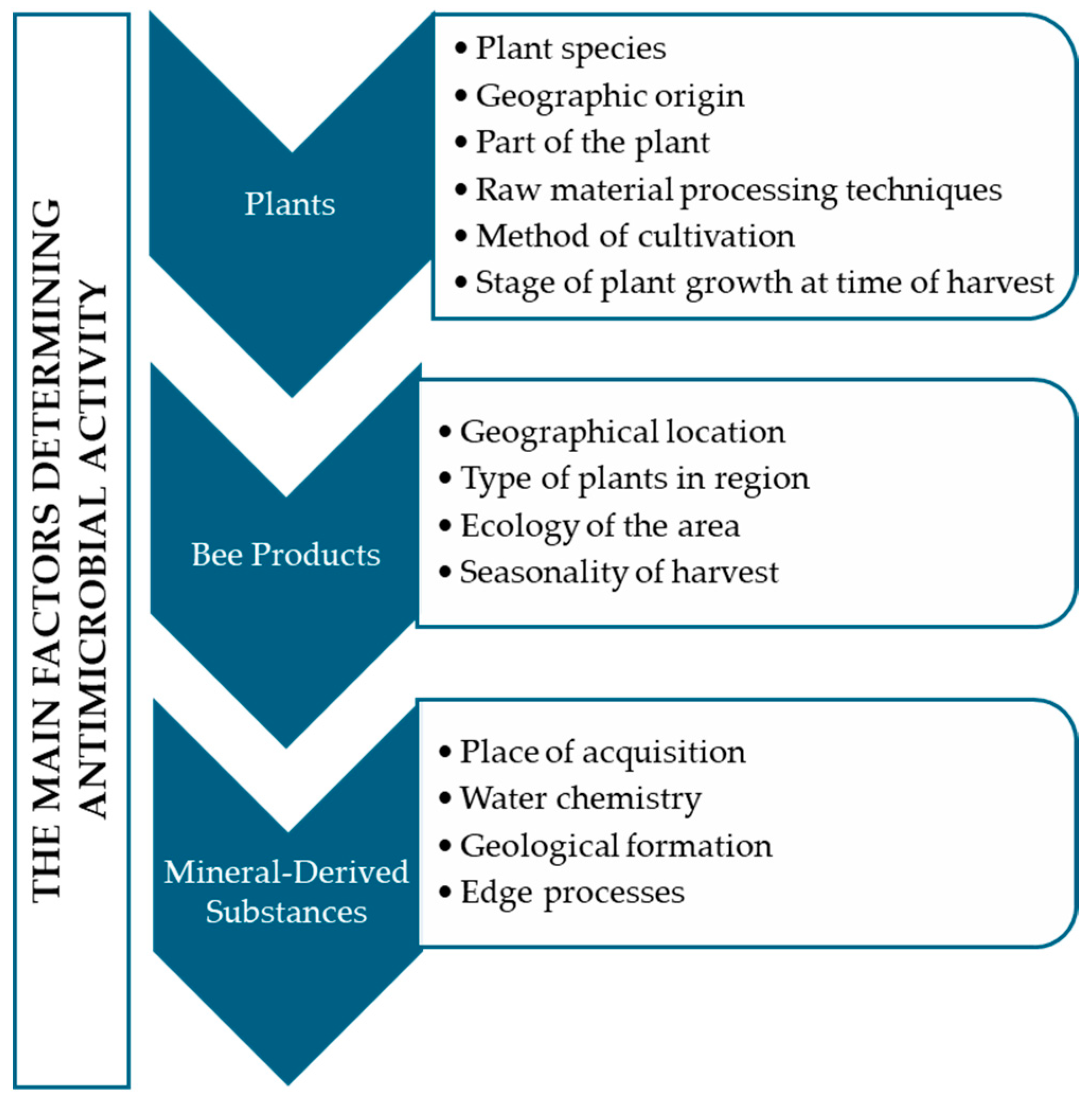
2. Materials and Methods

The study is a narrative review summarizing scientific reports regarding the antimicrobial activity of selected natural substances. This selection was based on a review of the raw materials used in formulations aimed at the prevention and adjunctive treatment of bacterial and fungal skin infections. Plant products and natural substances with antibacterial and antifungal effects in cosmetics available on the market were selected based on the analysis of two databases. CosIng (Cosmetic Ingredient Database)—a database maintained by the European Commission, containing detailed information on ingredients used in cosmetics within the European Union. It includes descriptions of natural substances, their uses, and legal restrictions. INCI (International Nomenclature of Cosmetic Ingredients)—a standard list of ingredient names used in cosmetics, allowing for the identification of natural and synthetic substances used worldwide. This work aimed to review available studies that could shed light on scientific evidence related to the microbiological activity of substances used in cosmetology. A search was conducted across PubMed and Scopus databases following the recommendations of the PRISMA statement [29]. The databases were searched up to August 2024. Substances that demonstrated antifungal and antibacterial activity were included. The search terms included “bacterial skin infections”, “fungal infections”, “skin pathogens”, “antibacterial substances”, “antifungal substances”, “natural substances in dermatology”, “microbiological activity of substances”, “garlic”, “lavender”, “sage”, “oregano”, “tea tree oil”, “aloe”, “fenugreek”, “black cumin”, “mud with microbiological activity”, “clays”, “mineral waters effects on skin”, “honey antibacterial and antifungal properties”, “manuka honey”, and “propolis”.
The retrieved articles were evaluated for confirmed antifungal and antibacterial activity. Duplicate articles, articles without available full texts, letters to the editor, and case reports were excluded.

3. Plant-Derived Natural Substances

Plant-derived natural substances, specifically isolated compounds and secondary metabolites, exhibit activity against fungal and bacterial pathogens that cause superficial skin infections. Plant preparations can be used to support local treatment of chronic dermatological diseases or in their prevention [30]. Presented below are selected extracts and essential oils present in cosmetic products with antimicrobial activity.

3.1. Garlic (Allium)

Plants from the genus Allium L., belonging to the Amaryllidaceae family, are known worldwide for their health benefits. Due to the presence of bioactive compounds such as thiosulfinate, flavonoids, and polyphenols, garlic exhibits antimicrobial, anti-inflammatory, antioxidant, and anticancer properties. The antibacterial and antifungal potential is primarily attributed to allicin [31,32]. Barbu et al. concluded from a phytochemical analysis of six extracts that the chemical composition varied by species. The most notable were Allium sativum and Allium ursinum, the only ones demonstrating antimicrobial activity. HPLC-DAD analysis of these species revealed the presence of chlorogenic acid and p-coumaric acid, as well as high levels of thiosulfinate (alliin and allicin), which correlated with antimicrobial activity against Candida albicans, Candida parapsilosis, Staphylococcus aureus, and Escherichia coli. The activity against pathogens was determined using microdilution techniques (MIC and MBC) and disk diffusion methods, where inhibition zones were demonstrated [31].
Rahman et al. examined the inhibition zone for pure juice, aqueous extract, ethanolic extract, and dried powder against Aspergillus flavus, Aspergillus parasiticus, Bacillus cereus, Escherichia coli, and Staphylococcus aureus. Among the pathogens studied, Bacillus cereus showed resistance to all forms. The highest efficacy was noted for pure garlic juice, although the extracts and powder also exhibited antimicrobial activity [33].
The disk diffusion method used in the study by Marpaung et al. confirmed the effectiveness of ethanolic garlic extract against Trichophyton rubrum and Pityrosporum ovale (Malassezia furfur). Growth inhibition of both pathogens was observed at a concentration of 60%, with inhibition zones measuring 15 mm and 8.35 mm, respectively [34].
The antifungal activity of aqueous, ethanolic, and methanolic extracts from Allium sativum against Trichophyton rubrum was particularly satisfactory; however, efficacy was also noted against Microsporum gypseum, Trichophyton mentagrophytes, Trichophyton verrucosum, Microsporum canis, and Epidermophyton floccosum. The studied extracts demonstrated fungicidal activity comparable to nystatin and azole drugs [35,36]. It was noted that the aqueous extract potentially exhibited better efficacy against dermatophytes than fluconazole and was nearly as effective as ketoconazole. Additionally, combining the extract with studied drugs increased therapeutic efficacy. Thus, the simultaneous application of Allium sativum extract and azoles may reduce azole dosage, which could contribute to a decreased risk of adverse effects [35]. Similar in vitro studies confirmed the efficacy of isolated allicin against T. rubrum as an alternative to ketoconazole treatment for mild dermatophytic infections [37].
Patil et al. developed an anti-acne gel containing garlic and coriander extract, which was tested in vitro against Propionibacterium acnes and Staphylococcus epidermidis. The results indicated satisfactory effectiveness, determining inhibition zones of 21.6 mm for P. acnes and 20.7 mm for S. epidermidis [38]. A similar product was created using garlic juice and carboxymethylcellulose, with effectiveness against P. acnes noted at a concentration of 7.5% garlic juice [39].
In general, garlic is used in the treatment and care of dermatological problems such as psoriasis, alopecia areata, keloid scars, difficult-to-heal wounds, and viral and fungal infections [40,41]. Furthermore, it has demonstrated antioxidant, immunomodulatory, anticancer, anti-aging, and photoprotective effects against UVB and has shown improvement in skin microcirculation [42,43]. Due to these properties, extracts and oils from Allium sativum have been utilized in the production of toner [44], gels [45], as well as scalp care products [46].

3.2. Sage (Salvia L.)

Species from the genus Salvia L., belonging to the Lamiaceae family, are known for their medicinal properties. The flavonoids, terpenoids, and phenolic acids present in them are considered the main compounds exhibiting therapeutic effects. In the pharmaceutical and cosmetic industries, Salvia officinalis (sage) is commonly used. When applied topically, it primarily exhibits antioxidant, anti-inflammatory, and antimicrobial activities. The outcome is attributed to the essential oils, which contain a high amount of oxidized sesquiterpenes [47].
Alves-Silva et al. studied the antifungal activity of the essential oil from Salvia aurea against pathogenic dermatophytes using the macro-dilution method. The oil was obtained from the leaves of the plant, which underwent hydrodistillation. Analysis revealed the presence of compounds such as 1.8-cineole (16.7%), β-pinene (11.9%), cis-thujone (10.5%), camphor (9.5%), and (E)-caryophyllene (9.3%). The antifungal study assessed its effect on strains of Trichophyton, Epidermophyton, and Microsporum. The highest sensitivity to the essential oil was shown by T. mentagrophytes, T. rubrum, and Epidermophyton floccosum, presenting MIC (minimum inhibitory concentration) values of 5 μL/mL, 1.25 μL/mL, and 1.25 μL/mL, respectively [48].
In another study conducted by Longaray-Delamare et al., the antibacterial activity of the essential oil obtained from the aerial parts of S. officinalis and S. triloba was analyzed. It was shown that the main compounds present in both oils were α-thujone, 1.8-cineole, and camphor. S. triloba was characterized by a high concentration of β-caryophyllene, α-humulene, and the presence of viridiflorol, while S. officinalis contained larger amounts of β-pinene, borneol, and δ-gurjunene. The MIC study of S. officinalis oil against pathogens indicated values of 5–10 mg/mL for E. coli, 5–10 mg/mL for Pseudomonas aeruginosa, and 5–10 mg/mL for Staphylococcus aureus and Staphylococcus epidermidis. Similarly, the MIC for oil from S. triloba showed activity levels of 5 mg/mL, 5–10 mg/mL, 0.3 mg/mL, and 1 mg/mL [49].
Stanciu et al. confirmed the antibacterial activity of S. officinalis essential oil against Staphylococcus, Enterococcus, Klebsiella sp., Proteus sp., and E. coli, determining inhibition zones between 4 mm and 9.5 mm [50]. A study using the hydroalcoholic extract of Salvia officinalis (1:10 w/v) against Staphylococcus aureus demonstrated bacterial sensitivity to the extract. Using the disk diffusion method, the average inhibition zone was 12.6 mm ± 0.58 for 30 µL of extract, 18.6 mm ± 0.58 for 50 µL, and 22 mm ± 1 for 100 µL of extract. When compared to popular pharmacological agents, it was concluded that the 100 µL of extract of Salvia officinalis (L.) surpassed the inhibition zone for 30 µg of vancomycin, 30 µg of cefotaxime, 10 µg of gentamicin, and 1 µg of oxacillin. No inhibition zones were recorded for the pathogens of Streptococcus agalactiae, Candida tropicalis, and Candida albicans [51].
A similar analysis of antimicrobial activity was conducted by Levaya et al., using hydroethanolic extracts from the flowers and leaves of S. stepposa against the pathogens S. aureus, B. subtilis, E. coli, and C. albicans. It was found that all concentrations of extracts from both leaves and flowers exhibited potent activity against S. aureus and B. subtilis. Activity against E. coli was observed at 90% flower extract and 70% leaf extract concentration. Minor activity against C. albicans was noted at 30% and 90% flower extracts and 90% leaf extracts [52].
The cosmetic industry utilizes sage extract as an active ingredient in cosmetic formulations such as soaps [53], gels [54], and pure sage oil for topical application on smooth skin and hairy scalp [55].

3.3. Tea Tree Oil (Melaleuca alternifolia L.)

In the cosmetic and pharmaceutical industries, tea tree oil, derived from the leaves of Melaleuca alternifolia L. of the Myrtaceae family, is well known for its antimicrobial properties. The biologically active compounds primarily include terpinen-4-ol, γ- and α-terpinene, α-terpineol, 1,8-cineole, p-cymene, α-pinene, and limonene. In addition to antifungal and antibacterial properties, the topical application of the oil has anesthetic, anti-inflammatory, and antiviral effects and accelerates wound healing. This raw material has mainly been used in the production of anti-acne and antifungal preparations for nails and feet [56].
The effectiveness of tea tree oil was confirmed in in vitro studies against the dermatophytes T. rubrum and T. mentagrophytes isolated from infected nails. The fungistatic activity against T. rubrum was determined at a concentration of 0.04%, while a concentration of 0.07% inhibited the growth of T. mentagrophytes [57].
The antibacterial efficacy of the oil was demonstrated in studies against Aeromonas hydrophila, Bacillus subtilis, Escherichia coli, Klebsiella pneumoniae, Staphylococcus aureus, Streptococcus agalactiae, Streptococcus pneumoniae, Streptococcus pyogenes, Proteus vulgaris, and Pseudomonas aeruginosa. The largest inhibition zone (36.3 mm) was demonstrated for Aeromonas hydrophila and was higher than for the antibiotic cefepime (33 mm). Similar results were observed for P. aeruginosa, B. subtilis, and S. agalactiae, where the inhibition zone of tea tree oil exceeded the inhibitory action of cefepime. The oil was more effective against the pathogens S. pyogenes and P. aeruginosa than rifampicin, cephalexin, erythromycin, amoxicillin, sulfamethoxazole, doxycycline, and clindamycin. Moreover, tea tree oil demonstrated greater efficacy than lemongrass and eucalyptus oils [58].
A study investigating the oil’s effects on E. coli, S. aureus, and C. albicans showed that the use of 0.5% tea tree oil led to disruptions in membrane permeability in pathogen cells and inhibited respiration [59]. Additionally, an analysis of the effect of tea tree oil on spores of E. coli and S. epidermidis was conducted. The effective concentrations resulting in no visible growth of pathogens were 0.5% for E. coli and 1.0% for S. epidermidis. The assessment was based on a colorimetric test using erythrosine B, which positively stains bacteria with damaged membranes. Complete elimination of dormant E. coli cells was observed after 1 h, and for S. epidermidis, after 24 h. Analysis indicates that the compound responsible for the antibacterial action against dormant pathogen cells is terpinen-4-ol [60].
Esmael et al. evaluated the in vitro effects of essential oils on bacteria potentially causing acne. Tea tree oil exhibited the highest antibacterial activity among those tested. The minimum bactericidal concentration against S. aureus and S. epidermidis was 78 mg/L, while for C. acnes, it was 39 mg/L [61]. Mazzarello et al. created a cream containing 20% propolis, 10% aloe, and 3% tea tree oil, comparing it with a cream containing erythromycin and a placebo. The in vivo study was conducted on a group of 60 patients aged 14–34 with mild to moderate acne on their faces. The effectiveness of the preparations was assessed after 15 and 30 days through visual skin assessment, macrophotography (Canon G10), and instrumental measurements such as sebum quantity, pH, and erythema index using the Courage+ Khazaka Electronic GmbH device. It was concluded that the new herbal product was more effective in reducing lesions and decreasing erythema than the synthetic product with an antibiotic [62].
Melaleuca alternifolia oils and extracts are used in the production of gels [63], serums [64], and ointments [65].

3.4. Aloe vera (Aloe L.)

Aloe vera (A. barbadensis) and other species of the Aloe genus are the most commonly used plants from the Liliaceae family. The extensive use of this raw material in the pharmaceutical and cosmetic industries stems from its health benefits. The primary applications are the oral consumption of aloe juice and the topical application of gel on affected areas. The properties of aloe related to skin application include mainly anti-inflammatory effects and accelerated wound healing, enabling its use in most dermatoses accompanied by epidermal damage. In addition to its therapeutic effect, the raw material exhibits antioxidant and photoprotective properties for the skin [66,67].
The antibacterial effects of the gel and the extract from the leaves (excluding the pulp) of A. vera were compared, revealing that, unlike the gel, the leaf extract did not exhibit activity against gram-negative and gram-positive bacteria isolated from the skin [66].
Spectrophotometric analysis of secondary metabolites in A. arborescens, A. aristata, and A. ferox showed the presence of phenols, flavonoids, and proanthocyanidins. After conducting antibacterial and antifungal tests, studied samples exhibited activity against skin pathogens, which correlated with the presence of phenolic compounds, flavonoids, and proanthocyanidins. All three extracts showed activity against T. rubrum, T. mentagrophytes, C. albicans, and C. tropicalis, determining the minimum inhibitory concentration at an average level of 0.60 μg/mL. The antibacterial activity of the extracts was noted against pathogens such as Actinomycetes brasiliensis, B. subtilis, Micrococcus spp., S. aureus, S. epidermidis, S. pneumoniae, S. pyogenes, E. aerogenes, K. pneumoniae, P. aeruginosa, Shigella sonnei, Proteus mirabilis, and P. vulgaris [68].
Danish et al. assessed the antimicrobial activity of ethanolic extracts from the leaves and roots of A. vera using the disk diffusion method. Extracts of varying concentrations (15, 20, 25, 30 µg) were prepared, and the inhibition zones were proportional to the concentration. The highest concentration extract from the leaves exhibited inhibition zones of 15 mm for B. subtilis, 18 mm for E. coli and Agrobacterium tumefaciens, 18.5 mm for Fusarium oxysporum, and 18 mm for A. niger. The root extract at the same concentration showed similar effects: 16 mm for B. subtilis, B. megaterium, and E. coli; 17.5 mm for A. tumefaciens; 18 mm for A. niger; 17 mm for F. oxysporum; and 15 mm for C. albicans. Additionally, all concentrations of both extracts exhibited antimicrobial activity against B. cereus, S. aureus, Streptococcus pyogenes, Proteus mirabilis, and Pseudomonas aeruginosa [69].
In vitro studies of hydroalcoholic extracts from A. vera leaves confirmed their action against C. albicans. It was concluded that a 75% leaf extract (average inhibition zone of 7.56 mm) could serve as a natural alternative to nystatin (average inhibition zone of 9 mm) [70]. The effectiveness of combining gentamicin sulfate with the aloe leaf extract was noted in an in vitro study, showing an inhibition zone of 7.63 mm against S. aureus [71].
Beneficial effects of aloe were demonstrated against multidrug-resistant clinical strains of P. aeruginosa and Acinetobacter baumannii. Modified aloe polysaccharide reduced bacterial viability depending on the concentration of the extract. At a dose of 1000 µg/mL, the survival rates were 42.9% for P. aeruginosa and 68.34% for A. baumannii [72].
Studies indicate that aloe acts against Propionibacterium acnes. Analysis of A. ferox indicated high activity of a 95% ethanolic extract against P. acnes [73]. In addition, a gel preparation containing 10% ethanol extract from A. vera showed an inhibition zone of 12.9 mm, which was classified as strong [74]. It was shown that the antibacterial activity of Aloe vera gel depends on the formulations and gelling agents used [75].
Aloe demonstrates a broad spectrum of antimicrobial properties, establishing it as a highly valued ingredient in dermatological and cosmetic formulations. Additionally, its anti-inflammatory, antioxidant, and regenerative properties offer benefits for skin affected by dermatoses characterized by both seborrhea and dryness. Aloe-containing cosmetic preparations primarily include after-sun products [76], anti-acne formulations [77], and those designed for dry skin [78].

3.5. Lavender (Lavandula L.)

Lavender is a plant from the Lamiaceae family, with the most commonly used species in the cosmetic industry being Lavandula angustifolia (syn. L. officinalis, L. vera) and Lavandula latifolia (syn. L. spica). The herbal raw material is the lavender flower, from which the essential oil is extracted, a valued cosmetic ingredient. Lavender essential oil is one of the most well-known aromatherapy oils, affecting the nervous and respiratory systems. It regenerates the skin, has immunomodulatory, antioxidant, and anti-inflammatory properties, acts antiseptically, and is also used as a preservative in cosmetics [79,80].
The chemical profile of the essential oil from L. angustifolia (Robertet®) is predominantly characterized by linalyl acetate (36.7%), linalool (31.4%), and terpinen-4-ol (14.9%) [81]. Similarly, another study analyzed the composition of lavender oil (Pollena-Aroma), identifying linalool (34.1%) and linalyl acetate (33.3%) as its major constituents [82]. Białon et al. compared the chemical profile of oil from L. angustifolia by EJTA and Crimean lavender oil (L. angustifolia). The EJTA oil consisted primarily of monoterpenoids (76.7%) and monoterpenes (22.7%), characterized by a higher concentration of linalool (41.8%), linalyl acetate (32.7%), and limonene (19.0%). The Crimean oil mainly contained monoterpenoids (80.1% according to HP-5MS), monoterpenes (5.8%), and sesquiterpenes (8.0%); analysis showed the highest content of linalool (34.1% according to HP-5MS), linalyl acetate (23.3%), and eucalyptol (5%). These results were correlated with studies on antibacterial activity against skin microbiota from oily facial skin. Dominant pathogens were identified, and inhibition zones for oils at concentrations of 40–80 μL/cm3 were determined. None of the oils exhibited activity against Enterococcus faecium. It was found that the commercial oil had higher antibacterial activity against the remaining pathogens: B. cereus, B. subtilis, B. mycoides, Staphylococcus warneri, Micrococcus luteus, and Corynebacterium spp. The Crimean oil showed activity against B. cereus (80 μL/cm3), M. luteus, Corynebacterium spp., as well as E. coli. The researchers concluded that the superior activity of the EJTA oil is due to its higher concentration of monoterpenoids and monoterpenes [79].
The evaluation of lavender oil (from Natural Aromas, Poland) against bacterial pathogens showed the largest inhibition zones for Enterococcus faecalis (21.78–23.15 mm), S. aureus (13.44 mm), and S. aureus (MRSA) (16.1 mm) [83]. Antibacterial activity against P. acnes was confirmed in a study where the minimum inhibitory concentration was equal to the minimum bactericidal concentration, measuring 0.125% for commercial essential oil from L. stoechas (Xiamen Denyla Essential Ooil Co., Ltd., Xiamen, China) [84].
Two lavender essential oils extracted from the leafy stems of L. angustifolia—the ‘Blue River’ (BR) and the ‘Ellegance Purple’ (EP)—were also studied. Both essential oils showed activity against S. aureus, S. aureus (MRSA), and E. coli [85]. It was reported that lavender essential oil from L. angustifolia demonstrated synergistic effects with the dichloride of octenidine, enhancing its action against methicillin-resistant S. aureus [82]. Increased antimicrobial effects were also observed from the combination of L. angustifolia essential oil with ciprofloxacin against S. aureus and with chloramphenicol against P. aeruginosa [81].
In addition, antifungal activity was noted for lavender oil (Lavandula angustifolia Mill), with 100% inhibition observed for Candida albicans and Trichophyton [86]. Another study focusing on the essential oil from L. multifida, which has a high content of monoterpenes, including carvacrol and cis-β-ocimene, demonstrated its antifungal activity. The minimum lethal concentration (MLC) was set at 0.64 μL/mL for C. albicans, C. tropicalis, C. krusei, C. parapsilosis, and Aspergillus fumigatus. The MLC for Candida guilliermondii, Cryptococcus neoformans, T. mentagrophytes, T. rubrum, Trichophyton verrucosum, M. canis, M. gypseum, and Epidermophyton floccosum was 0.32 μL/mL and 1.25 μL/mL for A. niger and A. flavus. Flow cytometry tests with propidium iodide indicated that 3 h of incubation with the studied essential oil at a concentration of 5.0 μL/mL led to membrane disruption and cell death in C. albicans [87].
Lavender essential oil is most commonly used to produce formulations for sensitive and acne-prone skin. As a raw material, it is utilized in shampoos [88], toners/mists [89], creams [90], and gels [91].

3.6. Fenugreek (Trigonella L.)

Fenugreek is a plant belonging to the Fabaceae family. The most commonly used species is T. foenum-graecum, and the raw materials are its leaves and seeds. In cosmetics, it is mainly used in the production of preparations for seborrheic and acne-prone skin. In addition to its antimicrobial activity, it exhibits anti-inflammatory, antioxidant, anticancer, and antidiabetic effects [92].
The assessment of the chemical composition of aqueous and ethanolic extracts from fenugreek leaves revealed the presence of saponins, steroid saponins, flavonoids, phenols, and proteins. Additionally, the aqueous extract contained carbohydrates and alkaloids [93]. Phytochemical analysis of extracts from T. foenum-graecum leaves showed a higher content of polyphenols and flavonoids in ethanolic and methanolic extracts compared to aqueous ones. The antibacterial activity was highest for the ethanolic extracts, with inhibition zones measuring 14 mm for S. aureus, 13 mm for P. aeruginosa, 12 mm for P. vulgaris, 12 mm for E. coli, 11 mm for Enterobacter aerogenes, and 10 mm for Klebsiella sp. [92].
The antimicrobial activity of methanolic extract from fenugreek seeds was confirmed by Majumdar et al., showing the highest inhibition zones for a concentration of 2 mg/mL against B. cereus (16.9 mm), E. coli (16.1 mm), P. aeruginosa (14.6 mm), S. aureus (13.2 mm), and S. aureus (MRSA) (12.5 mm). These results were compared to amoxicillin at a concentration of 10 µg/mL, yielding inhibition zones of 21.2 mm, 20.1 mm, 16.5 mm, 18.6 mm, and 15.1 mm for the respective strains. The same extract showed antifungal activity against A. flavus (19.6 mm), C. albicans (19.1 mm), and T. rubrum (17.3 mm) compared to amphotericin B at 10 µg/mL, which inhibited fungal growth by 23 mm, 22.1 mm, and 21 mm, respectively. Although higher inhibition zones were recorded for the antimicrobial agents, the methanolic extract from fenugreek seeds may serve as support against these pathogens [94].
The activity of aqueous and ethanolic extracts from T. foenum-graecum leaves against M. furfur was determined based on the inhibition zone, with a positive control of 2% ketoconazole. Analysis revealed greater activity from 0.3 mL of the aqueous extract than the ethanolic one, comparable to the inhibition zone of 0.1 mL of the drug. A gel preparation containing a 30% concentration of the aqueous extract was shown to be effective against pathogens residing on both dry and excessively oily scalps. In the same study, the effectiveness of the extracts at 0.3 mL against A. niger and C. albicans was indicated, showing greater effectiveness from the aqueous extract, comparable to 0.1 mL of 2% ketoconazole [93].
Minor activity against P. acnes and S. epidermidis was observed while determining the inhibition zone for boiled aqueous extract from fenugreek seeds at a concentration of 500 mg/mL, measuring 12 mm and 17 mm, respectively [95]. The activity of 150 μL of essential oil derived from powdered fenugreek seeds was demonstrated against P. vulgaris (15 mm), E. coli (15 mm), and B. subtilis (14.0 mm) [96]. As a cosmetic raw material, fenugreek is found in products for hair and scalp [97,98], facial gels [99], and creams [100].

3.7. Oregano (Origanum L.)

Oregano is a plant with a wide range of applications in the food and cosmetic industries. It belongs to the Lamiaceae family, with the essential oil derived from the aerial parts of common oregano (O. vulgare L.) being the most commonly used in cosmetology. Oregano exhibits biological activity on the skin, including anti-inflammatory, antioxidant, and antimicrobial effects [101].
While the antimicrobial activity has been confirmed, significant differences in chemical compositions exist, making it difficult to clearly identify the compound responsible for these effects. Taleb et al. analyzed the volatile components of the essential oil from O. vulgare using GC-MS, finding thymol at 99.44% as the main component responsible for its antimicrobial activity, while noting the absence of carvacrol. Antibacterial activity against P. acnes and S. epidermidis was tested using the disk diffusion method, showing a greater inhibition zone for the oil than for erythromycin and clindamycin. The minimum inhibitory concentration was 0.34 mg/mL for P. acnes and 0.67 mg/mL for S. epidermidis, with bactericidal concentrations of 0.672 mg/mL and 1.34 mg/mL, respectively [102].
Vale-Silva et al. evaluated the correlations of the chemical composition of essential oils from O. vulgare subsp. virens obtained from various geographic locations in Portugal. The phytochemical profile was assessed using GC-MS, revealing quantitative differences in their composition. The most significant contributions to the profile were noted for α-terpineol (0.1–65.1%), γ-terpinene (0.3–34.2%), carvacrol (0–34.2%), and linalool (2.0–27.4%). All samples exhibited antifungal activity, with varying inhibition strengths against C. albicans, C. tropicalis, C. krusei, C. guillermondii, C. parapsilosis, Cryptococcus neoformans, T. rubrum, T. mentagrophytes, M. canis, M. gypseum, E. floccosum, A. niger, Aspergillus fumigatus, and A. flavus. The highest effectiveness was found for the sample containing 34.2% carvacrol [101].
Souza et al. confirmed the antifungal activity of the essential oil from O. vulgare leaves against C. albicans, C. tropicalis, C. neoformans, T. rubrum, M. canis, A. flavus, A. fumigatus, and Cladosporium herbarium, with an MIC of 80 µL/mL. Complete cell death for T. rubrum and M. canis was observed after 2 days of application of the oil at a concentration of 80 µL/mL. Resistance to the studied essential oil was exhibited by strains of C. krusei, T. mentagrophytes, and M. gypseum. Similarly, a study on essential oil from the leaves of O. majorana showed a MIC of 160 µL/mL against C. albicans, C. tropicalis, C. neoformans, T. mentagrophytes, M. gypseum, and A. flavus, with other pathogens demonstrating resistance. Comparing the effects of both essential oil and ketoconazole against C. albicans, no differences in fungicidal action were noted after 4 h of exposure [103].
Malassezia sympodialis and Malassezia furfur also showed sensitivity to the essential oil from O. vulgare [104]. Antibacterial studies of essential oil obtained from the leaves and stems of oregano against clinical strains of E. coli and S. aureus indicated an average MIC of 128 μg/mL for S. aureus and 256 μg/mL for E. coli. The bactericidal activity was represented by average MBC values of 64 μg/mL and 128 μg/mL for S. aureus and E. coli, respectively [105].
A strong activity of oregano essential oil against S. aureus was demonstrated based on the measurement of the inhibition zone, which was 30.33 mm. In comparison, a 100% ethanolic extract from oregano inhibited the activity of the pathogen at 14.33 mm. The same essential oil and extract were tested against K. pneumoniae, yielding inhibition zones of 18 mm and 6.4 mm, respectively [106]. An in vivo study of nanoemulsion containing <5% oregano essential oil was conducted on mouse models infected with P. acnes. The inflammatory response was inhibited by >60%, significantly exceeding the positive control for 2% erythromycin, which showed 20% efficacy [102]. This study confirms the anti-acne action of oregano and explains the application of this raw material in the production of hydrosols [107] and soaps [108] for acne-prone skin. This raw material has also found wide application as an antifungal agent in foot and nail care cosmetics [109].

3.8. Black Cumin (Nigella L.)

Black cumin is a plant belonging to the Ranunculaceae family, considered a medicinal plant due to the wide application of its oil and extract derived from its seeds. In the cosmetic industry, the raw materials are primarily used for their anti-psoriatic, antioxidant, anti-inflammatory, and antimicrobial properties [110]. Gas chromatography analysis of the ethanolic extract from N. sativa seeds identified the highest proportion of thymoquinone (40.23%) and thymol (17.23%) among all major compounds. Other compounds identified included 7.51% 1,2-diphenylethylamine, 6.9% o-xylene, and 6.15% ethylbenzene, establishing thymoquinone and thymol as the primary compounds responsible for antimicrobial activity [111].
The phytochemical profile of extracts from Nigella sativa seeds varies by type, showing the presence of terpenoids, steroids, and alkaloids in all examined extracts. Phenolic compounds, flavonoids, and tannins were noted in methanol and water extracts, glycosides only in methanol, and saponins in water. GC-MS analysis of the seed oil confirmed the presence of toluene and m-cymene in the hexane-extracted oil and o-xylene in the methanol extract. These compounds have confirmed antimicrobial effects. The most effective antibacterial activity was exhibited by undiluted methanolic extract, where disk diffusion testing showed inhibition zones for P. aeruginosa, E. coli, B. subtilis, and S. aureus [110]. Shah et al. confirmed the activity of methanolic extract against S. aureus, with an average inhibition zone of 48 mm at a concentration of 30 µg/mL of N. sativa extract. In comparison, chloramphenicol (30 μg disc) inhibited S. aureus activity at an average of 24 mm [112]. Methicillin-resistant S. aureus isolated from patient wounds showed an inhibition zone of 17.75 mm for a methanolic extract at a concentration of 166 mg/mL [113]. The antibacterial action was also confirmed for a gel formulation containing 15% extract from Nigella sativa seeds, with average inhibition zones of 10.6 mm against S. aureus and 9.0 mm against P. acnes, compared to a commercial anti-acne gel that had inhibition zones of 8.3 mm for S. aureus and 10.5 mm for P. acnes [114]. An in vivo study of a hydrogel formula created from the hydroalcoholic extract of N. sativa confirmed the plant’s anti-acne effects, involving 60 patients diagnosed with mild to moderate acne. A reduction in acne lesions was demonstrated after 8 weeks compared to the placebo group [115]. The minimum inhibitory concentration of ether extract was shown for T. rubrum, T. mentagrophytes, and E. floccosum at 40 mg/mL and for M. canis at 10 mg/mL, confirming its activity against dermatophytes [116]. The multifaceted action of extracts from black cumin seeds allows for their use in anti-acne formulations [117] and soaps [118].

4. Animal-Derived Natural Substances

Animal-derived natural substances exhibiting broad antimicrobial properties primarily include bee products. In the cosmetic and pharmaceutical industries, various types of honey and propolis are mainly utilized. Bee-derived products are used externally in dermatology, otolaryngology, surgery, dentistry, gynecology, and ophthalmology [119].

4.1. Bee Honey

Honey also exhibits anti-inflammatory, antioxidant, and nourishing properties and promotes wound healing, which contributes to the benefits of local application of this raw material [120,121]. Analyzing the antimicrobial activity of various types of honey could represent a distinct area of research due to the large amount of published data. According to reviews of available cosmetic products on the market, manuka honey is of the greatest interest to manufacturers as an ingredient for problematic skin preparations [122] therefore, it has been included in this review.

Manuka Honey (Leptospermum scoparium)

Manuka honey is a monofloral honey derived from the nectar of the Leptospermum scoparium plant, belonging to the Myrtaceae family. It is naturally found in New Zealand and Australia. Particularly well-known for its antibacterial, antifungal, antioxidant, and anticancer properties, it accelerates the healing of wounds and diabetic ulcers [123,124]. This honey is rich in amino acids, proteins, enzymes, minerals, vitamins, as well as macro- and micronutrients. The therapeutic effects are mainly attributed to secondary metabolites such as flavonoids, phenolic acids, and 1,2-dicarbonyl compounds, with the most significant being methylglyoxal (MGO) from the perspective of antimicrobial activity. In commerce, the concentration of this compound is reflected in the Unique Manuka Factor (UMF) classification system [125,126,127]. In a previous study on manuka honey, sensitivity was noted for pathogens: S. aureus, S. aureus (MRSA), Staphylococcus spp., Pseudomonas aeruginosa, and Enterobacteriaceae. Paradoxically, the study showed that a higher classification (15+) does not guarantee better activity of the honey [127]. Bouacha et al. confirmed the antibacterial activity of honey against E. coli, P. aeruginosa, K. pneumoniae, S. aureus, Staphylococcus saprophyticus, and E. faecalis. It was noted that MIC values ranged from 5% to 20% (v/v) for Gram-positive bacteria and from 20% to 40% (v/v) for Gram-negative bacteria [128]. Manuka honey, with a declared MGO value of 400 mg/kg, underwent testing for minimum inhibitory concentration against S. pyogenes, S. aureus, and coagulase-negative Staphylococci, yielding MIC values of 8% (v/v), 7% (v/v), and 8% (v/v), respectively [129]. The honey’s action against E. coli producing extended-spectrum beta-lactamase was confirmed with an MIC of 12.5% (v/v) and an MBC of 25% (v/v) [130]. The combination of manuka honey (MGO70 and MGO85) and azithromycin demonstrated bactericidal activity against Mycobacterium abscessus, utilizing 0.037 g/mL of honey and <4 µg/mL of azithromycin, indicating added inhibitory value at lower concentrations [131]. The synergistic effectiveness of manuka honey (UMF 20+ and MGO 830+) with antibiotics (gentamicin, rifampicin, and vancomycin) was presented in a study by Liang et al., showing better susceptibility of bacteria S. aureus, S. epidermidis, and S. lugdunensis to the combination than when using only the antibiotic [132]. The activity of honey against dermatophytes causing fungal infections was examined by Brady et al., who conducted an agar well diffusion test. Growth inhibition was noted for Epidermophyton floccosum at 25% (v/v), Microsporum canis at 25% (v/v), Microsporum gypseum at 55% (v/v), Trichophyton mentagrophytes var. interdigitale at 45% (v/v), Trichophyton mentagrophytes var. mentagrophytes at 25% (v/v), Trichophyton rubrum at 20% (v/v), and Trichophyton tonsurans at 25% (v/v) [133].
Manuka honey is mainly directed toward the treatment of burn wounds, chronic ulcers [134], cancerous wounds, post-radiation treatment wounds, and those infected with bacterial pathogens. It is used in home and hospital treatments. In the cosmetic industry, manuka honey is utilized in gels [135], masks [136] and products for hair or scalp [137].

4.2. Propolis (Propolis cera)

Propolis is a mixture of secretions from bees and plant resin. Its composition depends on various factors, primarily the season and geographical area of collection. According to researchers, the most significant compounds regarding antimicrobial and antioxidant activity are phenolic acids and flavonoids. An analysis of Brazilian honey samples regarding the correlation of these compounds with the minimum inhibitory concentration demonstrated a relationship between antimicrobial activity and phenolic acids and flavonoids in variable proportions [138].
Ethanolic extracts of propolis sourced from poplar buds, originating from Austria, France, and Germany, exhibited variable chemical compositions. Identification via thin-layer chromatography (TLC) revealed the presence of flavonoids and phenolic esters in all tested samples—albeit in varying proportions [139]. Castro et al. documented the seasonal effects of Brazilian propolis on its antibacterial activity and phenolic composition [122]. An analysis of several Brazilian propolis samples showed correlations between the strength of action against S. aureus and the concentration of phenolic compounds [140].
The antibacterial activity of four ethanol extracts of propolis from Turkey was confirmed using macro-dilution methods. The minimum inhibitory concentrations for skin pathogens were determined as follows: Streptococcus sobrinus and Enterococcus faecalis at 2 μg/mL, M. luteus, C. albicans, and C. krusei at 4 μg/mL, Streptococcus mutans, Staphylococcus aureus, Staphylococcus epidermidis, and Enterobacter aerogenes at 8 μg/mL, Escherichia coli and C. tropicalis at 16 μg/mL, and P. aeruginosa at 32 μg/mL [141].
The activity against S. aureus and P. aeruginosa was confirmed using the disk diffusion method for a 20% alcohol tincture, showing inhibition zones of 10 mm and 16.66 mm, respectively; however, no effect of the propolis tincture was noted against E. coli [142]. Activity against P. acnes was demonstrated for a 30% ethanolic extract of Bulgarian propolis using the agar diffusion method against 13 strains. Significant antibacterial effects were noted with 30 μL of extract, inhibiting 12 out of 13 strains of P. acnes, with an average inhibition diameter of 12.6 mm [143].
An interesting study on the activity of 10%, 15%, and 20% ointments made from Brazilian propolis against T. mentagrophytes was conducted by Kusumaningtyas et al., showing inhibition zones for this dermatophyte. In vivo, the effect of the ointment was observed on infected rabbit skin over 18 days, comparing it to 2% miconazole treatment. On day 18, complete healing was confirmed in 100% of rabbits treated with propolis compared to 66% treated with miconazole [144].
The antifungal activity of aqueous and alcoholic extracts demonstrated greater effectiveness against T. mentagrophytes, T. interdigitale, M. canis, C. albicans, and Aspergillus nidulans for most concentrations of ethanolic extracts. For the lowest tested concentration (0.2%) of the ethanolic extract, the average diameter of the inhibition zone for the pathogens was 9.75 mm, 12 mm, 12 mm, 13.125 mm, and 12.75 mm, respectively. This activity was also confirmed against A. flavus, although it was slightly lower than that for the aqueous extract of propolis [145].
An important pathogen from a dermatological perspective is Malassezia globosa, whose growth was inhibited by Thai propolis extract (IC50, 1.22 mg/mL) [146]. As the above studies demonstrate, propolis exhibits high antimicrobial activity, confirming its therapeutic action against skin infections. Propolis is widely used in medicine, primarily as a means to accelerate the healing of the wounds and lesions resulting from dermatoses such as impetigo, pustular dermatitis, paronychia, psoriasis, and boils. It is utilized in both home and hospital settings, particularly after burns, surgeries, and radiation damage [147,148].
Manufacturers of over-the-counter cosmetics containing propolis claim that it soothes irritation [149] and accelerates the regeneration of the epidermis [150], making it a natural preventive measure for managing mild dermatoses [151].

5. Mineral-Derived Natural Substances

The therapeutic effects of mineral substances are primarily utilized in health resort medicine. Warm compresses and baths are mainly recommended in physiotherapy, orthopedics, and rheumatology due to their analgesic and anti-inflammatory properties. The cosmetic applications of natural mineral-derived substances have been found for clays, natural mineral waters, and mud substances. Mineral raw materials are of interest to the cosmetic industry because of their high content of minerals and macro- and micronutrients. The demonstration of additional antimicrobial activity has expanded the use of these raw materials in the industry; however, there is still a lack of large clinical studies confirming their in vivo efficacy.

5.1. Cosmetic Mud

Muds sourced from various parts of the world are utilized for cosmetic purposes, primarily due to their detoxifying, anti-inflammatory, moisturizing, and anti-aging activities [152]. Depending on the area of collection, mud substances vary in their chemical profile and applications. Among the mud raw materials available in the global retail market, Dead Sea mud was selected as it met the criteria for this review. Regional spa cosmetics containing muds from selected geological areas were not included.

Dead Sea Mud

This raw material is obtained from the lake at the border of Israel, Palestine, and Jordan, characterized by a high degree of salinity. Spa regions offer wraps and baths recommended for conditions such as psoriasis, atopic dermatitis, and rheumatic diseases. The composition of the mud was analyzed by comparing 24 samples from three different locations along the eastern shore of Jordan. The pH values ranged from 5.7 to 6.1. Depending on the sampling location, its profile varied due to different phenomena occurring in the three locations. The analysis revealed a homogeneous mineral composition, including clay, dolomite, gypsum, kaolin, pyrite, quartz, carbonates, and feldspar in varying ratios. Additionally, the presence of Al2O3, CaO, CO2, Fe2O3, H2O, MgO, MnO, K2O, SiO2, Na2O, SO3, TiO2, and trace amounts of metals such as Br, Cd, Co, Cu, Pb, Li, Sr, V, and Zn was confirmed [153].
Zeev Ma’or et al. investigated the antibacterial and antifungal properties of both raw and sterilized Dead Sea mud. The test was conducted using the disk diffusion method. The plates were incubated at 30 degrees Celsius for 36 h. The inhibition zones for both sterilized and raw mud were equal for the following pathogens: A. niger, P. acnes, and E. coli, measuring 10 mm, 7 mm, and 7 mm, respectively. A higher inhibition zone was noted for raw mud against C. albicans (10 mm) compared to sterilized mud (8 mm). No inhibition zone was observed for S. aureus. The authors concluded that the antimicrobial effect of Dead Sea mud may be due to its high concentration of salts and sulfides, as well as its low pH [154]. A 4-week in vivo study demonstrated the safety of using various types of mud on the forearms of healthy volunteers every two days. The absence of irritation and disruption of the hydrolipid barrier confirmed the safety of using mud on the skin [155].
Dead Sea mud possesses therapeutic properties for the skin: it stimulates circulation, acts as a cleanser, supports wound healing, enhances barrier function, and reduces inflammation. These properties allow for a wide range of applications in cosmetic preparations such as masks [156], soaps [157], and products for hair and scalp [158].

5.2. Cosmetic Clays

Clays applied to the skin for aesthetic purposes can have multifaceted effects. They are found as active ingredients in masks, peels, gels, shampoos, and photoprotective products. Clays exhibit cleansing, exfoliating, and excess sebum-absorbing properties, as well as astringent, lifting, and moisturizing effects, and they can reduce skin inflammation. The determining factors for the application of clays are their chemical composition, the type of clay material, and the structure of the clay [159].
It is important to note that variations in geological formation will differentiate clays in their properties. Clay minerals, as mineral constituents, determine the function of the product and mainly include Si, Al, Fe, Ti, Mg, Ca, K, Na, layered silicates, oxides, carbonates, kaolinite, chlorides, and trace elements like Na, K, Ca, Mg, Ti, Fe, Al, and Si. The presence of ZnO and MgO improves the appearance of the skin, while TiO2 provides protection against UV as a physical filter, and kaolinite offers moisturizing, soothing, and regenerating effects for the epidermis. Fe2O3 stimulates cells for renewal and acts as an antiseptic, a turbidity agent, and a pigment [160].
The antimicrobial activity of clays is thought to be due to the presence of sulfuric acid or the combination of aluminosilicates/mineral clays with reduced metals/ions such as Fe, Ag, Au, Cu, and Zn. Illite- and smectite-containing ferrous iron have been confirmed to exhibit bactericidal activity [161,162].
A significant factor in using clays for antibacterial purposes is hydration, as a dry state limits their antimicrobial activity [162]. Hydrated French green clay exhibited bactericidal activity against Mycobacterium ulcerans, the pathogen responsible for Buruli ulcer. Treatment involved daily applications of clay wraps on affected skin, leading to noticeable healing and cleansing of wounds over several days. The Buruli ulcer healed without scarring after a few months [163].
Adusumilli et al. confirmed the bactericidal activity in vitro against M. ulcerans after 7, 14, 21, and 28 days of incubation. After this period, in vivo studies were conducted on mice, where hydrated clay wraps were applied once daily for 22 days. The clay wraps were found to promote healing in infected mice and reduce the pathogen load in wounds [164].
Haydel et al. conducted a study on the effects of two iron-rich clay minerals (CrArO2 and CsAgO2) against pathogenic bacteria. The sample of illite and smectite clay enriched with magnesium and potassium demonstrated bactericidal activity against E. coli, extended-spectrum β-lactamase-producing E. coli, P. aeruginosa, Mycobacterium marinum, S. aureus, S. aureus (MRSA), and Mycobacterium smegmatis and remained thermally stable [165].
The analysis of the composition of clays showed similar mineralogy, dominated by clay materials along with quartz, feldspar, and calcite. The clay that exhibited antibacterial activity (CsAgO2) was characterized by a higher concentration of illite (along with dioctahedral mica and smectite) and ferrous iron symmetrite, as well as a higher overall content of oxides. It is presumed that the antibacterial effect is also influenced by the chemistry of the water in contact with the clay and the chemical and morphological differences of the crystals [166]. Kisameet clay, sourced from the coast of British Columbia in Canada, exhibited in vitro activity against E. faecium, S. aureus, K. pneumoniae, A. baumannii, P. aeruginosa, Enterobacter spp., C. albicans, C. neoformans, M. marinum, and M. ulcerans. This clay has a low mineral content and a significantly higher illite content. Notably, the clay contains Actinobacteria, which may contribute to its antimicrobial activity [161,167]. An in vivo study was conducted on a ready-to-use mask based on clay and jojoba oil, applied by 194 participants over a period of 6 weeks, 2–3 times a week (for 15–20 min each time). At the end of the study, a 54% reduction in the total number of lesions was observed. It was concluded that this preparation may be used to assist in the treatment of acne lesions [168]. French green clay can be found in products such as powders [169], masks [170], and gels [171].

6. Use of Natural Substances in the Cosmetics Industry

Natural and organic cosmetics have gained significant popularity due to consumer perceptions of their safety and superiority over synthetic products. However, it is important to note that the use of such products is not without potential risks. Natural ingredients, such as essential oils and plant extracts, may cause skin hypersensitivity and allergic reactions, leading to symptoms such as redness, itching, or rashes. Citrus extracts and certain essential oils can also increase photosensitivity, making the skin more vulnerable to sunlight exposure [172,173]. Natural cosmetics may pose a higher risk of microbial contamination due to the absence of synthetic preservatives [174]. Moreover, the lack of adequate legal regulations regarding the composition and concentrations of these cosmetics poses significant health risks to consumers. One of the main issues is the lack of a uniform definition and regulations regarding the labels “natural” and “organic” in cosmetics. In some countries, such as Thailand, the absence of appropriate regulations allows companies to label their products as organic, despite containing minimal amounts of organic ingredients. The lack of standardized regulations in this regard leads to the misuse of terminology, which can mislead consumers [175]. Current standards, such as ISO 16128, attempt to quantify the content of natural ingredients but do not require comprehensive toxicological studies. As a result, many products reach the market without a thorough evaluation of their safety and impact on health [176].
It should also be noted that the effectiveness of a natural substance in a cosmetic product cannot be fully determined. The efficacy of raw material is primarily influenced by its chemical composition. This composition is variable and, in the case of plant-based raw materials, depends on the species, geographical region of origin, cultivation methods, and the plant’s growth stage at the time of harvest [31,110]. In addition, the composition of bee products will be determined by factors such as geographical location, seasonality, the ecological condition of plants visited by bees, and the types of plants growing in the area [122,138]. In the case of mineral raw materials, the composition will depend on the source location, coastal processes, geological formations, and water chemistry [153,159,166]. These findings indicate that the effect of natural raw materials on the skin depends on their chemical composition and concentration. Particularly in the context of antimicrobial activity, the mere presence of a natural substance should not be considered a definitive indicator of a product’s effectiveness against skin pathogens.
The main factors influencing the antimicrobial activity of studied natural substances are presented in Figure 1.
In light of the above, the natural cosmetics market requires specific legal regulations and the implementation of effective safety standards. These should include mandatory toxicological testing for natural cosmetics before their market introduction, as well as the development of international standards that would comprehensively regulate the composition and concentration of substances used in natural cosmetics.
Extracts and essential oils obtained from plants, animal-derived substances, and mineral-derived natural substances have contributed to the development of the pharmaceutical and cosmetic industries. New formulations primarily support the topical treatment of chronic dermatological diseases. It is noteworthy that the phytochemical profile of plants determines their microbiological activity. Commercially available plant-based preparations, which contain high-quality raw materials, when applied topically, present a good skin tolerance.
Presented below (Table 2) are studied raw materials derived from plants, animals, and minerals with antimicrobial potential and examples of cosmetic formulations available on the market containing studied substances.

7. Conclusions

The increasing number of infections and the rise in pathogen resistance to standard medications necessitate the search for new alternatives in natural substances. The presented raw materials derived from plants, animals, and minerals exhibit antibacterial and antifungal activities, and when combined with synthetic drugs, allow for reduced dosages while achieving broader inhibition zones. A limitation of using natural substances is the potential for local irritation, particularly concerning essential oils and alcoholic plant extracts, which must be diluted before application to the skin. The number of plant-derived natural substances with antimicrobial activity surpasses that of animal and mineral substances. However, there is still a lack of in vivo studies confirming the effectiveness and safety of these substances for skin application. Available cosmetic products rich in plant, animal, and mineral antimicrobial substances typically belong to the natural products category. These substances can serve as natural preservatives, exhibit a range of beneficial effects on the skin, or characterize a product as antibacterial and/or antifungal. This review enabled the analysis of available raw materials on the market that demonstrate antibacterial and antifungal activity against skin pathogens and their application in cosmetic products. In cases of mild infectious skin conditions and their appendages, the use of natural substances may serve as an alternative to antibiotic and antifungal treatments, support the treatment of infections, and provide prophylaxis against recurrent conditions. The potential of natural substances is remarkable and encourages the exploration of new raw materials with antimicrobial properties.

Author Contributions

Conceptualization, K.K.-S. and M.W.; writing—original draft preparation, K.K.-S. and M.W.; writing—review and editing, K.K.-S., M.W., M.K.-S., E.Z. and E.B.-S.; supervision, M.W. All authors have read and agreed to the published version of the manuscript.

Funding

This work was supported by funding from the Ministry of Education and Science in Poland (Grant SKN29/24, entitled Targeting the Bacterial SOS Response as an Opportunity for Effective Therapy) and the Medical University of Warsaw (Grant F/MG/N/24, entitled Control of the Quorum Sensing System in Pseudomonas aeruginosa as a Potential Strategy to Combat Antibiotic Resistance), and supported by grant from Jan Kochanowski University (no. SUPB.RN.24.001).

Conflicts of Interest

The authors declare no conflicts of interest.

References

  1. Tiwari, S.; Nanda, M.; Pattanaik, S.; Shivakumar, G.C.; Sunila, B.S.; Cicciù, M.; Minervini, G. Analytical Study on Current Trends in the Clinico-Mycological Profile among Patients with Superficial Mycoses. J. Clin. Med. 2023, 12, 3051. [Google Scholar] [CrossRef]
  2. Mayslich, C.; Grange, P.A.; Dupin, N. Cutibacterium acnes as an Opportunistic Pathogen: An Update of Its Virulence-Associated Factors. Microorganisms. 2021, 9, 303. [Google Scholar] [CrossRef]
  3. Laureano, A.C.; Schwartz, R.A.; Cohen, P.J. Facial Bacterial Infections: Folliculitis. Clin. Dermatol. 2014, 32, 711–714. [Google Scholar] [CrossRef] [PubMed]
  4. Ibler, K.S.; Kromann, C.B. Recurrent Furunculosis—Challenges and Management: A Review. Clin. Cosmet. Investig. Dermatol. 2014, 7, 59–64. [Google Scholar] [CrossRef]
  5. Johnson, M.K. Impetigo. Adv. Emerg. Nurs. J. 2020, 42, 262–269. [Google Scholar] [CrossRef]
  6. Sullivan, T.; de Barra, E. Diagnosis and Management of Cellulitis. Clin. Med. 2018, 18, 160–163. [Google Scholar] [CrossRef] [PubMed]
  7. Jendoubi, F.; Rohde, M.; Prinz, J.C. Intracellular Streptococcal Uptake and Persistence: A Potential Cause of Erysipelas Recurrence. Front. Med. 2019, 6, 6. [Google Scholar] [CrossRef]
  8. Lamb, R.C.; Dawn, G. Cutaneous Non-Tuberculous Mycobacterial Infections. Int. J. Dermatol. 2014, 53, 1197–1204. [Google Scholar] [CrossRef]
  9. Salemi, S.Z.; Memar, M.Y.; Kafil, H.S.; Sadeghi, J.; Ghadim, H.H.; Alamdari, H.A.; Nezhadi, J.; Ghotaslou, R. The Prevalence and Antibiotics Susceptibility Patterns of Corynebacterium Minutissimum Isolates from Skin Lesions of Patients with Suspected Erythrasma from Tabriz, Iran. Can. J. Infect. Dis. Med. Microbiol. 2022, 2022, 4016173. [Google Scholar] [CrossRef] [PubMed]
  10. de Almeida, H.L.; Siqueira, R.N.; Meireles, R.D.S.; Rampon, G.; de Castro, L.A.S.; e Silva, R.M. Pitted Keratolysis. An. Bras. Dermatol. 2016, 91, 106–108. [Google Scholar] [CrossRef] [PubMed]
  11. Martinez-Rossi, N.M.; Peres, N.T.A.; Bitencourt, T.A.; Martins, M.P.; Rossi, A. State-of-the-Art Dermatophyte Infections: Epidemiology Aspects, Pathophysiology, and Resistance Mechanisms. J. Fungi 2021, 7, 629. [Google Scholar] [CrossRef]
  12. Bae, Y.; Lee, G.M.; Sim, J.H.; Lee, S.; Lee, S.Y.; Park, Y.L. Green Nail Syndrome Treated with the Application of Tobramycin Eye Drop. Ann. Dermatol. 2014, 26, 514–516. [Google Scholar] [CrossRef] [PubMed]
  13. Talapko, J.; Juzbašić, M.; Matijević, T.; Pustijanac, E.; Bekić, S.; Kotris, I.; Škrlec, I. Candida Albicans-the Virulence Factors and Clinical Manifestations of Infection. J. Fungi 2021, 7, 79. [Google Scholar] [CrossRef]
  14. Devendrappa, K.; Javed, M.W. Clinical Profile of Patients with Tinea Versicolor. Int. J. Res. Dermatol. 2018, 4, 33. [Google Scholar] [CrossRef]
  15. Van Dissel, J.T.; Kuijper, E.J. Rapidly Growing Mycobacteria: Emerging Pathogens in Cosmetic Procedures of the Skin. Clin. Infect. Dis. 2009, 49, 1365–1368. [Google Scholar] [CrossRef]
  16. Kroumpouzos, G.; Harris, S.; Bhargava, S.; Wortsman, X. Complications of Fillers in the Lips and Perioral Area: Prevention, Assessment, and Management Focusing on Ultrasound Guidance. J. Plast. Reconstr. Aesthetic Surg. 2023, 84, 656–669. [Google Scholar] [CrossRef] [PubMed]
  17. Yakupu, A.; Aimaier, R.; Yuan, B.; Chen, B.; Cheng, J.; Zhao, Y.; Peng, Y.; Dong, J.; Lu, S. The Burden of Skin and Subcutaneous Diseases: Findings from the Global Burden of Disease Study 2019. Front. Public Health 2023, 11, 1145513. [Google Scholar] [CrossRef] [PubMed]
  18. Xue, Y.; Zhou, J.; Xu, B.N.; Li, Y.; Bao, W.; Cheng, X.L.; He, Y.; Xu, C.P.; Ren, J.; Zheng, Y.R.; et al. Global Burden of Bacterial Skin Diseases: A Systematic Analysis Combined With Sociodemographic Index, 1990–2019. Front. Med. 2022, 9, 861115. [Google Scholar] [CrossRef] [PubMed]
  19. Andres, M.; Jaworek, A.; Stec-Polak, M.; Radzimowska, J.; Wojas-Pelc, A. Superficial Mycoses—Analysis of Mycological Examinations from Mycology Laboratory in Krakow in Years 2010–2014. Prz. Lek. 2015, 72, 253–256. [Google Scholar]
  20. Abid Khan, R.M.; Dodani, S.K.; Nadeem, A.; Jamil, S.; Zafar, M.N. Bacterial Isolates and Their Antimicrobial Susceptibility Profile of Superficial and Deep-Seated Skin and Soft Tissue Infections. Asian Biomed. 2023, 17, 55–63. [Google Scholar] [CrossRef]
  21. Nwankwo, I.U.; Edward, K.C.; Nwoba, C.N.; Okwudiri, C.V. Evaluation of Bacterial Species in Patients with Skin Infection and Their Antibiogram. South Asian J. Res. Microbiol. 2021, 9, 10–16. [Google Scholar] [CrossRef]
  22. Gadzhieva, L.; Kireeva, E.; Demchenkov, N.; Sorokovikova, T.; Morozov, A.; Bocharova, E. Nosocomial Skin and Soft Tissue Pathogens in Eastern Russia: Relevance and Antimicrobial Resistance. Arch. Euromedica 2023, 13, 1–7. [Google Scholar] [CrossRef]
  23. Iancu, A.V.; Maftei, N.M.; Dumitru, C.; Baroiu, L.; Gurau, G.; Elisei, A.M.; Stefan, C.S.; Tatu, A.L.; Iancu, A.F.; Arbune, M. Prevalence of Multidrug Resistance Pathogens in Dermatology: A Retrospective Study in Romania, 2018–2022. Electron. J. Gen. Med. 2024, 21, em582. [Google Scholar] [CrossRef]
  24. Koh, X.Q.; Pan, J.Y. Recalcitrant Cutaneous Fungal Infections—A Growing Problem. Australas. J. Dermatol. 2023, 64, 315–321. [Google Scholar] [CrossRef] [PubMed]
  25. Kaye, K.S.; Petty, L.A.; Shorr, A.F.; Zilberberg, M.D. Current Epidemiology, Etiology, and Burden of Acute Skin Infections in the United States. Clin. Infect. Dis. 2019, 68, S193–S199. [Google Scholar] [CrossRef]
  26. Zilberberg, M.D.; Shorr, A.F.; Micek, S.T.; Chen, J.; Ramsey, A.M.; Hoban, A.P.; Pham, V.; Doherty, J.A.; Mody, S.H.; Kollef, M.H. Hospitalizations with Healthcare-Associated Complicated Skin and Skin Structure Infections: Impact of Inappropriate Empiric Therapy on Outcomes. J. Hosp. Med. 2010, 5, 535–540. [Google Scholar] [CrossRef] [PubMed]
  27. Filatov, V.A.; Kulyak, O.Y.; Kalenikova, E.I. Chemical Composition and Antimicrobial Potential of a Plant-Based Substance for the Treatment of Seborrheic Dermatitis. Pharmaceuticals 2023, 16, 328. [Google Scholar] [CrossRef] [PubMed]
  28. Subramanian, S.; Shenoy, P.A.; Pai, V. Antimicrobial Activity of Some Essential Oils and Extracts from Natural Sources on Skin and Soft Tissue Infection Causing Microbes: An in-Vitro Study. Res. J. Pharm. Technol. 2021, 14, 3603–3609. [Google Scholar] [CrossRef]
  29. Page, M.J.; McKenzie, J.E.; Bossuyt, P.M.; Boutron, I.; Hoffmann, T.C.; Mulrow, C.D.; Shamseer, L.; Tetzlaff, J.M.; Akl, E.A.; Brennan, S.E.; et al. The PRISMA 2020 Statement: An Updated Guideline for Reporting Systematic Reviews. BMJ 2021, 372, n71. [Google Scholar] [CrossRef]
  30. Allemann, I.B.; Baumann, L. Botanicals in Skin Care Products. Int. J. Dermatol. 2009, 48, 923–934. [Google Scholar] [CrossRef]
  31. Barbu, I.A.; Ciorîță, A.; Carpa, R.; Moț, A.C.; Butiuc-Keul, A.; Pârvu, M. Phytochemical Characterization and Antimicrobial Activity of Several Allium Extracts. Molecules 2023, 28, 3980. [Google Scholar] [CrossRef] [PubMed]
  32. Pyun, M.S.; Shin, S. Antifungal Effects of the Volatile Oils from Allium Plants against Trichophyton Species and Synergism of the Oils with Ketoconazole. Phytomedicine 2006, 13, 394–400. [Google Scholar] [CrossRef]
  33. Rahman, Z.; Afsheen, Z.; Hussain, A.; Khan, M. Antibacterial and Antifungal Activities of Garlic (Allium sativum) against Common Pathogens. BioScientific Rev. 2022, 4, 30–40. [Google Scholar] [CrossRef]
  34. Marpaung, Y.; Adeline, A.; Budi, A.; Alvonsine, G. Garlic (Allium sativum) Extract Effectiveness Test Against Trichophyton Rubrum and Pityrosporum Ovale Mushrooms. Jambura J. Health Sci. Res. 2022, 4, 621–631. [Google Scholar] [CrossRef]
  35. Aala, F.; Yusuf, U.K.; Rezaie, S.; Davari, B.; Aala, F. In Vitro Antifungal Effects of Aqueous Garlic Extract Alone and in Combination with Azoles Against Dermatophytic Fungi 1. Int. Res. J Biochem. Bioinformat. 2011, 1, 226–231. [Google Scholar]
  36. Mercy, K.A.; Ijeoma, I.; Emmanuel, K.J. Anti-Dermatophytic Activity of Garlic (Allium sativum) Extracts on Some Dermatophytic Fungi. Int. Lett. Nat. Sci. 2014, 24, 34–40. [Google Scholar] [CrossRef]
  37. Aala, F.; Yusuf, U.K.; Jamal, F.; Rezaie, S. Antimicrobial Effects of Allicin and Ketoconazole on Trichophyton Rubrum Under In Vitro Condition. Braz. J. Microbiol. 2012, 43, 786–792. [Google Scholar] [CrossRef] [PubMed]
  38. Patil, M.S.S. Formulations and Evaluations of Herbal Anti-Acne Gel from Coriander and Garlic. Int. J. Res. Appl. Sci. Eng. Technol. 2023, 11, 3302–3310. [Google Scholar] [CrossRef]
  39. Saptarini, N.M.; Herawati, I.E. Development and Evaluation of Anti-Acne Gel Containing Garlic (Allium sativum) against Propionibacterium Acnes. Asian J. Pharm. Clin. Res. 2017, 10, 260–262. [Google Scholar] [CrossRef]
  40. Pazyar, N.; Feily, A. Garlic in Dermatology. Dermatol. Rep. 2011, 3, e4. [Google Scholar] [CrossRef] [PubMed]
  41. Maluki, A.; Al-Hajjar, Q.N. Treatment of Alopecia Areata With Topical Garlic Extract. Kufa Med. J. 2009, 12, 312–318. [Google Scholar]
  42. Abe, K.; Yamamoto, K.; Myoda, T.; Fujii, T.; Niwa, K. Protective Effects of Volatile Components of Aged Garlic Extract against Ultraviolet B-Induced Apoptosis in Human Skin Fibroblasts. J. Food Biochem. 2022, 46, e14482. [Google Scholar] [CrossRef] [PubMed]
  43. Jo, Y.J.; Shin, T.W.; Lee, J.; Jeong, H.S. High Temperature and Pressure Treated Garlic: Antioxidant and Antiaging Effect on Skin. J. Korean Soc. Food Sci. Nutr. 2022, 51, 737–742. [Google Scholar] [CrossRef]
  44. Available online: https://incidecoder.com/products/etude-house-ac-clean-up-toner (accessed on 14 November 2024).
  45. Available online: https://incidecoder.com/products/gelcatriz-gel-body-face (accessed on 14 November 2024).
  46. Available online: https://Www.Farmasi.Pl/Farmasi/Product/Detail/Dr-c-Tuna-Reviving-Regeneruj%C4%85cy-Olejek-Do-W%C5%82os%C3%B3w-30-Ml?Pid=1000311&srsltid=AfmBOoqS3cWhTNFKS77WQrZHIcWDfA64vOST5D6HQ0wSIVvJ7WuTXRx2 (accessed on 14 November 2024).
  47. Djaković Sekulić, T.; Božin, B.; Smoliński, A. Chemometric Study of Biological Activities of 10 Aromatic Lamiaceae Species’ Essential Oils. J. Chemom. 2016, 30, 188–196. [Google Scholar] [CrossRef]
  48. Alves-Silva, J.M.; Maccioni, D.; Cocco, E.; Gonçalves, M.J.; Porcedda, S.; Piras, A.; Cruz, M.T.; Salgueiro, L.; Maxia, A. Advances in the Phytochemical Characterisation and Bioactivities of Salvia Aurea L. Essential Oil. Plants 2023, 12, 1247. [Google Scholar] [CrossRef] [PubMed]
  49. Longaray Delamare, A.P.; Moschen-Pistorello, I.T.; Artico, L.; Atti-Serafini, L.; Echeverrigaray, S. Antibacterial Activity of the Essential Oils of Salvia officinalis L. and Salvia triloba L. Cultivated in South Brazil. Food Chem. 2007, 100, 603–608. [Google Scholar] [CrossRef]
  50. Stanciu, G.; Lupsor, S.; Oancea, E.; Mititelu, M. Biological Activity of Essential Sage Oil. J. Sci. Arts 2022, 22, 211–218. [Google Scholar] [CrossRef]
  51. Silvestin Celi Garcia, C.; Roesch Ely, M.; Adelfo Wasum, R.; Catarina de Antoni Zoppa, B.; Wollheim, C.; Ângela Neves, G.; Weiss Angeli, V.; Cristhinia Borges de Souza, K. Assessment of Salvia officinalis (L.) Hydroalcoholic Extract for Possible Use in Cosmetic Formulation as Inhibitor of Pathogens in the Skin. J. Basic Appl. Pharm. Sci. Rev. Ciências Farm. Básica Apl. 2012, 33, 509–514. [Google Scholar]
  52. Levaya, Y.K.; Zholdasbaev, M.E.; Atazhanova, G.A.; Akhmetova, S.B. Antibacterial Activity of Ultrasonic Extracts of Salvia Stepposa Growing in Kazakhstan. Bull. Karaganda Univ. Biol. Med. Geogr. Ser. 2021, 101, 45–49. [Google Scholar] [CrossRef]
  53. Available online: https://novaclear.eu/produkt/mydlo-pielegnacyjne-o-dzialaniu-antybakteryjnym/ (accessed on 14 November 2024).
  54. Available online: https://incidecoder.com/products/nutribiotic-super-shower-gel (accessed on 14 November 2024).
  55. Available online: https://ecospa.pl/olejek-szalwii-muszkatolowej (accessed on 14 November 2024).
  56. Carson, C.F.; Hammer, K.A.; Riley, T.V. Melaleuca Alternifolia (Tea Tree) Oil: A Review of Antimicrobial and Other Medicinal Properties. Clin. Microbiol. Rev. 2006, 19, 50–62. [Google Scholar] [CrossRef] [PubMed]
  57. Marcos-Tejedor, F.; González-García, P.; Mayordomo, R. Solubilization in Vitro of Tea Tree Oil and First Results of Antifungal Effect in Onychomycosis. Enfermedades Infecc. Y Microbiol. Clin. 2021, 39, 395–398. [Google Scholar] [CrossRef] [PubMed]
  58. Kabir Mumu, S.; Mahboob Hossain, M. Antimicrobial Activity of Tea Tree Oil against Pathogenic Bacteria and Comparison of Its Effectiveness with Eucalyptus Oil, Lemongrass Oil and Conventional Antibiotics. Am. J. Microbiol. Res. 2018, 6, 73–78. [Google Scholar] [CrossRef]
  59. Cox, S.D.; Mann, C.M.; Markham, J.L.; Gustafson, J.E.; Warmington, J.R.; Wyllie, S.G. Determining the Antimicrobial Actions of Tea Tree Oil. Molecules 2001, 6, 87–91. [Google Scholar] [CrossRef]
  60. Nguyen, L.A.; DeVico, B.; Mannan, M.; Chang, M.; Rada Santacruz, C.; Siragusa, C.; Everhart, S.; Fazen, C.H. Tea Tree Essential Oil Kills Escherichia Coli and Staphylococcus Epidermidis Persisters. Biomolecules 2023, 13, 1404. [Google Scholar] [CrossRef]
  61. Esmael, A.; Hassan, M.G.; Amer, M.M.; Abdelrahman, S.; Hamed, A.M.; Abd-raboh, H.A.; Foda, M.F. Antimicrobial Activity of Certain Natural-Based Plant Oils against the Antibiotic-Resistant Acne Bacteria. Saudi J. Biol. Sci. 2020, 27, 448–455. [Google Scholar] [CrossRef]
  62. Mazzarello, V.; Donadu, M.G.; Ferrari, M.; Piga, G.; Usai, D.; Zanetti, S.; Sotgiu, M.A. Treatment of Acne with a Combination of Propolis, Tea Tree Oil, and Aloe vera Compared to Erythromycin Cream: Two Double-Blind Investigations. Clin. Pharmacol. 2018, 10, 175–181. [Google Scholar] [CrossRef] [PubMed]
  63. Available online: https://incidecoder.com/products/aromatica-tea-tree-calming-gel (accessed on 15 November 2024).
  64. Available online: https://aarkada.com/produkt/serum-aarkada-tc16-11-ml/ (accessed on 15 November 2024).
  65. Available online: https://teatreetherapy.com/antiseptic-ointment (accessed on 15 November 2024).
  66. Bashir, A.; Saeed, B.; Mujahid, T.Y.; Jehan, N. Comparative Study of Antimicrobial Activities of Aloe vera Extracts and Antibiotics against Isolates from Skin Infections. Afr. J. Biotechnol. 2011, 10, 3835–3840. [Google Scholar]
  67. Pradhan, B. Phytochemistry, Pharmacology and Toxicity of Aloe vera: A Versatile Plant with Extensive Therapeutic Potential. Plant Arch. 2023, 23, 327–333. [Google Scholar] [CrossRef]
  68. Ghuman, S.; Ncube, B.; Finnie, J.F.; McGaw, L.J.; Coopoosamy, R.M.; Van Staden, J. Antimicrobial Activity, Phenolic Content, and Cytotoxicity of Medicinal Plant Extracts Used for Treating Dermatological Diseases and Wound Healing in KwaZulu-Natal, South Africa. Front. Pharmacol. 2016, 7, 320. [Google Scholar] [CrossRef] [PubMed]
  69. Danish, P.; Ali, Q.; Hafeez, M.; Malik, A. Antifungal and Antibacterial Activity of Aloe vera Plant Extract. Biol. Clin. Sci. Res. J. 2020, 1, 1–8. [Google Scholar] [CrossRef]
  70. Pouyafard, A.; Jabbaripour, N.; Jafari, A.A.; Owlia, F. Investigating the Anti-Fungal Activity of Different Concentrations of Aloe vera in Candida Albicans Infection under In Vitro Conditions. J. Adv. Med. Biomed. Res. 2023, 31, 268–274. [Google Scholar] [CrossRef]
  71. Amalia, R.; Sari, R.; Kedokteran, F.; Tanjungpura, U.; Hadari Nawawi Pontianak, J.H. Penentuan Nilai FICI Kombinasi Ekstrak Kulit Daun Lidah Buaya (Aloe vera (L.) Burm.f.) Dan Gentamisin Sulfat Terhadap Bakteri Staphylococcus Aureus Determination of FICI of Ethanolic Extract of Aloe vera Skin Leaves (Aloe vera (L.) Burm.f.) and Gentamicin Sulphate Againts Staphylococcus Aureus. Tradit. Med. J. 2017, 22, 175–181. [Google Scholar]
  72. Choi, S.H.; Shin, H.S. Anti-Inflammatory and Anti-Bacterial Effects of Aloe vera MAP against Multidrug-Resistant Bacteria. Nat. Product. Sci. 2017, 23, 286–290. [Google Scholar] [CrossRef]
  73. Jeong, W.Y.; Kim, K. Anti-Propionibacterium Acnes and the Anti-Inflammatory Effect of Aloe Ferox Miller Components. J. Herb. Med. 2017, 9, 53–59. [Google Scholar] [CrossRef]
  74. Bilal, M.; Lubis, M.S.; Yuniarti, R.; Nasution, H.M. Formulation Of Anti-Acne Extract Aloe vera (Aloe vera (L.) Burm.f.) In Hibiting The Activity Of Propionibacterium Acnes. Int. J. Health Pharm. 2022, 3, 241–248. [Google Scholar] [CrossRef]
  75. Firmansyah, F.; Vajrika, S.A.; Muhtadi, W.K. Effect of Combination of Carbopol-940 Base and HPMC Gel Extract of Aloe vera Flesh on Physical Properties and Antibacterial Activity of Propionibacterium Acnes. Malahayati Nurs. J. 2022, 4, 3347–3357. [Google Scholar] [CrossRef]
  76. Available online: https://www.esi.it/en/aloe-vera-gel-pure/ (accessed on 15 November 2024).
  77. Available online: https://nacomi-shop.pl/pl/p/serum-zelowe-aloesowe-do-twarzy-50-ml/126 (accessed on 15 November 2024).
  78. Available online: https://incidecoder.com/products/aloe-vera-australia-aloe-skin-hair-gel (accessed on 15 November 2024).
  79. Białon, M.; Krzysko-Łupicka, T.; Nowakowska-Bogdan, E.; Wieczorek, P.P. Chemical Composition of Two Different Lavender Essential Oils and Their Effect on Facial Skin Microbiota. Molecules 2019, 24, 3270. [Google Scholar] [CrossRef]
  80. da Silva, G.L.; Luft, C.; Lunardelli, A.; Amaral, R.H.; da Silva Melo, D.A.; Donadio, M.V.F.; Nunes, F.B.; de Azambuja, M.S.; Santana, J.C.; Moraes, C.M.B.; et al. Antioxidant, Analgesic and Anti-Inflammatory Effects of Lavender Essential Oil. An. Acad. Bras. Cienc. 2015, 87, 1397–1408. [Google Scholar] [CrossRef]
  81. De Rapper, S.; Viljoen, A.; Van Vuuren, S. The in Vitro Antimicrobial Effects of Lavandula Angustifolia Essential Oil in Combination with Conventional Antimicrobial Agents. Evid.-Based Complement. Altern. Med. 2016, 2016, 2752739. [Google Scholar] [CrossRef]
  82. Kwiatkowski, P.; Łopusiewicz, Ł.; Kostek, M.; Drozłowska, E.; Pruss, A.; Wojciuk, B.; Sienkiewicz, M.; Zielínska-Bliźniewska, H.; Dołegowska, B. The Antibacterial Activity of Lavender Essential Oil Alone and in Combination with Octenidine Dihydrochloride against MRSA Strains. Molecules 2019, 25, 95. [Google Scholar] [CrossRef]
  83. Tkachenko, H.; Opryshko, M.; Gyrenko, O.; Maryniuk, M.; Buyun, L.; Kurhaluk, N. Antibacterial Properties of Commercial Lavender Essential Oil against Some Gram-Positive and Gram-Negative Bacteria. Agrobiodivers. Improv. Nutr. Health Life Qual. 2022, 6, 220–228. [Google Scholar] [CrossRef]
  84. Zu, Y.; Yu, H.; Liang, L.; Fu, Y.; Efferth, T.; Liu, X.; Wu, N. Activities of Ten Essential Oils towards Propionibacterium Acnes and PC-3, A-549 and MCF-7 Cancer Cells. Molecules 2010, 15, 3200–3210. [Google Scholar] [CrossRef]
  85. Adaszyńska-Skwirzyńska, M.; Swarcewicz, M.; Dobrowolska, A. The Potential of Use Lavender from Vegetable Waste as Effective Antibacterial and Sedative Agents. Med. Chem. 2014, 4, 734–737. [Google Scholar] [CrossRef]
  86. Ahmad, M.R.; Ahmad, K. Antimicrobial Properties of Some Plant Essential Oils against Two Human Pathogens. Int. J. Pharm. Chem. Anal. 2023, 9, 184–187. [Google Scholar] [CrossRef]
  87. Zuzarte, M.; Vale-Silva, L.; Gonçalves, M.J.; Cavaleiro, C.; Vaz, S.; Canhoto, J.; Pinto, E.; Salgueiro, L. Antifungal Activity of Phenolic-Rich Lavandula Multifida L. Essential Oil. Eur. J. Clin. Microbiol. Infect. Dis. 2012, 31, 1359–1366. [Google Scholar] [CrossRef]
  88. Available online: https://inaessentials.pl/products/naturalny-szampon-lawendowy-przeciw-lupiezowi-do-przetluszczajacych-sie-wlosow-250-ml-z-olejkiem-lawendowym-1 (accessed on 15 November 2024).
  89. Available online: https://heritagestore.com/products/lavender-water-w-atomizer?_pos=4&_psq=lave&_ss=e&_v=1.0 (accessed on 15 November 2024).
  90. Available online: https://Www.Illavandetodiassisi.Com/Catalogo-Vendita-Online-d/Productidn/1866171/Lawendowy-Krem-Przeciwtrdzikowy-Do-Twarzy (accessed on 15 November 2024).
  91. Available online: https://incidecoder.com/products/avalon-organics-intense-defense-cleansing-gel (accessed on 15 November 2024).
  92. Ramya Premanath, J.; Sudisha, N.; Lakshmi Devi, S.M. Aradhya Antibacterial and Anti-Oxidant Activities of Fenugreek (Trigonella foenum graecum L.) Leaves. Res. J. Med. Plants 2011, 5, 695–705. [Google Scholar]
  93. Kulkarni, M.; Hastak, V.; Jadhav, V.; Date, A.A. Fenugreek Leaf Extract and Its Gel Formulation Show Activity against Malassezia Furfur. Assay. Drug Dev. Technol. 2020, 18, 45–55. [Google Scholar] [CrossRef] [PubMed]
  94. Alluri, N.; Majumdar, M. Phytochemical Analysis and in Vitro Antimicrobial Activity of Calotropis Gigantea, Lawsonia Inermis and Trigonella Foecum-Graecum. Int. J. Pharm. Pharm. Sci. 2014, 6, 524–527. [Google Scholar]
  95. Subramaniam, G.; Cootee, R.R.P.; Han, C.C.; Sivasamugham, L.A. Anti-Bacterial Activity of Trigonella Foenum-Graecum against Skin Pathogens. J. Exp. Biol. Agric. Sci. 2021, 9, S110–S115. [Google Scholar] [CrossRef]
  96. Moniruzzaman, M.; Shahinuzzaman, M.; Haque, A.; Khatun, R.; Yaakob, Z. Gas Chromatography Mass Spectrometry Analysis and in Vitro Antibacterial Activity of Essential Oil from Trigonella Foenum-Graecum. Asian Pac. J. Trop. Biomed. 2015, 5, 1033–1036. [Google Scholar] [CrossRef]
  97. Available online: https://incidecoder.com/products/reequil-anti-recurrence-dandruff-lotion (accessed on 18 November 2024).
  98. Available online: https://www.bandi.pl/sklep/ekstrakty/tricho-wcierka-ekstrakt-przeciw-przetluszczaniu-sie-skory-glowy-i-wlosow-957/ (accessed on 18 November 2024).
  99. Available online: https://incidecoder.com/products/idraet-purifying-gel-cleanser (accessed on 18 November 2024).
  100. Available online: https://incidecoder.com/products/ilcsi-fenugreek-eye-contour-cream (accessed on 18 November 2024).
  101. Vale-Silva, L.; Silva, M.J.; Oliveira, D.; Gonçalves, M.J.; Cavaleiro, C.; Salgueiro, L.; Pinto, E. Correlation of the Chemical Composition of Essential Oils from Origanum Vulgare Subsp. Virens with Their in Vitro Activity against Pathogenic Yeasts and Filamentous Fungi. J. Med. Microbiol. 2012, 61, 252–260. [Google Scholar] [CrossRef] [PubMed]
  102. Taleb, M.H.; Abdeltawab, N.F.; Shamma, R.N.; Abdelgayed, S.S.; Mohamed, S.S.; Farag, M.A.; Ramadan, M.A. Origanum Vulgare L. Essential Oil as a Potential Anti-Acne Topical Nanoemulsion—In Vitro and in Vivo Study. Molecules 2018, 23, 2164. [Google Scholar] [CrossRef] [PubMed]
  103. Almeida, N.; Souza, B.; De Oliveira Lima, E.; Nunes Guedes, D.; De Oliveira Pereira, F.; Leite De Souza, E.; Barbosa De Sousa, F. Efficacy of Origanum Essential Oils for Inhibition of Potentially Pathogenic Fungi. Braz. J. Pharm. Sci. 2010, 46, 499–508. [Google Scholar]
  104. Angiolella, L.; Rojas, F.; Mussin, J.; Giusiano, G. Modulatory Effect of Origanum Vulgare Essential Oil and Carvacrol on Malassezia Spp. Virulence Factors. Med. Mycol. 2023, 61, myad026. [Google Scholar] [CrossRef] [PubMed]
  105. Alexopoulos, A.; Plessas, S.; Kimbaris, A.; Varvatou, M.; Mantzourani, I.; Fournomiti, M.; Tzouti, V.; Nerantzaki, A.; Bezirtzoglou, E. Mode of Antimicrobial Action of Origanum Vulgare Essential Oil against Clinical Pathogens. Curr. Res. Nutr. Food Sci. 2017, 5, 109–115. [Google Scholar] [CrossRef]
  106. Amri, I.A.; Ramadani, N.F.; Hamidah, F.; Dameanti, F.N.A.E.P.; Adrenalin, S.L. Potential Antibacterial Effects of Ethanol Extract and Essential Oil of Origanum Vulgare on Klebsiella Pneumonia and Staphylococcus Aureus. World’s Vet. J. 2023, 13, 486–491. [Google Scholar] [CrossRef]
  107. Available online: https://bewit.love/gb/produkt/esencialni-voda-z-oregana?variant=4747 (accessed on 18 November 2024).
  108. Available online: https://incidecoder.com/products/fungisol-antibacterial-soap (accessed on 18 November 2024).
  109. Available online: https://incidecoder.com/products/margaret-dabbs-london-nail-cuticle-treatment (accessed on 20 November 2024).
  110. Shafodino, F.S.; Lusilao, J.M.; Mwapagha, L.M. Phytochemical Characterization and Antimicrobial Activity of Nigella sativa Seeds. PLoS ONE 2022, 17, e0272457. [Google Scholar] [CrossRef]
  111. Rahman, A.U.; Abdullah, A.; Faisal, S.; Mansour, B.; Yahya, G. Unlocking the Therapeutic Potential of Nigella sativa Extract: Phytochemical Analysis and Revealing Antimicrobial and Antioxidant Marvels. BMC Complement. Med. Ther. 2024, 24, 266. [Google Scholar] [CrossRef] [PubMed]
  112. Shah, R.K.; Upadhyay, B.; Buragohain, J.; Rai, M. Phytochemical Analysis, Antioxidant, Antimicrobial and Anticancer Activity of Nigella sativa and Oroxylum Indicum. Proc. Natl. Acad. Sci. India Sect. B-Biol. Sci. 2024, 94, 1059–1065. [Google Scholar] [CrossRef]
  113. Sharikh, M.; Udayalaxmi, J.; Rao, P.; Student, M. A Study of Antibacterial Effect of Nigella sativa Seed Extract on Clinical Isolates of Methicillin-Resistant Staphylococcus Aureus (MRSA). Indian J. Public Health Res. Dev. 2020, 11, 1516–1520. [Google Scholar]
  114. Nawarathne, N.W.; Wijesekera, K.; Wijayaratne, W.M.D.G.B.; Napagoda, M. Development of Novel Topical Cosmeceutical Formulations from Nigella sativa L. with Antimicrobial Activity against Acne-Causing Microorganisms. Sci. World J. 2019, 2019, 5985207. [Google Scholar] [CrossRef] [PubMed]
  115. Soleymani, S.; Zargaran, A.; Farzaei, M.H.; Iranpanah, A.; Heydarpour, F.; Najafi, F.; Rahimi, R. The Effect of a Hydrogel Made by Nigella sativa L. on Acne Vulgaris: A Randomized Double-Blind Clinical Trial. Phytother. Res. 2020, 34, 3052–3062. [Google Scholar] [CrossRef] [PubMed]
  116. Aljabre, S.H.M.; Randhawa, M.A.; Akhtar, N.; Alakloby, O.M.; Alqurashi, A.M.; Aldossary, A. Antidermatophyte Activity of Ether Extract of Nigella sativa and Its Active Principle, Thymoquinone. J. Ethnopharmacol. 2005, 101, 116–119. [Google Scholar] [CrossRef] [PubMed]
  117. Available online: https://incidecoder.com/products/blume-meltdown-acne-oil (accessed on 20 November 2024).
  118. Available online: https://arganove.pl/products/naturalne-mydlo-z-czarnuszka-antybakteryjne?_pos=8&_sid=edbf64ae7&_ss=r (accessed on 20 November 2024).
  119. Salatino, A. Perspectives for Uses of Propolis in Therapy against Infectious Diseases. Molecules 2022, 27, 4594. [Google Scholar] [CrossRef] [PubMed]
  120. Ajobiewe, P.T.; Malann, Y.D.; Ajobiewe, H.F.; Ajobiewe, J.O.; Udefuna, P.A.; Ogundeji, A.A.; Yashim, A.N.; Alau, K.K.; Ibrahim, A.E.; Abioye, J.O.K.; et al. Critical Appraisal of the Action of Honey on Skin Infection, a Case Study of Honeys from Four Different Locations in Nigeria. Sch. J. Appl. Med. Sci. 2022, 10, 389–392. [Google Scholar] [CrossRef]
  121. Ariani, Y.; Aliyatur, T.; Wicaksono, B. Literature Review: The Effect of Honey in Pressure Ulcer Wound Healing Acceleration. J. Rekonstr. Dan Estet. 2022, 7, 37–42. [Google Scholar] [CrossRef]
  122. Castro, M.L.; Cury, J.A.; Rosalen, P.L.; Alencar, S.M.; Duarte, S.; Koo, H. Própolis do Sudeste e Nordeste do Brasil: Influência da Sazonalidade na Atividade Antibacteriana e Composição Fenólica. Química Nova 2007, 30, 1512–1516. [Google Scholar] [CrossRef]
  123. Wahyuningtyas, E.S.; Rizkiyani, A.D.; Handayani, E. Madu Manuka Sebagai Terapi Penyembuhan Luka Pada Pasien Ulkus Diabetik: Literature Review. J. Keperawatan Widya Gantari Indones. 2024, 8, 63–74. [Google Scholar] [CrossRef]
  124. Bazaid, A.S.; Alamri, A.; Almashjary, M.N.; Qanash, H.; Almishaal, A.A.; Amin, J.; Binsaleh, N.K.; Kraiem, J.; Aldarhami, A.; Alafnan, A. Antioxidant, Anticancer, Antibacterial, Antibiofilm Properties and Gas Chromatography and Mass Spectrometry Analysis of Manuka Honey: A Nature’s Bioactive Honey. Appl. Sci. 2022, 12, 9928. [Google Scholar] [CrossRef]
  125. Kaźmierczak-Barańska, J.; Karwowski, B.T. The Antioxidant Potential of Commercial Manuka Honey from New Zealand—Biochemical and Cellular Studies. Curr. Issues Mol. Biol. 2024, 46, 6366–6376. [Google Scholar] [CrossRef] [PubMed]
  126. El-Senduny, F.F.; Hegazi, N.M.; Abd Elghani, G.E.; Farag, M.A. Manuka Honey, a Unique Mono-Floral Honey. A Comprehensive Review of Its Bioactives, Metabolism, Action Mechanisms, and Therapeutic Merits. Food Biosci. 2021, 42, 101038. [Google Scholar] [CrossRef]
  127. Girma, A.; Seo, W.; SheI, R.C. Antibacterial Activity of Varying UMF-Graded Manuka Honeys. PLoS ONE 2019, 14, e0224495. [Google Scholar] [CrossRef]
  128. Bouacha, M.; Besnaci, S.; Boudiar, I. Comparative Study of the Antibacterial Activity of Algerian Honeys and Manuka Honey Toward Pathogenic Bacteria from Burn Wound Infections. Mikrobiol. Zh 2023, 85, 26–36. [Google Scholar] [CrossRef]
  129. Chhawchharia, A.; Haines, R.R.; Green, K.J.; Barnett, T.C.; Bowen, A.C.; Hammer, K.A. In Vitro Antibacterial Activity of Western Australian Honeys, and Manuka Honey, against Bacteria Implicated in Impetigo. Complement. Ther. Clin. Pract. 2022, 49, 101640. [Google Scholar] [CrossRef] [PubMed]
  130. Idris, A.R.; Afegbua, S.L. Single and Joint Antibacterial Activity of Aqueous Garlic Extract and Manuka Honey on Extended-Spectrum Beta-Lactamase-Producing Escherichia Coli. Trans. R. Soc. Trop. Med. Hyg. 2017, 111, 472–478. [Google Scholar] [CrossRef]
  131. Nolan, V.C.; Harrison, J.; Cox, J.A.G. Manuka Honey in Combination with Azithromycin Shows Potential for Improved Activity against Mycobacterium Abscessus. Cell Surf. 2022, 8, 100090. [Google Scholar] [CrossRef]
  132. Liang, J.; Adeleye, M.; Onyango, L.A. Combinatorial Efficacy of Manuka Honey and Antibiotics in the in Vitro Control of Staphylococci and Their Small Colony Variants. Front. Cell Infect. Microbiol. 2023, 13, 1219984. [Google Scholar] [CrossRef] [PubMed]
  133. Brady, N.F.; Molan, P.C.; Harfoot, C.G. The Sensitivity of Dermatophytes to the Antimicrobial Activity of Manuka Honey and Other Honey. Pharm. Pharmacol. Commun. 1996, 2, 471–473. [Google Scholar]
  134. Available online: https://Kikgel.Com.Pl/Produkty/Manuka/#manuka-Ig (accessed on 20 November 2024).
  135. Available online: https://manukahealth.shop/en/products/manuka-blemish-spot-gel (accessed on 20 November 2024).
  136. Available online: https://incidecoder.com/products/la-bella-figura-purifying-manuka-mask (accessed on 20 November 2024).
  137. Available online: https://incidecoder.com/products/arata-anti-dandruff-hair-tonic (accessed on 20 November 2024).
  138. da Silva, J.F.M.; de Souza, M.C.; Matta, S.R.; de Andrade, M.R.; Vidal, F.V.N. Correlation Analysis between Phenolic Levels of Brazilian Propolis Extracts and Their Antimicrobial and Antioxidant Activities. Food Chem. 2006, 99, 431–435. [Google Scholar] [CrossRef]
  139. Hegazi, A.G.; Abd, F.K.; Hadyb, E.; Abd, F.A.M. Chemical Composition and Antimicrobial Activity of European Propolis. Z. Naturforschung C 2000, 55, 70–75. [Google Scholar] [CrossRef]
  140. Machado, B.A.S.; Silva, R.P.D.; Barreto, G.D.A.; Costa, S.S.; Da Silva, D.F.; Brandão, H.N.; Da Rocha, J.L.C.; Dellagostin, O.A.; Henriques, J.A.P.; Umsza-Guez, M.A.; et al. Chemical Composition and Biological Activity of Extracts Obtained by Supercritical Extraction and Ethanolic Extraction of Brown, Green and Red Propolis Derived from Different Geographic Regions in Brazil. PLoS ONE 2016, 11, e0145954. [Google Scholar] [CrossRef]
  141. Uzel, A.; Sorkun, K.; Önçaǧ, Ö.; Çoǧulu, D.; Gençay, Ö.; Salih, B. Chemical Compositions and Antimicrobial Activities of Four Different Anatolian Propolis Samples. Microbiol. Res. 2005, 160, 189–195. [Google Scholar] [CrossRef] [PubMed]
  142. Kalaba, V.; Golić, B.; Tanja, I.L.I.Ć.; Kalaba, D.; Zrnić, N. Antibacterial Action of Propolis on Selected Bacterial Reference Strains. Vet. J. Repub. Srp. 2020, 20, 173–182. [Google Scholar] [CrossRef]
  143. Boyanova, L.; Kolarov, R.; Gergova, G.; Mitov, I. In Vitro Activity of Bulgarian Propolis against 94 Clinical Isolates of Anaerobic Bacteria. Anaerobe 2006, 12, 173–177. [Google Scholar] [CrossRef] [PubMed]
  144. Kusumaningtyas, E.; Endrawati, D.; Siswandi, R. The Use of Propolis in Ointment Ingredient for the Treatment of Dermatophytosis Infection. IOP Conf. Ser. Earth Environ. Sci. 2023, 1271, 012073. [Google Scholar] [CrossRef]
  145. Hamdan, I.A.M.; Khalaf, T.M. The Effectiveness of Propolis as a Natural Antibiotic against Some Skin Fungi Pathogenic to Humans. Int. J. Pharm. Bio Med. Sci. 2024, 4, 115–121. [Google Scholar] [CrossRef]
  146. Konsila, K.; Assavalapsakul, W.; Phuwapraisirisan, P.; Chanchao, C. Anti-Malassezia Globosa (MYA-4889, ATCC) Activity of Thai Propolis from the Stingless Bee Geniotrigona Thoracica. Heliyon 2024, 10, e29421. [Google Scholar] [CrossRef] [PubMed]
  147. Güneş, Ü.Y.; Eşer, I. Effectiveness of a Honey Dressing for Healing Pressure Ulcers. J. Wound Ostomy Cont. Nurs. 2007, 34, 184–190. [Google Scholar] [CrossRef] [PubMed]
  148. Moolenaar, M.; Louwrens Poorter, R.; Van Der Toorn, P.P.G.; Willem Lenderink, A.; Poortmans, P.; Cornelis Gerardus Egberts, A. The Effect of Honey Compared to Conventional Treatment on Healing of Radiotherapy-Induced Skin Toxicity in Breast Cancer Patients. Acta Oncol. 2006, 45, 623–624. [Google Scholar] [CrossRef] [PubMed]
  149. Available online: https://Apipol.Com.Pl/Produkt/Masc-Propolisowa-3/ (accessed on 21 November 2024).
  150. Available online: https://Kerpro.Pl/Remmeles-Propolis-Spray-Do-Dezynfekcji-Stop-o-Dzialaniu-Odswiezajacym-500-Ml/ (accessed on 21 November 2024).
  151. Available online: https://incidecoder.com/products/ample-n-acne-shot-ampoule (accessed on 21 November 2024).
  152. Spilioti, E.; Vargiami, M.; Letsiou, S.; Gardikis, K.; Sygouni, V.; Koutsoukos, P.; Chinou, I.; Kassi, E.; Moutsatsou, P. Biological Properties of Mud Extracts Derived from Various Spa Resorts. Environ. Geochem. Health 2017, 39, 821–833. [Google Scholar] [CrossRef]
  153. Khlaifat, A.; Al-Khashman, O.; Qutob, H. Physical and Chemical Characterization of Dead Sea Mud. Mater. Charact. 2010, 61, 564–568. [Google Scholar] [CrossRef]
  154. Ma’or, Z.; Henis, Y.; Alon, Y.; Orlov, E.; Sørensen, K.B.; Oren, A. Antimicrobial Properties of Dead Sea Black Mineral Mud. Int. J. Dermatol. 2006, 45, 504–511. [Google Scholar] [CrossRef] [PubMed]
  155. Hamed, S.; Almalty, A.M.; Alkhatib, H.S. The Cutaneous Effects of Long-Term Use of Dead Sea Mud on Healthy Skin: A 4-Week Study. Int. J. Dermatol. 2021, 60, 332–339. [Google Scholar] [CrossRef] [PubMed]
  156. Available online: https://bingospa.eu/pl/p/TRADZIK-LOJOTOK-Redual-Bloto-Morze-Martwe-100-BINGOSPA/35 (accessed on 21 November 2024).
  157. Available online: https://incidecoder.com/products/mg217-psoriasis-dead-sea-soap (accessed on 21 November 2024).
  158. Available online: https://Puremineral.ca/Product/Mud-Hair-Shampoo/ (accessed on 21 November 2024).
  159. Sarruf, F.D.; Contreras, V.J.P.; Martinez, R.M.; Velasco, M.V.R.; Baby, A.R. The Scenario of Clays and Clay Minerals Use in Cosmetics/Dermocosmetics. Cosmetics 2024, 11, 7. [Google Scholar] [CrossRef]
  160. Daneluz, J.; da Silva Favero, J.; dos Santos, V.; Weiss-Angeli, V.; Gomes, L.B.; Mexias, A.S.; Bergmann, C.P. The Influence of Different Concentrations of a Natural Clay Material as Active Principle in Cosmetic Formulations. Mater. Res. 2020, 23, e20190572. [Google Scholar] [CrossRef]
  161. Behroozian, S.; Svensson, S.L.; Li, L.Y.; Davies, J.E. Broad-Spectrum Antimicrobial and Antibiofilm Activity of a Natural Clay Mineral from British Columbia, Canada. mBio 2020, 11, e02350-20. [Google Scholar] [CrossRef]
  162. Gomes, C.F.; Gomes, J.H.; da Silva, E.F. Bacteriostatic and Bactericidal Clays: An Overview. Environ. Geochem. Health 2020, 42, 3507–3527. [Google Scholar] [CrossRef] [PubMed]
  163. Williams, L.; Holland, M.; Eberl, D.D.; Brunet, T.; Burnet de Courrsou, L. Killer clays! Natural antibacterial clay minerals. Mineral. Soc. Bull. 2004, 139, 3–8. [Google Scholar]
  164. Adusumilli, S.; Haydel, S.E. In Vitro Antibacterial Activity and in Vivo Efficacy of Hydrated Clays on Mycobacterium ulcerans Growth. BMC Complement. Altern. Med. 2016, 16, 40. [Google Scholar] [CrossRef] [PubMed]
  165. Haydel, S.E.; Remenih, C.M.; Williams, L.B. Broad-Spectrum in Vitro Antibacterial Activities of Clay Minerals against Antibiotic-Susceptible and Antibiotic-Resistant Bacterial Pathogens. J. Antimicrob. Chemother. 2008, 61, 353–361. [Google Scholar] [CrossRef]
  166. Williams, L.B.; Haydel, S.E.; Giese, R.F.; Eberl, D.D. Chemical and Mineralogical Characteristics of French Green Clays Used for Healing. Clays Clay Miner. 2008, 56, 437–452. [Google Scholar] [CrossRef] [PubMed]
  167. Behroozian, S.; Svensson, S.L.; Davies, J. Kisameet Clay Exhibits Potent Antibacterial Activity against the ESKAPE Pathogens. mBio 2016, 7, e01842-15. [Google Scholar] [CrossRef]
  168. Meier, L.; Stange, R.; Michalsen, A.; Uehleke, B. Clay Jojoba Oil Facial Mask for Lesioned Skin and Mild Acne-Results of a Prospective, Observational Pilot Study. Forsch. Komplementarmed 2012, 19, 75–79. [Google Scholar] [CrossRef]
  169. Available online: https://ecospa.pl/francuska-glinka-zielona-montmorillonite (accessed on 22 November 2024).
  170. Available online: https://www.cattier-paris.com/en/visage/masque-visage/mask-a-clay-green-new-formula.html (accessed on 22 November 2024).
  171. Available online: https://incidecoder.com/products/hema-hydrating-face-wash-with-green-clay (accessed on 22 November 2024).
  172. Hussain, F.; Pathan, S.; Sahu, K.; Gupta, B. Herbs as Cosmetics for Natural Care: A Review. GSC Biol. Pharm. Sci. 2022, 19, 316–322. [Google Scholar] [CrossRef]
  173. Klaschka, U. Naturally Toxic: Natural Substances Used in Personal Care Products. Environ. Sci. Eur. 2015, 27, 1. [Google Scholar] [CrossRef]
  174. Warke, S.P.; Patil, P.R.; Sarode, P.; Sachdev, S.; Ingale, K.S.; Bhirud, M.R. Design and Evaluation of Natural Face Pack. Int. J. Multidiscip. Res. 2024, 6, 1–8. [Google Scholar] [CrossRef]
  175. Yuwaniyom, P. Legal Problem Arising from Organic Cosmetic. Ph.D. Thesis, Faculty of Law, Thammasat University, Bangkok, Thailand, 2015. [Google Scholar]
  176. Lauriola, M.M.; Corazza, M. The Wild Market of Natural Cosmetics of Obscure Safety. Dermatology 2019, 235, 527–528. [Google Scholar] [CrossRef]
  177. Available online: https://incidecoder.com/products/yari-100-pure-natural-garlic-oil-for-body-and-hair (accessed on 28 November 2024).
  178. Available online: https://incidecoder.com/products/meideme-green-salvia-multi-soothing-gel (accessed on 28 November 2024).
  179. Available online: https://incidecoder.com/products/butuh-wetless-deodorant-antiperspirant (accessed on 28 November 2024).
Figure 1. The Influence of Various Factors on the Antimicrobial Activity of Natural Substances [31,110,122,138,153,159,166].
Cosmetics 12 00001 g001
Table 1. Microorganisms causing skin diseases of bacterial and fungal etiology.
Main Skin and Subcutaneous Tissue DiseasesPathogenRef
Acne vulgarisCutibacterium acnes,
Propionibacterium acnes
[2]
FolliculitisStaphylococcus aureus[3]
Furuncles (or Carbuncles)Staphylococcus aureus[4]
ImpetigoStaphylococcus aureus,
Streptococcus pyogenes
[5]
CellulitisStreptococcus spp.,
Staphylococcus aureus
[6]
ErysipelasStreptococcus pyogenes[7]
Cutaneous mycobacterial infectionsMycobacterium[8]
Erythematosquamous dermatitisCorynebacterium minutissimum[9]
Pitted keratolysisCorynebacterium sp.,
Micrococcus sedentarius,
Dermatophilus congolensis
[10]
Dermatophytosis
(glabrous skin/hairy skin/nails)
Trichophyton spp.,
Epidermophyton spp.,
Microsporum spp.
[11]
Green nail syndromePseudomonas aeruginosa[12]
Cutaneous candidiasisCandida spp.[13]
Pityriasis versicolorMalassezia furfur[14]
Infections after aesthetic medical proceduresMycobacterium,
Staphylococcus aureus,
Streptococcus pyogenes,
Escherichia coli
[15,16]
Table 2. Examples of cosmetic products containing the raw materials with antimicrobial potential.
Raw MaterialsProducerCountry of OriginTrade NameCosmetic FormType of Substance in the Cosmetic Composition (INCI)Properties of the Cosmetic According to the ProducerRef.Ref. Antimicrobial Activity
Allium L.Farmasi Dr.C. TunaTurkeyReviving Hair Oil
Hair and Scalp
SerumAllium Sativum Bulb Oil
  • nourishes
  • strengthens
  • reduces breakage
  • antibacterial
[46][31,32,33,34,35,36,37,38,39]
YariNetherlands100% Pure Natural Garlic Oil for Body and HairOilAllium Sativum Bulb Extract
  • reduces hair loss
  • disinfects
  • anti-dandruff
  • anti-acne
[177]
Etude HouseKoreanAc Clean Up TonerTonerAllium Sativum Bulb Extract
  • soothing
  • reduces irritations
  • cares for skin lesions
[44]
Salvia L.NutriBioticUSASuper Shower GelGelSalvia Officinalis (Sage)
  • deep cleansing
  • unclogs clogged pores
[54][47,48,49,50,51,52]
DiagnosisPolandNovaclear Handsclear
Antibacterial Care Soap
SoapSalvia Officinalis Flower/Leaf/Stem Extract
  • antibacterial
[53]
MedumeKoreaGreen Salvia Multi Soothing GelGelSalvia Plebeia Extract
  • moisturizing
  • anti-inflammatory
  • antibiotic effect
[178]
Melaleuca alternifolia L.AROMATICAKoreaTea Tree Calming GelGelMelaleuca Alternifolia Leaf Extract
  • soothing
  • anti-acne
[63][56,57,58,59,60,61,62]
ArrkadaPolandSerum TC16 Arkada for Skin and Nail RegenerationSerumMelaleuca Alternifolia Leaf Oil
  • regenerates
  • antiseptic effect
  • antibacterial
  • antifungal
[64]
Tree Tea TherapyUnited StatesAntiseptic OintmentOintment(Tea Tree) leaf oil Melaleuca alternifolia
  • protective
  • moisturizing
  • regenerates
[65]
Aloe L.ESI
Aloe Vera Gel
ItalyPure Aloe Vera GelGelAloe Barbadensis Leaf Juice
  • soothing
  • protects damaged skin
  • moisturizing
  • elasticizing
[76][66,67,68,69,71,72,73,74,75]
Aloe Vera AustraliaAustraliaAloe Skin and Hair GelGelAloe Vera Juice
  • soothing
  • moisturizing
  • protective
  • skin barrier repair
  • accelerates healing
[78]
NacomiPolandSoothing Gel FormulaSerumAloe Barbadensis Leaf Juice, Aloe Barbadensis Leaf Extract
  • anti-inflammatory
  • antiseptic effect
  • moisturizing
  • regenerates
[77]
Lavendula L.InEssentialsPolandNatural Lavender Shampoo ShampooLavandula Angustifolia Flower Water
  • cleansing
  • anti-dandruff
  • reduces irritation
  • reduces itching
  • reduces flaking of the skin
[88][79,81,83,84,85,86,87]
Heritage StoreUnited StatesLavender WaterMistLavandula Angustifolia (Lavender) Oil
  • reduces imperfections
  • smoothing
  • give shine
[89]
Il Lavandeto Di AssisiItalyLavender Anti-Acne Face CreamCreamLavandula Angustifolia Distillate Water
  • anti-acne
  • moisturizing
[90]
Trigonella L.IdraetBrazilPurifying Gel CleanserGelTrigonella Foenum-Graecum
  • cleansing
  • anti-shine
[99][92,93,94,96]
BandiPolandBandi Tricho-EstheticSerumTrigonella Foenum - Graecum Seed Extract
  • reduces oiliness of the scalp and hair
[98]
Re’equilIndiaAnti-Recurrence Dandruff LotionLotionTrigonella Foenum-Graecum Seed Oil
  • anti-dandruff
  • reduces itching
  • reduces redness
  • anti- inflammatory
[97]
Origanum L.FungisolPhilippinesFun.G Antibacterial SoapSoapOriganum Vulgare (Oregano) Extract
  • antibacterial
  • gently cleansing
[108][101,102,103,104,105,106]
BEWIT BulgariaOregano HydrolateHydrolatOriganum Vulgare Flower Water
  • anti-acne
  • soothing
  • reduces irritation
  • moisturizing
  • toning
[107]
Margaret Dabbs LondonGreat BritainNail and Cuticle TreatmentOilOriganum Vulgare (Oregano) Leaf Oil
  • protective
  • regenerates
[109]
Nigella L.ButuhIndonesiaWeightless Deodorant AntiperspirantAntiperspirantNigella Sativa Seed Extract
  • perfuming
  • antiperspirant
[179][110,111,112,113,114,115,116]
ArganovePolandAntibacterial SoapSoapNigella Sativa Seed Oil, Nigella Sativa Powder
  • cleansing
  • antibacterial
  • soothing imperfections
[118]
BlumeCanadaMeltdown Acne OilOilBlack Cumin Seed Oil
  • anti-acne
[117]
Leptospermum scopariumManuka Health New ZealandManuka Blemish Spot GelGelLeptospermum scoparium Branch/Leaf Oil
  • reduces acne lesions
  • reduces irritation
[135][127,128,129,130,132,133]
ArataIndiaAnti-dandruff Hair TonicTonicLeptospermum scoparium (Manuka) Branch/Leaf Oil
  • reduces dandruff
  • reduces itching
  • moisturizing
[137]
La Bella FiguraUnited StatesPurifying Manuka MaskMaskOrganic Leptospermum Scoparium (Raw Manuka Honey)
  • cleansing
  • antibacterial
[136]
Propolis ceraAMPLE:NKoreaAcne Shot AmpouleSerumPropolis Extract
  • anti-acne
  • soothing
[151][122,140,141,142,143,144,145,146,147,148]
FarminaPolandPropolis Ointment 3%Ointment Propolis Extract
  • reduces irritation
  • accelerates healing
[149]
Remmele’s
Propolis
GermanyPropolis Balsam SpraySprayPropolis Cera
  • regenerates
  • antifungal
  • softens
  • disinfects
[150]
Dead Sea MudBINGOSPAPolandRedual+MaskMaris Limus
  • anti-acne
  • reduces seborrhea
  • exfoliates
[156][154,155]
MG217IsraelPsoriasis Dead Sea SoapSoapMaris Sal (Dead Sea Salt), Maris Limus (Dead Sea Mud)
  • cleansing
  • protective
[157]
Pure MineralIzraelMud Hair ShampooShampooMaris Limus
  • cleansing
  • reduces irritation
  • nourishes
  • scalp protective
[158]
French Green ClayECOSPAPoland100% Natural French Green ClayPowderMontmorillonite, Illite French Green Clay
  • antibacterial
  • anti-acne
  • regenerates
  • narrows pores
  • soothes
[169][161,162,165,166,167,168]
CATTIER PARISFranceGreen Clay MaskMaskKaolin, Illite, Montmorillonite
  • cleansing
  • remineralizing
  • seboregulating effect
[170]
HemaNetherlandsHydrating Face Wash with Green ClayGelIllite, Kaolin,
Montmorillonite
  • cleansing
  • moisturizing
[171]
Disclaimer/Publisher’s Note: The statements, opinions and data contained in all publications are solely those of the individual author(s) and contributor(s) and not of MDPI and/or the editor(s). MDPI and/or the editor(s) disclaim responsibility for any injury to people or property resulting from any ideas, methods, instructions or products referred to in the content.

