Review

The Evolutionary Role of Urate in Humans Revisited: Relevance to Gout and Metabolic Syndrome

1
Department of Medicine, David Geffen School of Medicine, University of California Los Angeles, Los Angeles, CA 90095, USA
2
Department of Medicine, Division of Rheumatology, Greater Los Angeles Veterans Health Administration, Los Angeles, CA 90073, USA
*
Author to whom correspondence should be addressed.
Gout Urate Cryst. Depos. Dis. 2026, 4(3), 17; https://doi.org/10.3390/gucdd4030017
Submission received: 26 February 2026 / Revised: 7 August 2026 / Accepted: 18 August 2026 / Published: 26 August 2026

Abstract

Humans and other great apes are unique among animals in lacking functional uricase, an enzyme that breaks down urate and allows its excretion. Modern-day humans have elevated serum urate, which is associated with various chronic health issues, including gout, metabolic syndrome, and hypertension. This prompts the question: what is the evolutionary benefit of the loss of uricase function, given that it may confer an increased risk of these chronic diseases? In this review, we explore numerous possible advantages, including some less commonly discussed, such as urate’s function as an antioxidant, its roles in ancestral survival, and its metabolic role in the context of our evolutionary history and the environmental pressures that have shaped human biology. We go on to examine the tradeoffs of urate’s evolutionary advantages, and how these features may be a double-edged sword for our health in our modern environment characterized by a caloric abundance of key nutrients, such as fructose, which are intimately linked with urate production and/or metabolism. Moving forward, we suggest that future work should explore the role of our evolutionary history in the rising prevalence of gout and other modern-day metabolic diseases. We further recommend that drugs and therapeutic strategies for diseases such as gout be informed by an understanding of the rich evolutionary context from which these maladies emerged and the unifying role of serum urate across these disease states.

1. Background

Gout is a highly prevalent condition in humans, affecting around 3.9% of the US population, with the number of self-reported gout cases increasing from 8.3 to 9.2 million in recent years [1,2]. While elevated serum urate (SU) levels are key to gout pathophysiology, not every individual with hyperuricemia has gout, as evidenced by the fact that 14.6% of the US population has hyperuricemia, while only 3.9% go on to develop gout [3]. Though multifactorial, the pathophysiology of gout is thought to rely on urate accumulation and precipitation into monosodium urate (MSU) crystals in or around joints [4]. In turn, MSU crystals act as danger-associated molecular patterns (DAMPs) that activate the NLRP3 inflammasome in innate immune cells, leading to caspase-1–mediated cleavage of pro–IL-1β into active IL-1β [5]. In humans, SU accumulation is facilitated by the fact that humans lack a functional urate oxidase (UOX) enzyme, also commonly referred to as uricase [6]. UOX oxidizes urate into 5-hydroxyisourate, which then breaks down into allantoin either enzymatically or spontaneously [7]. Allantoin is a readily excreted by-product that is more soluble than urate [7]. There are relatively few other species or groups of animals that lack UOX (including bats, some reptiles and birds), and therefore few other species where hyperuricemia or gout occurs other than apes and humans [8]. Thus, humans have a unique relationship with urate compared to most other organisms. Given the morbidity associated with gout and hyperuricemia [9], there is interest in the selective pressures that led to the evolutionary loss of UOX. The unique evolutionary drivers impressed upon great apes compared to other animals, ultimately leading to UOX inactivation, will be explored further in this review.
Loss of UOX function in the ancestors of humans is thought to have occurred ~15–20 million years ago (MYA) during the Miocene Epoch. While a single disabling mutation, F222S, is thought to have abolished all UOX activity and thus could have allowed for propagation via the founder effect, there were multiple other mutations and steps that targeted genetic elements thoroughly, cementing the loss of a functional UOX gene [10,11,12]. The early Miocene, wherein UOX was inactivated, was characterized by warmer climates, greater plant diversity, higher fruit abundance, and crucially, with high seasonal variation in availability, resulting in increased exploration and utilization of new habitats by our early hominoid ancestors [13,14]. These factors likely contributed to the set of pressures that favored UOX pseudogenization.
While a range of different biological strategies emerged in response to similar evolutionary pressures during the early Miocene [15], UOX inactivation was uniquely suited to support ape survival due to apes’ relatively slow and long life histories, dependence on adult survival, late reproduction, extended longevity, and other reasons discussed further in this review. The first disabling mutations were frameshift and nonsense mutations that rendered UOX a pseudogene [16]. Additional frameshift and splice site mutations accumulated, further disrupting gene function. Finally, mutations in promoter regions reduced and ultimately silenced expression [12], completing UOX pseudogenization. The progressive accumulation of these multiple deleterious mutations suggests that the loss of UOX function conferred a clear but slowly developing evolutionary advantage in humans and other great apes.
Early hominid (pre-UOX inactivation) SU levels would have been nearly undetectable, similar to other species with a functional UOX enzyme (Figure 1). The slow, multi-step inactivation of UOX would have led to a gradual rise in SU levels over evolutionary time in the hominid lineage. Importantly, modern non-human great apes including chimpanzees, gorillas and orangutans, which diverged from humans 15-5 mya, have elevated SU levels (2–4 mg/dL) compared to the lesser apes (macaque, baboon) but not as high as modern-day humans (2.6–7.2 mg/dL) [17,18] (Figure 1). Of note, some modern human populations such as Polynesian individuals are known to have an increased prevalence of gout and higher urate levels largely explained by genetic predisposition [19], but an extensive discussion of the factors determining this in different populations is beyond the scope of this article. The harmful effects of hyperuricemia have become most apparent with the advent of caloric abundance associated with modern dietary changes. Today, SU levels have the potential to be chronically high and induce chronic diseases such as gout (Figure 1). Notably, the earliest evidence of gout comes from the ancient Egyptians (around 2640 B.C.), and was thought to be uncommon during this time, being most prevalent in individuals with unique access to large amounts of calories [20]. It is important to point out that gout is not likely part of human evolution, but a recent consequence of metabolic and environmental factors. While the harmful effects of the loss of UOX are apparent, there are many hypotheses for the potential evolutionary values and evolutionary trade-offs of losing UOX activity. These hypotheses are reviewed to provide context for how such trade-offs may confer increased susceptibility to diseases in modern humans. Further, we expand on these ideas to generate hypotheses linking the loss of UOX to the risk of metabolic syndrome and gout in humans.

2. Co-Evolution of the Urate Transporter URAT1

Urate excretion is a complex process mediated by multiple organs, including the kidneys and intestines [21], but a full discussion of the evolution of urate handling in these organ systems in response to UOX inactivation is beyond the scope of this article. However, it is important to acknowledge the co-evolution of the urate transporter URAT1 with UOX inactivation, as the evolutionary modification of the URAT1 sequence would be synergistic to achieve higher SU levels from UOX mutations. Modern URAT1 works in the proximal tubules of the kidney to reabsorb about 90% of excreted urate and maintain SU levels between 3.5 and 7 mg/dL, which is over threefold higher than in most other organisms with an intact UOX enzyme [22,23]. In early mammals (~200 MYA), URAT1 is thought to have had low affinity but high capacity, meaning it bound weakly to urate but could move large amounts of it, despite UOX not yet having been lost, thus still being able to break down SU into allantoin [24]. Low-affinity, high-capacity binding is consistent with early mammals’ ability to salvage a waste product (urate) during times of scarcity. As an example, this functionality is similar to the features of rodent URAT1 today [24].
URAT1 modification in early hominids is thought to have occurred over two major steps. First, around 77–42 MYA, URAT1 developed a higher affinity for urate, and then accumulated mutations leading to reduced capacity around 42–27 MYA, closer to the timeframe when UOX became fully inactivated (~20 MYA) [24]. These changes suggest a genetic movement toward more carefully regulating urate handling as SU levels increased and UOX lost functionality. Some have speculated that this not only produced a more stable increase in SU levels but was also protective against the rapid accumulation of SU, which could have provoked crystallization [24]. The co-evolution of URAT1 and the loss of UOX function is consistent with SU emerging as a key regulated metabolite with high evolutionary value to retain, yet requiring increasingly tight regulation.

3. Serum Urate Effects on Blood Pressure

A commonly proposed explanation for the evolutionary loss of UOX is that higher SU levels would help maintain blood pressure in larger-sized primates in environments with low salt intake, helping prevent hypotension and conferring a survival benefit [25]. Indeed, multiple lines of evidence indicate a relationship between increased SU and high blood pressure, in both correlational [26] and mechanistic studies demonstrating that SU can stimulate the renin–angiotensin system and cause vasoconstriction [25]. However, this hypothesis has limitations.
Logical extension of the SU-blood pressure association would imply that larger land-based beings would have a selective pressure for UOX inactivation. However, primate size alone does not correlate with UOX inactivation. Gibbons, the smallest apes (around 30 lbs), lack UOX, while mandrills, large monkeys (up to 110 lbs), have a functional UOX enzyme. Giraffes, the tallest land herbivores, developed strategies to increase blood pressure, such as ventricular hypertrophy, dynamic regulation of blood vessels in the brain, and vessel structure tolerant of high pressures [27,28]. While also living in salt-poor conditions, they are thought to retain a functional UOX [29], suggesting that UOX inactivation is not a requirement for increasing blood pressure regardless of evolutionary pressure. There is also mixed data regarding the effects of SU on blood pressure, with research showing that reduced SU levels with allopurinol in adolescent humans lead to a significant reduction in blood pressure, underscoring the association between high SU levels and high blood pressure in today’s relatively salt-rich environments [30,31]. In contrast, a well-powered randomized clinical trial showed that urate-lowering therapy did not lower blood pressure in young adults [32]. Perhaps the relationship between urate and blood pressure may only manifest in the later stages of life and may be explained by age-associated factors such as endothelial dysfunction.

4. Urate as an Antioxidant

In humans today, SU is a potent free radical scavenger and provides over half of the antioxidant activity of plasma [33]. Allantoin and reactive oxygen species are the by-products of SU metabolism by UOX activity. Allantoin does not offer the same antioxidant properties as SU [34]. Oxidative stress is a well-known driver of cellular damage, and antioxidant levels are positively associated with longevity in organisms with slow life histories, including primates [35,36]. Of note, antioxidants still play a key role in the life histories of shorter-lived mammals and other organisms, but this is geared more toward acutely protecting cellular machinery during high-output metabolism [37,38] and ensuring successful reproduction by protecting sperm integrity and offspring development at the expense of oxidative damage to the somatic cells of the parents [39,40]. Thus, achieving long-term high and stable antioxidant levels in the blood through UOX inactivation suggests a distinct role for antioxidants in humans that is more in line with lifespan prolongation. Intriguingly, the knockout of UOX in murine models prolongs lifespan, but also renders individuals susceptible to SU-induced diseases such as gout [41]. If SU contributed to lengthening lifespan, this may have also paved the way for more complex behaviors to develop in hominoid ancestors, such as endurance hunting or long-distance foraging [42] and caring for multiple generations of offspring [43].

5. Urate as Neuro-Cognitive Antioxidant

Increased antioxidant activity through SU accumulation may have had an added protective effect on neurons, potentially facilitating the evolution of the increasingly large brains of early human ancestors. Larger-brained species are associated with more complex social behaviors, such as parental care, which are hallmarks of human evolution [44,45]. Multiple studies [46] suggest that higher SU levels (above or equal to 5.5 mg/dL) are associated with measures of greater cognitive performance. On the other hand, in elderly individuals [47,48] higher SU levels are associated with an increased risk of dementia and reduced cognitive functioning, which may be due in part to vascular dementia as a component of metabolic syndrome [49], a condition often comorbid with hyperuricemia [50]. The development of metabolic syndrome in the modern era is likely a result of novel environmental conditions such as fructose abundance and complex social changes. Importantly, our ancestors had shorter lifespans compared to modern times, as low as 33 years on average, though this was highly variable due to high rates of neonatal death [42,51]. Many studies demonstrating an association between increased SU levels and enhanced cognition investigated individuals of reproductive age, when fecundity and fitness are most relevant. Thus, SU may exert its valuable effects on enhancing cognition and survival in individuals of reproductive age, while having less evolutionary reason to support cognition in elderly individuals, for whom survival and fecundity are no longer as evolutionarily relevant.

6. Urate as a Metabolic Regulator

The loss of UOX may have helped early humans take advantage of variable nutrient abundance by supporting the conversion of fructose into fat, leading to increased survival in the nutrient-limited and relatively fructose-abundant “feast or famine” period of the early Miocene [52]. A propensity for this conversion is particularly relevant to the susceptibility of humans to modern-day metabolic disease. An in-depth description of the ecological factors characterizing the early Miocene (the timeframe when UOX activity was lost) is beyond the scope of this article, but this period was characterized by the global concurrence of fruit abundance and high seasonal variability, leading to periods of nutrient shortage [53]. There are many reasons why such an environment would select for species with increased fat storage in response to fructose consumption. Increased utilization of stored fat as a fuel source may increase the availability of metabolically produced water and requires less oxygen compared to adenosine triphosphate (ATP) production by exclusively mitochondrial means due to the ability of lipids to be metabolized by glycolysis [54]. The phosphorylation of fructose by fructokinase, a key early step of fructose metabolism, rapidly consumes ATP, leading to increased adenosine monophosphate (AMP) formation in a nearly unregulated fashion, and ultimately to purine breakdown and SU production [55]. SU then triggers oxidative stress, leading to suppressed metabolic lipid oxidation, increased hepatic lipid synthesis, and lipid accumulation by activation of SREBP-1c [56], directly increasing fat storage and hepatic fat deposition. This would have led to the ability to take advantage of fructose when available by leveraging SU as a key intermediate to efficiently store nutrients from a fructose-rich meal as fat to better survive during periods of food scarcity.
During the early Miocene, other animals developed strategies unrelated to urate to manage periods of intermittent nutrient scarcity and abundance. For instance, foragers like cervids were thought to maintain their foraging habits, but with a preference for high-nutrient forage when available, while also developing the ability to turn to grasses during forage-poor periods [57]. Conversely, some predators developed dentition to facilitate bone-cracking and marrow consumption during this period of variable nutrient abundance, perhaps to better extract as many nutrients as possible from a kill during times of scarcity [58]. While the evolutionary pressures placed upon most animals within the early Miocene followed similar trends (i.e., periods of seasonal nutrient abundance and periods of scarcity), apes were somewhat unique in their reliance on bursts of fruit availability compared to other animals that focused more on constant food acquisition (i.e., grazing/foraging behavior or predation). The ability to store fructose and ethanol (to be explored further) as fat efficiently, facilitated by UOX inactivation, is a strategy uniquely well suited to hominoid biology, given that our hominoid ancestors were characterized by longer life histories and prioritizing long-term survival with considerable flexibility in nutrient sourcing.

7. Urate and Vitamin C

The consumption of fructose in modern times has a complex relationship with gout. While the consumption of fructose-rich beverages is associated with a higher risk of developing gout [59,60], the consumption of whole fruit, which contains fiber and other compounds not found in most beverages, is associated with a lower risk of recurrent gout attacks [61,62]. This suggests that there may be a nutrient profile found in whole fruits that mitigates some elements of gout pathogenesis. Vitamin C consumption is independently associated with a lower risk for gout [63], possibly by helping to increase SU excretion, although conclusions on whether interventions with vitamin C can reduce gout development are still controversial [64].
Hominoid ancestors lost the ability to synthesize vitamin C, an antioxidant like urate, about 40 MYA [65]. This finding has led to the evolutionary proposal that high SU serves to replace hominids’ ability to synthesize vitamin C endogenously [66]. However, some lines of evidence indicate that the loss of endogenous vitamin C production does not necessitate UOX inactivation. For instance, lemurs, a group of primates that eat mostly fruit, which is high in vitamin C, retain a functional UOX enzyme [67,68]. Further, the loss of L-gulono-lactone oxidase, the enzyme responsible for endogenous vitamin C synthesis, preceded the loss of UOX activity by ~20 million years, suggesting that SU was not immediately required to compensate for the loss of vitamin C synthesis and uncoupling the two gene inactivation events to an extent [65].
Vitamin C serves as a key antioxidant, although it is not nearly as potent as SU in modern humans [69]. Vitamin C’s antioxidant activities are uncoupled from fructose-stimulated lipid synthesis and fat retention, unlike those of SU. A complex interplay exists between vitamin C and SU, wherein vitamin C can increase SU excretion, reduce SU production by inhibiting purine breakdown, and ultimately blunt SU’s effects on metabolism while partially usurping antioxidant activity from SU [69,70]. This relationship makes sense from an evolutionary perspective: when fruit consumption is sustained, exogenous vitamin C can supply antioxidant activity in place of SU, while SU continues stimulating lipid synthesis when vitamin C is accompanied by fructose.
However, as a cautionary note, in contrast to whole fruit, high-fructose beverages and snacks lack fiber. Fiber slows fructose uptake [71], potentially mitigating the unregulated generation of AMP by fructokinase and its downstream lipogenic effects, thus limiting the increased production of SU. This is supported by evidence that the consumption of fruit juices, but not the consumption of whole fruits, is associated with gout flares [60]. Additionally, modern fruits have been selectively bred to contain more concentrated fructose than their ancient or wild counterparts, while their fiber content is thought to have been reduced [72] It is therefore likely that consuming a targeted balance of vitamin C and fiber with lower concentrations of fructose can reduce the SU-generating effects of fructose and limit SU’s lipogenic effects.

8. Urate and Ethanol

Gout is historically described as being associated with wealth during periods when gout primarily affected individuals with access to large amounts of rich foods and alcohol/ethanol [20,73]. It is thought that small but regular amounts of ethanol consumption were a feature of our early frugivorous diet [73], as fruit fermentation naturally produces ethanol and occurs relatively quickly in warm, humid conditions, coinciding with conditions thought to characterize the early Miocene. Even modern-day chimpanzees have been observed consuming naturally occurring ethanol in the wild [74]. The ability to metabolize ethanol likely developed in response to dietary ethanol exposure during the early- to mid-Miocene period, facilitated by an activating mutation in alcohol dehydrogenase class IV [73]. It is thought that this mutation developed shortly after the first inactivating mutations of UOX.
Importantly, ethanol consumption increases SU levels by increasing purine breakdown and limiting SU excretion [75]. It is likely that the ability to metabolize EtOH and fructose together improved survival by increasing urate-mediated fat storage, given that both nutrients increase SU levels and likely co-occurred in the same wild sources (fruits). Consuming high amounts of ethanol in one sitting (0.5 g ethanol/kg, binge drinking) increased serum SU levels only in individuals with daily ethanol consumption > 60 g but not in those with occasional ethanol use [76]. Under conditions consistent with early Miocene evolutionary pressures (i.e., small but regular amounts of ethanol consumption), hominid ancestors would have been primed to produce moderately higher SU levels in response to EtOH consumption. However, some modern-day ethanol-containing beverages contain other compounds, such as high levels of exogenous purines in beer, which is thought to partly explain why beer consumption is associated with higher SU levels than the consumption of other alcoholic beverages [77]. Of note, daily drinking of moderate amounts of wine, which may more closely resemble fermented fruit than other alcoholic drinks, does not increase serum SU levels in some studies [77]. Thus, the use of modern ethanol-containing beverages with uniquely high purine content, consumed in greater quantities and more sporadically than ancestral patterns of consumption, might cause detrimental spikes of high serum SU, similar to the consumption of modern fructose-rich, fiber-reduced fruits.
It is important to note that many of the aforementioned effects of foods and alcohol on producing gout may do so not by increasing SU levels per se, but instead may provoke gout once hyperuricemia is already established through promoting other cardiometabolic conditions or the rapid fluctuation of SU levels [78]. Supporting this paradigm, some data suggests that SU levels are largely determined by genetics [79]. Further, it is clear that SU variations in already hyperuricemic patients may be a major driver of gout [80]. One study demonstrated that acute purine intake was associated with a higher risk of recurrent gout flares in hyperuricemic patients even when on allopurinol, suggesting that there may be other factors leading to gout in predisposed patients, possibly along genetic lines [81]. More work remains to be completed to understand the interplay of factors leading to gout pathogenesis and the relative contributions of genetics, hyperuricemia and other cardiometabolic conditions.

9. Pro-Inflammatory Effects of Urate

MSU crystals are known to instigate gout flares due to their immunogenic accumulation in the joint space, and SU levels are central to gout pathogenesis as high levels of SU can lead to MSU crystallization. However, some lines of evidence suggest that SU itself can act as a beneficial immune system enhancer in other contexts. SU has been shown to directly activate T-cells and promote their proliferation, a key feature for cellular immunity [82]. Outside of the context of MSU crystals driving gout pathophysiology in modern-day humans, urate and MSU crystals can collect directly in cells in conditions of acute inflammation or injury [83]. Thus, the release of urate and MSU due to cell lysis or infection may signal cellular damage and augment the appropriate immune response, which could have improved survival in our hominid ancestors. Specifically, SU augments dendritic cell tumor-killing activity [83] and activates the NLRP3 inflammasome [84,85], which triggers inflammation. Further supporting this idea, there is a large body of work examining MSU and SU’s roles in driving trained immunity, which is essentially the concept that through repeated exposure to MSU, the innate immune system can be trained to respond with a heightened pro-inflammatory response that may drive chronic low-grade inflammation [86]. This is an extensively evaluated topic with intricacies beyond the scope of this review [87], but ultimately an augmented inflammatory response due to elevated SU and possibly MSU may have conferred significant survival advantage during infections, since tissue damage would cause cells to release MSU crystals and urate that help trigger immune activation, facilitating clearance of infection and subsequent tissue repair. However, this once beneficial adaptation may currently manifest as maladaptive with increased chronic low-grade inflammation and gout flares, as SU levels remain high and we age beyond our evolutionarily expected timeframes, with other metabolic diseases further pushing humans towards a chronic and damaging pro-inflammatory state. Ultimately, the interplay between the immune system and SU is complex but may have long-term species survival benefits in addition to SU’s role in the pathogenesis of modern diseases such as gout.

10. Conclusions and Future Directions

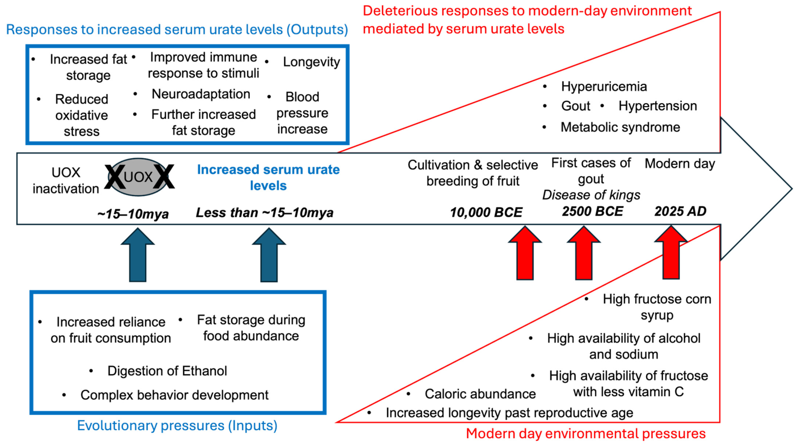
Evolutionarily, many of the features that define humans as a species (high intelligence, complex behaviors dependent on long life histories, a slow life cycle, a propensity for fat storage, and robust adaptive immune responses) may have arisen in part due to the loss of UOX activity and the consequently higher serum SU levels (Figure 2). SU has therefore been a long-term evolutionary companion of humans, which has shaped our physiology through the modern day. In light of the above hypotheses and findings, we propose that due to the rapid increase in the availability of fructose and ethanol, alongside increasing human longevity, humans are currently left with a susceptibility to diseases across multiple organ systems mediated in part by SU, which was once essential for survival throughout periods of environmental harshness (Figure 2). Modern-day humans experience various consequences of high SU levels that seem to track with increasing age. Gout prevalence increases sharply with each decade of life [88]. Further, hypertension and chronic kidney disease, both of which are associated with aging [89], are highly comorbid with gout [88]. Together, these data suggest that gout may represent the long-term cost of SU’s evolutionary role in promoting human longevity, manifesting in the accumulation of comorbidities with prolonged lifespan, when the evolutionary fitness of the individual becomes less relevant to reproduction and population dynamics.
The evolutionary importance of UOX inactivation on purine metabolism needs to be considered in light of other evolutionary changes and genetic mutations that affect urate homeostasis. Although mutations to the URAT1 gene occurred about the same time as UOX inactivation [24] and have become fixed in the great apes, the accumulation of genetic variation continues to affect urate transporter genes. For example, among people with African ancestry, a rare novel loss-of-function mutation, G65W (rs12800450) has occurred, and genetic variations in ABCG2 (e.g., the 141K missense variant), and GLUT9 impact urate excretion [90] in ancestrally diverse populations [91]. Moving forward, efforts should be made to design drugs and treat diseases such as gout with an understanding of the rich human evolutionary context these modern-day maladies have emerged from, and the unifying role of SU across these disease states.

Author Contributions

M.J.N. conceived of the topic, conducted the literature review, created the figures, and wrote and edited the manuscript. A.R. and J.D.F. revised the manuscript and provided key guidance on figure design and the scope of the article. All authors have read and agreed to the published version of the manuscript.

Funding

Salary support for Amir Razmjou and John D. FitzGerald for their mentoring roles is provided by the Greater Los Angeles VA and UCLA. The views expressed in this manuscript do not represent the views of either employer, the official position of the Department of Veterans Affairs or the U.S. government.

Institutional Review Board Statement

Not applicable.

Data Availability Statement

No new data were created or analyzed in this study. Data sharing is not applicable to this article.

Acknowledgments

We thank the UCLA physician-scientist training program and internal medicine residency program for facilitating varied and comprehensive clinical experiences for their trainees, ultimately making this manuscript possible. We thank Brian Grant for his grammatical review and editing.

Conflicts of Interest

The authors have no competing interests to report.

Abbreviations

The following abbreviations are used in this manuscript: SU; serum urate, UOX; uricase, ATP; adenosine triphosphate, AMP; adenosine monophosphate, EtOH; ethanol, MSU; monosodium urate.

References

  1. Singh, G.; Lingala, B.; Mithal, A. Gout and hyperuricaemia in the USA: Prevalence and trends. Rheumatology 2019, 58, 2177–2180. [Google Scholar] [CrossRef] [Scilit] [PubMed]
  2. Yokose, C.; McCormick, N.; Lu, N.; Tanikella, S.; Lin, K.; Joshi, A.D.; Raffield, L.M.; Warner, E.; Merriman, T.; Hsu, J.; et al. Trends in Prevalence of Gout Among US Asian Adults, 2011–2018. JAMA Netw. Open 2023, 6, e239501. [Google Scholar] [CrossRef] [Scilit] [PubMed]
  3. Chen-Xu, M.; Yokose, C.; Rai, S.K.; Pillinger, M.H.; Choi, H.K. Contemporary Prevalence of Gout and Hyperuricemia in the United States and Decadal Trends: The National Health and Nutrition Examination Survey, 2007–2016. Arthritis Rheumatol. 2019, 71, 991–999. [Google Scholar] [CrossRef] [Scilit] [PubMed]
  4. Ahn, E.Y.; So, M.W. The pathogenesis of gout. J. Rheum. Dis. 2025, 32, 8–16. [Google Scholar] [CrossRef] [Scilit] [PubMed]
  5. Dalbeth, N.; Merriman, T.R.; Stamp, L.K. Gout. Lancet 2016, 388, 2039–2052. [Google Scholar] [CrossRef] [Scilit] [PubMed]
  6. Roman, Y.M. The Role of Uric Acid in Human Health: Insights from the Uricase Gene. J. Pers. Med. 2023, 13, 1409. [Google Scholar] [CrossRef] [Scilit] [PubMed]
  7. Maiuolo, J.; Oppedisano, F.; Gratteri, S.; Muscoli, C.; Mollace, V. Regulation of uric acid metabolism and excretion. Int. J. Cardiol. 2016, 213, 8–14. [Google Scholar] [CrossRef] [Scilit] [PubMed]
  8. Siegmund, O.H.; Aiello, S.E. The Merck Veterinary Manual; Merck & Co.: Rahway, NJ, USA, 1955. [Google Scholar]
  9. Kuo, C.F.; Grainge, M.J.; Mallen, C.; Zhang, W.; Doherty, M. Comorbidities in patients with gout prior to and following diagnosis: Case-control study. Ann. Rheum. Dis. 2016, 75, 210–217. [Google Scholar] [CrossRef] [Scilit] [PubMed]
  10. Wu, X.W.; Lee, C.C.; Muzny, D.M.; Caskey, C.T. Urate oxidase: Primary structure and evolutionary implications. Proc. Natl. Acad. Sci. USA 1989, 86, 9412–9416. [Google Scholar] [CrossRef] [Scilit] [PubMed]
  11. Oda, M.; Satta, Y.; Takenaka, O.; Takahata, N. Loss of urate oxidase activity in hominoids and its evolutionary implications. Mol. Biol. Evol. 2002, 19, 640–653. [Google Scholar] [CrossRef] [Scilit] [PubMed]
  12. Kratzer, J.T.; Lanaspa, M.A.; Murphy, M.N.; Cicerchi, C.; Graves, C.L.; Tipton, P.A.; Ortlund, E.A.; Johnson, R.J.; Gaucher, E.A. Evolutionary history and metabolic insights of ancient mammalian uricases. Proc. Natl. Acad. Sci. USA 2014, 111, 3763–3768. [Google Scholar] [CrossRef] [Scilit] [PubMed]
  13. Zhamangara, A.; Akmagambet, S.; Nigmatova, S.; Madyarova, I.; Kashaganov, K.; Zadagali, A.; Seidali, A.; Bayshashov, B. The Early Miocene Paleoclimate of Erzhilansay: Interpretation of Climatic Parameters Using Modern Methods. Sustainability 2025, 17, 143. [Google Scholar] [CrossRef] [Scilit]
  14. Habinger, S.G.; Chavasseau, O.; Jaeger, J.J.; Chaimanee, Y.; Soe, A.N.; Sein, C.; Bocherens, H. Evolutionary ecology of Miocene hominoid primates in Southeast Asia. Sci. Rep. 2022, 12, 11841. [Google Scholar] [CrossRef] [Scilit] [PubMed]
  15. Li, C.; Wang, S.Q.; Wang, Y.; Deng, T.; Ma, J.; Wang, B.; Jiangzuo, Q.; Sun, D. Savanna ecosystems and mammalian adaptations in Mid-Miocene Northern China. Sci. Rep. 2025, 15, 25586. [Google Scholar] [CrossRef] [Scilit] [PubMed]
  16. Wu, X.W.; Muzny, D.M.; Lee, C.C.; Caskey, C.T. Two independent mutational events in the loss of urate oxidase during hominoid evolution. J. Mol. Evol. 1992, 34, 78–84. [Google Scholar] [CrossRef] [Scilit] [PubMed]
  17. Videan, E.N.; Heward, C.B.; Chowdhury, K.; Plummer, J.; Su, Y.; Cutler, R.G. Comparison of biomarkers of oxidative stress and cardiovascular disease in humans and chimpanzees (Pan troglodytes). Comp. Med. 2009, 59, 287–296. [Google Scholar] [PubMed]
  18. Desideri, G.; Castaldo, G.; Lombardi, A.; Mussap, M.; Testa, A.; Pontremoli, R.; Punzi, L.; Borghi, C. Is it time to revise the normal range of serum uric acid levels? Eur. Rev. Med. Pharmacol. Sci. 2014, 18, 1295–1306. [Google Scholar] [PubMed]
  19. Gosling, A.L.; Matisoo-Smith, E.; Merriman, T.R. Hyperuricaemia in the Pacific: Why the elevated serum urate levels? Rheumatol. Int. 2014, 34, 743–757. [Google Scholar] [CrossRef] [Scilit] [PubMed]
  20. Nuki, G.; Simkin, P.A. A concise history of gout and hyperuricemia and their treatment. Arthritis Res. Ther. 2006, 8, S1. [Google Scholar] [CrossRef] [Scilit] [PubMed]
  21. Hyndman, D.; Liu, S.; Miner, J.N. Urate Handling in the Human Body. Curr. Rheumatol. Rep. 2016, 18, 34. [Google Scholar] [CrossRef] [Scilit] [PubMed]
  22. Enomoto, A.; Kimura, H.; Chairoungdua, A.; Shigeta, Y.; Jutabha, P.; Cha, S.H.; Hosoyamada, M.; Takeda, M.; Sekine, T.; Igarashi, T.; et al. Molecular identification of a renal urate anion exchanger that regulates blood urate levels. Nature 2002, 417, 447–452. [Google Scholar] [CrossRef] [Scilit] [PubMed]
  23. Percudani, R.; Capelli, C.; Sassi, G.; Mori, G. Evolutionary Differences in Uric Acid Metabolism Inform about the Risks of Pig-to-Human Kidney Transplant. J. Am. Soc. Nephrol. 2024, 35, 1281–1283. [Google Scholar] [CrossRef] [Scilit] [PubMed]
  24. Tan, P.K.; Farrar, J.E.; Gaucher, E.A.; Miner, J.N. Coevolution of URAT1 and Uricase during Primate Evolution: Implications for Serum Urate Homeostasis and Gout. Mol. Biol. Evol. 2016, 33, 2193–2200. [Google Scholar] [CrossRef] [Scilit] [PubMed]
  25. Watanabe, S.; Kang, D.H.; Feng, L.; Nakagawa, T.; Kanellis, J.; Lan, H.; Mazzali, M.; Johnson, R.J. Uric acid, hominoid evolution, and the pathogenesis of salt-sensitivity. Hypertension 2002, 40, 355–360. [Google Scholar] [CrossRef] [Scilit] [PubMed]
  26. Yokokawa, H.; Fukuda, H.; Suzuki, A.; Fujibayashi, K.; Naito, T.; Uehara, Y.; Nakayama, A.; Matsuo, H.; Sanada, H.; Jose, P.A.; et al. Association Between Serum Uric Acid Levels/Hyperuricemia and Hypertension Among 85,286 Japanese Workers. J. Clin. Hypertens. 2016, 18, 53–59. [Google Scholar] [CrossRef] [Scilit] [PubMed]
  27. Natterson-Horowitz, B.; Baccouche, B.M.; Mary, J.; Shivkumar, T.; Bertelsen, M.F.; Aalkjaer, C.; Smerup, M.H.; Ajijola, O.A.; Hadaya, J.; Wang, T. Did giraffe cardiovascular evolution solve the problem of heart failure with preserved ejection fraction? Evol. Med. Public Health 2021, 9, 248–255. [Google Scholar] [CrossRef] [Scilit] [PubMed]
  28. Aalkjaer, C.; Wang, T. The Remarkable Cardiovascular System of Giraffes. Annu. Rev. Physiol. 2021, 83, 1–15. [Google Scholar] [CrossRef] [Scilit] [PubMed]
  29. Agaba, M.; Ishengoma, E.; Miller, W.C.; McGrath, B.C.; Hudson, C.N.; Bedoya Reina, O.C.; Ratan, A.; Burhans, R.; Chikhi, R.; Medvedev, P.; et al. Giraffe genome sequence reveals clues to its unique morphology and physiology. Nat. Commun. 2016, 7, 11519. [Google Scholar] [CrossRef] [Scilit] [PubMed]
  30. Agarwal, V.; Hans, N.; Messerli, F.H. Effect of allopurinol on blood pressure: A systematic review and meta-analysis. J. Clin. Hypertens. 2013, 15, 435–442. [Google Scholar] [CrossRef] [Scilit] [PubMed]
  31. Feig, D.I.; Soletsky, B.; Johnson, R.J. Effect of allopurinol on blood pressure of adolescents with newly diagnosed essential hypertension: A randomized trial. JAMA 2008, 300, 924–932. [Google Scholar] [CrossRef] [Scilit] [PubMed]
  32. Gaffo, A.L.; Calhoun, D.A.; Rahn, E.J.; Oparil, S.; Li, P.; Dudenbostel, T.; Feig, D.I.; Redden, D.T.; Muntner, P.; Foster, P.J.; et al. Effect of Serum Urate Lowering With Allopurinol on Blood Pressure in Young Adults: A Randomized, Controlled, Crossover Trial. Arthritis Rheumatol. 2021, 73, 1514–1522. [Google Scholar] [CrossRef] [Scilit] [PubMed]
  33. Fabbrini, E.; Serafini, M.; Colic Baric, I.; Hazen, S.L.; Klein, S. Effect of plasma uric acid on antioxidant capacity, oxidative stress, and insulin sensitivity in obese subjects. Diabetes 2014, 63, 976–981. [Google Scholar] [CrossRef] [Scilit] [PubMed]
  34. Kandar, R.; Stramova, X.; Drabkova, P.; Krenkova, J. A monitoring of allantoin, uric acid, and malondialdehyde levels in plasma and erythrocytes after ten minutes of running activity. Physiol. Res. 2014, 63, 753–762. [Google Scholar] [CrossRef] [Scilit] [PubMed]
  35. Ames, B.N.; Cathcart, R.; Schwiers, E.; Hochstein, P. Uric acid provides an antioxidant defense in humans against oxidant- and radical-caused aging and cancer: A hypothesis. Proc. Natl. Acad. Sci. USA 1981, 78, 6858–6862. [Google Scholar] [CrossRef] [Scilit] [PubMed]
  36. Santos, A.L.; Sinha, S.; Lindner, A.B. The Good, the Bad, and the Ugly of ROS: New Insights on Aging and Aging-Related Diseases from Eukaryotic and Prokaryotic Model Organisms. Oxidative Med. Cell. Longev. 2018, 2018, 1941285. [Google Scholar] [CrossRef] [Scilit] [PubMed]
  37. Latta, L.C.t.; Tucker, K.N.; Haney, R.A. The relationship between oxidative stress, reproduction, and survival in a bdelloid rotifer. BMC Ecol. 2019, 19, 7. [Google Scholar] [CrossRef] [Scilit] [PubMed]
  38. Garratt, M.; Vasilaki, A.; Stockley, P.; McArdle, F.; Jackson, M.; Hurst, J.L. Is oxidative stress a physiological cost of reproduction? An experimental test in house mice. Proc. Biol. Sci. 2011, 278, 1098–1106. [Google Scholar] [CrossRef] [Scilit] [PubMed]
  39. Stier, A.; Reichert, S.; Massemin, S.; Bize, P.; Criscuolo, F. Constraint and cost of oxidative stress on reproduction: Correlative evidence in laboratory mice and review of the literature. Front. Zool. 2012, 9, 37. [Google Scholar] [CrossRef] [Scilit] [PubMed]
  40. Costantini, D. Meta-analysis reveals that reproductive strategies are associated with sexual differences in oxidative balance across vertebrates. Curr. Zool. 2018, 64, 1–11. [Google Scholar] [CrossRef] [Scilit] [PubMed]
  41. Cutler, R.G.; Camandola, S.; Feldman, N.H.; Yoon, J.S.; Haran, J.B.; Arguelles, S.; Mattson, M.P. Uric acid enhances longevity and endurance and protects the brain against ischemia. Neurobiol. Aging 2019, 75, 159–168. [Google Scholar] [CrossRef] [Scilit] [PubMed]
  42. Raichlen, D.A.; Alexander, G.E. Exercise, APOE genotype, and the evolution of the human lifespan. Trends Neurosci. 2014, 37, 247–255. [Google Scholar] [CrossRef] [Scilit] [PubMed]
  43. Hawkes, K.; O’Connell, J.F.; Jones, N.G.; Alvarez, H.; Charnov, E.L. Grandmothering, menopause, and the evolution of human life histories. Proc. Natl. Acad. Sci. USA 1998, 95, 1336–1339. [Google Scholar] [CrossRef] [Scilit] [PubMed]
  44. Crotty, G.F.; Ascherio, A.; Schwarzschild, M.A. Targeting urate to reduce oxidative stress in Parkinson disease. Exp. Neurol. 2017, 298, 210–224. [Google Scholar] [CrossRef] [Scilit] [PubMed]
  45. Kramer, K.L. The evolution of human parental care and recruitment of juvenile help. Trends Ecol. Evol. 2011, 26, 533–540. [Google Scholar] [CrossRef] [Scilit] [PubMed]
  46. Stabouli, S.; Chrysaidou, K.; Chainoglou, A.; Gidaris, D.; Kotsis, V.; Zafeiriou, D. Uric Acid Associates With Executive Function in Children and Adolescents With Hypertension. Hypertension 2021, 77, 1737–1744. [Google Scholar] [CrossRef] [Scilit] [PubMed]
  47. Schretlen, D.J.; Inscore, A.B.; Jinnah, H.A.; Rao, V.; Gordon, B.; Pearlson, G.D. Serum uric acid and cognitive function in community-dwelling older adults. Neuropsychology 2007, 21, 136–140. [Google Scholar] [CrossRef] [Scilit] [PubMed]
  48. Latourte, A.; Soumare, A.; Bardin, T.; Perez-Ruiz, F.; Debette, S.; Richette, P. Uric acid and incident dementia over 12 years of follow-up: A population-based cohort study. Ann. Rheum. Dis. 2018, 77, 328–335. [Google Scholar] [CrossRef] [Scilit] [PubMed]
  49. Raffaitin, C.; Gin, H.; Empana, J.P.; Helmer, C.; Berr, C.; Tzourio, C.; Portet, F.; Dartigues, J.F.; Alperovitch, A.; Barberger-Gateau, P. Metabolic syndrome and risk for incident Alzheimer’s disease or vascular dementia: The Three-City Study. Diabetes Care 2009, 32, 169–174. [Google Scholar] [CrossRef] [Scilit] [PubMed]
  50. Yuan, H.; Yu, C.; Li, X.; Sun, L.; Zhu, X.; Zhao, C.; Zhang, Z.; Yang, Z. Serum Uric Acid Levels and Risk of Metabolic Syndrome: A Dose-Response Meta-Analysis of Prospective Studies. J. Clin. Endocrinol. Metab. 2015, 100, 4198–4207. [Google Scholar] [CrossRef] [Scilit] [PubMed]
  51. Goldman, L. Three Stages of Health Encounters Over 8000 Human Generations and How They Inform Future Public Health. Am. J. Public Health 2018, 108, 60–62. [Google Scholar] [CrossRef] [Scilit] [PubMed]
  52. Johnson, R.J.; Sanchez-Lozada, L.G.; Nakagawa, T.; Rodriguez-Iturbe, B.; Tolan, D.; Gaucher, E.A.; Andrews, P.; Lanaspa, M.A. Do thrifty genes exist? Revisiting uricase. Obesity 2022, 30, 1917–1926. [Google Scholar] [CrossRef] [Scilit] [PubMed]
  53. Andrews, P.; Martin, L. Hominoid dietary evolution. Philos. Trans. R. Soc. Lond. B Biol. Sci. 1991, 334, 199–209, discussion 209. [Google Scholar] [CrossRef] [Scilit] [PubMed]
  54. Spengler, R.N., 3rd; Kienast, F.; Roberts, P.; Boivin, N.; Begun, D.R.; Ashastina, K.; Petraglia, M. Bearing Fruit: Miocene Apes and Rosaceous Fruit Evolution. Biol. Theory 2023, 18, 134–151. [Google Scholar] [CrossRef] [Scilit] [PubMed]
  55. Russo, E.; Leoncini, G.; Esposito, P.; Garibotto, G.; Pontremoli, R.; Viazzi, F. Fructose and Uric Acid: Major Mediators of Cardiovascular Disease Risk Starting at Pediatric Age. Int. J. Mol. Sci. 2020, 21, 4479. [Google Scholar] [CrossRef] [Scilit] [PubMed]
  56. Choi, Y.J.; Shin, H.S.; Choi, H.S.; Park, J.W.; Jo, I.; Oh, E.S.; Lee, K.Y.; Lee, B.H.; Johnson, R.J.; Kang, D.H. Uric acid induces fat accumulation via generation of endoplasmic reticulum stress and SREBP-1c activation in hepatocytes. Lab. Investig. 2014, 94, 1114–1125. [Google Scholar] [CrossRef] [Scilit] [PubMed]
  57. DeMiguel, D.; Fortelius, M.; Azanza, B.; Morales, J. Ancestral feeding state of ruminants reconsidered: Earliest grazing adaptation claims a mixed condition for Cervidae. BMC Evol. Biol. 2008, 8, 13. [Google Scholar] [CrossRef] [Scilit] [PubMed]
  58. Van Valkenburgh, B. Deja vu: The evolution of feeding morphologies in the Carnivora. Integr. Comp. Biol. 2007, 47, 147–163. [Google Scholar] [CrossRef] [Scilit] [PubMed]
  59. Choi, H.K.; Curhan, G. Soft drinks, fructose consumption, and the risk of gout in men: Prospective cohort study. BMJ 2008, 336, 309–312. [Google Scholar] [CrossRef] [Scilit] [PubMed]
  60. Choi, H.K.; Willett, W.; Curhan, G. Fructose-rich beverages and risk of gout in women. JAMA 2010, 304, 2270–2278. [Google Scholar] [CrossRef] [Scilit] [PubMed]
  61. Zhang, Y.; Neogi, T.; Chen, C.; Chaisson, C.; Hunter, D.J.; Choi, H.K. Cherry consumption and decreased risk of recurrent gout attacks. Arthritis Rheum. 2012, 64, 4004–4011. [Google Scholar] [CrossRef] [Scilit] [PubMed]
  62. Nakagawa, T.; Lanaspa, M.A.; Johnson, R.J. The effects of fruit consumption in patients with hyperuricaemia or gout. Rheumatology 2019, 58, 1133–1141. [Google Scholar] [CrossRef] [Scilit] [PubMed]
  63. Choi, H.K.; Gao, X.; Curhan, G. Vitamin C intake and the risk of gout in men: A prospective study. Arch. Intern. Med. 2009, 169, 502–507. [Google Scholar] [CrossRef] [Scilit] [PubMed]
  64. Brzezinska, O.; Styrzynski, F.; Makowska, J.; Walczak, K. Role of Vitamin C in Prophylaxis and Treatment of Gout-A Literature Review. Nutrients 2021, 13, 701. [Google Scholar] [CrossRef] [Scilit] [PubMed]
  65. Hornung, T.C.; Biesalski, H.K. Glut-1 explains the evolutionary advantage of the loss of endogenous vitamin C-synthesis: The electron transfer hypothesis. Evol. Med. Public Health 2019, 2019, 221–231. [Google Scholar] [CrossRef] [Scilit] [PubMed]
  66. Alvarez-Lario, B.; Macarron-Vicente, J. Uric acid and evolution. Rheumatology 2010, 49, 2010–2015. [Google Scholar] [CrossRef] [Scilit] [PubMed]
  67. Wright, P.C.; Tecot, S.R.; Erhart, E.M.; Baden, A.L.; King, S.J.; Grassi, C. Frugivory in four sympatric lemurs: Implications for the future of Madagascar’s forests. Am. J. Primatol. 2011, 73, 585–602. [Google Scholar] [CrossRef] [Scilit] [PubMed]
  68. Christen, P.; Peacock, W.C.; Christen, A.E.; Wacker, W.E. Urate oxidase in primate phylogenesis. Eur. J. Biochem. 1970, 12, 3–5. [Google Scholar] [CrossRef] [Scilit] [PubMed]
  69. Mahajan, M.; Kaur, S.; Mahajan, S.; Kant, R. Uric acid a better scavenger of free radicals than vitamin C in rheumatoid arthritis. Indian J. Clin. Biochem. 2009, 24, 205–207. [Google Scholar] [CrossRef] [Scilit] [PubMed]
  70. Zou, Y.; Liu, Y.; Li, S. Association between dietary vitamin C intake and gout among American adults. Front. Immunol. 2024, 15, 1431323. [Google Scholar] [CrossRef] [Scilit] [PubMed]
  71. Giuntini, E.B.; Sarda, F.A.H.; de Menezes, E.W. The Effects of Soluble Dietary Fibers on Glycemic Response: An Overview and Futures Perspectives. Foods 2022, 11, 3934. [Google Scholar] [CrossRef] [Scilit] [PubMed]
  72. Parker, I.M.; Lopez, I.; Petersen, J.J.; Anaya, N.; Cubilla-Rios, L.; Potter, D. Domestication Syndrome in Caimito (Chrysophyllum cainito L.): Fruit and Seed Characteristics. Econ. Bot. 2010, 64, 161–175. [Google Scholar] [CrossRef] [Scilit] [PubMed]
  73. Carrigan, M.A.; Uryasev, O.; Frye, C.B.; Eckman, B.L.; Myers, C.R.; Hurley, T.D.; Benner, S.A. Hominids adapted to metabolize ethanol long before human-directed fermentation. Proc. Natl. Acad. Sci. USA 2015, 112, 458–463. [Google Scholar] [CrossRef] [Scilit] [PubMed]
  74. Bowland, A.C.; Bersacola, E.; Ramon, M.; Bessa, J.; Melin, A.D.; Carrigan, M.A.; Harrison, X.A.; Hockings, K.J. Wild chimpanzees share fermented fruits. Curr. Biol. 2025, 35, R279–R280. [Google Scholar] [CrossRef] [Scilit] [PubMed]
  75. Yamamoto, T.; Moriwaki, Y.; Takahashi, S. Effect of ethanol on metabolism of purine bases (hypoxanthine, xanthine, and uric acid). Clin. Chim. Acta 2005, 356, 35–57. [Google Scholar] [CrossRef] [Scilit] [PubMed]
  76. Nishimura, T.; Shimizu, T.; Mineo, I.; Kawachi, M.; Ono, A.; Nakajima, H.; Kuwajima, M.; Kono, N.; Matsuzawa, Y. Influence of daily drinking habits on ethanol-induced hyperuricemia. Metabolism 1994, 43, 745–748. [Google Scholar] [CrossRef] [Scilit] [PubMed]
  77. Choi, H.K.; Curhan, G. Beer, liquor, and wine consumption and serum uric acid level: The Third National Health and Nutrition Examination Survey. Arthritis Rheum. 2004, 51, 1023–1029. [Google Scholar] [CrossRef] [Scilit] [PubMed]
  78. Yokose, C.; McCormick, N.; Choi, H.K. Dietary and Lifestyle-Centered Approach in Gout Care and Prevention. Curr. Rheumatol. Rep. 2021, 23, 51. [Google Scholar] [CrossRef] [Scilit] [PubMed]
  79. Li, X.; Meng, X.; He, Y.; Spiliopoulou, A.; Timofeeva, M.; Wei, W.Q.; Gifford, A.; Yang, T.; Varley, T.; Tzoulaki, I.; et al. Genetically determined serum urate levels and cardiovascular and other diseases in UK Biobank cohort: A phenome-wide mendelian randomization study. PLoS Med. 2019, 16, e1002937. [Google Scholar] [CrossRef] [Scilit] [PubMed]
  80. Uhlig, T.; Karoliussen, L.F.; Sexton, J.; Kvien, T.K.; Haavardsholm, E.A.; Perez-Ruiz, F.; Hammer, H.B. Fluctuation and change of serum urate levels and flares in gout: Results from the NOR-Gout study. Clin. Rheumatol. 2022, 41, 3817–3823. [Google Scholar] [CrossRef] [Scilit] [PubMed]
  81. Zhang, Y.; Chen, C.; Choi, H.; Chaisson, C.; Hunter, D.; Niu, J.; Neogi, T. Purine-rich foods intake and recurrent gout attacks. Ann. Rheum. Dis. 2012, 71, 1448–1453. [Google Scholar] [CrossRef] [Scilit] [PubMed]
  82. Webb, R.; Jeffries, M.; Sawalha, A.H. Uric acid directly promotes human T-cell activation. Am. J. Med. Sci. 2009, 337, 23–27. [Google Scholar] [CrossRef] [Scilit] [PubMed]
  83. Wang, Y.; Ma, X.; Su, C.; Peng, B.; Du, J.; Jia, H.; Luo, M.; Fang, C.; Wei, Y. Uric acid enhances the antitumor immunity of dendritic cell-based vaccine. Sci. Rep. 2015, 5, 16427. [Google Scholar] [CrossRef] [Scilit] [PubMed]
  84. Kim, S.K. The Mechanism of the NLRP3 Inflammasome Activation and Pathogenic Implication in the Pathogenesis of Gout. J. Rheum. Dis. 2022, 29, 140–153. [Google Scholar] [CrossRef] [Scilit] [PubMed]
  85. Braga, T.T.; Forni, M.F.; Correa-Costa, M.; Ramos, R.N.; Barbuto, J.A.; Branco, P.; Castoldi, A.; Hiyane, M.I.; Davanso, M.R.; Latz, E.; et al. Soluble Uric Acid Activates the NLRP3 Inflammasome. Sci. Rep. 2017, 7, 39884. [Google Scholar] [CrossRef] [Scilit] [PubMed]
  86. Wang, K.; Li, J.; Li, J.; Zeng, F.; Li, S.; Chen, P.; Xiong, H. Spatiotemporal immune gradients in gout: Immune response-driven activation of the NLRP3-IL-1beta axis and its transition to trained immunity. Front. Immunol. 2026, 17, 1776479. [Google Scholar] [CrossRef] [Scilit] [PubMed]
  87. Cabau, G.; Crisan, T.O.; Kluck, V.; Popp, R.A.; Joosten, L.A.B. Urate-induced immune programming: Consequences for gouty arthritis and hyperuricemia. Immunol. Rev. 2020, 294, 92–105. [Google Scholar] [CrossRef] [Scilit] [PubMed]
  88. Singh, J.A.; Gaffo, A. Gout epidemiology and comorbidities. Semin. Arthritis Rheum. 2020, 50, S11–S16. [Google Scholar] [CrossRef] [Scilit] [PubMed]
  89. Fischer, M.J.; O’Hare, A.M. Epidemiology of hypertension in the elderly with chronic kidney disease. Adv. Chronic Kidney Dis. 2010, 17, 329–340. [Google Scholar] [CrossRef] [Scilit] [PubMed]
  90. Flynn, T.J.; Phipps-Green, A.; Hollis-Moffatt, J.E.; Merriman, M.E.; Topless, R.; Montgomery, G.; Chapman, B.; Stamp, L.K.; Dalbeth, N.; Merriman, T.R. Association analysis of the SLC22A11 (organic anion transporter 4) and SLC22A12 (urate transporter 1) urate transporter locus with gout in New Zealand case-control sample sets reveals multiple ancestral-specific effects. Arthritis Res. Ther. 2013, 15, R220. [Google Scholar] [CrossRef] [Scilit] [PubMed]
  91. Tin, A.; Li, Y.; Brody, J.A.; Nutile, T.; Chu, A.Y.; Huffman, J.E.; Yang, Q.; Chen, M.H.; Robinson-Cohen, C.; Mace, A.; et al. Large-scale whole-exome sequencing association studies identify rare functional variants influencing serum urate levels. Nat. Commun. 2018, 9, 4228. [Google Scholar] [CrossRef] [Scilit] [PubMed]
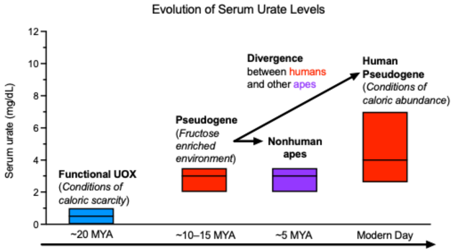
Figure 1. Theoretical changes in serum urate with changes in UOX function and food supply across an evolutionary timescale.
Gucdd 04 00017 g001
Figure 2. Evolutionary and modern-day pressures leading to physiological effects mediated by serum urate.
Gucdd 04 00017 g002
Disclaimer/Publisher’s Note: The statements, opinions and data contained in all publications are solely those of the individual author(s) and contributor(s) and not of MDPI and/or the editor(s). MDPI and/or the editor(s) disclaim responsibility for any injury to people or property resulting from any ideas, methods, instructions or products referred to in the content.