


Abstract
Background and Objectives: To study the bony compartmentalization of the jugular foramen in comparison with existing anatomical models. Materials and Methods: We carried out the study on 59 undamaged dry adult human skulls of known age and sex. Two independent examiners with 10 years of experience in anatomical studies by visual inspections determined the type of osseous septation at the inner surface of the jugular foramina. Relationships between jugular foramen morphology and laterality were assessed at the foraminal level, while associations with age groups and sex were evaluated at the skull level. The data obtained was analyzed using the chi-square statistical test to determine the possibility that two or more variables were associated. Results: Different types of jugular foramina were observed: a single foramen without bony spurs was rarely observed (0.9%). Bipartite and tripartite partial or total foramina were frequent (42.2% and 51.4%, respectively). Partial quadripartite foramina were more rarely observed (5.5%). Complete bony bridges were observed in 13 right and 11 left jugular foramina (24/109, 22.01%). Statistical analysis revealed no significant differences in the occurrence of bony bridges in relation to sex, age, laterality, or foramen partition. As regards the bipartite foramina on the left, we found that in males the frequency was higher in older subjects compared to younger ones; although statistical significance was not reached (p = 0.054). Furthermore, the absence of the intrajugular process from the occipital bone was much higher (45.9%) than that from the temporal bone (1.8%), and this difference was statistically significant (p = 0.0001). Conclusions: The jugular foramen from our studies appears mostly septate. These partitions fit well into the anatomical models existing in the literature for the jugular foramina. Our study fits into the anatomical models of the septum and subdivision of the jugular foramen, especially the bipartite and tripartite ones, even if there are differences related to the behavior of the meninges present in some current anatomical models. The existing literature relates primarily to ethnic groups other than our study, which was conducted on European skulls.
1. Introduction
The jugular foramen (JF), less known as the posterior foramen lacerum, is a large foramen located at the base of the cranium between the petrous part of the temporal bone and the basilar part of the occipital bone through which pass the glossopharyngeal cranial nerve (CN IX), vagus cranial nerve (CN X), and accessory cranial nerve (CN XI), two dural sinuses [sigmoid sinus (SS) and inferior petrosal sinus (IPS)], and the meningeal branches of the occipital and ascending pharyngeal arteries. The foramen is surmounted by a domed bony roof, indicating the presence of a jugular bulb. The two bones can form a bridge between the respective jugular processes, dividing the canal into two compartments, the anteromedial and the posterolateral compartments, in which the nerves and vascular elements are located. The IPS and the CN IX pass through the nervous compartment of the foramen: the anteromedial. The vascular compartment, posterolateral, contains the jugular bulb, the CN X and the spinal portion of the CN XI [1]. The presence of other processes in the foramen can make it tripartite or, in rare cases, quadripartite. Septa of the dura mater in the skull are inserted on the bony processes and contribute to the compartmentalization of the JF [2,3].
However, microanatomical studies have demonstrated that this compartmentalization of the JF in a vascular and nervous compartment is incorrect. Instead, it is fairer to describe the petrosal, sigmoid, and intrajugular portions of the JF. The petrosal portion contains the IPS. The sigmoid portion receives the SS. The intrajugular portion contains CNs IX, X and XI [4,5]. Because the lower cranial nerves pass through the JF, tumors in this region produce multiple cranial nerve palsies, i.e., JF syndromes. These lower cranial nerves could be displaced by pseudolesions arising in the jugular bulb, such as a protruding jugular bulb and arteriovenous malformation. The tumors can arise from the nerves themselves, such as schwannoma or neurofibroma. Finally, glomus jugular tumor can arise from paraganglionic tissue. Paragangliomas are the most common tumors of the JF region [6].
The displacement of the last CNs IX-X-XI by this tumor and other processes may give rise to Vernet’s syndrome [5]. This is characterized by loss of taste sensation in the posterior third of the tongue, paralysis of the vocal cords and soft palate, and weakness of the trapezius and sternocleidomastoid muscles [5].
The JF was a subject of many morphological studies for its importance in skull base neurosurgery.
Most of these osteological studies concern the morphology of the JF, its morphometry to capture statistical differences related to sex, race, and laterality [6,7]. Few focus on the partition, the modalities of bone formation and the types of partition, which can provide important details for the neurosurgery of the cranial base.
Therefore, we analyzed the frequency and morphological appearance of the JF partitions in a collection of Italian skulls and compared the partition of the JF observed with the anatomical model of partition present in the literature. Furthermore, we looked for an association between frequency of partitions and type of partition with the age, sex and laterality of the skulls. This is the first study on a population of skulls from Italy, since most of the skulls in many previous studies come from regions of South America, India and Africa [8,9,10,11,12,13]. Furthermore, most of these studies focus on the morphometry of the jugular foramen and not on the detailed septation as in our study.
2. Materials and Methods
2.1. Subjects
We carried out the study on 59 undamaged dry adult human skulls of known age and sex. The skulls were collected from the local ‘Leonetto Comparini’ Museum of Siena and belonged to individuals from Siena and its surroundings (1882–1932), therefore of European ethnicity. Approval by the ethics commission was not requested because the skulls have been the property of the Anatomical Museum since 1882. The number of skulls observed was 59, corresponding to a total of 118 jugular foramina. Nine foramina could not be assessed because they were damaged; therefore, 109 jugular foramina were included in the analysis belonging to 55 specimens, including 32 males (58.18%) and 23 females (41.82%). The youngest subject was 18 years old and the oldest was 85. We calculated the mean age by sex, and the data are as follows: mean age for males 58.8, mean age for females 58.5. Based on age, these subjects were divided into two groups: group 1, 18–62 years (26 individuals), and group 2, 63–85 years (29 individuals).
2.2. Jugular Foramina Analysis
Two independent examiners with 10 years of experience in anatomical studies independently performed visual inspection to determine the type of osseous septation, classified as complete or incomplete, at the inner surface of the jugular foramina. Inter-rater reliability was assessed using percent agreement, which was 83% between the two examiners.
Different types of jugular foramina were observed and grouped into four categories: Type 1, a single foramen without bony spurs; Type 2, partial bipartite (with variants) or total bipartite; Type 3, partial tripartite (with variants) or total tripartite; and Type 4, partial quadripartite (with variants). The frequency of jugular processes arising from the temporal and occipital bones was assessed and classified as no process, solitary process, double process, or triple process.
We also recorded the frequency of complete bony bridges that resulted in total partitioning of the jugular foramen. These bridges were formed either by a single elongated process arising from the temporal bone, a single elongated process from the occipital bone, or by two processes—one from each bone—that fused together. Total partition was defined as a bipartite or tripartite foramen and was assigned only when all bridges present were complete. Therefore, foramina with a single complete bridge adjacent to additional short processes were classified as tripartite or partial quadripartite.
The authors compared the compartmentalization observed in the JF with the current model present in the literature. A total of 4 models have been proposed to date [14]. These include the 2-part (bipartite) models by Hovelacque (1934) [15] and Shapiro (1972) [16] and the 3-part (tripartite) subdivisions by Katsuta (1997) [4] and Bernard (2018) [17]. Hovelaque [15] reported that the intrajugular process (IJP) and a fibrous bridge divided the JF in two compartments: a smaller anteromedial containing CN IX and IPS and a larger posterolateral containing CN X and CN XI, jugular bulb and postero meningeal artery. Shapiro’s model [16] can be considered a revised Hovelaque model [15]. In the model of Katsuta [4] there are present two venous compartments containing an anterior IPS and posterior internal jugular vein (IJV), separated by the IJP. One nervous compartment is intrajugular and contains IX, X and XI CNs. Bernard [17], in a microdissection study proposed a new tripartite model containing two neural compartments and an interperiosteodural compartment containing IPS, IJV, and extradural neuroaxis compartment (EDNAC). EDNAC is a space extending from the orbit to the coccyx and containing valveless veins, nerves, and adipose tissue.
We also assessed the presence or absence, in each JF, of a dome above the jugular fossa. A dome indicates the presence of a prominent superior jugular bulb. The dome is always posterolateral in the JF.
2.3. Statistics
We studied the relationships between jugular foramen morphology and laterality at the foraminal level, and the associations between morphological patterns and age groups or sex at the skull level, considering only specimens with available data for each variable. Categorical variables were analyzed using the chi-square test or Fisher’s exact test, as appropriate, depending on expected cell frequencies. Given the exploratory nature of the study and the predominantly descriptive aim, no formal correction for multiple comparisons was applied. The present study set its significance level at 5% (0.05). We employed the software GraphPad Prism 6, Dotmatics, England for statistical analysis.
3. Results
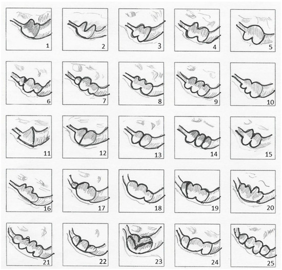
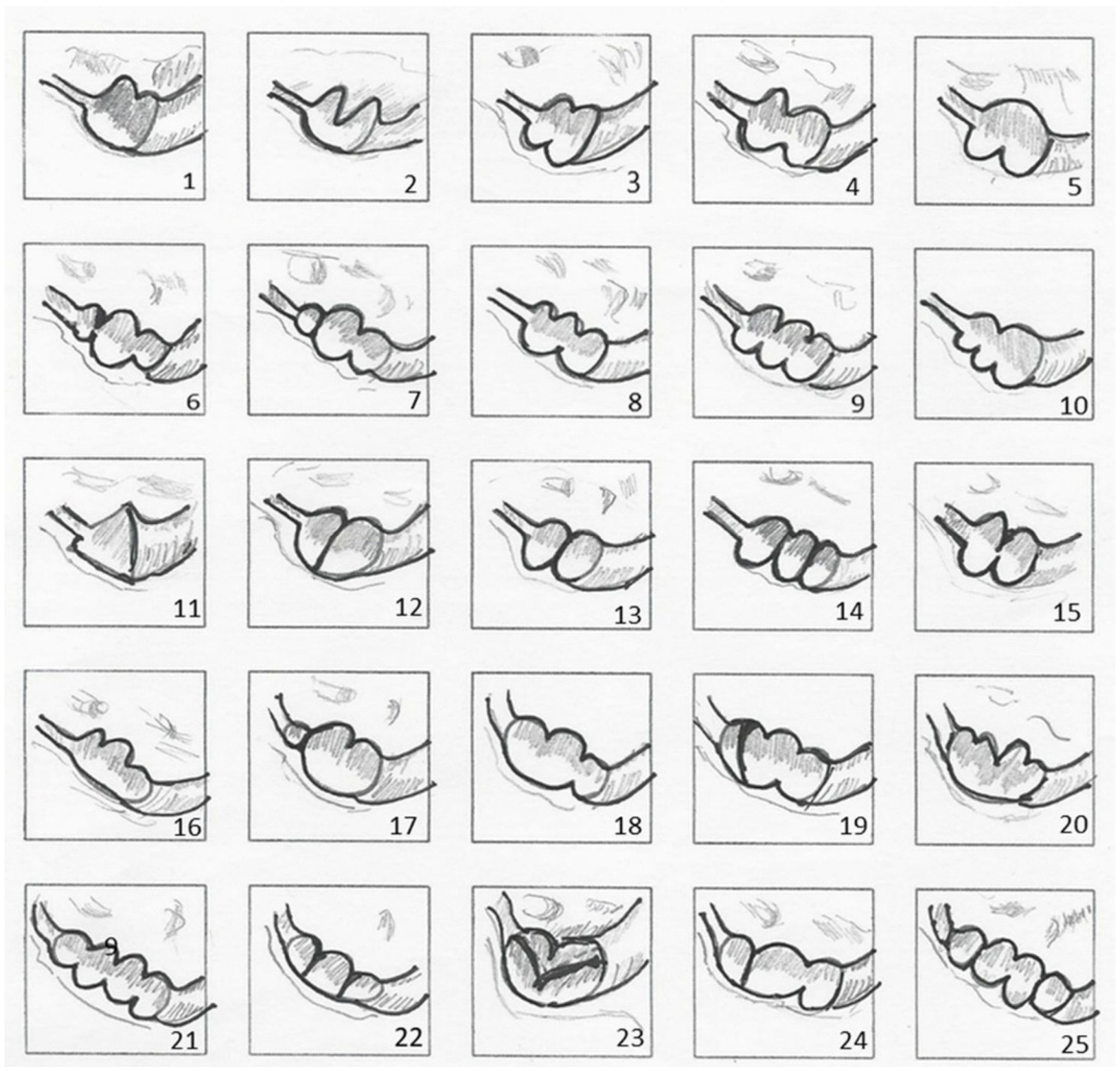
Twenty-five different types of jugular foramina were observed; a classification scheme was prepared to more clearly describe the varied types of the JF bony septa (Figure 1).
Figure 1.
Diagram of the right jugular foramen and septa as seen from the right endocranial surface. The septa visible at the top of the jugular foramen originated from the pyramid of the temporal bone, the septa visible at the bottom originated from the occipital bone. Twenty-five different types of foramina are shown. Specimens 1–5 exhibit partial bipartite foramina, whereas specimen 15 shows an almost complete bipartition. Specimens 12 and 13 demonstrate complete bipartition: the former resulting from a single bony spur arising from the temporal bone, and the latter from two opposing bony spurs originating from the temporal and occipital bones. Partial tripartite foramina are observed in specimens 6–10, 16–18, and 24, while specimens 14 and 22 display complete tripartition. Finally, specimens 19–21 and 25 present quadripartite foramina. Number 11 shows a wide single triangular jugular foramen observed in a single skull. Number 23 is a case of uncertain classification showing 2 bony spurs from the temporal bone aligned from front to back and no bony spurs from the occipital bone.
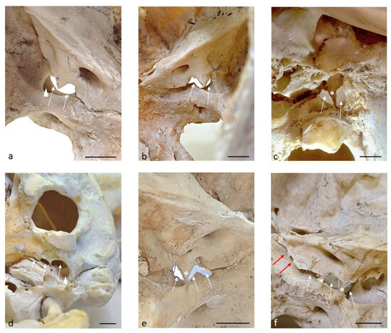
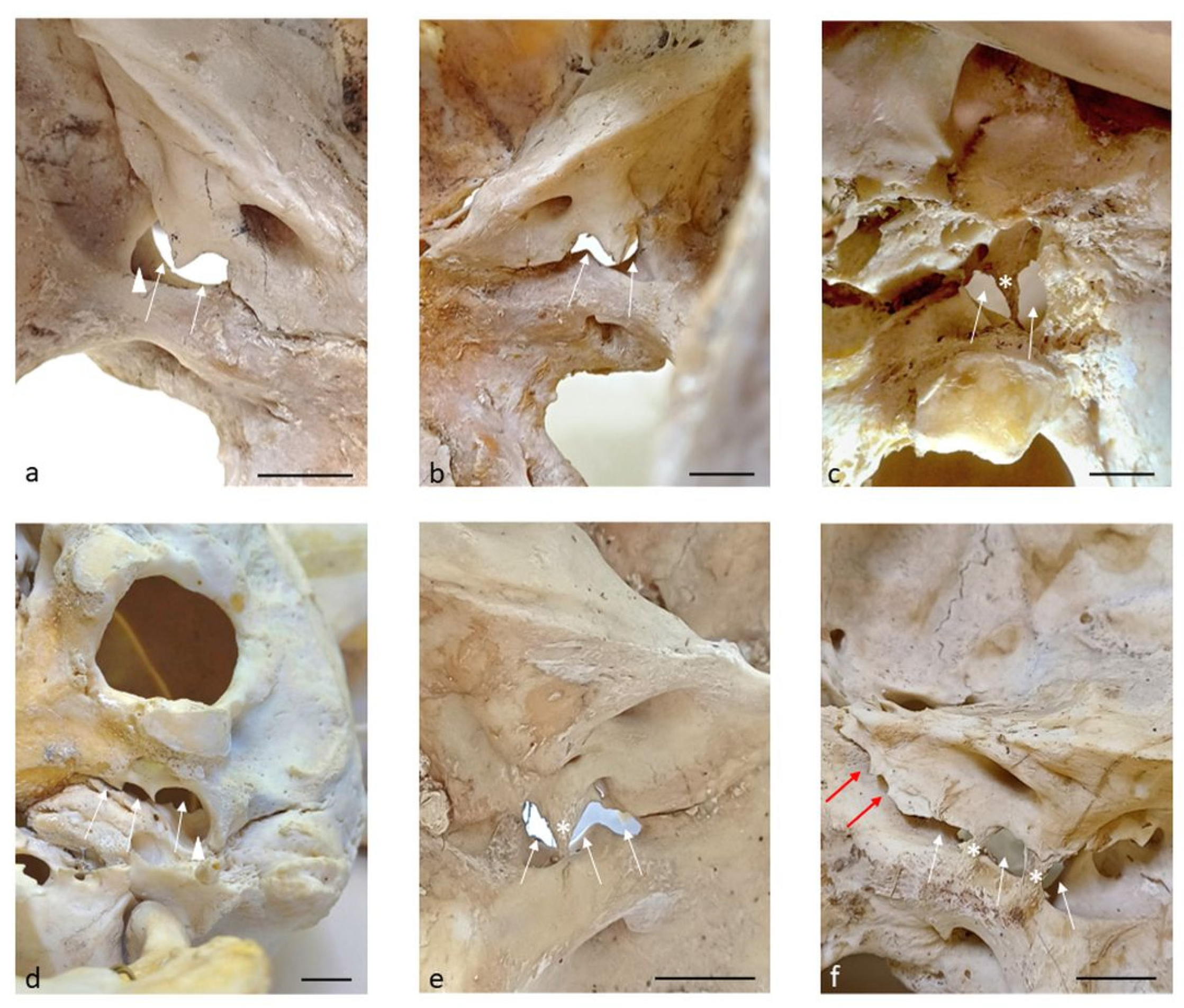
In Figure 2, some types of division of the jugular foramen are visible.
Figure 2.
(a) Endocranial view of the left JF in an 80-year-old male skull, showing a partially bipartite foramen. (b) Endocranial view of the right JF in the same 80-year-old male skull, showing a partially bipartite foramen. Panels (a,b) belong to the same subject. (c) Exocranial view of the left JF in an 82-year-old male skull, showing a totally bipartite foramen. (d) Exocranial view of the right JF in a 71-year-old male skull, showing a partially tripartite foramen. (e) Endocranial view of the left JF in a 75-year-old female skull, showing a partially tripartite foramen. (f) Endocranial view of the right JF in a 26-year-old female skull, showing a totally tripartite foramen. The white arrows show the compartments into which the JF was divided. The white asterisks indicate the complete bridges. White arrowheads show obvious and deep domes. Red arrows show the petro-occipital fissure. Scale bar = 1 cm.
Sex-related analyses were performed on the 54 skulls in which sex could be reliably determined. Age-related analyses were performed on the 55 skulls with reliable age information. At the foraminal level, the single foramen without bony spurs was rarely observed (0.9%).
Bipartite and tripartite partial or total foramina were frequent, 42.2% and 51.4% respectively. Partial or complete tripartite jugular foramina most commonly consisted of two bony spurs arising from the temporal bone and one from the occipital bone, or of two spurs from each bone. Less frequently, they were formed by two bony spurs originating from the temporal bone with no contribution from the occipital bone.
At the foraminal level, partial quadripartite foramina (5.5%) were more rarely observed. Quadripartite jugular foramina were separated by three bony spurs arising from the temporal bone and one from the occipital bone. In addition, two foramina had three bony spurs from the temporal bone and three from the occipital bone; one foramen had three bony spurs from the temporal bone and none from the occipital bone, and two quadripartite jugular foramina showed completely closed medial and lateral compartments with two partially closed central compartments.
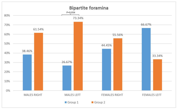
As regards the bipartite foramina on the left, we found that in males the frequency was higher in group 2 than in group 1; a trend toward a higher frequency was observed, although statistical significance was not reached (p = 0.054) (Table 1).
Table 1.
Distribution of bipartite jugular foramina according to sex, side, and age group (Group 1: 18–62 years; Group 2: 63–85 years). Percentages represent the proportion of bipartite foramina observed within each subgroup. Related graphic representation. In males on the left the frequency was higher in group 2 than in group 1; a trend toward a higher frequency was observed, although statistical significance was not reached (p = 0.054; SD = 0.49; SE Group 1 = 0.16, SE Group 2 = 0.11). The differences observed between the other data are not statistically significant.
As regards the frequency of the jugular process of the temporal bone, no process was observed in a low percentage (1.8%), a solitary process in 43.1%, a frequent double process in 46.8%, and a triple process in 7.3%. Compared with the temporal bone, the intrajugular process of the occipital bone showed a high frequency of no process (45.9%), and this difference was statistically significant (p = 0.0001). Complete bony bridges were observed in 13 right and 11 left jugular foramina, accounting for 24 of 109 foramina (22.01%). In some cases, complete partitioning of the jugular foramen was observed, resulting from the presence of a full bony bridge formed either by a single elongated process originating from the temporal bone or by two processes—arising from both the temporal and occipital bones—that fused together.
Jugular bulb domes are 49 on the right and 38 on the left: 87 total, 73.72% of which 56.32% on the right, 43.67% on the left; 53 domes in males: 60.91%, of which 32 are on the right (60.37%) and 21 are on the left (39.62%); 34 domes in females: 39.08%, of which 17 are on the right (50%) and 17 are on the left (50%).
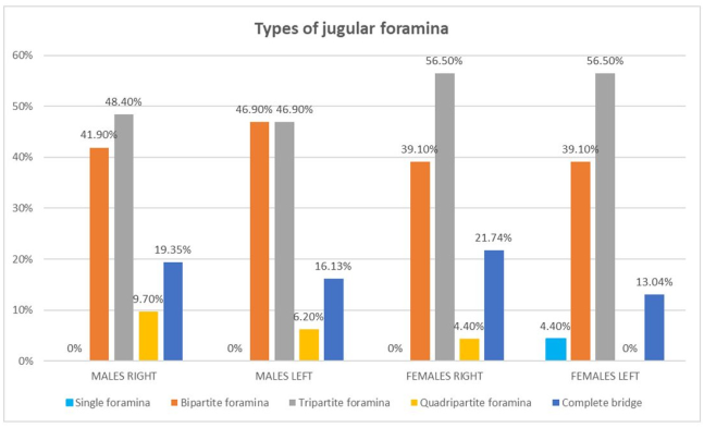
No statistically significant differences were observed in the distribution of jugular bulb domes or bony bridges in relation to sex, age group, laterality, or foramen partition. Table 2 summarizes the data relating to the percentages of the various types of jugular foramina by side and sex.
Table 2.
Distribution of jugular foramen morphological types (single, bipartite, tripartite, and quadripartite) and complete bony bridges according to sex and side. Values are expressed as percentages within each subgroup. Related graphic representation.
4. Discussion
The JF from our studies appears mostly septate. A foramen without septa is present in 0.9% of foramina; that is, in only one case. In most cases, it appears bipartite or tripartite (42.2% and 51.4%, respectively). In rare cases, a quadripartite foramen was observed (5.5%). In the work of N. Taprantzis et al. (2026) [14], the percentages of partial septation are close to those observed by us: “Partial right septation ranged from 37.7% to 40.9% (I2 ≈ 97%), partial left septation from 42.7% to 46.5% (I2 ≈ 97%), and partial bilateral septation from 5.9% to 8.6% (I2 ≈ 95%).” However, as regards the absence of septation, these authors report a much higher percentage than the one observed by us.
These partitions fit well into the anatomical models existing in the literature for the jugular foramina. In these models, the vascular-nervous structures find a precise localization and subdivision. Four models currently exist [18]: bipartite models by Hovelacque (1934) [15] and Shapiro (1972) [16] and the 3-part (tripartite) subdivisions by Katsuta (1997) [4] and Bernard (2018) [17]. Hovelacque [15] and Shapiro [16] divide the JF into an anteromedial and a posterolateral compartment in which the vascular and nervous structures are positioned differently in the two models. We note a similarity with these two models of our osteological model in which an IJP originating mainly from the temporal bone divides the JF into two compartments. Katsuta [4] divided the JF into an anterior petrosal part, containing the IPS; a middle intrajugular/neural part, transmitting CNs IX, X, and XI; and a posterior sigmoid part, which received venous drainage from the SS. After microdissection, Bernard [17] proposed a new compartmentalization of the JF that divides the foramen into two neural compartments and an interperiosteodural compartment. The division of the vascular and nervous structures is based not only on the intrajugular process of the other models but also on the existence of the EDNAC. The EDNAC is a compartment containing arteries, valveless veins, nerves, and adipose tissue, which extends uninterruptedly from the orbit to the coccyx [14,19]. We cannot compare our osteological study to that of Bernard [17], which is based on microdissection. In all models, the dividing structure of the JF is the jugular process, which we observed missing from the occipital bone (45.9%) in a significant percentage compared to that of the temporal bone (1.8%). This difference was highly significant (p = 0.0001). Therefore, the temporal bone with the intrajugular process contributes most significantly to the compartmentalization of the JF, similarly to the bipartite and tripartite models.
A variant of the JF is the rare quadripartite foramen (5.5%). Solter [20] in 300 skulls examined found the foramen divided into 4 parts by 3 bone bridges.
The subdivision of our quadripartite foramina appears more complex than that of Solter [20]. The quadripartite foramina are separated by 3 bony spurs of the temporal bone and 1 from the occipital bone in a foramen. In addition, there are 2 foramina that have 3 bony spurs from the temporal bone and 3 from the occipital bone; 1 foramen that has 3 bony spurs from the temporal bone and 0 from the occipital bone; and then there are 2 quadripartite jugular foramina that have completely closed medial and lateral foramina and 2 partially closed central foramina.
In some cases, we observed a total partition of the JF, which could be determined by the presence of a complete bridge generated by a single long process originating from the temporal bone and two processes originating from both bones that join together.
A complete bony bridge was observed in 22.01% of the analyzed jugular foramina, in agreement with previous reports [21,22]. However, Fang reports a lower percentage of bony bridges [23]. The sex difference in the frequency of the bridging trait was not statistically significant. Nor was the difference between the left and right sides statistically significant. Oliveira [24] observed significant sex-related differences, but he observed septation on the external face of the JF. One complete septation (i.e., the presence of two fully divided jugular compartments) occurred more often on the right side of female individuals (1 complete septation, male = 9.43%; 1 complete septation, female = 25.58%; p = 0.038 [21]). This data could be important to avoid injuring adjacent cranial nerves. Despite the advances in skull base surgery, postoperative lower cranial nerve deficits still occur. A bony septum can also complicate the manipulation of the access to nerves exiting the JF because it provides greater resistance than dural septa, increasing the risk of injury. Juxtacondylar approaches are indicated for neurinomas and meningiomas of the JF and paragangliomas that extend into the petrous bone. To visualize areas in front of the brainstem during surgery may be necessary [25], drilling into an intrajugular foramen, during which particular care must be taken to avoid injury to adjacent cranial nerves.
A preoperative radiological study is necessary to have a detailed anatomical radiological view of the JF. High-resolution computed tomography (HRCT) has been proposed as the method of choice in preoperative evaluation of JF lesions. The JF appeared as a canal with endocranial and exocranial apertures in all specimens. No bony partition of the JF was observed. A dural band consistently divided the JF into two parts: anterior to it was the CN IX, while the CN X and CN XI were located posteriorly [26]. Tekdemir et al. [26] used axial CT Sect. (1 mm slice thickness) to describe JF septation. A more recent study [27] of osteological and neuroradiological data overcomes this limitation by utilizing multiplanar reconstructions and 0.4–0.6 mm CT slices, revealing finer and more frequent septations, particularly incomplete IJPs (subtype 1a), which may have been overlooked in older imaging protocols.
The existence of a transparent roof of the jugular fossa dome was observed: 49 on the right and 38 on the left, 87 total, 73.72%, of which 56.32% were on the right, and 43.67% on the left; 53 domes in males, 60.91%, of which 32 were at right (60.37%) and 21 at left (39.62%); 34 domes in female, 39.08%, of which 17 were at right (50%) and 17 at left (50%).
The frequency was lower than that reported by Vlajkovic [28]. Rodrigues [21] reported that in 114 skulls (89%), 47.6% female and 41.4% male, have a unilateral presence of the dome, and 7 (55.4%) have it bilaterally.
We observed bilateral domes in 59.37% of males and 77.78% of females, while we observed a unilateral dome in 40.63% of males and 22.22% of females.
“Dome of the jugular,” or more accurately the jugular bulb (JB) dome (within the jugular fossa), refers to the superior, rounded, bony roof of the jugular fossa, which houses the superior bulb of the internal jugular vein. It acts as a major drainage pathway for deoxygenated blood from the brain, skull, and neck.
The JB connects the sigmoid sinus and internal jugular vein. Anatomically, the superior border of the JB lies below the lowest floor of the hypotympanum, with a bony plate separating the dome of the JB from the middle ear cavity. Vascular anomalies in the temporal bone include a high jugular bulb (HJB), a dehiscent JB, a diverticulum associated with JB, an anterior location of the sigmoid sinus, and a dehiscent internal carotid artery. The most common vascular anomaly of the petrous temporal bone is HJB.
4.1. Clinical Implications
The absence and variable anatomy of the dome may have clinically relevant implications. In cases where the dome has a thinner formation, direct contact between the mucosa of the tympanic cavity and the jugular bulb is feasible, potentially manifesting as multiple patient manifestations, such as pulsatile tinnitus, vertigo, and conductive hearing loss. This variation can potentially facilitate the entry of the glomus jugulare tumors into the tympanic cavity.
By contrast, the absence of the bony dome generally indicates an underdeveloped or absent superior jugular bulb. Without this bony structure contributing to the middle ear floor, the anatomical likelihood of tumor penetration into the middle ear is markedly reduced [14].
4.2. Future Directions of the Study
The study was conducted on skulls that were over 100 years old, but a portion of those that appeared to be in excellent condition were selected. Future development may include the study of skulls belonging to a more recent and larger population.
5. Conclusions
In conclusion, our osteological and morphometric study highlights the complexity of the JF septation by adding new data to the scarce data present in the literature on this topic. In fact, most of the studies focus on the morphometry of the JF and neglect the topic of the septation and the jugular processes. Unlike a previous study, we found no differences regarding sex and laterality, except for a greater frequency of the intrajugular process of the temporal bone in the septation. We emphasize that this is one of the few studies performed on skulls of subjects of known age and sex.
Our findings align with existing anatomical models describing septation and subdivision of the JF, particularly the bipartite and tripartite configurations. However, some differences remain, especially regarding the behavior of the meninges as described in certain contemporary models and highlighted by microdissection studies.
Finally, we would like to underline that this study is the first conducted on a sample of skulls of Italian origin of known sex and age.
Author Contributions
Conceptualization, P.L.; Methodology, W.M.R. and M.A.; Validation, R.C.; Formal analysis, D.F. and M.G.; Investigation, D.F., I.S., W.M.R. and M.A.; Writing—original draft, P.L., D.F., M.G., G.M. and I.S.; Writing—review and editing, P.L., M.G., G.M., R.C., I.S. and M.A.; Visualization, G.M.; Supervision, P.L., R.C. and W.M.R.; Project administration, P.L. and M.A. All authors have read and agreed to the published version of the manuscript.
Funding
This research received no external funding.
Institutional Review Board Statement
Not applicable.
Informed Consent Statement
Consent to publication not applicable.
Data Availability Statement
All the skulls are at the local Museum ‘’Leonetto Comparini’’ of the University of Siena.
Conflicts of Interest
The authors declare that they have no conflicts of interest.
Abbreviations
The following abbreviations are used in this manuscript:
| JF | Jugular Foramen |
| CN IX | Glossopharyngeal cranial nerve |
| CN X | Vagus cranial nerve |
| CN XI | Accessory cranial nerve |
| SS | Sigmoid sinus |
| IPS | Inferior Petrosal Sinus |
| IJP | Intrajugular process |
| IJV | Internal Jugular Vein |
| EDNAC | Extradural neuroaxis compartment |
| HRCT | High-Resolution Computed Tomography |
References
- Tummala, R.P.; Coscarella, E.; Morcos, J.J. Surgical Anatomy of the Jugular Foramen. Oper. Tech. Neurosurg. 2005, 8, 2–5. [Google Scholar] [CrossRef] [Scilit]
- Tubbs, R.S.; Griessenauer, C.J.; Bilal, M.; Raborn, J.; Loukas, M.; Cohen-Gadol, A.A. Dural Septation on the Inner Surface of the Jugular Foramen: An Anatomical Study. J. Neurol. Surg. B Skull Base 2015, 76, 214–217. [Google Scholar] [CrossRef] [Scilit] [PubMed]
- Ackdag, U.B.; Ogut, E.; Barut, C. Intraforaminal Dural Septations of the Jugular Foramen: A Cadaveric Study. World Neurosurg. 2020, 141, e718–e727. [Google Scholar] [CrossRef] [Scilit] [PubMed]
- Katsuta, T.; Rhoton, A.L., Jr.; Matsushima, T. The Jugular Foramen: Microsurgical Anatomy and Operative Approaches. Neurosurgery 1997, 41, 149–202. [Google Scholar] [CrossRef] [Scilit]
- Skrzat, J.; Mróz, I.; Spulber, A.; Walocha, J. Morphology, Topography and Clinical Significance of the Jugular Foramen. Folia Med. Cracov. 2016, 56, 71–80. [Google Scholar]
- Ramina, R.; Maniglia, J.J.; Fernandes, Y.B.; Paschoal, J.R.; Pfeilsticker, L.N.; Neto, M.C.; Borges, G. Jugular Foramen Tumors: Diagnosis and Treatment. J. Neurosurg. 2004, 17, 31–40. [Google Scholar] [CrossRef] [Scilit]
- Idowu, O.E. The Jugular Foramen—A Morphometric Study. Folia Morphol. 2004, 63, 419–422. [Google Scholar]
- Hamzaoglu, V.; Ozalp, H.; Beger, O.; Erdogan, O.; Kara, E.; Vayisoglu, Y.; Kayan, G.; Bobus, A.; Dagtekin, A.; Bagdatoglu, C.; et al. Quantitative Analysis of the Vascular and Neural Compartments of the Jugular Foramen in Dry Skulls. Turk. Neurosurg. 2020, 30, 832–840. [Google Scholar] [CrossRef] [Scilit]
- Pereira, G.A.M.; Lopes, P.T.C.; Santos, A.; Krebs, W.D. Morphometric Aspects of the Jugular Foramen in Dry Skulls of Adult Individuals in Southern Brazil. J. Morphol. Sci. 2010, 27, 3–5. [Google Scholar]
- Patel, R.; Mehta, C.D. Morphometric Study of Jugular Foramen at Base of the Skull in South Gujarat Region. J. Dent. Med. Sci. 2014, 13, 58–61. [Google Scholar] [CrossRef] [Scilit]
- Das, S.S.; Saluja, S.; Vasudeva, N. Complete Morphometric Analysis of Jugular Foramen and its Clinical Implications. J. Craniovertebral Junction Spine 2016, 7, 257–264. [Google Scholar] [CrossRef] [Scilit]
- Ukoha, U.U.; Okeke, C.M.; Ukoha, C.; Obazie, I.F.; Nwankwo, H.C.; Ekezie, J. Morphometric Study of the Jugular Foramen in Dry Nigerian Skulls. Estudio morfométrico del Foramen Yugular en Cráneos Nigerianos Secos. Rev. Argent Anat. Clin. 2018, 10, 112–119. [Google Scholar] [CrossRef] [Scilit]
- Jyothi, Y.; Kumar Pranay, M. A Study of Structural Variations of Jugular Foramen of the Dried Adult Human Skull. IJTPR 2022, 12, 105–111. [Google Scholar]
- Taprantzis, N.; Chrysikos, D.; Shihada, A.; Piagkou, M.; Troupis, T. Clinical and surgical significance of the jugular foramen anatomy: Systematic review with meta-analysis. J. Clin. Neurosci. 2026, 146, 111881. [Google Scholar] [CrossRef] [Scilit] [PubMed]
- Hovelacque, A. Le crâne dans son ensemble. In Ostéologie; G. Doin and Cie: Paris, France, 1934; Volume 2, pp. 155–156. [Google Scholar]
- Shapiro, R. Compartmentation of the Jugular Foramen. J. Neurosurg. 1972, 36, 340–343. [Google Scholar] [CrossRef] [Scilit]
- Bernard, F.; Zemmoura, I.; Cottier, J.P.; Fournier, H.D.; Terrier, L.M.; Velut, S. The Interperiosteodural Concept Applied to the Jugular Foramen and its Compartmentalization. J. Neurosurg. 2017, 129, 770–778. [Google Scholar] [CrossRef] [Scilit]
- Bond, J.D.; Zhang, M. Compartmental Subdivisions of the Jugular Foramen. A Review of the Current Models. World Neurosurg. 2020, 136, 49–57. [Google Scholar] [CrossRef] [Scilit]
- Parkinson, D. Extradural neural axis compartment. J. Neurosurg. 2000, 92, 585–588. [Google Scholar] [CrossRef] [Scilit]
- Solter, M.; Paljan, D. Variations in shape and dimensions of sigmoid groove, venous portion of jugular foramen, jugular fossa, condylar and mastoid foramina classified by age, sex and body side. Z. Anat. Entwickl. Gesch. 1973, 140, 319–335. [Google Scholar] [CrossRef] [Scilit]
- Rodrigues, R.R.; de Nascimento Neto, D.F.; Fernandes, J.V.A.; Barreto, L.d.O.; Amaral, V.B.M.D.; Deca, D.K.d.A.; Figueiredo, V.L.F.d.A.; de Lucena, J.D.; da Silva, I.B.; Sales, T.H.d.A.; et al. Morphological analysis of the jugular foramen in dry human skulls in northeastern Brazil. Anat. Cell Biol. 2024, 57, 213–220. [Google Scholar] [CrossRef] [Scilit]
- Dodo, Y. Observations on the bony bridging of the jugular foramen in man. J. Anat. 1986, 144, 153–165. [Google Scholar]
- Fang, Y.A.; Olewnik, L.; Iwanaga, J.; Loukas, M.; Dumont, A.S.; Tubbs, R.S. Variations and Classification of Bony Septations of the Jugular Foramen: An Anatomic and Histologic Study With Application to Imaging and Surgery of the Skull Base. World Neurosurg. 2022, 163, e464–e470. [Google Scholar] [CrossRef] [Scilit]
- Oliveira-Neto, O.B.; Haddad, J.; Prado, F.B.; Rossi, A.C.; Monte-Bispo, R.F.; Sousa-Rodrigues, C.F.; Olave, E.; Freire, A.R. Presence of Complete or Incomplete Septa in Human Jugular Foramens. Int. J. Morphol. 2021, 39, 45–49. [Google Scholar] [CrossRef] [Scilit]
- Griessenauer, C.J.; McGrew, B.; Matusz, P.; De Caro, R.; Loukas, M.; Tubbs, R.S. Surgical Approaches to the Jugular Foramen: A Comprehensive Review. J. Neurol. Surg. B Skull Base 2016, 77, 260–264. [Google Scholar]
- Tekdemir, I.; Tuccar, E.; Aslan, A.; Elhan, A.; Deda, H.; Ciftci, E.; Akyar, S. The Jugular Foramen: A Comparative Radioanatomic Study. Surg. Neurol. 1998, 50, 557–562. [Google Scholar] [CrossRef] [Scilit]
- Triantafyllou, G.; Paschopoulos, I.; Papadopoulos-Manolarakis, P.; Karangeli, N.; Demetriou, F.; Tsakotos, G.; Piagkou, M. Morphological variability of the jugular foramen: A comprehensive anatomical-imaging study emphasizing its compartmentalization. Neurosurg. Rev. 2025, 48, 527. [Google Scholar] [CrossRef] [Scilit]
- Vlajković, S.; Vasović, L.; Daković-Bjelaković, M.; Stanković, S.; Popović, J.; Cukuranović, R. Human Bony Jugular Foramen: Some Additional Morphological and Morphometric Features. Med. Sci. Monit. Int. Med. J. Exp. Clin. Res. 2010, 16, BR140–BR146. [Google Scholar]

Disclaimer/Publisher’s Note: The statements, opinions and data contained in all publications are solely those of the individual author(s) and contributor(s) and not of MDPI and/or the editor(s). MDPI and/or the editor(s) disclaim responsibility for any injury to people or property resulting from any ideas, methods, instructions or products referred to in the content. |
© 2026 by the authors. Licensee MDPI, Basel, Switzerland. This article is an open access article distributed under the terms and conditions of the Creative Commons Attribution (CC BY) license.