Case Report

Experimental Long-Term Conservation of an Infant-Sized Piglet—Efficacy of a Late 17th Century Embalming Procedure


by
Andreas G. Nerlich
1,*,
Stephanie Panzer
1,2 and
Oliver K. Peschel
1
1
Institute of Forensic Medicine, Ludwig-Maximilians University, Nussbaumstrasse 26, D-80336 Munich, Germany
2
Department of Radiology, University Hospital Salzburg, Paracelsus Medical University, A-5020 Salzburg, Austria
*
Author to whom correspondence should be addressed.
Anatomia 2026, 5(2), 10; https://doi.org/10.3390/anatomia5020010
Submission received: 19 February 2026 / Revised: 24 March 2026 / Accepted: 31 March 2026 / Published: 7 April 2026

Abstract

In this experiment, we used a late 17th century embalming protocol for the long-term preservation (7.2 years) of an infant-sized piglet in order to evaluate the success of this technique over a period of several years. According to the description of the French anatomist Penicher (published in 1699), an 8.8 kg female piglet corpse was treated with a broad spectrum of herbs, seeds, leaves, flowers and dried berries, along with an alcohol-based fluid following subtotal exenteration and a reduction in peripheral muscle mass. The further process of this dry embalming technique was monitored by visual, tactile and olfactory evaluation of the embalmed body, along with a record of the body weight. Repeatedly taking samples from the skin and soft tissues provided insight into eventual changes on a histomorphological level and two whole-body CT scans complemented the evaluation of the internal changes within the corpse, which was eventually examined at autopsy. On the macroscopic level, we recorded slight signs of autolysis and very mild putrefaction within the first few weeks and a very well preserved and stable body over the subsequent years of evaluation. In parallel, we noted a gradual loss of fluid, as shown by a reduction in the body weight. This occurred faster in the first year (reduction in body weight by ca. 25%) than in the following ca. 4 years (with another ca. 25% loss of body weight). The CT scans showed stable osseous and soft tissue structures, while the few remaining internal organs that had been left inside the body after initial evisceration (such as kidneys and internal genitalia) had already completely disappeared after approx. 1 year. On the histological level, the histoanatomy of skin, subcutis and muscle remained intact over the entire observation period. A loss of epidermal cell nuclei was not noted before day 1772 and there were only slight signs of adipocire formation of fat tissue at the end point of observation (day 2634). In summary, we can confirm that excellent body preservation of external skin and soft tissue was maintained over a considerably long period (in this case, 7.2 years) using the applied protocol of dry embalming, but a complete loss of residual internal organs/structures beyond skin, subcutaneous fat tissue and muscle. Previous observations of the excellent preservation of an infant mummy that underwent the dry embalming procedure are very plausible.

1. Introduction

The embalming of human bodies has been practiced in various cultures for various reasons since ancient times. Therefore, different techniques have been developed for this purpose; they either mimicked natural mummification through dehydration, utilized the application of chemical substances to prevent autolysis, or used a combination of both techniques [1,2]. The most widespread use of embalming took place in ancient Egypt, where almost all individuals were subjected to some kind of body preservation, mostly via dehydration and the application of natural preservative substances. In most instances, excellent preservation of the dead body was achieved [3,4].
With the decline of Pharaonic Egypt, burial practices changed. The Greeks and Romans usually cremated their deceased. During the early-to-late Middle Ages, Christian beliefs did not attribute much importance to the human body and, hence, its conservation [5]. Artificial mummification was applied only in very rare cases—and the outcome was poor [6]. Following these periods, embalming reoccurred in Europe, where, in time, it became a widely used practice in high-social-class burials for reasons related to the transport or laying-out of the corpse. In particular, as the Middle Ages came to an end, embalming of human bodies was used with increasing frequency. Descriptions of methods used in Europe have been preserved in the writings of contemporary physicians, such as Peter Forestus (1522–1597) and Ambroise Paré (1510–1590) [2] (for details, see Supplementary Material S1). Interestingly, these authors used elements of ancient embalming procedures and experimented with novel components. Accordingly, Forestus described his procedure as follows: eviscerate the body, wash with cold water and aqua vita, fill cavities with consecutive layers of aqua vita-moistened cotton and powder, sew the corpse, and finally wrap the corpse in waxed cloth and other things. Ambroise Paré’s technique was similar [2].
Accordingly, embalming included evisceration, application of alcohol to the body, insertion of preservative herbs into incisions previously made in the fleshy parts of the body, and wrapping the body in tarred or waxed sheets. This was also known as “dry embalming” [7]. During the Renaissance period, embalming became influenced by scientific developments in medicine [8]. Bodies were needed for dissection purposes and preservation required more refined embalming techniques. Among these new techniques, there was injection into hollow structures of the body, but normally not into the vascular system. This changed with the discovery of blood circulation, at which point new techniques were added: the injection of alcoholic and/or heavy metal-containing substances (lead, arsenic, mercury) led to the production of permanent preparations—albeit with the risk of poisoning the embalmer. These techniques were known as “wet embalming” [2].
Recent palaeopathological investigations of human bodies from Central European crypts—some of which have been prepared by artificial techniques—raised the question as to the efficacy of the techniques applied in general [5]. This applies, in particular, to children’s bodies, for reasons of human affection [9,10,11]. In a search of a protocol that may have been applied in those cases, we detected the precise protocol of a “dry embalming” performed by the French anatomist Louis Penicher [12], who provided a detailed description of an artificial embalming at the end of the 17th century. The description of Penicher closely resembles the methods used by Paré and other contemporary embalmers [13]; this protocol is advantageous, as it provides not only a very detailed prescription of the procedures, but indicates the precise amounts of each substance used (precisely listed in [14]).
Infant mummies are particularly interesting, since these are scarce, but when available they are often in an excellent state of preservation. We therefore focused our experiments on an infant mummy with the main aim of testing the plausibility of the preservation methods firsthand and reproducing preservation patterns; even more importantly, we aimed to assess the long-term structural stability as a kind of “proof-of-concept” for a “standard” Renaissance type of human body embalming. This is the first experiment on this topic, as no such study has been performed before.
In the present report, we describe an experimental approach that was designed to test the Penicher protocol in an animal model of infant size. Beyond the potential clarification of the outcome of a Renaissance state-of-the-art embalming of an infant, the limited size of an infant mummy made the experiment much easier. This design was also used since we had detected an extraordinarily well-preserved human infant mummy of ca. 1 ½ years old with clear signs of dry embalming, suggesting that a Penicher protocol-type procedure had been used [10]. We adapted the Penicher protocol to be used on a piglet, making the adjustments in size and weight for a ca. 1.5–2 years old infant. Our experiment should cover a long-term observation period of more than 7 years in order to evaluate not only the short-term outcome, but also to record eventual long-term changes in such a mummy—such as those recently performed for an ancient Egyptian animal model which also had used a pig model for embalming [4]. A preliminary report on the infant mummy and the experimental setting has been presented previously as a short conference report [10] reporting the short-term observations made over a period of several months; this has since been extended with a precise description of the technique and the publication of the complete observation period of more than 7 years.

2. Materials and Methods

2.1. The Animal and the Embalming Procedure

The experiment was started at the Institute of Pathology, Academic Clinic Schwabing, Ludwig-Maximilians-University Munich on the 25 November 2017 (under, at that time, director Prof. Dr. Andreas G. Nerlich) and concluded in July 2025 (for logistical reasons). A porcine model was chosen as it resembles the human body closely, especially with respect to the body surface [15]. Despite obvious differences in anatomy and some differences in the body composition, especially a slightly higher fat content, this model comes close to humans and is notable, as it is the chosen animal for xenograft research human transplant surgery [16,17,18].
Since we wanted to simulate the changes in a child’s body, we used a piglet weighing 8.8 kg, which corresponds to a child aged approximately 1.5 to 2 years. The animal was provided by the Department of Molecular Animal Breeding and Biotechnology, Faculty of Veterinary Medicine of the University, as it required euthanizing because of an eye infection. Under EU law, no further regulatory or ethical approval was required for the study. The Department of Molecular Animal Breeding and Biotechnology, however, confirmed that all necessary ethical regulations had been observed. The cadaver was obtained within one day after death and the embalming procedure was performed on the next day so that the post mortem interval was less than 48 h (see Figure 1A). This is also well within the time frame of skin allograft transplantation [19].
The embalming protocol followed Penicher’s specifications which had been adapted to a corpse of ca. 9 kg weight (see above). A detailed list of the materials used is provided as Supplementary Material S2 (most of the substances had already been ground to powder when obtained). The materials all came from present-day commercial sources (Kräuter Schulte, Gernsbach, Germany). They formed two powders: a coarsely ground powder of various aromatic herbs, i.e., their roots or tubers, leaves, seeds, flowers, or fruit/dried berries which was intended to fill the large body cavities after the removal of the organs (termed powder “A”). A second, finely ground powder was made from various aromatic herbs that have an intense aroma and preservative properties. This powder is intended for the spaces in the meat created by the necessary incisions, but was also used to fill the body cavities (termed powder “B”). Finally, a rub emulsion (termed emulsion “C”) was prepared—mostly with alcohol and oils—to treat the body surface. The prepared body was then wrapped with linen bandages in order to keep its outer shape and to avoid a loss of filling material between sutures.
The steps of the embalming procedure are given in Supplementary Material S3 with the following minor changes/modifications:
In our experiment, the following slight modifications of the aforementioned Penicher’s protocol were applied (partly due to the small size of the animal and especially due to differences in the animal’s anatomy): We did not make incisions on the animal’s face (#6), the abdomen (#8), neck, back and loins (#11) and used the fine powder (“B”) only for the incisions of the front and hind legs, so that the remaining fine powder was added to the cranial and abdomino-thoracal fillings. Accordingly, we did not use any kind of special night-cap.
In addition to the aforementioned preparation steps, the following measures were carried out: Initially, the body had been carefully washed first with running water. Subsequently, the bowel and chest were opened by a longitudinal incision from the pubis to the xiphoid with a scalpel and most inner organs were removed. These comprised the lungs, heart, liver, spleen and the gastrointestinal tract. The removed organs were weighed together. Internal genitalia and both kidneys, as well as the adrenal glands and all structures of bone and soft tissue, were left in place. Similarly, the skin and bone of the skull were opened at the occiput; the brain was fragmented and also removed. Subsequently, the prescriptions for body embalming given by Louis Penicher were followed as described above. The embalmed corpse was placed in a dry room with a constant temperature of 18 °C and good ventilation. After a 6-week period, the corpse was finally unwrapped in order to record and evaluate any possible surface changes.

2.2. Evaluation of the Embalming Outcome

The outcome of the embalming procedure was monitored mainly by frequent control of the body weight, visual evaluation of changes in body surface, firmness of the skin/soft tissues and smell, and finally by occasional whole-body CT scans and repeated histological analysis of the skin/subcutaneous tissue.
The body weight was monitored by frequent weighing. For the first 6 weeks, the body was weighed daily or every second day, then for the next 6 months it was weighed twice weekly, for the next 4 ½ years monthly and finally—for some logistical reasons—for the final almost 2 years, once every three months. The experiment was terminated after 2634 days (ca. 7.2 years). Weighing was performed on the same scale throughout the experiment. All measurements were performed to the nearest gram.
The body surface was evaluated by macroscopic inspection following unwrapping after 6 weeks. Changes in skin, soft tissue—especially the firmness of skin and soft tissue—and eventual changes in the smell of the corpse were recorded using a 4-degree scheme: 0 = no change up to 4 = signs of significant decay for each of the criteria (Table 1). These were then added (0–max. 20 points), and finally, the resulting value was divided by 5 (for the five criteria) so that a mean score value could be calculated for each day of evaluation ranging between 0 and 4.
This evaluation was, in most instances, performed by two observers, and any differences during the evaluations were immediately discussed and resolved jointly. Furthermore, during the first 6 weeks following unwrapping of the body, the evaluations were repeatedly performed by all three researchers in order to reach a sufficient degree of reproducibility.
A whole-body CT scan was performed on day 295 (ca. 0.8 years) and a second CT evaluation was obtained briefly prior to the termination of the experiment (on day 2586, ca. 7 years). The scans (Siemens Healthcare, Erlangen, Germany) were undertaken in the supine position with a slice thickness of 0.625 mm, 120 kV and 200 mA according to the standard algorithm previously determined for mummies [20]. The CT data of the two examinations were finally compared on a workstation with full access to approx. 1400 body slices and 3D-rendering techniques [20].
For histological analysis, a baseline skin sample was taken on day 2. Further samples for histology were procured at days 365 (exactly 1 year), 1025 (ca. 2.8 years), 1385 (ca. 3.8 years), 1772 (ca. 4.8 years) and finally at the end of the experiment (day 2634, ca. 7.2 years). At these time points, tissue samples were obtained with a small ca. 1 cm excision (made using a scalpel) on the dorsal lower abdomen of the body. The samples were then rehydrated and fixed in 4% buffered formaldehyde, pH 7.4, as previously described in detail for human mummified samples [21]. Following routine processing and embedding into paraffin wax, histological sections (2–3 µm thickness) were prepared on a microtome and stained according to routine protocols [21,22]. In addition to the routine staining with haematoxylin and eosin (HE), a connective tissue stain, the Elastica van Gieson’s stain, and staining for glycoproteins, which also delineates fungi and microparasites, the Periodic acid–Schiff’s reagent stain (PAS), were applied. Finally, the histological slides were evaluated under a microscope.

3. Results

3.1. Preparation of Embalming Substances and Body and Visual, Tactile and Olfactory Evaluation of the Embalming Progress

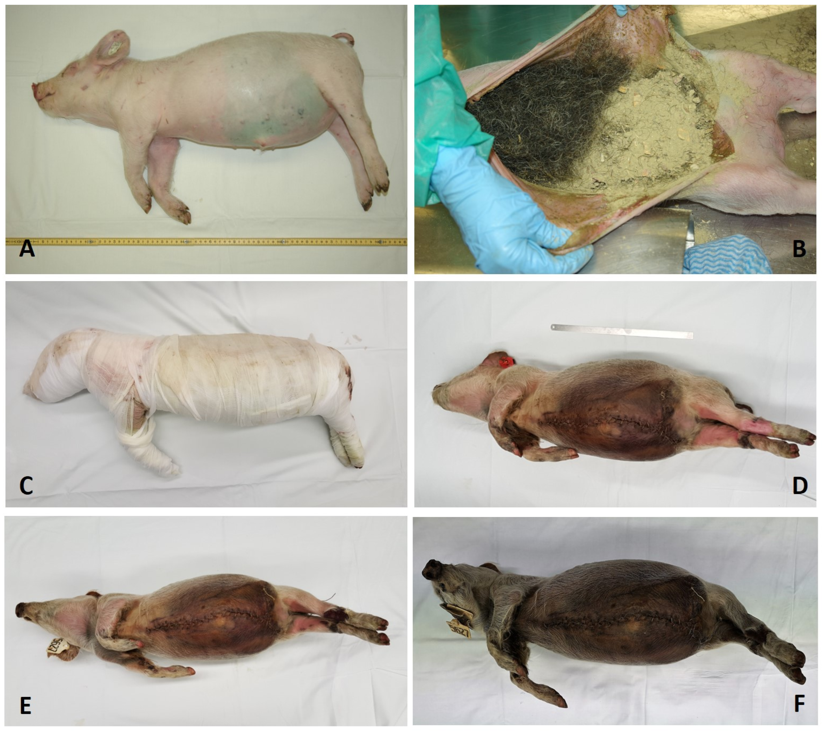
The “surgical part” of the embalming procedure was comparably easy and quick to perform. Accordingly, the opening of the thorax/bowel from the ventral side and the skull from the dorsal side, the removal of major internal organs and the filling of the emptied body cavities were executed without issue, as described by Penicher (see Figure 1B). The empty body cavities had previously carefully been cleaned and all blood residues had been removed to the greatest extent possible. All other manipulations followed Penicher’s instructions (see the Section 2/Supplementary Material S3).
As described in the Section 2, the embalmed corpse had initially been covered with linen bindings—mostly to keep the body in shape and to avoid any loss of embalming substances from the body’s surface or at the sutured sites. This was maintained for 6 weeks. During this period, the linen bindings showed small focal areas of liquid impregnation of brown fluid (Figure 1C), mostly at the rectal orifice, but also on the abdomen, thorax, lower back and skull. This was presumably a mixture of body fluids that had leaked out, together with some embalming fluid residues (the “rub-in emulsion”). The body surface could easily be impressed to a small extent without evidence of any defects. The intensely aromatic (positive) smell of the embalming powders mixed with the slightly sweet odour of tissue decay.
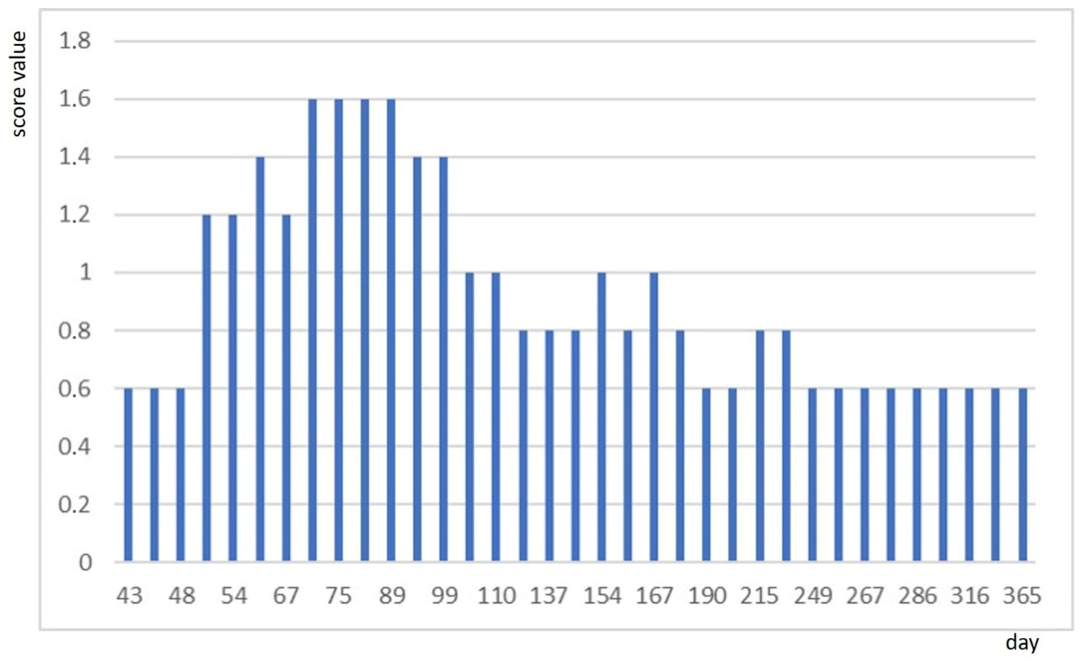
After the removal of the bindings after 6 weeks, we evaluated the status of the body according to the criteria given in the Section 2 (see Table 1). The surface of the body had turned brown over the abdomen (Figure 1D), and there were occasional and very small foci of superficial white fungi on the body surface, such as at both axillae, which remained unchanged over longer periods of time. The standardized evaluation is shown in Figure 2 for the time period between day 43 (following unwrapping) and day 365 (1 year of follow-up). There were no further changes in the observation over the further follow-up period of an additional 6.2 years with a constant value of 0.6 due to the progressive brown coloration of the whole body, which continued until completion (Figure 1E,F). Part of the mentioned small foci of fungi disappeared (although these had not been removed mechanically or “treated” by any chemical substance) while few other foci appeared, though there was no evidence of progression in size nor in tissue dissolution. The very aromatic smell persisted even until the time of autopsy (7.2 years), although the intensity of the smell slowly diminished with time.

3.2. Changes in the Body Weight

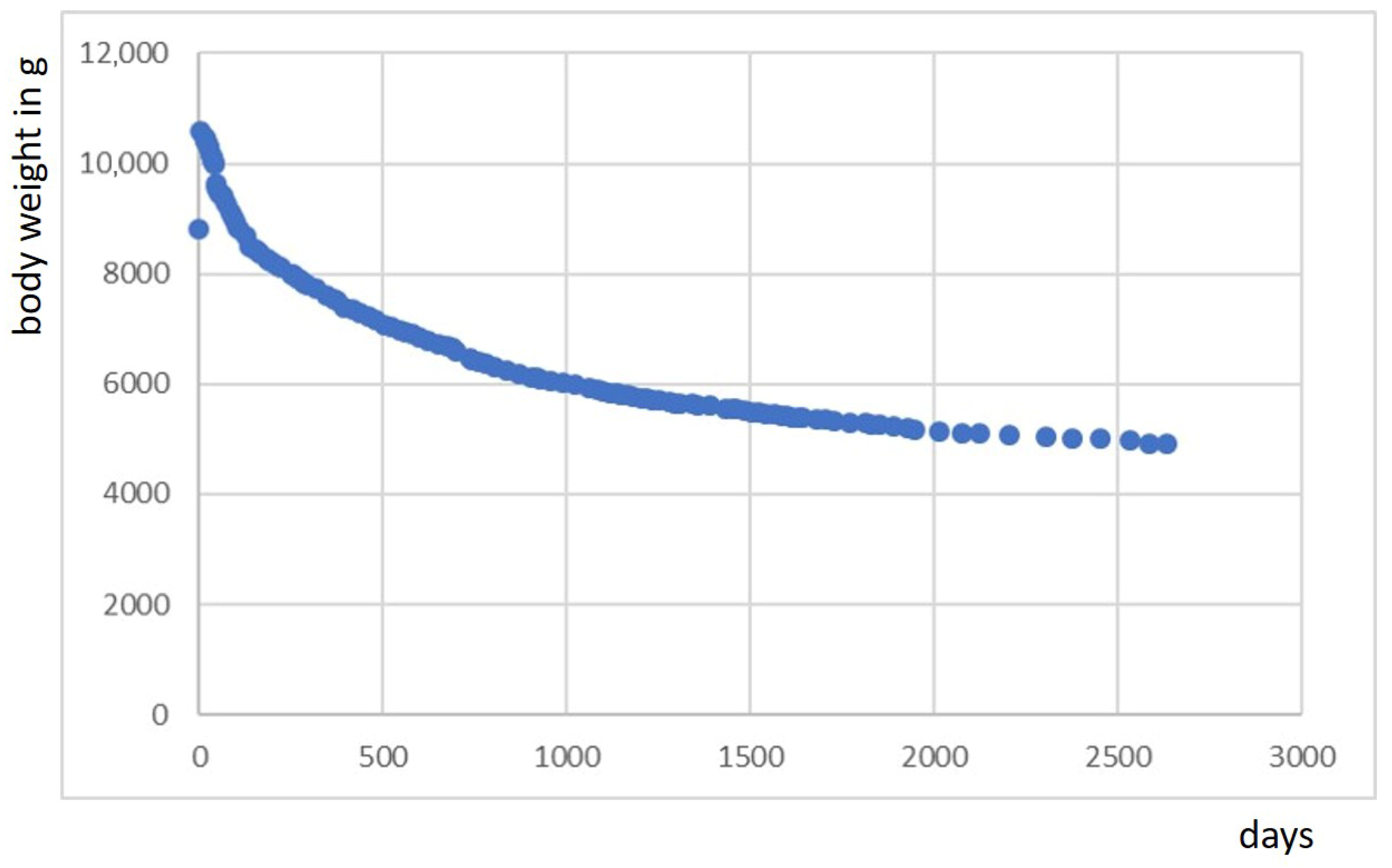
The simplest and most informative parameter for any progress of the embalming was the monitoring of the body weight. Starting from a weight of exactly 8811 g, the application of embalming substances (after the removal of most of the internal organs) resulted in a maximum body weight of 10,604 g after preparation (and removal of the aforementioned internal organs, which together weighed ca. 1000 g). The subsequent course is shown in Figure 3. Following enhanced dehydration in the first year (resulting in a weight loss of approx. 25% in this period), this process slowed down to a rate of body weight loss of another ca. 25% after 5 years of embalming. When we terminated the experiment on day 2634, the body weight was 4927 g (46.4%), of which 2297 g could be attributed to filling material.

3.3. CT Investigations of the Embalmed Body

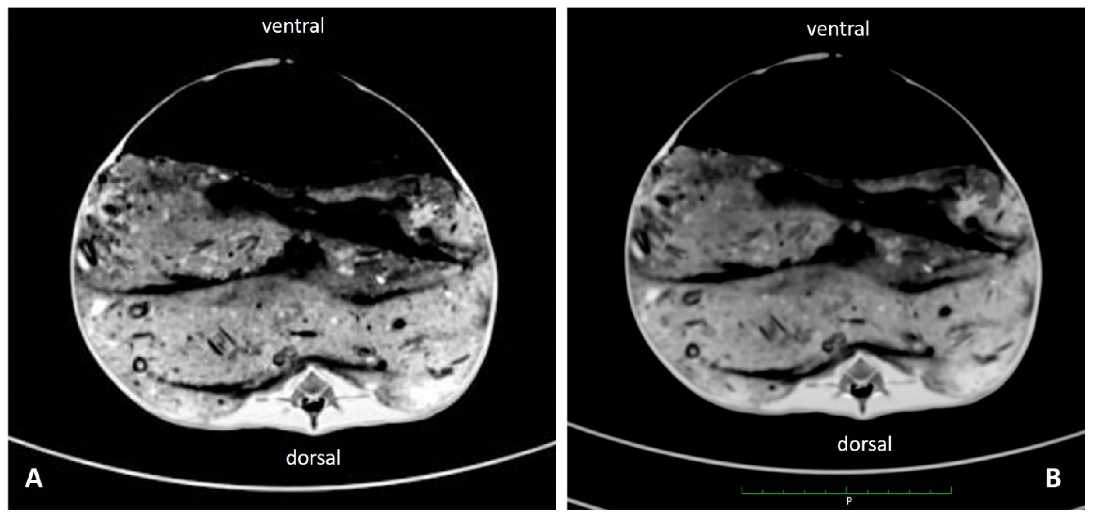
The two CT scans of the complete body showed almost identical features, although there was a time difference of 6 years between the two analyses (Figure 4). The scans showed a regular skeletal system, significantly shrunken soft tissues of all body parts and, in thoracic and abdominal cavities, filling with the embalming material. There was no evidence of any tissue destruction, such as air inclusions or changes in soft tissue density. As early as the first examination, none of the internal organs that had been left inside the corpse (kidneys, internal genitalia) could be identified.

3.4. Histological Analysis

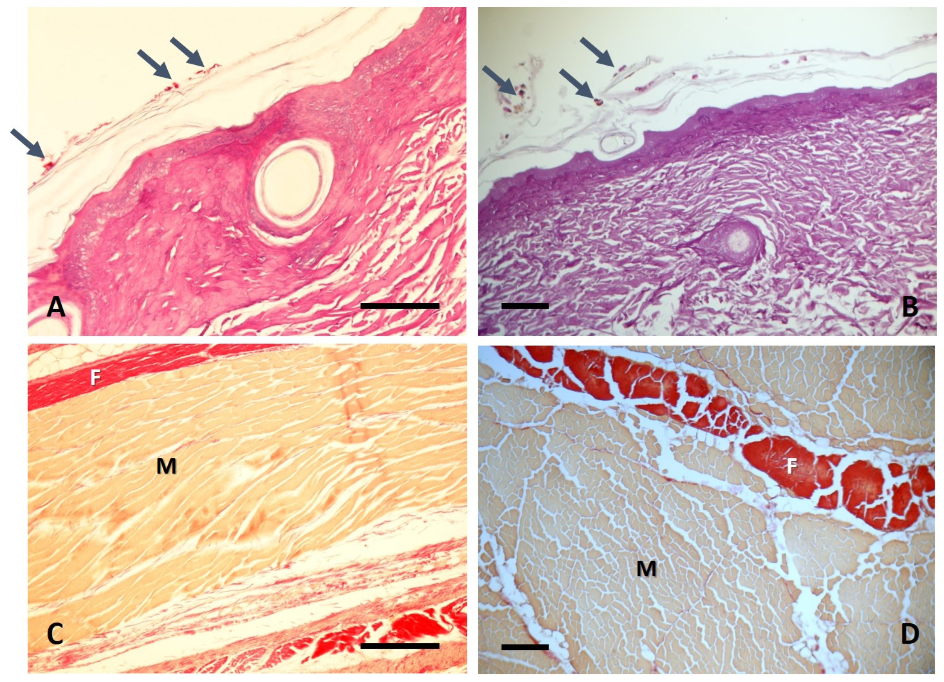
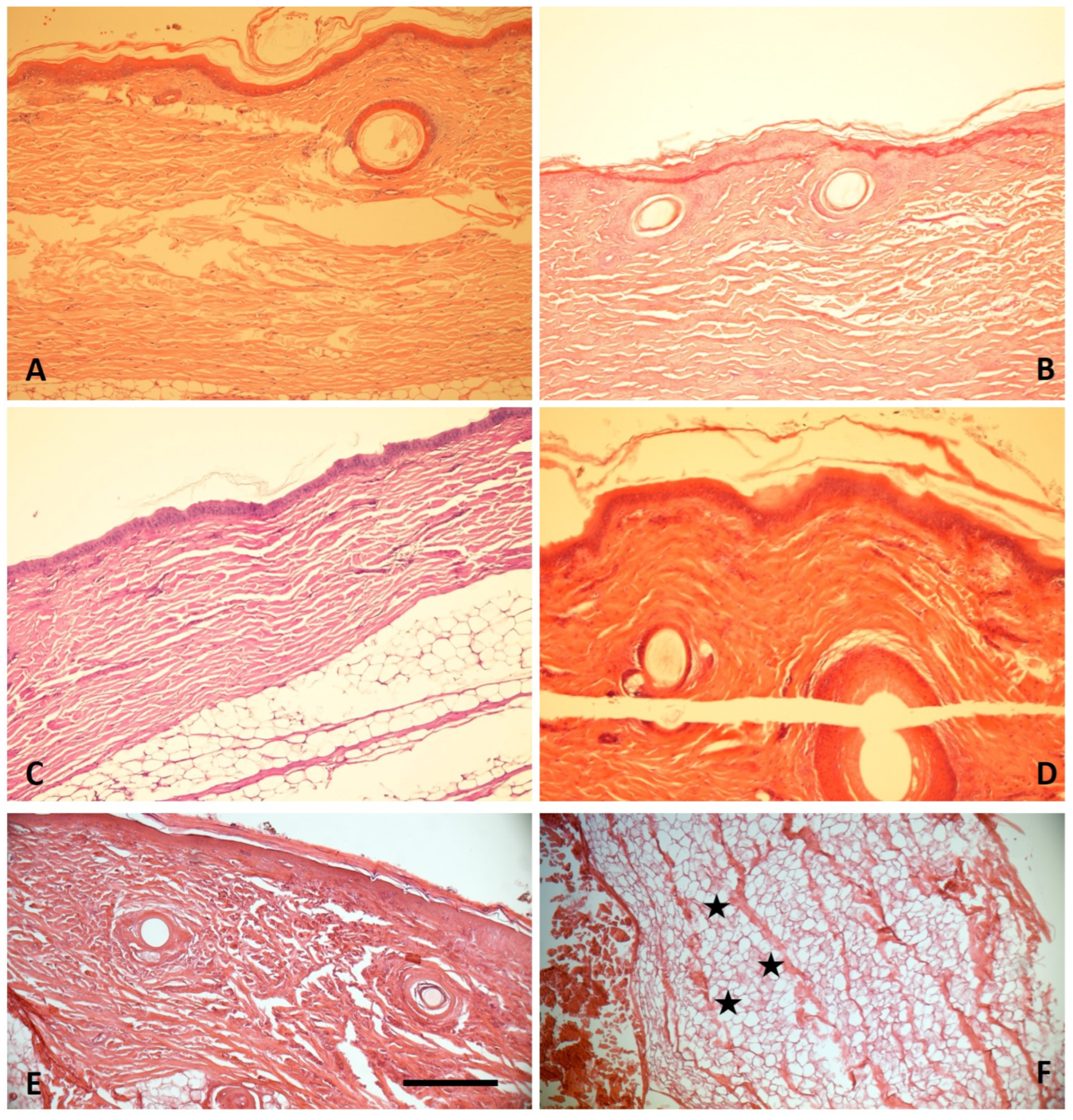
Repeated histological investigations were performed on skin and subcutaneous tissue samples, which occasionally also included skeletal muscle and fascial structures. The samples did not reveal significant differences in the presence of various dermal/subcutaneous tissues (see Figure 5 and Figure 6). In particular, the epidermal layer was intact in all specimens (Figure 5A–E) and even residues of nuclei of the epidermal cells (keratinocytes) were detectable up to day 1772. Only the final sample, taken at autopsy after 2634 days of this experiment, was free of such cell nuclei. The dermal collagen was also excellently preserved in all samples including its birefringence (not shown). There was no evidence for any tissue dissolution, gas bubbles, bacterial or fungal growth within the tissue. The surface of the horn layer of the epidermis contained focal fungal spores (see Figure 6A,B), but there was no evidence of fungal invasion into the tissue (this was especially shown in the Periodic acid–Schiff’s reagent staining (PAS)). Other special stains, such as the Elastica van Gieson staining, confirmed the excellent preservation of the dermal tissue (not shown), but also of fascial structures that envelop a very well preserved skeletal muscle (Figure 6C,D). In the very final samples taken at the autopsy time point, some adipocire transformation of the subcutaneous fat tissue was seen (Figure 5F).
We did not detect any residues of internal organs at autopsy, which was well in line with the CT scans, and even several histological samples from the pelvic cavity and the retroperitoneum did not contain any preserved organ remnants. Similarly, the autopsy did not show residues of the trachea or the esophagus; again, this was confirmed by histological analysis of several samples from presumed regions.

4. Discussion

The efforts to achieve long-term preservation of human bodies were not only prevalent in ancient high cultures, such as in ancient Egypt, but also in various South American populations (e.g., the Chinchorros), Chinese populations, and others, leading to remarkably well-preserved human mummies being created for hundreds and thousands of years [1]. In particular, following the Middle Ages, embalming techniques experienced some revival in Southern and Central Europe when wealthy aristocrats, citizens and merchants started to preserve the bodies of their ancestors and relatives [2]. Several reports provide good evidence that at least some of those embalmed bodies were excellently preserved over considerably long periods of time. Likewise, it has been reported that the French surgeon Ambroise Paré, personal physician of King Louis XIV, stored a very well-prepared body of a young female in his house over several years [23] without evidence of any significant decay. From written evidence, we know that Paré applied a series of substances, especially plant extracts and various herbs, in order to achieve such a perfect result (see Supplementary Material S1). It is also reported that Paré had immersed the body for several weeks in an alcoholic solution [23], although unfortunately the exact compositions of Paré’s fluids and powders (weights and mixing ratios) have not been preserved.
Recent studies, especially of infant mummies, focused on very well-preserved human bodies from a time period between ca. 1780 and 1820 AD, with evidence of dry embalming procedures [9,10,11,24]. The present study was designed in order to find out if typical contemporaneous embalming techniques are successful on a long-term basis of several years. We therefore used a piglet that resembled an infant of ca. 1.5 to 2 years in size and weight. A piglet was chosen because these animals have a similar body surface and a composition of tissues that comes close that of to humans [15,17]. However, there are of course considerable differences between the model and humans, especially with respect to the body anatomy. Our experiment was designed for a period of several years in order to find out the long-term effects of the manipulations. This has never been investigated before.
When selecting an embalming protocol, we came across the method described by Penicher [12] in 1699 AD. This was a method that provided precise weight and volume prescriptions that we could use to replicate the embalming process in an experimental setting [14]. Furthermore, previous reports focused on Penicher’s protocol [7], while other referenced embalming recipes, such as those created by Paré or others, are limited by incomplete information or a lack of precise descriptions of the manipulations and/or the amounts of material applied. Dry embalming was performed in historic times until ca. 1830 AD [23].
As with many historic descriptions, however, no data exist as to the efficiency and long-term outcome of such protocols, so we decided to perform a long-term experiment on an animal model using the method described by Penicher, following his method as closely as possible in order to test this particular protocol as a “proof-of-concept” for the long-term outcome in infant mummies. In this respect, we have, however, to admit that we used—for logistical reasons—only one animal and only substances that are available from modern sources, so our observations are limited to this particular setting. Accordingly, we do not want to speculate on any possible outcome in corpses of different size (such as those from adult individuals) nor those embalmed using any other type of manipulation than we used here. This includes potential variations in the environmental conditions, animal-to-human anatomical differences, and variability in historical ingredient quality.
The most time-consuming and laborious aspect of this dry embalming protocol is the procurement of the embalming substances. Likewise, the preparation of the various ingredients of the two different powders (which fortunately nowadays are mostly available in powdered form) required significant time and financial resources. All non-powdered material had to be ground by hand, mostly with a mortar and pestle. It is understandable that in previous times, procurement of the various materials specified by Penicher was time-consuming and expensive. In historical times, however, a large and comprehensively stocked pharmacy certainly facilitated embalming of the kind described here. In this regard, it is remarkable that Penicher used a very broad spectrum of herbs, leaves, flowers, seeds, fruit and berries—all in dried conditions. The reason might be a combination of efficient and rapid “chemical” stabilization and fixation of the tissue (mostly via dehydrating and antimicrobial effects) along with an excellent aromatic and very positive olfactory effect on the specimen, even in the first time period, which seems essential for the embalming outcome. This “good smell” obviously masked all negative odours that may have occurred in the initial stage of decay and eventual putrid tissue dissolution.
In order to evaluate the stabilizing and preserving effect, we applied a semiquantitative scoring system that took various aspects into account; we established a macroscopic—visual, tactile and olfactory—analysis which should monitor the stability of the animal’s surface, signs for superficial decay or putrefaction and finally the smell. Although we tried to standardize the evaluation of these parameters, we have to admit that the scale implies a certain subjectivity. Using this scheme, we had to record minor evidence of tissue changes especially in the time period between days ca. 60 and 100—but this resolved into a very stable condition thereafter. Accordingly, we did not note any signs of external (nor internal) dissolution of the body—only the progressive brown coloration of the skin surface, along with some shrinkage of the body, had to be noted. In summary, this part of the evaluation indicates that the embalming process had a good outcome over a period of several years. Furthermore, the “good, aromatic smell” persisted over the complete period of observation of more than 7 years; although this positive olfactory effect slightly diminished over this time, the embalmed animal corpse still exuded a very pleasant scent at autopsy.
In addition to the aforementioned subjective observation, we identified various parameters that should be used to record the condition of the embalmed tissue on an objective level. This comprised monitoring of body weight changes over the complete period of observation, two whole-body CT scans and repeated histological tissue analysis.
The easiest parameter to record was the body weight, which was reduced by approx. 25% within ca. one year and another 25% within the subsequent ca. 4 years. This clearly mirrored the dehydration of the body, which seemed to be enhanced in the initial period of embalming, but which proceeded over the whole period of observation. Since the process of desiccation was noticed even after more than 7 years, we may assume that this process had not finished.
The second approach was a whole-body CT scan taken several months after the embalming. This confirmed the layered filling of the body cavities with the embalming material along with well-preserved osseous and soft tissues of the inside of the body. However, the few internal organs that had been left inside the abdominal and pelvic cavities, such as kidneys and internal genitalia, could not be identified—although there was no evidence of tissue destruction by decay, such as cystic transformation or changes in tissue density. Similarly, there were no residues of the trachea visible that had been left inside the upper thorax during evisceration. A repetition of the CT scan 6 years later showed identical structures—again without evidence of putrefaction or major decay.
Finally, we performed repeated histological analyses of skin and adjacent soft tissues via sampling of small tissue pieces, which also showed excellent preservation of the tissue. An important measure of the success of the embalming progress is the presence of an intact epidermal layer over the complete observation period of more than 7 years. Even cellular structures such as the cell nuclei of the epidermis were retained for several years, but then finally disappeared. Skeletal muscle showed cross-striation of myotubes in all samples, which is also a sign of excellent preservation, although the cell nuclei of all stroma cells and of the myotubes had disappeared in the first tissue sample (taken at the 1-year mark). Furthermore, histology showed some fungal infestation which was restricted to the very upper layer of epidermal horn and which did not infiltrate the epidermis nor dermis or subcutaneous tissue.
With respect to the presence of epidermal cell nuclei, even after 1772 days there was only very limited data on this issue. A long-term animal experiment involving an ancient Egyptian-style mummification showed remnants of fragmented epidermal cell nuclei after 7 years of long-term observation [4], which is comparable to our Renaissance-type embalming model. Histological studies on skin samples from ancient Egyptian mummies (which of course have thousands of years of embalming history) did not show cell nuclei even in those rare instances where the epidermal layer was present [25]. In conclusion, the loss of epidermal cell nuclei under embalming conditions seems to be a much slower process than that of organ/soft tissue cell nuclei; this might be due to the preserving substances having direct access to the skin surface—which may also be responsible for another observation of this experiment:
As mentioned before, it was a very surprising finding that the remaining internal organs had disappeared while the skin and subcutaneous tissues, such as fat and muscle, were excellently preserved. The aforementioned “lack” of residual internal organs must have happened during the first few months of embalming. We assume that the preserving effects of the specially treated body surface (with emulsion “C”) was superior to the effect of the body cavity filling (with substance “A” and oakum and horse hair). Therefore, we cannot exclude some that minimal tissue dissolution of the internal organ structures had occurred that may not have been fully prevented by the body cavity fillings.
In summary, we present here the results of the first experimental embalming performed according to a presumably frequently applied embalming protocol from the late 17th century. The outcome was very good even over a period of several years, at least under the controlled conditions of this experiment. Experimental embalming tests have been applied to ancient Egyptian embalming situations [4,26,27,28,29,30,31], which showed considerable differences from the Renaissance period embalming protocols of, e.g., Paré, Penicher and others. Our previous study on experimental ancient Egyptian embalming also used a pig model (of adult size) with an excellent outcome after 13 years [4]. As such, we can confirm that pig models can be used as adequate replacement for experimental investigations of historic embalming techniques from different time periods and cultures. Furthermore, we can state that the Renaissance period embalming protocol described by Penicher may provide stable long-term preservation of human bodies and that reported successful embalming performed using this method is therefore plausible.

Supplementary Materials

The following supporting information can be downloaded at https://www.mdpi.com/article/10.3390/anatomia5020010/s1.

Author Contributions

Conceptualization, A.G.N. and O.K.P.; methodology, A.G.N.; validation, S.P. and O.K.P.; formal analysis, A.G.N. and S.P.; investigation, A.G.N., S.P. and O.K.P.; resources, A.G.N. and O.K.P.; data curation, A.G.N., S.P. and O.K.P.; writing—original draft preparation, A.G.N.; writing—review and editing, A.G.N., S.P. and O.K.P.; visualization, A.G.N. and S.P.; supervision, A.G.N. and O.K.P.; project administration, A.G.N. All authors have read and agreed to the published version of the manuscript.

Funding

This research received no external funding.

Institutional Review Board Statement

The dead animal was provided by the Department of Molecular Animal Breeding and Biotechnology, Faculty of Veterinary Medicine of the University Munich as it required euthanizing because of an eye infection. The department ensured careful handling of the animal. Under EU law, no further regulatory or ethical approval was required for the study when it started in 2017.

Informed Consent Statement

Not applicable.

Data Availability Statement

All data presented in this study are openly available in the manuscript/Supplementary Materials.

Acknowledgments

The authors’ thanks go to the Department of Animal Breeding, Faculty of Veterinary Medicine, Ludwig-Maximilians-University Munich, B. Kessler, for providing us with the dead animal. The technical help of A. Riepertinger and R. Gillich, Institute of Pathology, Academic Clinic Munich-Schwabing, and C. Reischl, Institute of Forensic Medicine, University Munich, is appreciated.

Conflicts of Interest

The authors declare no conflicts of interest.

References

  1. Aufderheide, A.C. The Scientific Study of Mummies; Cambridge University Press: Cambridge, UK; New York, NY, USA, 2003. [Google Scholar]
  2. Brenner, J. Human body preservation—Old and new techniques. J. Anat. 2014, 224, 316–344. [Google Scholar] [CrossRef] [PubMed]
  3. Ikram, S.; Dodson, A. Mummy in Ancient Egypt: Equipping the Dead for Eternity; Thames & Hudchinson: London, UK, 1998. [Google Scholar]
  4. Nerlich, A.G.; Panzer, S.; Fischer, F.; Peschel, O.K. Experimental Ancient Egyptian Human Mummification Tested in a Porcine Model: Excellent Preservation at a 13-Year Follow-Up. Heritage 2025, 8, 194. [Google Scholar] [CrossRef]
  5. Nerlich, A.G.; Bianucci, R. Mummies in Crypts and Catacombs. In The Handbook of Mummy Studies; New Frontiers in Scientific and Cultural Perspectives; Shin, D.H., Bianucci, R., Eds.; Springer Nature: Singapore, 2021; pp. 741–776. [Google Scholar]
  6. Charlier, P. Embalming in France (from Twelfth to Nineteenth Century): Principle and Development of Techniques. In The Handbook of Mummy Studies; New Frontiers in Scientific and Cultural Perspectives; Shin, D.H., Bianucci, R., Eds.; Springer Nature: Singapore, 2021; pp. 908–928. [Google Scholar]
  7. Magnus, J. Das Einbalsamieren der Leichen in Alter und Neuer Zeit; Georg Westermann: Braunschweig, Germany, 1839. [Google Scholar]
  8. Ezugworie, J.; Anibeze, C.; Ozoemena, R. Trends in the development of embalming methods. Internet J. Altern. Med. 2009, 7, 1–6. [Google Scholar] [CrossRef]
  9. Marinozzi, S.; Gazzaniga, V.; Guifra, V.; Fornaciari, G. Historical sources about diseases, death and embalming regarding the family of Jean Antoin Michel Agar, minister of finance of Gioacchino Murat. Vesalius 2011, 17, 45–51. [Google Scholar] [PubMed]
  10. Nerlich, A.G.; Panzer, S.; Röcker, P.; Peschel, O. Making an infant mummy—Analysis and experimental investigations of how to prepare a 19th century infant mummy. Canar. Arqueol. 2021, 22, 437–447. [Google Scholar]
  11. Nerlich, A.G.; Loynes, R.D.; Jardiel Badia, A.; Begerock, A.; Delgado, D.; Gonzalez, M.; Bianucci, R. Intravital skin ulcer in a late 18th century Spanish child mummy: Evidence for septicemic death? Forensic Sci. Med. Pathol. 2025, 21, 813–818. [Google Scholar] [CrossRef] [PubMed]
  12. Penicher, L. Traité des Embaumements Selon les Anciens et les Modernes; B. Girin: Paris, France, 1699. [Google Scholar]
  13. Pettigrew, T.J. The History of Egyptian Mummies; Longman: London, UK, 1834; p. 256. [Google Scholar]
  14. Dufieu, J.F. Dictionnaire Raisonné D’anatomie et de Physiologie; Desaint: Paris, France, 1766; Volume 1, pp. 303–310. [Google Scholar]
  15. Debeer, S.; Le Luduec, J.-B.; Kaiserlian, D.; Laurent, P.; Nicolas, J.-F.; Dubois, B.; Kanitakis, J. Comparative histology and immunohistochemistry of porcine versus human skin. Eur. J. Dermatol. 2013, 23, 456–466. [Google Scholar] [CrossRef] [PubMed]
  16. Sachs, D.H. The pig as a potential xenograft donor. Vet. Immunol. Immunopathol. 1994, 43, 185–191. [Google Scholar] [CrossRef] [PubMed]
  17. Yamamoto, T.; Iwase, H.; King, T.W.; Hara, H.; Cooper, D.K.C. Skin xenotransplantation: Historical review and clinical potential. Burns 2018, 44, 1738–1749. [Google Scholar] [CrossRef] [PubMed]
  18. Xi, J.; Zheng, W.; Chen, M.; Zou, Q.; Tang, C.; Zhou, X. Genetically engineered pigs for xenotransplantation: Hopes and challenges. Front. Cell Dev. Biol. 2023, 10, 1093534. [Google Scholar] [CrossRef] [PubMed]
  19. Nather, A. The Scientific Basis for Transplantation; Advances in Tissue Banking, Volume 5; World Scientific Publishing Co.: Hackensack, NJ, USA; London, UK; Singapore; Kowloon, Hong Kong, 2021. [Google Scholar]
  20. Panzer, S.; Ketterl, S.; Bicker, R.; Schoske, S.; Nerlich, A.G. How to CT scan human mummies: Theoretical considerations and examples of use. Int. J. Paleopathol. 2019, 26, 122–124. [Google Scholar] [CrossRef] [PubMed]
  21. Grove, C.; Peschel, O.; Nerlich, A.G. A Systematic Approach to the Application of Soft Tissue Histopathology in Paleopathology. BioMed Res. Int. 2015, 2015, 631465. [Google Scholar] [CrossRef] [PubMed]
  22. Nerlich, A.G.; Parsche, F.; Wiest, I.; Schramel, P.; Löhrs, U. Extensive pulmonary haemorrhage in an Egyptian mummy. Virchows Arch. 1995, 427, 423–429. [Google Scholar] [CrossRef] [PubMed]
  23. Hawlik-van de Water, M. Der Schöne Tod. Zeremonialstrukturen des Wiener Hofes bei Tod und Begräbnis Zwischen 1640 und 1740; Herder: Freiburg im Breisgau, Germany, 1989; pp. 203–211. [Google Scholar]
  24. Squires, K.; Viner, M.; Hoban, W.; Loynes, R.; Van Schaik, K.; Piombino-Mascali, D. The “angioletti” of Palermo: The health and development of mummified non-adults in late modern Palermo, Sicily (1787–1880 CE). Front. Med. 2024, 11, 1443291. [Google Scholar] [CrossRef] [PubMed]
  25. Nerlich, A.G. Entwickelt und Enträtselt. Die Rolle von Mumien als Bioarchiv am Beispiel der Mumie aus der Orientalischen Sammlung des Museums Kloster Banz. In Human Remains. Ethische Herausforderungen für Ausstellungen; Fackler, G., Klotz, T.M., Menke, S., Eds.; WBG Academic: Darmstadt, Germany, 2022; pp. 323–334. [Google Scholar]
  26. Lucas, A. The use of natron in mummification. J. Egyptol. Archaeol. 1932, 18, 125–140. [Google Scholar] [CrossRef]
  27. Sandison, A.T. The Use of Natron in Mummification in Ancient Egypt. J. Near East. Stud. 1963, 22, 259–267. [Google Scholar] [CrossRef]
  28. Garner, R. Experimental mummification. In The Manchester Mummy Project; David, A.R., Ed.; Manchester University Press: Manchester, UK, 1979; pp. 19–24. [Google Scholar]
  29. Notman, D.N.H.; Aufderheide, A.C. Experimental mummification and computed imaging. In Actas I Congresso Mummy Studies; Museo Arqueologico y Etnografico de Tenerife: Tenerife, Spain, 1984; pp. 821–828. [Google Scholar]
  30. García-Jiménez, L. Application of oils and resins during the process of mummification: Experimental analysis and study problems. In Pharmacy and Medicine in Ancient Egypt; Dinarès Solà, R., Fernàndez Georges, M., Guasch-Jané, R.M., Eds.; Archaeopress: Oxford, UK, 2021; pp. 15–29. [Google Scholar]
  31. Ikram, S. (Ed.) Manufacturing Divinity: The Technology of Mummification. In Divine Creatures: Animal Mummies in Ancient Egypt; The American University in Cairo Press: Cairo, NY, USA, 2015; pp. 17–44. [Google Scholar]
Figure 1. Macroscopic aspects of the embalmed animal. (A) The body on the first day of the embalming procedure. (B) Opening of the bowel and filling of the thorax and abdominal cavity with the filling material (day 0). (C) The wrapped body on day 27 with some fluid impregnation of the bindings at the rectal orifice and slightly coloured bindings over the abdomen and back. (D) The animal after unwrapping (day 43) with brown coloration of the bowel. (E) The body after 1 year (day 365) and (F) the animal after 3 years (day 1011). The body is now completely brown coloured, but otherwise well preserved.
Anatomia 05 00010 g001
Figure 2. Evaluation of the progress of embalming according to a semiquantitative scoring system. The evaluation started after the unwrapping (day 43) and is shown until the end of the first year (day 365). All further evaluations remained at this level until day 2634. The Y-axis shows the mean score value as obtained by the addition of the five criteria of the evaluation (divided by 5; see the Section 2) and the X-axis refers to the days of evaluation starting with day 43 (the first day of unwrapping) (every second day of evaluation is indicated).
Anatomia 05 00010 g002
Figure 3. Weight curve of the embalmed body over the complete evaluation period of the experiment.
Anatomia 05 00010 g003
Figure 4. Axial CT sections of the abdomen of the experimental mummy. (A) Examination on day 295 and (B) on day 2568. The body cavities are filled with the embalming material in various layers, with some air inclusions. The bones and soft tissue residues are seen in a regular position; there is no evidence of remaining structures of internal organs.
Anatomia 05 00010 g004
Figure 5. Histological investigations of skin samples taken during the study period. (A) Samples take on day 365, (B) on day 1025, (C) on day 1358, (D) on day 1772 and (E) during the final autopsy on day 2634 all show an excellently preserved skin tissue with an intact epidermal layer that contains, until at least day 1772, nuclear residues of the epidermis, as well as very well preserved collagenous dermis and fat tissue of the subcutis. (F) Only at the final time point (day 2634) was some adipocire (asterisks) identified in the subcutaneous fat. All stainings: haematoxylin and eosin (HE); the magnification bar in (E) is valid for all images, comprising 100 µm size.
Anatomia 05 00010 g005
Figure 6. Histological aspects of special issues: On the surface of the epidermal horn layer, occasional fungal spores (arrows) are seen on day 1025 (A), as well as on day 1772 (B). Below the subcutis, intact and very well-preserved fascia (“F”) envelop excellently preserved skeletal muscle (“M”), on days 1358 (C) and 2634 (D). Staining: (A,B): PAS staining; (C,D): Elastica van Gieson’s connective tissue stain. Magnification bars: 25 µm each.
Anatomia 05 00010 g006
Table 1. Macroscopic evaluation scheme of the embalmed body.
Parameter/Points01234
Surface integrityIntactSmall defectsFocal defectsLarger defectsDissolved
Surface colourNormalFocal brownMostly brownComplete brownDark/black
Evidence for fungiNoSmall fociLarger fociExtensive—less than half of body coveredExtensive—more than half of body covered
Firmness of skin/soft tissueFirmFirm with small impressionsModerate impression possibleSoft—large impressions possibleDissolved or brittle
SmellAromaticA little aromaticSlightly sweet—putridModerately sweet—putridStrongly sweet—putrid
Disclaimer/Publisher’s Note: The statements, opinions and data contained in all publications are solely those of the individual author(s) and contributor(s) and not of MDPI and/or the editor(s). MDPI and/or the editor(s) disclaim responsibility for any injury to people or property resulting from any ideas, methods, instructions or products referred to in the content.