1. Introduction
Neurosurgical care in Sub-Saharan Africa and other developing regions faces critical challenges, including limited access to diagnostic imaging technologies, shortages of trained clinicians, and a lack of essential resources [
1,
2,
3]. These constraints impair the ability to accurately identify critical intracranial structures, increasing the risk of iatrogenic injury during surgical procedures. The transverse dural venous sinuses (TS), in particular, are vulnerable to injury during suboccipital approaches due to their variable anatomical course and proximity to commonly used burr hole sites [
4]. In settings where Magnetic Resonance Venography or Computed Tomography are unavailable, alternative anatomical methods to predict TS location and morphology are vital for safe surgical intervention [
5].
The TS are paired venous structures located at the attachment of the tentorium cerebelli, typically asymmetrical, with the right TS often wider and considered dominant [
6,
7]. Anatomical studies have reported considerable interindividual variability, including septations, hypoplasia, aplasia, and fenestration [
8,
9,
10,
11]. Prior investigations have employed angiographic or imaging-based techniques, with only a few incorporating cadaveric methods to directly examine the TS. However, most such studies have focused on multiple sinuses rather than isolating the TS, and the use of external cranial landmarks as surgical guides remains controversial due to inconsistent spatial correlations [
3,
7,
12,
13].
This study aims to provide a detailed anatomical description of the TS in adult human cadavers (of South Africa) by quantitatively assessing its length and width at key points along its course. The dural measurements were compared to the corresponding transverse sulcal impressions to evaluate the accuracy of bony landmarks in predicting venous sinus morphology. To our knowledge, no cadaveric studies to date have quantitatively compared TS width and shape using both meningeal and osseous landmarks in Sub-Saharan populations. By addressing this gap, our findings may improve neurosurgical planning and training, particularly in low-resource settings where reliance on anatomical reference points remains essential.
2. Materials and Methods
This study employed a descriptive, cross-sectional design to investigate the morphology of the TS in human adult cadavers (whole body donors). All dissections were conducted at the Department of Anatomy, University of Pretoria, in South Africa, under ethical approval from the Faculty of Health Science Research Ethics Committee (Reference number: 233/2021). This clearance pertains to the conduct of a cadaveric study and the use of data collected from brain specimens, which do not include any animals or living patients/volunteers. The study adhered to the ethical standards of the 1964 Declaration of Helsinki and its subsequent amendments, as well as South Africa’s National Health Act, 61 of 2003.
The sample consisted of 32 formalin-fixed human adult cadavers (20 male, 12 female), with an age range of 27 to 95 years (
Table S1). Ancestry, weight, and height were not used as exclusion criteria. Cadavers were excluded if the cranial cavity was poorly embalmed, damaged, previously subjected to surgery, or exhibited pathological changes in the regions of interest.
2.1. Procedure
A horizontal craniotomy was performed to access the cranial cavity while preserving the dura mater. The brain was carefully removed, and a single linear incision was made in the dural meningeal layer, along the course of the TS and sigmoid sinus, bilaterally. This exposed the TS for direct measurement. The internal anatomy of these venous sinuses was examined for the presence of duplication and additional communications with other venous structures. Following soft tissue measurements, the dura mater was removed to expose the underlying transverse sulcus for comparative bony analysis.
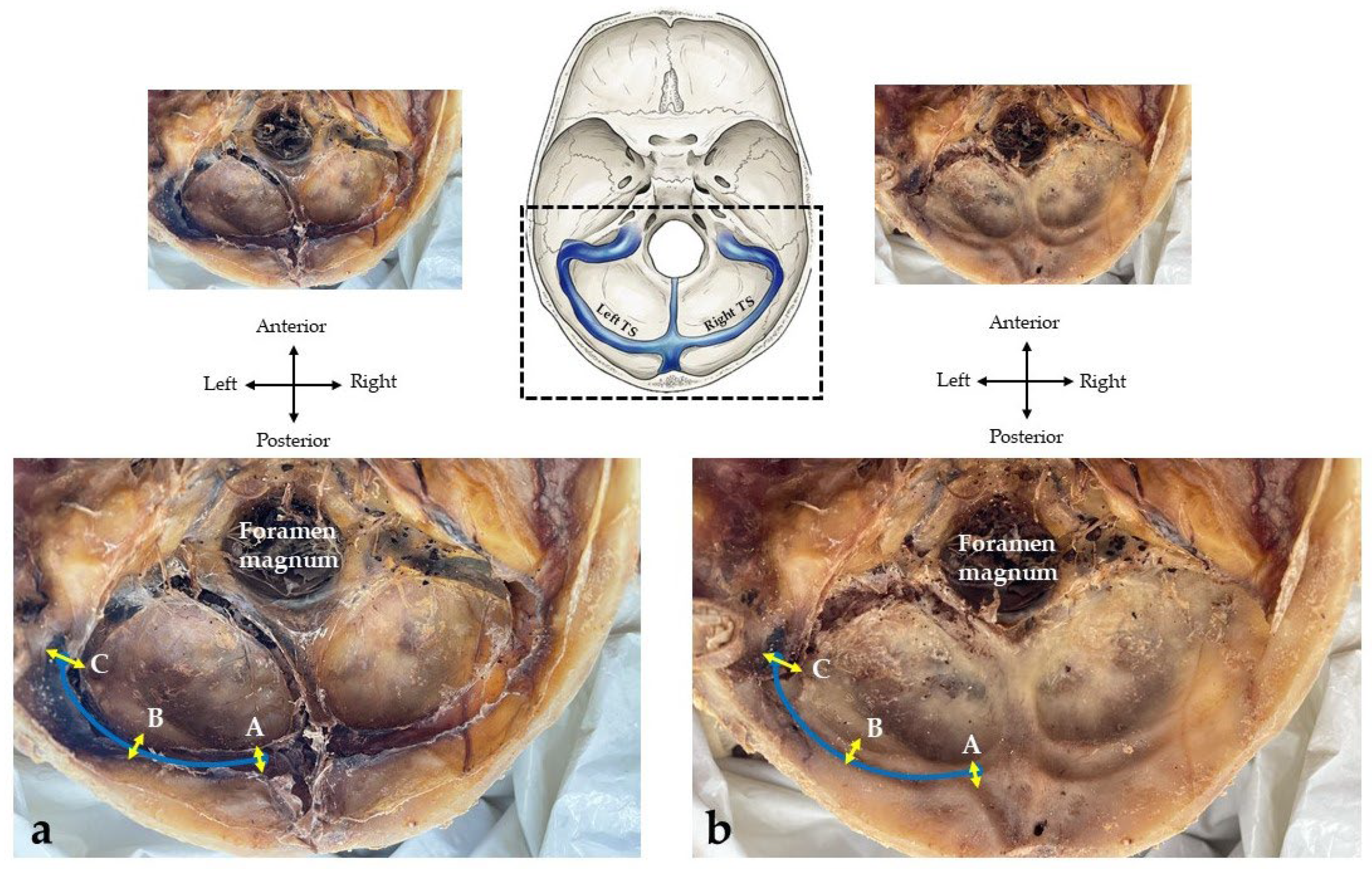
Measurements were performed bilaterally. The length of each TS was recorded by placing a flexible string along its curved course and measuring it with a calibrated ruler in millimeters. The width of the TS was measured at three defined anatomical points:
Origin: the junction with the superior sagittal sinus (right) or straight sinus (left).
Midpoint: the central region of the TS, with equal distances between the origin and termination.
Termination: the point where the TS transitions into the sigmoid sinus at the petrous part of the temporal bone.
The widths were measured in millimeters using a calibrated digital sliding caliper. Color-coded pins were used to mark origin, midpoint, and termination during the data collection. Following the removal of the dura mater, corresponding bony impressions in the transverse sulcus were identified, and similar measurements were repeated for comparative analysis (
Figure 1).
2.2. Statistical Analysis
Descriptive statistics included the mean, standard deviation, minimum, and maximum. The Shapiro–Wilk test was selected to assess the normality of the sample data.
Paired sample t-tests were used to compare measurements between the left and right sides, between the dural and osseous measurements within the same individuals, as well as between males and females. Intra-class correlation tests (ICC) were used to assess the reliability and repeatability of measurements. Statistical significance was set at p ≤ 0.05. All analyses were conducted using IBM SPSS Statistics for Windows, Version 22.0 (IBM Corp., Armonk, NY, USA).
3. Results
A Shapiro–Wilk test for normality was performed, and the majority of the data were normally distributed, as shown in
Table 1 and
Table 2. However, some data were not normally distributed, including the width of the right termination point along the transverse sulcus (
p = 0.000) and the average width at the midpoint of the transverse sulcus (
p = 0.012). Although these two measurements were significant, a Z-score analysis revealed no extreme outliers, and all values fell within an acceptable range. Intra-class correlation coefficient (ICC) analysis was performed to assess the reliability and repeatability of the measurements. The results demonstrated excellent agreement, with ICC values of 0.90 for both intra-observer and inter-observer comparisons. These findings confirm a high level of consistency and reliability in the measurement methodology and resulting dataset.
3.1. Length of the Transverse Sinuses
The mean length of the TS was 72.54 mm (±7.34) on the left and 70.23 mm (±7.59) on the right, as measured within the dura mater. No statistically significant differences were observed between the left and right TS within the dura mater (p = 0.292) or in the transverse sulcus (p = 0.082) within the same individual. Given the overlapping standard deviations and modest mean difference (~2 mm), these variations are not statistically significant and are likely attributed to natural anatomical asymmetry.
No significant differences were found between the dura mater and transverse sulcus measurements on either side (
p = 0.113), suggesting that bony grooves offer a relatively reliable approximation of the TS length. The details of the TS length measurements are summarized in
Table 1.
3.2. Width of the Transverse Sinuses
The width of the TS was measured at the origin, midpoint, and termination on both the left and right sides of the posterior cranial fossa. In 71.9% of cadavers, the right TS was wider in the dura mater, indicating a right-sided venous dominance. In contrast, right dominance in the transverse sulcus was observed in only 56.3% of cases, indicating partial correspondence between dural and osseous widths.
Statistically significant differences were found in the width of the midpoint (
p = 0.048) and termination (
p = 0.000) between left and right TS within the dura mater. These findings suggest a consistent asymmetry favoring the right side. The width of the transverse sulcus, however, showed no significant differences between sides at any of the three locations (
p > 0.05), calling into question the sulcus’s reliability in using the TS width in surgical planning. The details of these width measurements are summarized in
Table 2 and
Table 3.
To gain a more thorough understanding of the shape of the TS, the origin-width was compared with the midpoint-width and the termination-point width within the same cadaver using a paired-samples
t-test. The findings summarized in
Table 4 indicate an abrupt narrowing from the origin to the midpoint, followed by a marked widening towards the termination. This pattern was particularly evident in males, where all comparisons were significant (
p ≤ 0.05). In females, a more gradual narrowing was observed between the origin and midpoint (
p = 0.229), but the widening between the midpoint and termination remained significant (
p = 0.017).
t-tests concluded that neither the width nor the length of the TS could be linked to the sex of the cadaver.
4. Discussion
An accurate understanding of the anatomy and morphometry of the TS is critical for neurosurgical safety and efficacy. The TS serves as a major venous drainage route for the brain, and its variable morphology, dominance patterns, and anatomical relationships make it particularly vulnerable to iatrogenic injury during surgical interventions affecting the posterior cranial fossa [
14]. Knowledge of TS dimensions (including length, width, and trajectory) is especially crucial when advanced imaging modalities are unavailable or unreliable, as is often the case in low-resource settings [
3,
6,
7]. Precise morphometric data not only inform safer burr hole placement and craniotomy boundaries, but also enhance the interpretation of neuroimaging findings and improve the management of conditions such as venous thrombosis and idiopathic intracranial hypertension. The present study aimed to address gaps in TS morphometry by providing detailed cadaveric measurements and evaluating patterns of asymmetry and shape change, with implications for both clinical practice and anatomical education.
4.1. Length of the Transverse Sinuses
This study investigated the morphological characteristics of the transverse dural venous sinuses (TS) using direct cadaveric measurements. Despite the pooled mean length of the right TS being slightly shorter than the left, this difference was not statistically significant. These findings corroborate previous observations of right TS shortening, which have been attributed to a deviation of the superior sagittal sinus towards the right, a phenomenon observed in approximately 34% of cases [
15]. Such deviations can result in a more direct and thus shorter anatomical pathway into the right TS [
16].
Interestingly, the mean TS length observed in this sub-Saharan cadaveric sample (71 mm) is substantially greater than that reported in an Indian population (48 mm) [
17]. This disparity likely reflects differences in demographic anatomy or dissection methodology. Nonetheless, the consistency between dural and sulcal length measurements observed here supports the use of external bony landmarks in estimating the TS course and length.
4.2. Width of the Transverse Sinuses
The TS width showed significant variation along its course, with a narrowing at the midpoint and widening toward the termination. These findings were statistically significant and suggest that the TS does not maintain a uniform diameter but rather undergoes dynamic changes, both anatomical and potentially functionally significant. It further emphasized that the measurements obtained from the transverse sulcus may not be a true reflection of the actual width of the transverse sinuses.
Right-sided dominance in TS width was observed in 71.9% of specimens, consistent with previous findings [
10,
18,
19]. This dominance pattern has been confirmed in radiologic and cadaveric studies, with at least 11 cited investigations reporting that right TS dominance is more frequent than left dominance or symmetry [
15]. This has critical clinical implications, as the dominant sinus must not be occluded or injured during neurosurgical procedures, as this may lead to venous infarction or inadequate collateral flow [
4,
6].
The measured widths in this South African study were also notably larger than those reported using magnetic resonance venography [
20]. The discrepancy may arise from different measurement techniques and definitions of the “origin” point. The data from this study provide direct physical measurements, potentially offering a more accurate representation of anatomical width for surgical planning.
The TS demonstrated a consistent morphological pattern: narrowing from the origin to the midpoint, followed by widening toward the termination. This shape was more pronounced in males, and all comparisons were statistically significant. While narrowing of the middle part of the TS has often been associated with idiopathic intracranial hypertension, this data suggests that such narrowing may also represent a normal anatomical variant, particularly in males [
8,
20,
21].
Differentiating pathological stenosis from non-pathological narrowing is essential for avoiding misdiagnosis. In idiopathic intracranial hypertension, TS stenosis is typically bilateral and occurs in the middle part of the sinus [
21]. Consistent with this, this cadaveric sample revealed similar narrowing patterns at the midpoint of the TS in the absence of a pathological context, underscoring the importance of anatomical benchmarks when interpreting MRV or angiographic data.
4.3. Implications for Neurosurgical Planning in Resource-Limited Settings
In environments where imaging resources are scarce, as is often the case in Sub-Saharan Africa, a thorough anatomical understanding of the TS becomes indispensable. Our findings suggest that the transverse sulcus may not consistently reflect the internal width of the TS, particularly in the presence of sinus dominance or abrupt diameter changes. Consequently, training programs in such settings should emphasize direct anatomical correlations and variability in venous morphology. Enhanced neurosurgical training, grounded in anatomical and morphometric studies, can reduce iatrogenic injury during posterior cranial fossa procedures. Knowledge of TS dominance, as well as its abrupt narrowing and widening patterns, should inform burr hole placement and craniotomy planning, especially when imaging or neuronavigation is unavailable [
3,
7].
4.4. Limitations and Future Research
The present study has several limitations. The sample size was constrained by practical and logistical factors, as data collection occurred during the COVID-19 pandemic when access to anatomical laboratories and cadaveric material was restricted. These disruptions reduced the number of suitable specimens available for analysis and led to the exclusion of several cadavers due to tissue degradation during extended laboratory closures. In addition, cadaveric anatomical research is inherently dependent on the availability of donated bodies within institutional body donation programs, which may limit the achievable sample size.
Despite these constraints, the number of specimens analyzed is comparable to that used in many descriptive cadaveric anatomical investigations. Importantly, the primary objective of this study was not to establish population-level prevalence estimates but to examine morphological characteristics of the transverse sinuses and to compare measurements obtained from the dura mater with those derived from the corresponding osseous transverse sulcus within the same individuals. This comparative approach highlights potential limitations in relying solely on dried skull specimens to estimate venous sinus morphology.
Future studies should aim to include larger, more demographically diverse cadaveric samples and to integrate radiological imaging techniques to further correlate external morphometry with internal venous sinus architecture.
5. Conclusions
This study provides foundational morphometric data on the transverse dural venous sinuses, highlighting consistent patterns of asymmetry, dominance, and dimensional variation that have direct implications for neurosurgical safety and training. These findings are particularly relevant in Sub-Saharan and other low-resource contexts where access to advanced imaging is limited and anatomical knowledge remains the primary tool for surgical navigation. Future studies should build on this work by integrating cadaveric morphometry with magnetic resonance imaging and computed tomography data. Further investigation of the normal range and clinical relevance of TS narrowing, particularly at the midpoint, is needed to distinguish anatomical variants from pathological stenosis. Such research will contribute to the development of evidence-based guidelines for safer posterior fossa surgical approaches, ultimately improving patient outcomes.
Author Contributions
Conceptualization, G.V. and L.P.; Methodology, G.V., L.P. and J.J.G.; Formal Analysis, J.J.G.; Investigation, J.J.G.; Resources, J.J.G. and K.S.R.; Data Curation, J.J.G. and G.V.; Writing—Original Draft Preparation, All authors.; Writing—Review and Editing, All authors; Supervision, G.V. and L.P.; Project Administration, G.V. All authors have read and agreed to the published version of the manuscript.
Funding
This study received no funding.
Institutional Review Board Statement
The study was conducted in accordance with the Declaration of Helsinki, and approved by the Faculty of Health Science Research Ethics Committee of the University of Pretoria (protocol code: 233/2021, approved on 13 May 2021) for studies involving human cadavers.
Informed Consent Statement
As per the South African National Health Act, 61 of 2003, and the University of Pretoria’s body donation program, the consent statement of the body donor was taken prior to their demise.
Data Availability Statement
The original contributions presented in this study are included in the article and
Supplementary Materials. Further inquiries can be directed to the corresponding author.
Acknowledgments
The authors sincerely thank the individuals who donated their bodies to science, thereby making this research possible. Body donation programs constitute an essential and ethical foundation for anatomical education and research, advancing medical knowledge and clinical practice [
22]. Beyond enabling technical investigation, engagement with donated human material has also been shown to foster professional development, ethical awareness, and respect among health science trainees [
23]. The use of donated human bodies carries an ethical responsibility to ensure that all human remains are treated with dignity and respect throughout research and teaching activities [
24]. The authors acknowledge this contribution with deep gratitude. Disclaimer: In finalizing this manuscript, the authors used an AI-based language model (Grammarly v1.2.235) to support academic writing tasks, including clarity enhancement, syntax refinement, and organization. The AI tool was used solely as a writing and critical reading assistant; it did not contribute to conceptual development, data interpretation, or the drafting of original scholarly content. The authors take full responsibility for the content, accuracy, and integrity of the final manuscript.
Conflicts of Interest
The authors declare that no financial or personal relationship(s) have inappropriately influenced their writing of this article. The authors declare that there is no conflict of interest that is relevant to the content of this article.
References
- El-Fiki, M. African neurosurgery, the 21st-century challenge. World Neurosurg. 2010, 73, 254–258. [Google Scholar] [CrossRef] [PubMed]
- Punchak, M.; Mukhopadhyay, S.; Sachdev, S.; Hung, Y.C.; Peeters, S.; Rattani, A.; Dewan, M.; Johnson, W.D.; Park, K.B. Neurosurgical care: Availability and access in low-income and middle-income countries. World Neurosurg. 2018, 112, e240–e254. [Google Scholar] [CrossRef] [PubMed]
- Venter, G.; Prigge, L.; Viljoen, B.L.; De Beer, F.C.; Bam, L.C. Evaluation of the inion and asterion as neurosurgical landmarks for dural venous sinuses: Osteological study on a sample of South African skull specimens. J. Neurosurg. Sci. 2021, 65, 200–206. [Google Scholar] [CrossRef] [PubMed]
- Kim, Y.S.; Jung, S.H.; Lim, D.H.; Kim, T.S.; Kim, J.H.; Lee, J.K. Traumatic Dural Venous Sinus Injury. Korean J. Neurotrauma 2015, 11, 118–123. [Google Scholar] [CrossRef]
- Gökçe, E.; Acu, B.E.; Beyhan, M.; Celikyay, F.; Celikyay, R. Magnetic resonance imaging findings of developmental venous anomalies. Clin. Neuroradiol. 2014, 24, 135–143. [Google Scholar] [CrossRef]
- Bozbuga, M.; Boran, B.O.; Sahinoglu, K. Surface anatomy of the posterolateral cranium regarding the localization of the initial burr–hole for a retrosigmoid approach. Neurosurg. Rev. 2006, 29, 61–63. [Google Scholar] [CrossRef]
- Prigge, L.; Hoffman, J.W.; Bam, L.C.; Venter, G. The semispinalis capitis muscle skull attachment as a superficial external landmark for the location of the transverse dural venous sinuses. Transl. Res. Anat. 2024, 37, e100334. [Google Scholar] [CrossRef]
- Alper, F.; Kantarci, M.; Dane, S.; Gumustekin, K.; Onbas, O.; Durur, I. Importance of anatomical asymmetries of transverse sinuses: An MR venographic study. Cerebrovasc. Dis. 2004, 18, 236–239. [Google Scholar] [CrossRef]
- Strydom, M.A.; Briers, N.; Bosman, M.C.; Steyn, S. The anatomical basis of venographic filling defects of the transverse sinus. Clin. Anat. 2010, 23, 153–159. [Google Scholar] [CrossRef]
- Massrey, C.; Altafulla, J.J.; Iwanaga, J.; Litvack, Z.; Ishak, B.; Oskouian, R.J.; Loukas, M.; Tubbs, R.S. Variations of the transverse sinus: Review with an unusual case report. Cureus 2018, 10, e3248. [Google Scholar] [CrossRef]
- Iwanaga, J.; Courville, E.; Anand, M.K.; Khan, P.A.; Goren, O.; Lammle, M.; Bui, C.J.; Dumont, A.S.; Tubbs, R.S. Chordae Willisii within the transverse sinus: Morphologic study. World Neurosurg. 2020, 139, e38–e44. [Google Scholar] [CrossRef] [PubMed]
- Tubbs, R.S.; Salter, G.; Oakes, W.J. Superficial surgical landmarks for the transverse sinus and torcular herophili. J. Neurosurg. 2000, 93, 279–281. [Google Scholar] [CrossRef] [PubMed]
- Avci, E.; Kocaogullar, Y.; Fossett, D.; Caputy, A. Lateral posterior fossa venous sinus relationships to surface landmarks. Surg. Neurol. 2003, 59, 392–397. [Google Scholar] [CrossRef] [PubMed]
- Yu, Z.; Sun, J.; Li, S.; Bai, P.; Cheng, K.; Song, J.; Luo, Q.; Li, Y. Morphologic Study of positional relationship between transverse-sigmoid sinus and extracranial bony landmarks with reconstructed computed tomographic image. J. Craniofacial Surg. 2016, 27, 1849–1853. [Google Scholar] [CrossRef]
- Kitamura, M.A.; Costa, L.F.; de Araújo Silva, D.O.; Batista, L.L.; de Almeida Holanda, M.M.; Valença, M.M. Cranial venous sinus dominance: What to expect? Analysis of 100 cerebral angiographies. Arq. Neuro-Psiquiatr. 2017, 75, 295–300. [Google Scholar] [CrossRef]
- Curé, J.K.; Van Tassel, P.; Smith, M.T. Normal and variant anatomy of the dural venous sinuses. Semin. Ultrasound CT MRI 1994, 15, 499–519. [Google Scholar] [CrossRef]
- Thamke, S.; Kalra, S.; Khandelwal, A. Morphometric evaluation of dural venous sinuses: Anatomical study with its implications in surgical interventions. J. Morphol. Sci. 2016, 33, 083–089. [Google Scholar] [CrossRef]
- Durgun, B.; Ilgit, E.T.; Cizmeli, M.O.; Atasever, A. Evaluation by angiography of the lateral dominance of the drainage of the dural venous sinuses. Surg. Radiol. Anat. 1993, 15, 125–130. [Google Scholar] [CrossRef]
- Gökçe, E.; Pınarbaşılı, T.; Acu, B.; Fırat, M.M.; Erkorkmaz, Ü. Torcular Herophili classification and evaluation of dural venous sinus variations using digital subtraction angiography and magnetic resonance venographies. Surg. Radiol. Anat. 2014, 36, 527–536. [Google Scholar] [CrossRef]
- Larson, A.; Rinaldo, L.; Chen, J.J.; Cutsforth-Gregory, J.; Theiler, A.R.; Brinjikji, W. Reductions in bilateral transverse sinus pressure gradients with unilateral transverse venous sinus stenting for idiopathic intracranial hypertension. J. Neurointerv. Surg. 2021, 13, 187–190. [Google Scholar] [CrossRef]
- Connor, S.E.; Siddiqui, M.A.; Stewart, V.R.; O’Flynn, E.A. The relationship of transverse sinus stenosis to bony groove dimensions provides an insight into the aetiology of idiopathic intracranial hypertension. Neuroradiology 2008, 50, 999–1004. [Google Scholar] [CrossRef]
- Ghosh, S.K.; Walocha, J.A. Responsible research in the practice of anatomy: Attributes relevant to body donors and human tissues sourced from them. Ann. Anat.-Anat. Anz. 2024, 252, 152184. [Google Scholar] [CrossRef]
- Leeper, B.J.; Grachan, J.J.; Robinson, R.; Doll, J.; Stevens, K. Honoring human body donors: Five core themes to consider regarding ethical treatment and memorialization. Anat. Sci. Educ. 2024, 17, 483–498. [Google Scholar] [CrossRef]
- Choi, W.J. Teaching respect for body donors: A US medical student perspective. Anat. Sci. Educ. 2024, 17, 687–692. [Google Scholar] [CrossRef]
| Disclaimer/Publisher’s Note: The statements, opinions and data contained in all publications are solely those of the individual author(s) and contributor(s) and not of MDPI and/or the editor(s). MDPI and/or the editor(s) disclaim responsibility for any injury to people or property resulting from any ideas, methods, instructions or products referred to in the content. |