


Abstract
Introduction: Understanding superficial epigastric vessel anatomy is crucial for abdominal surgeries like laparoscopy, especially in neonates, to prevent injury. While standard courses are described, variations occur. This case report highlights a unique anatomical variation in the superficial epigastric artery found during the dissection of a stillborn neonatal cadaver. Case Report: In contrast to the usual origin from the femoral artery, this variation features the inferior epigastric artery penetrating the anterior abdominal wall near the umbilicus and branching superiorly to supply the superficial abdominal wall. Conclusions: This distinctive vascular configuration, which to the best of our knowledge has not been previously described in neonatal anatomical literature, diverges from the typical symmetrical arrangement and previously reported variations. The study stresses the clinical importance of this finding, especially for laparoscopic procedures in neonates. During trocar placement, surgeons should be cognizant of such variations to reduce the risk of iatrogenic injuries, including rectus sheath hematoma. The report highlights the need for further investigation to establish the prevalence of this variation and its potential effects on surgical safety and outcomes in a broader neonatal population, which may also reflect the dynamic vascular remodeling that occurs during early developmental stages.
1. Introduction
Anatomical knowledge and understanding of the position of abdominal vessels are fundamental considerations in surgical approaches to the abdominal wall, including laparoscopic procedures in neonates. The vascular supply is divided into superficial and deep parts. The superficial epigastric artery (TA2: arteria epigastrica superficialis) originates from the femoral artery, approximately 1 cm below the inguinal ligament in adults. It ascends within Camper’s fascia to supply the skin beneath the umbilicus and nearby lymph nodes. The superficial epigastric vein (TA2: vena epigastrica superficialis) is a significant tributary of the great saphenous vein [1,2]. The inferior epigastric artery (TA2: arteria epigastrica inferior) arises from the external iliac artery, runs medially past the deep inguinal ring, contributing to the formation of the lateral border of the inguinal (Hesselbach’s) triangle, and enters the rectus sheath to provide blood to the rectus abdominis muscle [2,3]. The superficial and deep vasculature communicate via perforating branches that pierce the anterior rectus sheath to anastomose in the subcutaneous tissue [4,5].
Most anatomical and surgical texts describe and illustrate the typical vascular configuration of the anterior abdominal wall. Knowing the general course of these vascular structures in the anterior abdominal wall, especially in reference to abdominal laparoscopic procedures, is essential to prevent or reduce inadvertent injury [6,7], but it is also crucial to be aware of potential variations. Several variations in the course of the superficial epigastric artery have been previously documented [8,9]. Thoma et al. [9] reported that the artery is missing or underdeveloped in 35% of their adult sample [9]. The authors would like to highlight, as noted by Rozen et al. [10] and Franchi et al. [11], that the nomenclature of the “deep” inferior epigastric artery has been accepted into plastic surgery literature, to distinguish itself from the “superficial” epigastric artery. However, it has yet to be adopted into other medical literature [10] and does not form part of the accepted terminology as per TA2. As such, the authors of this case report will make reference to the “inferior epigastric artery” and the “superficial epigastric artery” as per the accepted anatomical terminology.
This case report presents a vascular configuration that, to the best of our knowledge, has not previously been described in neonatal anatomical literature regarding the origin and course of the superficial epigastric artery. As anatomical variations are increasingly recognized and reported in the literature, it is essential to be cognizant of these variations to prevent, or at least reduce the risk of, iatrogenic injury during laparoscopic procedures, therefore providing safe and effective surgical outcomes during laparoscopic procedures for all patients [8,12].
2. Case Report
The variation was discovered during a research-based prosection of the anterior abdominal wall in one (n = 1) formalin-fixed (submersion) stillborn neonatal sample. The prosection was conducted within the dissection halls at the University of Pretoria, Faculty of Health Sciences, Pretoria, South Africa, during the autumn term of 2023. This neonate, part of the body donation program of the National Tissue Bank, Pretoria, Gauteng, South Africa, exhibited no evidence of prior abdominal surgeries or pathologies. The demographic profile of the neonate included the following information: sex: male, population group: black South African, age: 0 days (stillborn), height: 480 mm, weight: 2.3 kg. Unfortunately, the gestational age information for the neonatal donor was not available in the donor records and could not be included. The specimen had been preserved using formalin-based submersion fixation according to the standard protocol used in the Department of Anatomy for neonatal anatomical donors. No additional vascular injection, angiography, or histological analysis was performed on the neonatal donor, which represents a limitation of the present observation.
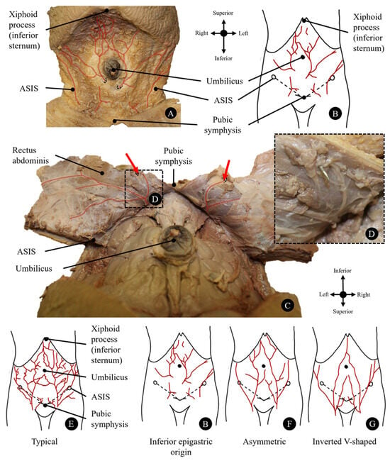
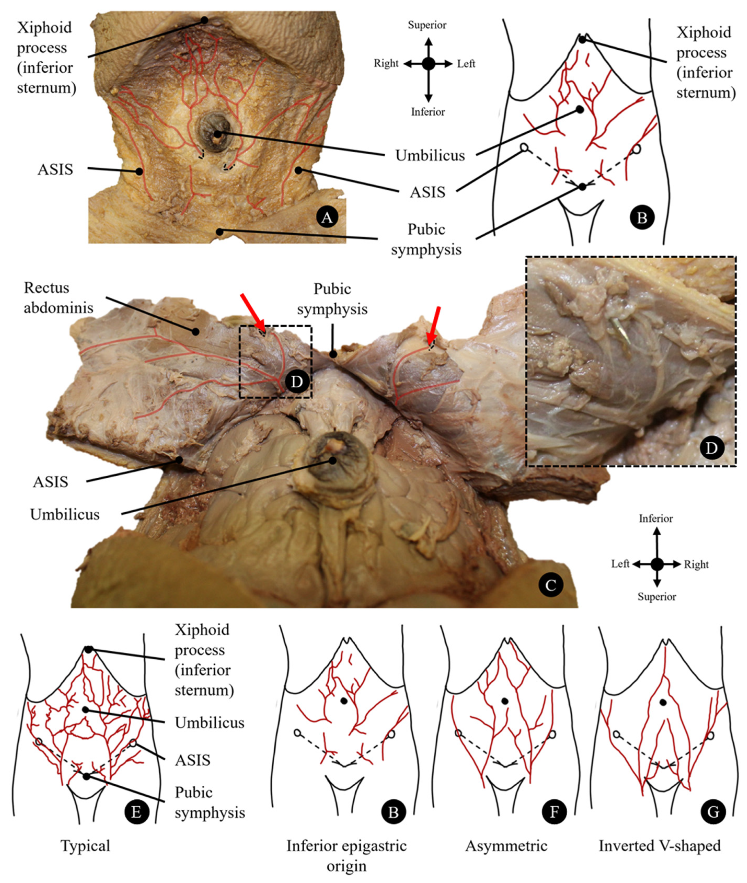
While dissecting the anterior abdominal wall to expose the superficial vascular structures through careful microdissection under direct visualization, there was a notable variation in the vascular course of these blood vessels. On further exploration, these superficial vessels appeared to radiate superiorly from the level of the umbilicus. As such, despite taking the greatest of care to preserve all the superficial vascular structures through precise micro-dissection, neither the left nor the right superficial epigastric artery was observed at its expected origin from the femoral artery. The femoral triangle was carefully explored during the dissection to identify the vessel; however, no artery consistent with the superficial epigastric artery could be identified arising from the femoral artery. Instead, the superficial vascular supply appeared to originate predominantly from perforating branches of the inferior epigastric artery emerging near the level of the umbilicus. A branch of the inferior epigastric artery could be seen penetrating through the anterior abdominal wall muscles, ultimately spreading over the entire superficial anterior abdominal wall (Figure 1A,B). These superficial abdominal arteries were tagged with a pin, and the abdomen was resected to identify the origin of the penetrating vessel. The inferior portions of the rectus abdominis were removed to see the inferior epigastric artery coursing superiorly, providing a significant contributing branch to the indicated (“red” arrow) tagged pins (Figure 1C). These branches from the inferior epigastric arteries were observed to pass through the anterior abdominal wall muscles, from internal to external (Figure 1D), to give rise to most of the superficial epigastric arteries to supply the superficial anterior abdominal wall. The vascular configuration observed in this specimen most closely corresponds to the schematic pattern illustrated in Figure 1B, in which branches originating from the inferior epigastric artery contribute directly to the superficial vascular supply of the anterior abdominal wall. However, in the present case, this arrangement appeared more pronounced, with the superficial arterial supply predominantly derived from perforating branches of the inferior epigastric artery emerging near the level of the umbilicus and radiating superiorly across the superficial abdominal wall.
Figure 1.
The major vascular supply of the superficial anterior abdominal wall from the inferior epigastric arteries. (A) Photograph and superimposed illustration of the cutaneous vascular branches of the superficial epigastric arteries and thoraco-epigastric veins. (B) Line diagram depicting the cutaneous vascular branches of the superficial epigastric arteries and thoraco-epigastric veins. (C) Photograph and superimposed illustration of the internal vascular branches of the inferior and superior epigastric arteries. The red arrows indicate the perforating branches of the inferior epigastric artery. (D) Photograph of the internal vascular branch of the inferior epigastric artery. (B,E–G) Line diagrams depicting the general vascular configuration of the superficial anterior abdominal wall. (D) Typical, symmetric pattern. (B) Superficial epigastric arteries that originate from the inferior epigastric artery. (E) Asymmetric pattern. (F) Inverted V-shaped midline crossover arborized below the inguinal ligament.
3. Discussion
This study demonstrated a vascular pattern of the inferior epigastric arteries in one neonate cadaver that, to the authors’ knowledge, has not been previously reported in the literature. This vascular pattern shows the inferior epigastric arteries penetrating the anterior abdominal wall muscles (deep to superficial) just below the umbilicus to supply the upper abdominal quadrants, in the absence of well-defined superficial epigastric vessels.
Vascular structures are important during laparoscopic surgery, especially in the neonatal population [6,7,8,13,14]. However, there is limited information on the superficial epigastric vessels in neonatal samples, particularly regarding their anatomical variations. Several authors have focused on the anatomy of the superficial epigastric vessels and the various patterns observed in adults [1,5,9,15,16]. However, according to these authors, none of the previous studies has described the variations observed in this case report. Park et al. [5] found, in adults, a typical, symmetrical pattern in 87.3% of the cases they observed (Figure 1E). In their angiogram analysis, they observed that 12.7% had an asymmetric pattern (Figure 1F), and in only 5.5%, a large, inverted V-shaped midline crossover with arborization below the inguinal ligament (Figure 1G) [5].
Mall in 1898 described the embryological development of the rectus abdominis muscle, which originates from the ventral longitudinal column of the ventral hypomere, derived from the lateral plate of the paraxial mesoderm [10,17]. This ventral longitudinal column separates into the two hemi-abdominal origins, attaching to the pubic symphysis and pubic crest. These two origins would run parallel and attach superiorly to the seventh costal cartilage [10]. The myotomes that have migrated into the embryo’s ventral wall, which are accompanied by nerve and vasculature cells, laterally contribute to the formation of intercostal nerves and their related blood vessels [17]. Rozen et al. [10] further elaborates that in the ventral midline, segmental arteries create deep epigastric arteries, supplying the muscles that emerge from the ventral longitudinal muscle column [10], likely providing each rectus abdominis muscle its own blood supply from its corresponding inferior epigastric artery, with one perforator row for each head, irrespective of the branching pattern of the inferior epigastric artery and the branches thereof, as described by Pfiztner in 1889 in the Anatomischer Anzeiger [4]. Schaverien et al. [16] evaluated the medial and lateral perforators from the inferior epigastric artery and vena comitans, establishing that there is a significant anastomosis between these vessels and the superficial epigastric vein [16], found in Scarpa’s fascia within the subcutaneous tissue [15]. These medial perforators often have a larger caliber [18,19,20]. This may provide a possible developmental explanation for the vascular configuration observed in the present case, in which both the inferior epigastric arteries penetrated the anterior abdominal wall to supply the cutaneous regions in the apparent absence of well-developed superficial epigastric arteries. As described by Taylor and Daniel [21], the vascular territories are dynamic; when one source is missing or underdeveloped, neighboring areas compensate for the reduced blood supply [21].
It should be noted that microscopic or histological confirmation of a hypoplastic or vestigial superficial epigastric artery was not performed. Therefore, the findings should be interpreted as an apparent absence of the typical vessel with functional predominance of perforating branches arising from the inferior epigastric artery. It should also be acknowledged that the specimen was preserved using formalin-based submersion fixation. Embalming methods may influence tissue elasticity, vascular lumen integrity, and the visibility of small superficial vessels, particularly in neonatal tissues, where vessels are delicate. Previous studies have demonstrated that embalmed tissues may show alterations in vascular lumen patency and microvascular visibility, which can influence the interpretation of fine vascular structures during dissection [22,23]. Therefore, although careful dissection was performed, the possibility that very small superficial vessels were collapsed or obscured cannot be completely excluded.
This anatomical variation highlights the importance of understanding the vascular anatomy of the anterior abdominal wall in neonates. While previous research has focused on adult vascular patterns [5,15,16], this case emphasizes the unique considerations in neonatal populations, where angiogenesis is ongoing. A thorough understanding of these variations is crucial for surgeons performing laparoscopic procedures in neonates, as the well-defined superficial vessels from the inferior epigastric arteries, as observed in this case, are at risk of iatrogenic injury during trocar placement [7], which, if injured, could lead to a rectus sheath hematoma. Authors such as Iwanaka et al. [14] found that using an open technique around the umbilicus for the first trocar placement for surgeries such as fundoplication, pyloromyotomy, and endorectal pull-through yielded no trocar-related complications [14]. This observed variation presented here contributes to the existing knowledge of the anatomy of the superficial epigastric arteries in neonates.
4. Conclusions
To the best of our knowledge, this vascular configuration has not been previously described in the neonatal anatomical literature, and it may contribute to a broader anatomical awareness relevant to laparoscopic procedures in neonates. The findings presented here contribute to the existing knowledge on the anatomy of the superficial epigastric artery and may have surgical implications in neonates. Further research is needed to determine the prevalence of these variations and their clinical significance.
Author Contributions
All authors contributed to the conception and design of the study. Material preparation, data collection and analysis were performed by D.J.v.T. and A.v.S. The first draft of the manuscript was written by D.J.v.T., and D.J.v.T., N.K., M.L.v.N. and A.v.S. commented on previous versions of the manuscript. All authors have read and agreed to the published version of the manuscript.
Funding
This work is based on the research supported by the National Research Foundation (NRF) of South Africa (Grant Number: 120410). The funding source had no involvement in the study design, data collection, analysis and interpretation of the data; in the writing of the report; or in the decision to submit the article for publication.
Institutional Review Board Statement
The authors state that every effort was made to comply
with all applicable local and international ethical guidelines and laws regarding the use of human
cadaveric donors in anatomical research [24]. Research and ethical approvals for this research (ethics
reference no: 224/2023; approval date: 12 July 2023) were granted by both the PhD and Ethical
Committees of the Faculty of Health Sciences at the University of Pretoria.
Informed Consent Statement
The prosection was
conducted on a bequeathed embalmed human neonatal cadaver, acquired through either a family
donation or as an unclaimed body, forming part of the Department of Anatomy’s cadaver collection.
In compliance with the National Health Act, 61 of 2003, and adhering to the ethical guidelines of
the Declaration of Helsinki (2024), no identifiable information was collected, ensuring consistency,
integrity, and quality throughout the research.
Data Availability Statement
The original contributions presented in this study are included in the article. Further inquiries can be directed to the corresponding author.
Acknowledgments
The authors sincerely thank those who donated their bodies to science and their families, as this allows for important anatomical research. Such research can enhance overall knowledge and lead to better patient care. For this reason, these donors and their families truly deserve our deepest gratitude. Additionally, the authors express their gratitude towards Dietrich Ernst Lorke for his valuable contribution to translating German and French texts. Furthermore, the authors thank Jade Sterling for her invaluable assistance with language editing. While preparing this work, the authors utilized Gemini (version 2.0 Flash, Google, 2025) and Grammarly (version 1.2.138.1610, Grammarly, 2025) to rephrase and clarify, and for basic grammar and spelling checks. Following the use of these services, the authors reviewed and edited the content as necessary and assumed complete responsibility for the publication’s content.
Conflicts of Interest
The authors declare that there is no conflict of interest regarding the publication of this paper.
Abbreviations
The following abbreviations are used in this manuscript:
| TA2 | Terminologia Anatomica Second Edition |
References
- Kostov, S.; Dineva, S.; Kornovski, Y.; Slavchev, S.; Ivanova, Y.; Yordanov, A. Vascular Anatomy and Variations of the Anterior Abdominal Wall-Significance in Abdominal Surgery. Prague Med. Rep. 2023, 124, 108–142. [Google Scholar] [CrossRef] [Scilit]
- Moore, K.L.; Dalley, A.F.; Agur, A.M.R. Clinically Oriented Anatomy, 8th ed.; Wolters Kluwer Health: Waltham, MA, USA, 2017. [Google Scholar]
- Kandinata, N.; Van Fossen, K. Anatomy, Abdomen and Pelvis: Epigastric Artery. In StatPearls [Internet]; StatPearls Publishing: Treasure Island, FL, USA, 2023. Available online: https://www.ncbi.nlm.nih.gov/books/NBK537156/ (accessed on 7 February 2026).
- Pfitzner, W. Über die Ursprungsverhältnisse der Arteria obturatoria. Anat Anz. 1886, 1, 504–514. [Google Scholar]
- Park, S.O.; Imanishi, N.; Chang, H. The Anatomic Features and Role of Superficial Inferior Epigastric Vein in Abdominal Flap. Arch. Plast. Surg. 2022, 49, 482. [Google Scholar] [CrossRef] [Scilit]
- Mechchat, A.; Bagan, P. Management of major vascular complications of laparoscopic surgery. J. Visc. Surg. 2010, 147, e145–e153. [Google Scholar] [CrossRef] [Scilit]
- Vasquez, J.M.; Demarque, A.M.; Diamond, M.P. Vascular complications of laparoscopic surgery. J. Am. Assoc. Gynecol. Laparosc. 1994, 1, 163–167. [Google Scholar] [CrossRef] [Scilit] [PubMed]
- Štádler, P.; Dorosh, J.; Dvořáček, L.; Vitásek, P.; Matouš, P.; Lin, J.C. Review and current update of robotic-assisted laparoscopic vascular surgery. Semin. Vasc. Surg. 2021, 34, 225–232. [Google Scholar] [CrossRef] [Scilit] [PubMed]
- Thoma, A.; Jansen, L.; Sprague, S.; Duku, E. A comparison of the superficial inferior epigastric artery flap and deep inferior epigastric perforator flap in postmastectomy reconstruction: A cost-effectiveness analysis. Can. J. Plast. Surg. 2008, 16, 77. [Google Scholar] [CrossRef] [Scilit] [PubMed]
- Rozen, W.M.; Kapila, S.; Donahoe, S. Why there are two rows of deep inferior epigastric artery perforators despite variability in the number of deep inferior epigastric artery trunks: An anatomical and embryological argument. Clin. Anat. 2011, 24, 786–788. [Google Scholar] [CrossRef] [Scilit]
- Franchi, A.; Patanè, L.; Hummel, C.H.; Jung, F. Confusion Regarding the Anatomy of the Superficial Inferior Epigastric Artery and the Superficial Circumflex Iliac Artery Superficial Branch. Plast. Reconstr. Surg. Glob. Open 2024, 12, e5714. [Google Scholar] [CrossRef] [Scilit]
- Langreen, S.; Ludwikowski, B.; Dingemann, J.; Ure, B.M.; Hofmann, A.D.; Kuebler, J.F. Laparoscopic pyeloplasty in neonates and infants is safe and efficient. Front. Pediatr. 2024, 12, 1397614. [Google Scholar] [CrossRef] [Scilit]
- Aguilera, A.; Gómez Rivas, J.; Álvarez-Maestro, M.; Martinez Piñeiro, L. How to deal with major complications during laparoscopic surgery? New training model for vascular lesions. Actas Urol. Esp. (Engl. Ed.) 2020, 44, 447–449. [Google Scholar] [CrossRef] [Scilit]
- Iwanaka, T.; Uchida, H.; Kawashima, H.; Nishi, A.; Kudou, S.; Satake, R. Complications of laparoscopic surgery in neonates and small infants. J. Pediatr. Surg. 2004, 39, 1838–1841. [Google Scholar] [CrossRef] [Scilit]
- Hester, T.R., Jr.; Nahai, F.; Beegle, P.E.; Bostwick, J. Blood Supply of the Abdomen Revisited, with Emphasis on the Superficial Inferior Epigastric Artery. Plast. Reconstr. Surg. 1984, 74, 657–666. Available online: https://journals.lww.com/plasreconsurg/fulltext/1984/11000/blood_supply_of_the_abdomen_revisited,_with.11.aspx (accessed on 7 February 2026). [CrossRef] [Scilit]
- Schaverien, M.; Saint-Cyr, M.; Arbique, G.; Brown, S.A. Arterial and venous anatomies of the deep inferior epigastric perforator and superficial inferior epigastric artery flaps. Plast. Reconstr. Surg. 2008, 121, 1909–1919. [Google Scholar] [CrossRef] [Scilit] [PubMed]
- Mall, F.P. Development of the ventral abdominal walls in man. J. Morphol. 1898, 14, 347–366. [Google Scholar] [CrossRef] [Scilit]
- Rozen, W.M.; Ashton, M.W.; Le Roux, C.M.; Pan, W.R.; Corlett, R.J. The perforator angiosome: A new concept in the design of deep inferior epigastric artery perforator flaps for breast reconstruction. Microsurgery 2010, 30, 1–7. [Google Scholar] [CrossRef] [Scilit] [PubMed]
- Li, Z.; Tang, Y. The DIEA Branching Pattern and Its Relationship to Perforators. In Oncoplastic Flap Surgery; Springer: Singapore, 2023; pp. 49–73. [Google Scholar] [CrossRef] [Scilit]
- Rozen, W.M.; Palmer, K.P.; Suami, H.; Pan, W.R.; Ashton, M.W.; Corlett, R.J.; Taylor, G.I. The DIEA branching pattern and its relationship to perforators: The importance of preoperative computed tomographic angiography for DIEA perforator flaps. Plast. Reconstr. Surg. 2008, 121, 367–373. [Google Scholar] [CrossRef] [Scilit]
- Taylor, G.; Daniel, R. The anatomy of several free flap donor sites. Plast. Reconstr. Surg. 1975, 53, 243–253. [Google Scholar] [CrossRef] [Scilit]
- Trucas, M.; Vincis, M.; Intini, C.; Johnston, D.; Diana, A.; Barry, D. The potential translational utility of embalmed cadaveric gastrointestinal tract specimens: A proof-of-concept study. Transl. Res. Anat. 2025, 39, 100404. [Google Scholar] [CrossRef] [Scilit]
- Trucas, M.; Dervan, A.; Quondamatteo, F. Are spinal cord and medulla samples from embalmed donors suitable for histological examination? A pilot study. J. Anat. 2025, 246, 1042–1052. [Google Scholar] [CrossRef] [Scilit]
- Iwanaga, J.; Singh, V.; Takeda, S.; Ogeng’o, J.; Kim, H.J.; Moryś, J.; Ravi, K.S.; Ribatti, D.; Trainor, P.A.; Sañudo, J.R.; et al. Standardized statement for the ethical use of human cadaveric tissues in anatomy research papers: Recommendations from Anatomical Journal Editors-in-Chief. Clin. Anat. 2022, 35, 526–528. [Google Scholar] [CrossRef] [Scilit] [PubMed]
Disclaimer/Publisher’s Note: The statements, opinions and data contained in all publications are solely those of the individual author(s) and contributor(s) and not of MDPI and/or the editor(s). MDPI and/or the editor(s) disclaim responsibility for any injury to people or property resulting from any ideas, methods, instructions or products referred to in the content. |
© 2026 by the authors. Licensee MDPI, Basel, Switzerland. This article is an open access article distributed under the terms and conditions of the Creative Commons Attribution (CC BY) license.