


Abstract
The biceps brachii muscle is a two-headed muscle located in the anterior compartment of the arm. Anatomical variations observed in the biceps brachii muscle are not uncommon. However, very few incidents are reported on a unilateral, rarely described accessory pectoral head in conjunction with the more commonly seen accessory humeral head. This anatomical variation was encountered during routine educational dissection and is the first case of this specific type of four-headed biceps brachii muscle to be described in detail. Variations in the biceps brachii morphology are linked to neurovascular compression, kinematic irregularities and variations in the musculocutaneous nerve. Knowledge of anatomical variations in the biceps brachii muscle is essential for understanding kinematic abnormalities, preventing surgical complications, and diagnosing symptoms of neurovascular compression.
1. Introduction
The biceps brachii muscle (BBM) is a multi-headed fusiform muscle that is situated in the anterior compartment of the arm. It plays a primary role in forearm flexion and supination at the elbow joint, as well as contributing to arm flexion at the shoulder joint [1,2,3,4,5].
Anatomically, the BBM typically consists of two heads. The tendon of the long head originates from the supraglenoid tubercle of the scapula, while the short head arises from the coracoid process as part of a conjoint tendon with the coracobrachialis muscle [1,6,7,8]. The two tendons continue into elongated muscle bellies that unite distally to form the bicipital tendon, which inserts on the radial tuberosity. The bicipital aponeurosis is a broad medial expansion of the bicipital tendon that fuses with the deep fascia of the forearm and reinforces the roof of the cubital fossa [1,3,4].
The BBM is innervated by the musculocutaneous nerve (MCN), which arises from the lateral cord of the brachial plexus [9,10]. This nerve branch is consistently accompanied by a vascular pedicle, which should be noted during surgical intervention, especially when this nerve needs to be isolated [1]. In addition, variations in innervation patterns have been linked to accessory muscle heads, underscoring the clinical significance of this muscle.
In addition to the typical two-headed configuration of the biceps brachii muscle, a range of anatomical variations has been documented in the literature [1,2,3,4,5,6,7,8,9,10]. Supernumerary or accessory heads are the most frequently reported variant, with studies reporting that these additional heads can range from a third to as many as seven in certain individuals, although three heads are most commonly observed [8,11,12]. These supernumerary heads are most often located on the humeral shaft and may originate from various nearby structures, including the coracobrachialis, or even the pectoral muscle tendons, indicating considerable morphological diversity [5,13,14]. In certain cases, supernumerary heads of the biceps brachii muscle are associated with longer upper limb lengths [15]. The reported prevalence of accessory heads varies among populations, with incidences of a third head ranging approximately 8–10% in certain populations, and rare configurations of four heads or more, documented sporadically in the literature [1,15,16]. Awareness of these variations is important, as they have implications for surgical approaches, imaging interpretation and the potential for altered relationships with surrounding neurovascular structures [1,17].
The presence of supernumerary heads in the biceps brachii can be understood within the broader context of limb muscle development. Skeletal muscles of the upper limb arise embryologically from mesenchymal cells derived from the somatic layer of the lateral plate mesoderm and adjacent myotomes. These precursor cells proliferate and differentiate into muscle masses that subsequently undergo segmentation, fusion, and migration to form the definitive muscle architecture [18]. During this process, transient muscle primordia and atavistic muscle bundles may be present, and variations such as accessory muscle heads are thought to arise when these primordia persist or fail to regress during normal development. Such ontogenetic mechanisms provide a plausible basis for the morphological variability observed in adult musculature, including supernumerary biceps heads, and may reflect both developmental and phylogenetic factors in muscle formation [19].
This case report aims to describe a unilateral four-headed BBM that includes both a humeral and a rarely reported pectoral head, and to highlight the clinical and embryological significance of this anatomical variation.
2. Case Report
During routine teaching dissections, performed at the Department of Anatomy, by second-year medical students at the University of Pretoria, a unilateral anatomical variation in the left BBM was observed. This 62-year-old, white, South African male cadaver had an unrelated cardiopulmonary arrest as the reported cause of death. No additional anatomical abnormalities or potential pathologies related to the BBM variation were observed.
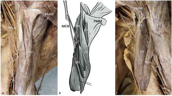
The dissection of the cadaver followed the standard anterior dissection technique, where the skin and superficial fascia of the anterior compartment of the left brachium were reflected, and the biceps brachii and coracobrachialis muscles were exposed. Bilaterally, the long and short heads of both BBMs were unremarkable and followed their normal anatomical origin and insertion pattern, while two supernumerary heads of the biceps brachii muscle (SHBBM) were observed on the left side, as seen in Figure 1A.
Figure 1.
(A) Photograph of the laterally reflected pectoralis major and biceps brachii muscles in anatomical position, indicating the origin and insertion of the pectoral head. (B) Schematic illustration of the medial view of BBM, indicating the location of both the humeral and pectoral heads. (C) Photograph of the medially rotated BBM, from the lateral view of the arm, indicating the origin and insertion of the humeral head. LH = Long head of BBM, SH = short head, PH = pectoral head, HH = humeral head, MCN = musculocutaneous nerve, PMM = pectoralis major muscle.
The humeral head originated between the distal insertion of the coracobrachialis muscle and the proximal origin of the brachialis muscle on the anterior aspect of the humeral shaft. This head extended distally, joining the short head of the BBM, proximal to the brachialis muscle and inserted via the bicipital tendon. A branch from the MCN was noted entering this additional head on the distal third (1/3) on the medial side of the muscle (Figure 1C).
The pectoral head originated from the posterior aspect of the tendinous insertion of pectoralis major, near its insertion at the crest of the greater tubercle (lateral lip of intertubercular sulcus) of the humerus. This supernumerary head descended first laterally and then ventrally to the long head of BBM and inserted, with the other muscle bellies, on the bicipital tendon distally. No clear branch from the MCN was observed directed to this additional head (Figure 1A).
The BBM bellies of the left arm were measured in situ, using a digital calliper, from the musculotendinous intersections to the point of muscle fibre termination. The long head measured 238 mm, and the short head was 261 mm. The accessory humeral head measured 152 mm, while the pectoral head was recorded at 172 mm, between the visible ends.
3. Discussion
The BBM is one of the most anatomically variable muscles in the body [2,3,6,7,8,9,11,14,20]. The variability of this muscle encompasses variations in the origin, insertion, and number of heads [2,3,8]. The supernumerary variant is the most commonly reported type of bicipital variant, with BBMs reported in the literature ranging from three to seven heads. [6,8,9,11]. Among documented points of origin, the pectoralis major muscle is among the least documented, with only three reported cases [5,13,14] and brief mentions by Grieg and colleagues [7] and Szewczyk and co-authors [11].
The estimated global prevalence of BBM variability is approximately 8.4% but may range from 2% to 37.5% depending on the population and ethnicity [4]. The prevalence of BBM variability may be attributed to differences in functional adaptations and epigenetic factors across populations and ethnic groups [4]. Certain ethnicities have higher predispositions to BBM variations than others, with the black South African population having one of the highest in the world, with a 20.5% prevalence, with a significant absence from the Zulu population and an 8.3% prevalence in the white South African population [12].
Variability of BBM morphology is often accompanied by variations in the course of and presence of the MCN [6,8,10,14,21,22]. The now-altered nerve pathway has implications for anaesthesia, specifically regional nerve blocks aimed at the MCN, which may result in failure of administration, or the nerve block may affect unwanted areas if another terminal branch of the brachial plexus assumes the innervation of the now missing MCN [13].
The importance of a study into SHBBMs lies in the clinical manifestations of this variation. The main pathology accompanying SHBBMs is neurovascular compression due to both the presence of the muscle and/or impingement of neurovascular bundles during contraction [3,4,6,8,9,11,13,20], leading to idiopathic compression symptoms [3,4,13]. During surgery, additional abnormal forces exerted on various bones by the SHBBMs may cause irregular bone displacement, leading to surgical complications [8,11,12,13]. Additionally, a benign SHBBM may be misdiagnosed on ultrasound imaging as either a longitudinal tear of the long head of BBM or a soft-tissue tumour [4,6,23]. Variations, such as those described in this case report, serve as important differential diagnoses for other suspected pathologies.
Multiple theories have been proposed to explain the mechanisms underlying the morphogenesis of BBM variations; however, no specific functional explanation has been proposed for these variations [12]. Anomalous muscles may be attributed to disordered embryological development, stemming from inappropriate separations of myotome masses in utero. These separations may be influenced by the development of adjacent neurovascular structures [4]. Phylogenetically, the emergence of anomalous muscles, especially those in the upper limb, may be explained by the regression of vestigial muscles during the development of bipedalism [8]. It is hypothesised that, during the evolutionary transition from quadrupedal to bipedal locomotion in hominins, certain minor vestigial muscles of the upper limb were lost due to a decreased functional demand for weight-bearing and locomotion [4]. Therefore, the authors hypothesise that these atavistic muscles may transiently appear during embryological development but typically regress before birth. An example of an atavistic muscle is the long head of the coracobrachialis, which, in certain non-human primates, inserts onto the radial tuberosity, a feature notably absent in humans [3,4,12]. If muscles such as these persist during foetal development, they may present as supernumerary muscles postnatally [4].
In conclusion, the presence of a four-headed biceps brachii muscle, although rare, highlights the variability of human muscular anatomy. Awareness of such anatomical variations is clinically important, particularly in surgical planning, imaging interpretation, and the diagnosis of neurovascular compression syndromes. This case contributes to the existing body of literature by reinforcing the need for vigilance when encountering atypical presentations in the upper limb and underscoring the value of detailed anatomical knowledge in both clinical and educational settings.
Author Contributions
Conceptualization: L.P.; methodology and investigation: S.M.; writing—original draft preparation: S.M.; writing—review and editing: L.P.; supervision: L.P. All authors have read and agreed to the published version of the manuscript.
Funding
No funding was received for conducting this study.
Institutional Review Board Statement
The authors affirm that all applicable local and international ethical guidelines, including the 1964 Declaration of Helsinki and its subsequent amendments, were strictly followed in the conduct of this study. The authors hereby confirm that every effort was made and that all procedures involving human cadaveric donors complied with relevant legal and ethical standards for anatomical research. Ethical approval for this manuscript was obtained from the Ethics Committee of the University of Pretoria (Reference: 657/2025) prior to publication, ensuring full adherence to all relevant ethical requirements.
Informed Consent Statement
The specimen used in this study was obtained through the institutional body donation program, in which donors provide informed consent prior to death for the use of their bodies in teaching and research.
Data Availability Statement
No datasets were generated or analyzed during the current study; therefore, data sharing is not applicable.
Acknowledgments
The authors would like to acknowledge and thank Kate Peterkin for the valuable illustration she made. The authors sincerely thank those who have donated their bodies to science, enabling anatomical research to be performed. Results from such research can potentially increase mankind’s overall knowledge, which can then improve patient care. Therefore, these donors and their families deserve our highest gratitude.
Conflicts of Interest
The authors declare no conflicts of interest.
References
- Standring, S. Gray’s Anatomy. The Anatomical Basis of Clinical Practice, 42nd ed.; Elsevier: Philadelphia, PA, USA, 2021; pp. 1403–1405, 1415, 2308. [Google Scholar]
- Alraddadi, A.S. The morphometric parameters of the biceps brachii: Cadaveric study. Surg. Radiol. Anat. 2024, 46, 463–472. [Google Scholar] [CrossRef] [Scilit]
- Bharambe, V.; Kanaskar, N.; Arole, V. A study of biceps brachii muscle: Anatomical considerations and clinical implications. Sahel. Med. J. 2015, 18, 31–37. [Google Scholar] [CrossRef] [Scilit]
- Manjatika, A.T.; Davimes, J.G.; Mazengenya, P. The third head of the biceps brachii muscle exhibiting variable shape presentation: Prevalence, variability and clinical considerations. Transl. Res. Anat. 2024, 34, 100282. [Google Scholar] [CrossRef] [Scilit]
- Sargon, M.F.; Tuncali, D.; Çelik, H. An unusual origin for the accessory head of biceps brachii muscle. Clin. Anat. 1996, 9, 160–162. Available online: https://onlinelibrary.wiley.com/doi/pdf/10.1002/(SICI)1098-2353(1996)9:3%3C160::AID-CA4%3E3.0.CO;2-K. [CrossRef] [Scilit]
- Gheno, R.; Zoner, C.S.; Buck, F.M.; Nico, M.A.C.; Haghighi, P.; Trudell, D.J.; Resnick, D. Accessory head of biceps brachii muscle: Anatomy, histology, and MRI in cadavers. Am. J. Roentgenol. 2010, 194, W80–W83. [Google Scholar] [CrossRef] [Scilit]
- Grieg, H.W.; Anson, B.J.; Budinger, J.M. Variations in the form and attachments of the biceps brachii muscle. Q. Bull. Northwest Univ. Med. Sch. 1952, 26, 241–244. [Google Scholar] [PubMed] [PubMed Central]
- Rodríguez Niedenführ, M.; Vázquez, T.; Choi, D.; Parkin, I.; Sañudo, J.R. Supernumerary humeral heads of the biceps brachii muscle revisited. Clin. Anat. 2003, 16, 197–203. [Google Scholar] [CrossRef] [Scilit] [PubMed]
- Abu-Hijleh, M.F. Three-headed biceps brachii muscle associated with duplicated musculocutaneous nerve. Clin. Anat. 2005, 18, 376–379. [Google Scholar] [CrossRef] [Scilit] [PubMed]
- Arora, L.; Dhingra, R. Absence of musculocutaneous nerve and accessory head of biceps brachii: A case report. Indian J. Plast. Surg. 2005, 38, 114–146. [Google Scholar] [CrossRef] [Scilit]
- Szewczyk, B.; Sanudo, J.R.; Podgórski, M.; Zielinska, N.; Pires, M.B.; Aragonés, P.; Olewnik, L. A Proposal for a New Classification of the Supernumerary Heads of the Biceps Brachii Muscle. BioMed Res. Int. 2022, 1510363. [Google Scholar] [CrossRef] [Scilit]
- Asvat, R.; Candler, P.; Sarmiento, E.E. High incidence of the third head of biceps brachii in South African populations. J. Anat. 1993, 182, 101–104. [Google Scholar] [PubMed] [PubMed Central]
- Fraser, P.R.; Howard, L.W.; Rosales, A.A.; Guttmann, G.D. Bilateral symmetrical supernumerary heads of biceps brachii with rare pectoralis major insertion. Surg. Radiol. Anat. 2015, 37, 299–302. [Google Scholar] [CrossRef] [Scilit] [PubMed]
- Je, S.S.; Park, B.; Kim, J.; Yoon, S.P. Five-headed biceps brachii muscle with a rare origin from the tendon of pectoralis major muscle. Anat. Sci. Int. 2016, 91, 110–113. [Google Scholar] [CrossRef] [Scilit]
- Heo, Y.-R.; Lee, H.; Lee, S.-W.; Kim, B.-S.; Kim, H.-T.; Lee, J.-H. Supernumerary Head of the Biceps Brachii Muscle Influences the Topography of the Coracobrachialis and Biceps Brachii Muscles. Medicina 2024, 60, 1726. [Google Scholar] [CrossRef] [Scilit]
- Benes, M.; Kachlik, D.; Lev, D.; Kunc, V. Accessory heads of the biceps brachii muscle: A systematic review and meta-analysis. J. Anat. 2022, 241, 461–477. [Google Scholar] [CrossRef] [Scilit]
- Ravi, P.K.; Gaikwad, M.R.; Tirpude, A.P.; Panda, M.; Mishra, P.R. Morphological Variations of Biceps Brachii Muscle in Eastern Indian Population: A Cadaveric Study. Natl. J. Clin. Anat. 2020, 9, 31–35. [Google Scholar] [CrossRef] [Scilit]
- Moore, K.L.; Persuad, T.V.N.; Tochia, M.G. Before We Are Born, 9th ed.; Elsevier: Philadelphia, PA, USA, 2016; pp. 239–245. [Google Scholar]
- Diogo, R.; Siomava, N.; Gitton, Y. Development of human limb muscles based on whole-mount immunostaining and the links between ontogeny and evolution. Development 2019, 146, dev180349. [Google Scholar] [CrossRef] [Scilit]
- Nakatani, T.; Tanaka, S.; Mizukami, S. Bilateral four-headed biceps brachii muscles: The median nerve and brachial artery passing through a tunnel formed by a muscle slip from the accessory head. Clin. Anat. 1998, 11, 209–212. [Google Scholar] [CrossRef]
- Vazquez, T.; Rodríguez-Niedenführ, M.; Parkin, I.; Sañudo, J.R. A rare case of a four-headed biceps brachii muscle with a double piercing by the musculocutaneous nerve. Surg. Radiol. Anat. 2003, 25, 462–464. [Google Scholar] [CrossRef] [Scilit] [PubMed]
- Wysiadecki, G.; Snow, E.; Wysocki, P.; Przybycień, W.; Haładaj, N.; Haładaj, R.; Grzonkowska, M.; Żytkowski, A. A neuroanatomical analysis of the link between muscle variants and innervation patterns: The accessory head of the biceps brachii muscle as a case example. Transl. Res. Anat. 2025, 13, 100446. [Google Scholar] [CrossRef] [Scilit]
- Lutterbach-Penna, R.A.; Brigido, M.K.; Robertson, B.; Kim, S.M.; Jacobson, J.A.; Fessel, D.P. Sonography of the Accessory Head of the Biceps Brachii. J. Ultrasound Med. 2014, 33, 1851–1854. [Google Scholar] [CrossRef] [Scilit] [PubMed]
Disclaimer/Publisher’s Note: The statements, opinions and data contained in all publications are solely those of the individual author(s) and contributor(s) and not of MDPI and/or the editor(s). MDPI and/or the editor(s) disclaim responsibility for any injury to people or property resulting from any ideas, methods, instructions or products referred to in the content. |
© 2026 by the authors. Licensee MDPI, Basel, Switzerland. This article is an open access article distributed under the terms and conditions of the Creative Commons Attribution (CC BY) license.