Review

Ascorbic Acid in Seeds, Priming and Beyond

by
Mattia Terzaghi
1 and
Mario C. De Tullio
2,*
1
Department of Biosciences, Biotechnologies and Environment, University of Bari, 70126 Bari, Italy
2
Department of Earth and Environmental Sciences, University of Bari, 70126 Bari, Italy
*
Author to whom correspondence should be addressed.
Seeds 2023, 2(4), 421-435; https://doi.org/10.3390/seeds2040032
Submission received: 9 January 2023 / Revised: 26 October 2023 / Accepted: 27 October 2023 / Published: 1 November 2023
(This article belongs to the Special Issue Seed Priming Approaches That Achieve Environmental Stress Tolerance)

Abstract

:
Ascorbic acid (AsA) is mainly known as an antioxidant. However, if the peculiar features of the AsA system in the different stages of seed development and germinationare taken into consideration, it can be concluded that the function of AsA goes far beyond its antioxidant properties. The possible involvement of AsA in the regulation of hormone synthesis and in the epigenetic control of gene expression opens new directions to further research. In recent years, seed priming with AsA has been successfully used as a strategy to improve germination and plant productivity. Beneficial effects of seed AsA priming could be observed in several crop species, but the underlying molecular mechanism(s) are still unclear. The available evidence suggests that AsA priming induces a wide range of coordinated responses allowing primed seeds to overcome adverse environmental conditions.

1. Introduction

The capability of producing seeds is one of the most successful features that appeared during plant evolution [1]. Seed-producing plants (spermatophytes) significantly improved the chances for their progeny to thrive even under adverse environmental conditions. Besides providing mechanical protection to the embryo and storing reserves that will be used for embryo development, seeds ensure embryo survival due to their capability of withstanding desiccation and reducing metabolic activity until suitable conditions for germination occur [2]. The implementation of the developmental program leading to seedling establishment requires extensive epigenetic and hormonal reprogramming [3,4]. Moreover, the transition from quiescence to active development is full of hidden perils and could lead to severe damaging of the embryo itself unless proper protection is prepared before seed dehydration or activated in parallel with the “awakening” of the germinating seed [5]. An increasing number of studies points at ascorbic acid (AsA) as a key player involved in all those processes. The dynamic regulation of the AsA system (including AsA production, utilization, and recycling of its oxidized forms) along the different stages of seed development, desiccation, after-ripening and germination supports the view that AsA availability varies as a function of the specific needs of each stage. Recent reports also show that seed priming using AsA treatment improves germination and plant performance, especially under stress conditions. The aim of the present contribution is providing a novel viewpoint on the different aspects of AsA function in seeds.

2. The AsA System

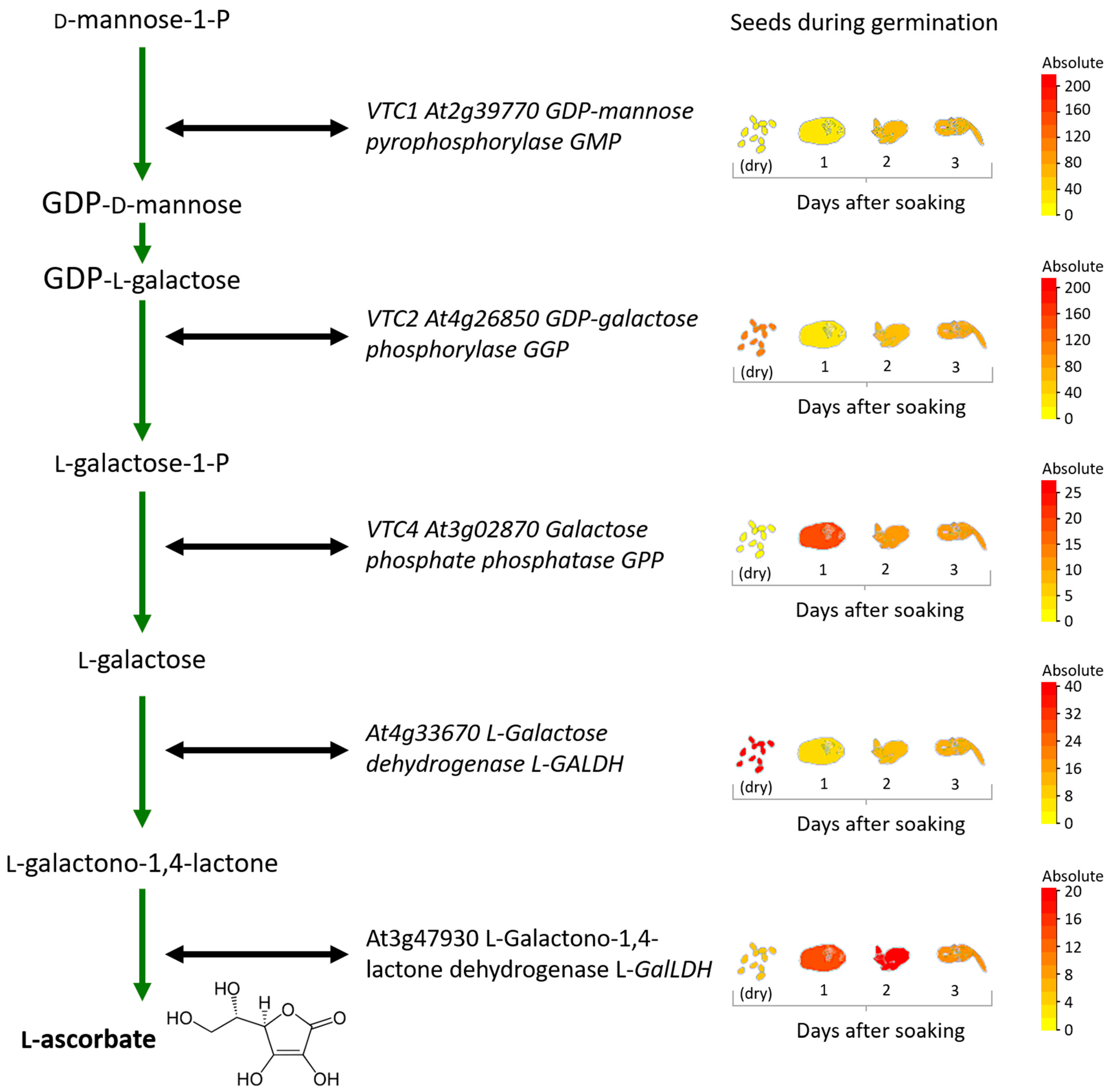
2.1. AsA Biosynthesis

All plants synthesize AsA following a biosynthetic route known as the d-mannose l-galactose (Smirnoff–Wheeler) pathway [6]. Additional entry points for AsA production may occur under specific conditions using the myo-inositol [7] and the galacturonate [8] pathways. The biosynthesis appears strictly controlled [9], with an AsA-dependent feedback mechanism inhibiting the expression of at least three key genes in the Smirnoff–Wheeler pathway, including the GDP-l-galactose phosphorylase (GGP) gene (known as VTC2 in Arabidopsis) [10]. AsA biosynthesis is also regulated by different cues, including light and hormones [11]. Notably, AsA biosynthesis occurs in the cytosol, excepting for the terminal step, which takes place at the mitochondrial inner membrane and is catalyzed by the enzyme l-galactono-1,4-lactone dehydrogenase (l-GalLDH) using oxidized cytochrome c as the electron acceptor [12]. The acquisition of this mitochondrial step is a major difference between the way in which animals [13] and plants synthesize AsA and is likely to be an adaptation to a photoautotrophic lifestyle [14].

2.2. AsA Transport and Intracellular Distribution

Once synthesized in the mitochondria, AsA must be transported to all other organelles and cell compartments, where it is unevenly distributed. Immunocytochemical detection in Arabidopsis leaves shows higher AsA content in peroxisomes and the cytosol, a bit less in the nuclei, plastids and mitochondria, with vacuoles presenting the lowest AsA content [15]. There is also indication of a relatively small but significant apoplastic AsA pool possibly involved in redox sensing of extracellular cues [16]. The mechanism of AsA transport from the site of synthesis is not fully understood. It is known that AsA can cross lipid bilayers by means of passive diffusion [17], but it is generally assumed that the oxidized form dehydroascorbic acid (DHA, see below) diffuses more efficiently [16]. AsA transporters GLUT and SVTC have been detected and characterized in animal cells [18]. Much less is known about plant transporters. A mitochondrial ascorbic acid (MAT) transporter, apparently different from GLUT and SVTC ones, from rat liver and potato tuber has been partially characterized [19]. In addition, the Arabidopsis PHT4;4 protein, a member of the phosphate transporter 4 family, is responsible for chloride-dependent AsA transport into chloroplasts [20]. Additional transporters are likely to regulate AsA intracellular distribution.

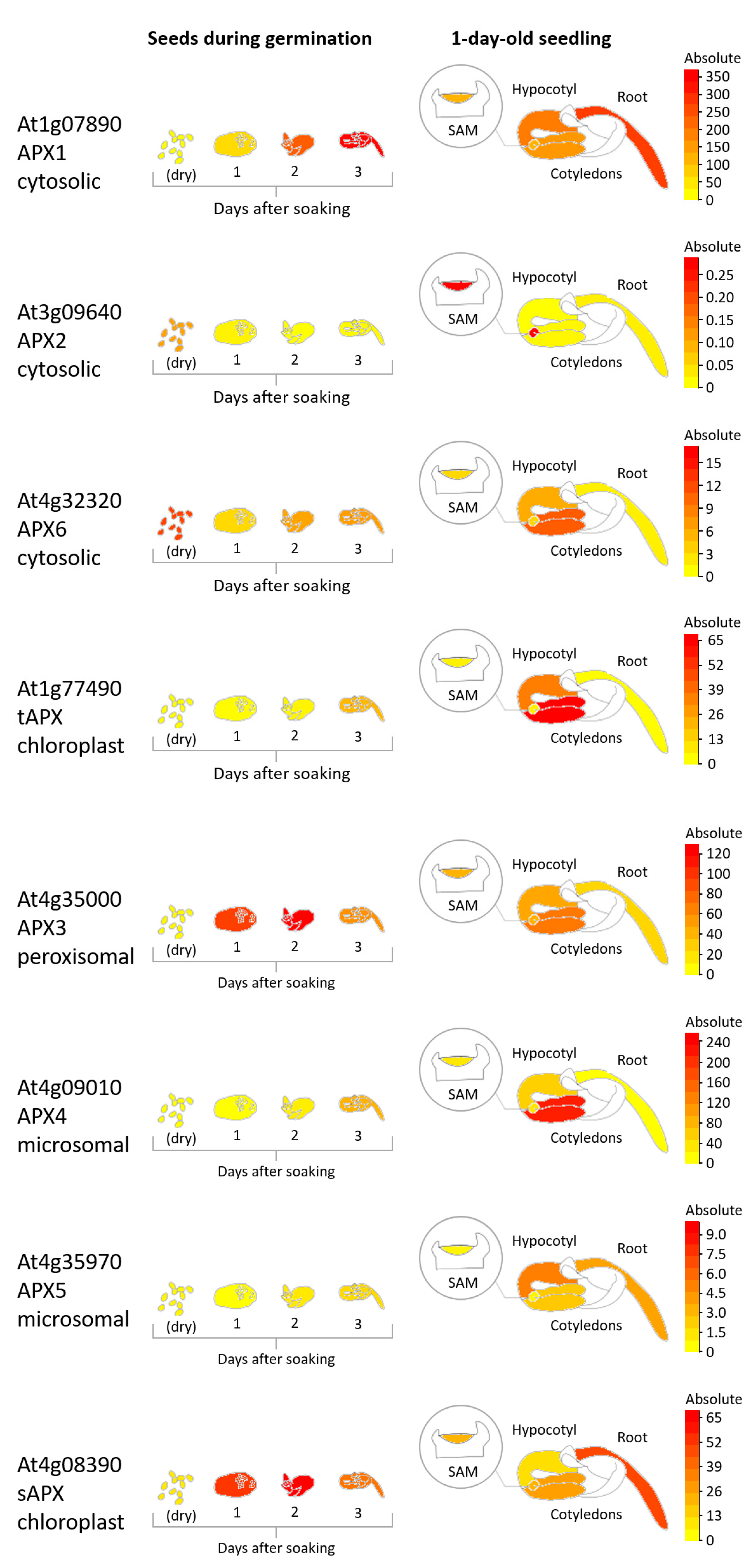
2.3. AsA Utilization

Plant-specific AsA peroxidases (APX, EC 1.11.1.11) have been detected in the cytosol [21], mitochondria [22], peroxisomes [23], and chloroplasts, where two distinct APX forms (stromal and thylakoidal, respectively) occur [24]. The enzyme catalyzes hydrogen peroxide conversion to water and O2, specifically using AsA as the electron donor. Rather than scavengers, APXs are responsible for the fine-tuning of hydrogen peroxide content [24], which is essential for signaling purposes [25]. Recent studies have also suggested that, at least in some plant species (including Oryza sativa, Glycine max, Zea mays, and species of the orchid genus Oncidium), cytosolic APX can also use glutathione (GSH) as an electron donor [26].
The blue-copper enyzme AsA oxidase (AO, EC 1.10.3.3) is another AsA-dependent enzyme whose physiological function has not been fully understood, although it is possibly involved in cell elongation [27] and in the establishment of root symbioses with arbuscular mycorrhizal fungi and rhizobacteria [28]. A search in the TAIR database (www.arabidopsis.org, accessed on 26 October 2023) retrieves four genes encoding putative AOs in the Arabidopsis genome: AAO1 (At4g39830), AAO2 (At5g21100), AAO3 (At5g21105), and recently added SRG1 (At1g17020).
Several members of the large class of 2-oxoglutarate-dependent dioxygenases (2-ODDs) utilize AsA in a complex reaction mechanism requiring, besides AsA and 2-oxoglutarate, also molecular oxygen and Fe2+ [29,30]. Different 2-ODDs catalyze a variety of reactions, including hydroxylation, epoxidation, and desaturation of specific substrates involved in the biosynthesis or the catabolism of plant hormones/regulators (ethylene, abscisic acid, gibberellins, auxin, salicylic acid) and a number of secondary metabolites. A very special subsection of 2-ODDs has been identified, involved in epigenetic mechanisms. These include TET hydroxylases, catalyzing the demethylation of methyl cytosine, and the Jumonji group of histone demethylases. TET hydroxylases have been fully characterized in animal cells, but indirect evidence suggests that plants also have them [31]. Histone demethylation activity catalyzed by specific 2-ODDs occurs during plant developmental processes and in response to stress conditions [32,33]. The actual amount of AsA consumed by each 2-ODD activity is hardly measurable. An indirect estimate has been obtained by measuring AsA content in Arabidopsis insertion lines in which different putative 2-ODDs had been inactivated. At least in some of those lines, AsA content was more than doubled [34], suggesting that some 2-ODDs could be responsible for a significant, if not massive, use of AsA.
Besides catalyzed utilization, AsA is known to react non-enzymically with reactive oxygen species (ROS) and with metal ions. According to available experimental evidence, AsA reacts preferentially with copper (II) and iron (III), rather than ROS [35]. On the other hand, it is unlikely that highly reactive radicals just freely travel around the cells, also considering the low permeability of polar molecules across biological membranes [36]. Therefore, AsA’s direct interaction with ROS should not always be taken for granted.

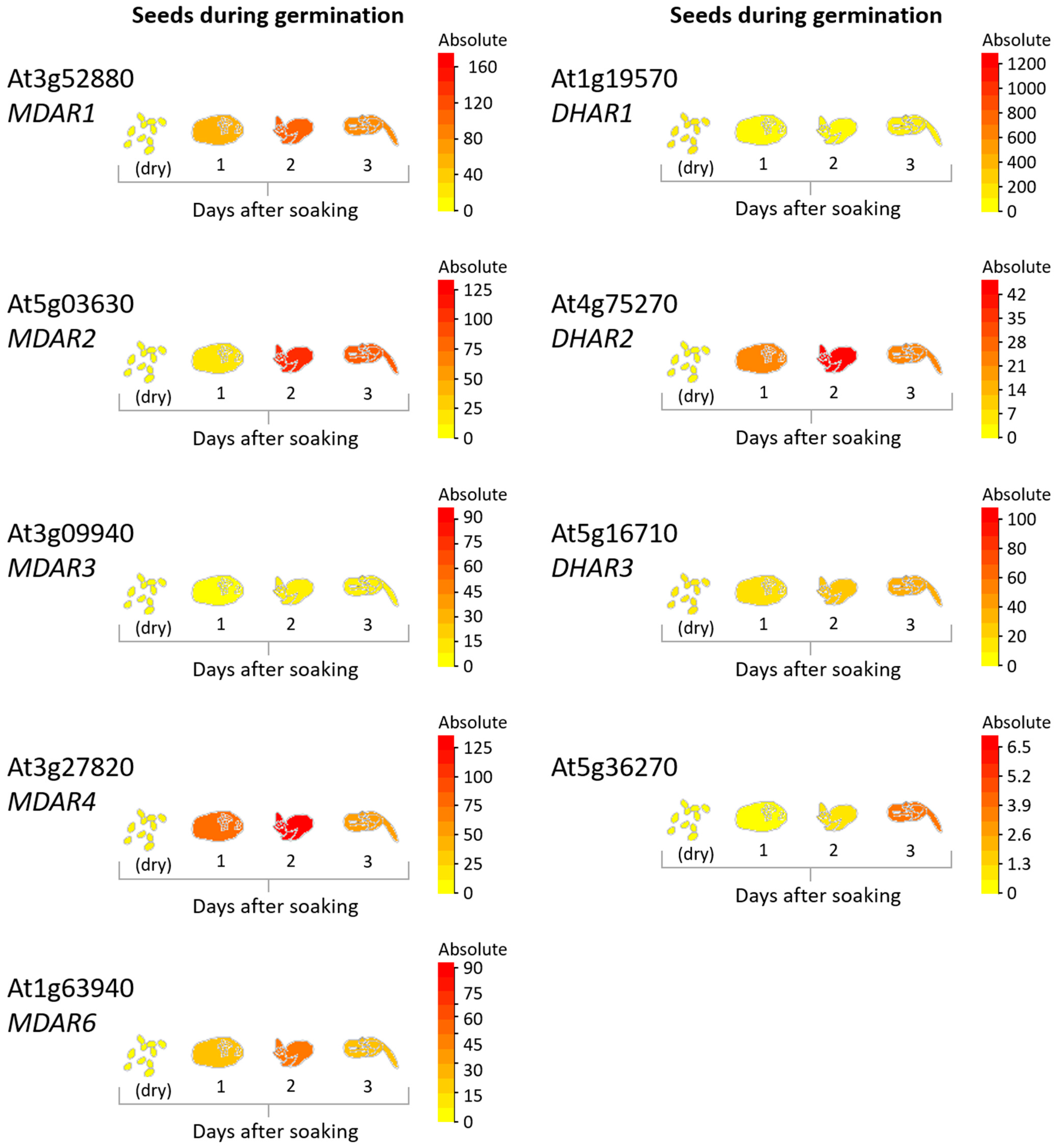
2.4. Recycling of AsA Oxidized Forms

One-electron AsA oxidation produces monodehydroascorbate (MDHA, also known as ascorbate free radical). This free radical is short-lived and disproportionates to AsA and dehydroascorbic acid (DHA) [37]. Alternatively, MDHA can be reduced enzymatically by the NADH-dependent enzyme MDHA reductase (MDHAR, EC 1.6.5.4) located in the cytosol, mitochondria, chloroplasts, and peroxisomes [38]. Fully oxidized DHA, in turn, can be re-reduced to AsA by DHA reductase (DHAR, EC 1.8.5.1), using reduced glutathione (GSH) as the electron donor [39]. MDHAR and DHAR are considered “recycling enzymes”, reconverting oxidized AsA forms back to AsA. However, recycling alone, in the absence of new biosynthesis, is apparently unable to keep up with the pace of AsA consumption [39,40]. This is also confirmed by the observation that seeds of the Arabidopsis vtc2/vtc5 double mutant, which is not capable of de novo AsA synthesis, can start the germination process due to the recycling activity, but the seedlings are not viable in the absence of AsA supplementation [41]. It should also be considered that DHA reductase activity is performed by several proteins characterized by the C-X-X-C motif, including protein disulfide isomerase and glutaredoxins [42].

2.5. Possible DHA Signaling and Further Catabolism

In comparison with the huge amount of literature dealing with AsA biosynthesis and functions, relatively little attention has been given to the products of its degradation and catabolism. As mentioned above, the product of AsA oxidation is DHA, which is usually, but erroneously, represented as a three-carbonyl molecule, whereas it is preferentially in the dimeric form [43]. There is indication that DHA, rather than just the end product of AsA oxidation, should be considered a relevant signaling molecule [44,45,46], possibly in connection with its capability of reacting with thiols to form disulfide bonds [47]. A low AsA/DHA ratio in the apoplast is generally considered a proxy of stress [48].
If not recycled back to AsA (see above, Section 2.4), DHA undergoes irreversible degradation following two possible routes: either by hydrolysis yielding diketogulonic acid, or by oxidation, with the consequent production of oxalylthreonate, oxalate, and threonate [49,50,51,52]. Remarkably, different oxidizing agents produce different AsA degradation products, suggesting the possibility that they act as molecular signals [53]. Enzyme activities are involved in catabolism, but to our knowledge, the enzymes responsible have not been characterized yet. Interestingly, the inactivation of Arabidopis AtFAHD1a, the gene encoding a fumarylacetoacetate hydrolase (FAH) domain-containing protein 1a, highly expressed in developing seeds, results in AsA, DHA, and threonic acid accumulation [54]. Recently, a new bacterial pathway of AsA degradation has been identified, involving novel enzymes and a FAH family member catalyzing the conversion of 2-keto-3-deoxy-l-lyxonate into 2-oxoglutarate (α-ketoglutarate) [55]. Hopefully, the full disclosure of the details of AsA catabolism in plants is quite close. This will possibly also help in understanding the physiological role of AsA degradation products, as in the case of the observed oxalyltransferase activity responsible for the transfer of oxalate groups from oxalylthreonate to carbohydrates [56].

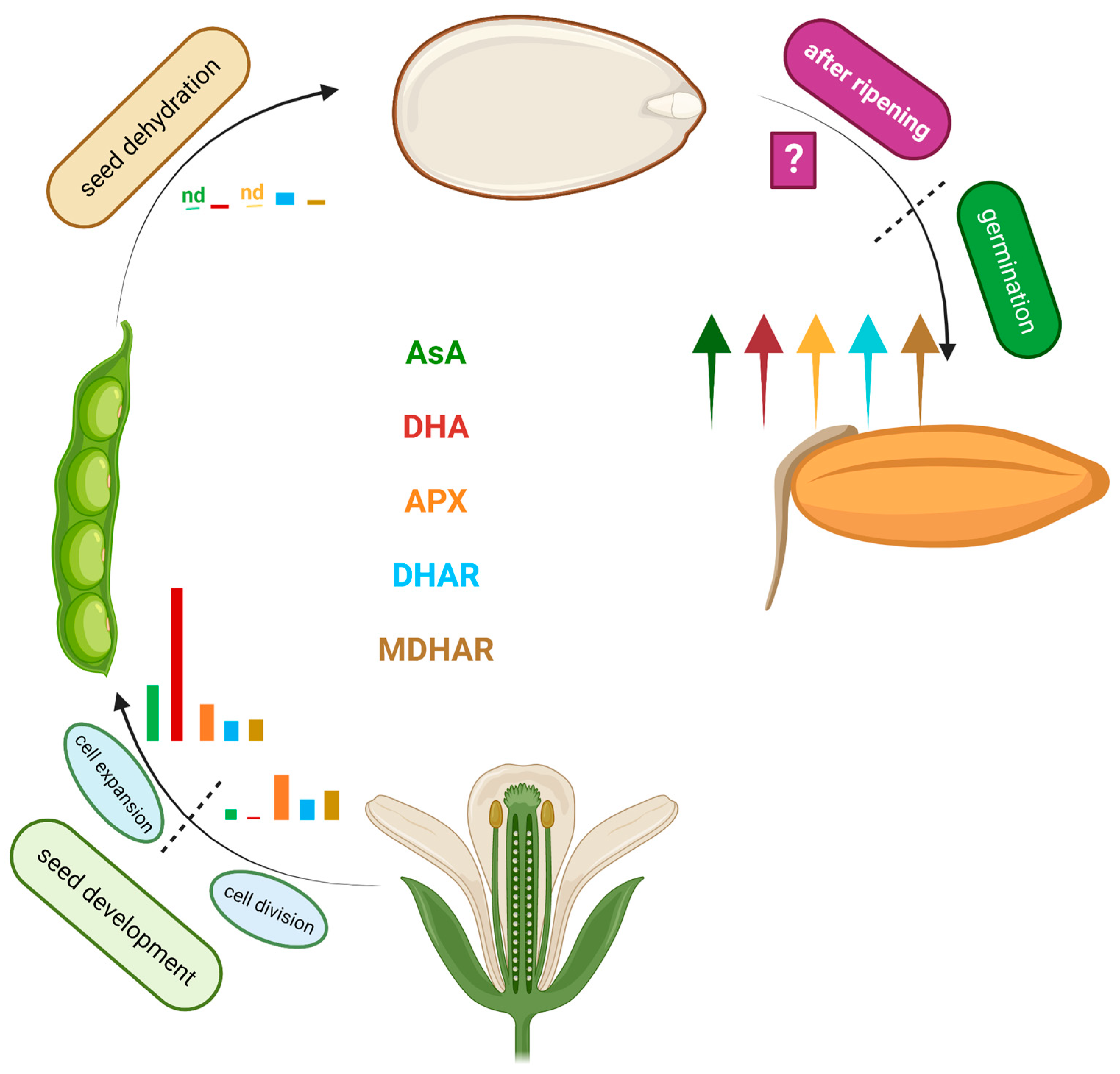
3. Dynamic Regulation of the AsA System during Seed Development

During the different stages of their life, seeds apparently activate different items of the wide toolkit forming the AsA system [5]. The scheme in Figure 1 summarizes the main changes in the AsA system occurring during orthodox seed development, quiescence, and early germination stages. AsA content increases during seed development, then dramatically drops below detectability during dehydration. DHA content is very high during the cell elongation stage of seed development, then markedly decreases, although still remains detectable, in dry seeds [57,58]. No APX activity can be measured in dry seeds, but some DHAR and MDHAR activity is still observed. AsA oxidase is undetectable at all stages.
It is worth noting that mature recalcitrant seeds, which undergo partial or no dehydration, retain AsA and APX activity [59]. Therefore, the absence of AsA and APX in orthodox seeds, in parallel with a low DHA supply and limited activity of the recycling enzymes, appears correlated with seed quiescence and longer viability.
The analysis of available Arabidopsis RNA-sequencing data [60] allows us to further characterize the changes occurring in seeds during the early stages of germination. By comparing the expression of the genes involved in the main AsA biosynthetic (Smirnoff-Wheeler) pathway (Figure 2), early activation of l-galactono-1,4-lactone dehydrogenase (l-GalLDH) occurs, in accordance with the observation that seeds become capable of converting l-GalL into AsA early during imbibition [5]. However, the expression of the main regulator of AsA biosynthesis, the VTC2 gene encoding a GDP-l-galactose phosphorylase, appears rather low at this stage, suggesting that full-rate AsA biosynthesis is likely to occur only later in germination. Expression analysis of putative DHAR and MDAR genes (Figure 3) confirms the observation that recycling activities provide the small but essential AsA amount necessary to restart metabolic activity in germinating seeds, before de novo AsA biosynthesis becomes fully operational. Among putative MDAR genes, MDAR1 and MDAR4, both encoding peroxisomal isoforms of the MDHA reductase enzyme, show early expression, paralleled by cytosolic DHAR2. It should, however, be considered that different plant species, or even different cultivars apparently manage the recycling of oxidized AsA forms in different ways. As an example, in germinating Pisum sativum cv. Alaska seeds, DHAR activity could not be detected [61], whereas it was 30-fold lower than MDAR activity in the cv. Lincoln [62].

4. AsA in Seed Dehydration, Dormancy, and Germination: To Be or Not to Be (There)?

The dynamic regulation of the AsA system outlined above shows that AsA and DHA are relevant players during seed development, and especially at the cell elongation stage, where DHA takes the lead. It should be considered that the entire process of development is controlled by the coordinated action of gibberellins and ABA [63]. AsA-dependent 2-ODDs (see Section 2.2 above) are involved in the biosynthesis of both hormones [64,65] and increased AsA content induces the expression of the ABA biosynthesis gene 9-cis-epoxycarotenoid dioxygenase NCED3 [66]. Due to its capability to oxidize protein thiols [47], DHA is also possibly responsible of the redox shift occurring in storage proteins, which at seed maturity are mostly in their oxidized (disulfide) state [67]. In the next stage (dehydrated seeds), AsA is totally absent, and only little DHA remains, suggesting not only that AsA is not required at this stage, but possibly that its presence could also negatively affect the dehydration step. Desiccation tolerance in living tissues is a complex process, not yet fully understood, but for sure it requires the interaction of several different players [68]. For orthodox seeds, desiccation precedes dormancy, which is the implementation of the safety mechanism(s) by which seeds do not germinate under “deceptive” favorable conditions occurring episodically, as in a short mild-weather period before full winter comes [2]. The establishment of dormancy is an ABA-dependent process regulated by the activity of NCED dioxygenases [65], requiring the presence of the co-substrate AsA [69], but the subsequent maintenance of dormancy apparently requires AsA removal. This possibility is strongly suggested by both old and recent findings. Besides the well-known presence of AsA and APX in recalcitrant (non-dormant) seeds [59], recent work by Gerna et al. [54] further supports this eventuality. As mentioned above regarding AsA catabolism (Section 2.5), the Arabidopsis mutant Atfahd1a-1, lacking fumarylacetoacetate hydrolase (FAH) activity, is apparently impaired in the AsA catabolic pathway and accumulates AsA, DHA, and threonic acid. Interestingly, the mutant shows shallower thermo-dormancy, together with increased seed longevity and a shift of the seed redox poise towards a reduced state. In the wild type, the FAHD1a gene is highly expressed in the embryo of fully mature and desiccated seeds. The total loss of AsA and the almost complete disappearance of DHA in dry seeds could be explained with the high activity of the FAHD1a enzyme (and possibly other uncharacterized AsA catabolic enzymes) at this stage. Oxalate, another product of AsA catabolism, is often accumulated in dry seeds, possibly regulating calcium uptake [70].
The fact that AsA content progressively decreases during seed maturation, so that dry seeds are devoid of AsA, is unlikely to be accidental. In a way, the absence of AsA in dry seeds is counterintuitive, especially in the general pervasive view that antioxidants, and AsA for one, always have a protective effect against any form of unfavorable environmental conditions, and seeds obviously need to be protected to increase their chances of survival. A possible explanation to this apparent contradiction could be the involvement of AsA in epigenetic mechanisms, namely in methyl-cytosine demethylation, in analogy with the well-characterized TET dioxygenases of animal cells [71]. The DNA methylation pattern markedly increases in developing embryos, keeps steady in quiescent seeds, and is then dramatically reversed with extensive demethylation at the very beginning of the germination process [72], in parallel with the recovery of AsA regeneration and de novo biosynthesis. It is tempting to hypothesize a causal relationship between these two events and a direct involvement of AsA in widespread DNA demethylation also in the cells of plant embryos, similarly to what is known to occur in human stem cells [73]. A second possibility to explain AsA absence in dry seeds is the well-known involvement of ROS in dormancy release [74,75]. Although known for years, only recently the mechanism of ROS-dependent dormancy alleviation has been better characterized at the cellular and molecular levels [76,77]. If AsA was stored in dry seeds, it could interfere with early ROS production that is key to starting germination, so its antioxidant action would be a burden rather than an advantage. Exciting new findings on dormancy and dormancy release have been reported in the last few years [78]. The identification of DELAY OF GERMINATION-1 (DOG1) as a master regulator of dormancy opened new and unexpected directions in ongoing research on seed biology. The DOG1 protein appears involved in a complex signaling system involving ABA and possibly more, still uncharacterized, players [4,79]. Surprisingly, the DOG1 system is strictly connected to the expression of genes previously characterized for their involvement in the control of flowering time, including FLOWERING LOCUS C (FLC) and FLOWERING LOCUS T (FT) [80], the latter acting in two opposite configurations to regulate either flowering or dormancy release [81]. Histones associated with DOG1 and FLC undergo extensive changes in their methylation patterns at the transition from dormancy to germination [82]. Indeed, both flowering and germination processes share the necessity of avoiding “false starts” that would jeopardize plant survival and life-cycle completion. It is worth mentioning that increased AsA content delays flowering and the expression of the LEAFY gene, which is expressed downstream of FT in the specification of floral organs [83].
Once germination starts, metabolic activity is fully recovered. Mitochondrial metabolism has a pivotal role at this stage [75,77]. Early ROS accumulation occurs in the mitochondria and is caused by the activation of the respiratory electron transport chain [84]. High expression of the gene encoding l-GalLDH, the mitochondria-located enzyme catalyzing the last step of AsA biosynthesis, occurs at this stage (Figure 2). As far as germination proceeds, ROS are found in the nuclei, where they possibly operate in the mechanism of chromatin decompaction, and later on in the peroxisomes [77]. It is conceivable that at this stage, AsA and APX become crucial to avoiding ROS overproduction. The expression of the six Arabidopsis APX genes during germination and in the seedlings is reported in Figure 4. APX3, encoding a peroxisomal APX, is expressed during germination. Early expression of the gene coding for the APX of chloroplast stroma is also observed, in a stage characterized by gibberellin-regulated rapid proplastid differentiation [85].
The control of ROS production by AsA and APX becomes more relevant under stress conditions. Inhibition of Arabidopsis seed germination in the presence of excess salt is regulated by a ROS-mediated signaling module connecting the transcription factor ABI4 with the NADPH oxidase gene RbohD and the AsA biosynthesis gene VTC2 [86], confirming the centrality of AsA in the germination process. However, it should be considered that exogenous administration of AsA in excess inhibits rice seed germination, in a mechanism involving ABA and GA [87]. This means that endogenous AsA content must be carefully controlled to balance hormonal production and action.

5. Priming with AsA

Seed priming is an empirical practice known since antiquity [88] that proved to be a powerful tool in improving seed germination and plant performance, especially under adverse environmental conditions [89,90,91]. The term antioxidant priming is used to indicate seed treatment with different molecules potentially reacting with ROS, including AsA. Although a simple internet search using together the words “seed”, “priming”, and “ascorbic” retrieves a large number of papers whose title and abstract suggest a clear-cut positive effect of AsA priming on many different parameters of plant productivity, a closer look into some of those articles reveals a lack of proper controls and other flaws in the experimental design, thus making the data of those reports difficult to interpret. Even not considering those flawed papers, a substantial amount of sound experimental data confirm that priming seeds with AsA at different concentrations is beneficial to plant growth, development, and productivity. A very short list of papers comparing the effects of AsA priming on mean emergence time (MET) is presented in Table 1.
Plant materials (cultivars) and treatment conditions vary in the different experiments reported in Table 1, but a tendency of early seedling emergence in AsA-primed seeds is generally observed. AsA priming also increases, although to a different extent, germination percentage, germination uniformity, and vegetative and reproductive growth in a variety of model and non-model plant species, or even improves nutrient profiles in seeds harvested from plants originally subjected to AsA priming at the seed stage [97]. However, the most convincing results are obtained when the germination of AsA-primed seeds takes place under stress conditions. As an example, the germination percentage of wheat plants in the presence of 200 mM of NaCl is 55 ± 6.5 in unprimed controls, but increases to 73 ± 6.6 when the seeds are primed with a 150 mg/L AsA solution [98]. Unfortunately, not many attempts have been made to explain the molecular mechanisms underlying the beneficial effects of AsA priming. Most studies simply advance the hypothesis that AsA priming improves general antioxidant defenses [99,100,101,102]. Only a few studies tried to go deeper into detail. An accurate analysis in artificially aged oat seeds has shown a repair effect of AsA and GSH priming on damaged mitochondria [103]. An interesting study analyzing the effect of AsA priming (0.5 mmol/L for 12 h) on wheat seed proteome, with or without NaCl (250 mmol/L solution), has shown altered expression of 167 proteins, the majority of which were under-regulated [104]. Most interestingly, AsA priming impacted negatively defense-related proteins, including antioxidants superoxide dismutase and AsA peroxidases. Such proteins were less represented in primed seeds (in both embryo and surrounding tissues) as compared to controls, and even less in primed seeds treated with NaCl. The presence of the AsA biosynthetic enzyme GDP-mannose-3,5-epimerase was also lower in primed seeds. This is in clear contrast with the claim that AsA priming is effective because it improves antioxidant defenses. Proteome data suggest that AsA priming induces a complex response that upregulates proteins involved in metabolism/energy and downregulates defense-related proteins, a picture that cannot be explained with simplistic considerations and unsupported assumptions based on AsA antioxidant properties. The effect of AsA priming on the after-ripening stage remains to be investigated.

6. What’s Next?

Environmental stresses caused by climate change pose new constraints to plant growth and productivity. Investigating the mechanisms regulating seed germination under stress conditions will help in selecting tolerant genotypes of crop plants able to grow and reproduce in unfavorable environments. Several lines of reasoning point at a central role of AsA as a multi-level regulator of seed developmental and germination processes, making this peculiar molecule a promising target for further investigations. Unfortunately, AsA suffers the prejudice of being essentially categorized as an antioxidant, which is probably only a small part of its complex biochemical function. As discussed in the previous sections, there are sufficient indications that AsA is required for hormone synthesis and epigenetic regulation of gene expression (DNA and histone demethylation), although the details of this functional AsA dependency are still little known. The positive effects of seed priming with AsA, especially under unfavorable environmental conditions, offers an outstanding opportunity to deepen our understanding of the mechanisms controlling seed dormancy and germination. As discussed by Munns and Gilliham [105], plants growing in saline soils (or other stressful situations) pay their dues in terms of energy costs, at the expenses of their progeny (which, from an agricultural point of view, means at the expenses of plant productivity). Studies in the animal field suggest that epigenetic mechanisms can bring selective advantages, so that an organism endangered by environmental conditions invests in the improvement of progeny fitness [106]. AsA priming apparently goes in this direction, as suggested by the observation that corn plants derived from AsA-primed seeds produce seeds with improved vigor and protein content in the next generation [97], which is good both for the plants and for the heterotrophs consuming those seeds. It is especially interesting that a surprising shift occurs in the proteome of AsA-primed wheat seeds germinating in the presence of NaCl, with an increase of energy and metabolism-related proteins and a decrease in defense-related proteins [104]. The idea of a trade-off between energy and defense (immunity) is at the basis of ecological immunology, an area of research investigating how individual defense responses are integrated in the framework of environmental cues [107]. Ecological epigenetics is also a field in rapid expansion that will possibly provide answers to many questions currently under debate [108]. For sure, in order to untangle the complex interrelation of cues involved in the crucial process of germination and understand the actual contribution of AsA within this process, we need to think out of the box and explore new directions. This will be a challenge for the years to come.

Author Contributions

Conceptualization, original draft preparation, M.C.D.T. Review and editing, preparation of Figure 2, Figure 3 and Figure 4, M.T. All authors have read and agreed to the published version of the manuscript.

Funding

This research was partly supported by the Italian Ministry of University and Research, PRIN grant 2022LLATJH.

Data Availability Statement

No new data were produced.

Acknowledgments

Original figures were created with BioRender.com, accessed on 26 October 2023.

Conflicts of Interest

The authors declare no conflict of interest.

References

  1. Linkies, A.; Graeber, K.; Knight, C.; Leubner-Metzger, G. The evolution of seeds. New Phytol. 2010, 186, 817–831. [Google Scholar] [CrossRef] [PubMed]
  2. Finch-Savage, W.E.; Leubner-Metzger, G. Seed dormancy and the control of germination. New Phytol. 2006, 171, 501–523. [Google Scholar] [CrossRef] [PubMed]
  3. Kravets, O.P.; Sokolova, D.O. Epigenetic mechanisms regulating seed germination rate. Cytol. Genet. 2017, 51, 346–351. [Google Scholar] [CrossRef]
  4. Smolikova, G.; Strygina, K.; Krylova, E.; Leonova, T.; Frolov, A.; Khlestkina, E.; Medvedev, S. Transition from Seeds to Seedlings: Hormonal and Epigenetic Aspects. Plants 2021, 10, 1884. [Google Scholar] [CrossRef]
  5. De Tullio, M.C.; Arrigoni, O. The ascorbic acid system in seeds: To protect and to serve. Seed Sci. Res. 2003, 13, 249–260. [Google Scholar] [CrossRef]
  6. Wheeler, G.L.; Jones, M.A.; Smirnoff, N. The biosynthetic pathway of vitamin C in higher plants. Nature 1998, 393, 365–369. [Google Scholar] [CrossRef]
  7. Lorence, A.; Chevone, B.I.; Mendes, P.; Nessler, C.L. myo-inositol oxygenase offers a possible entry point into plant ascorbate biosynthesis. Plant Physiol. 2004, 134, 1200–1205. [Google Scholar] [CrossRef]
  8. Agius, F.; González-Lamothe, R.; Caballero, J.L.; Muñoz-Blanco, J.; Botella, M.A.; Valpuesta, V. Engineering increased vitamin C levels in plants by overexpression of a D-galacturonic acid reductase. Nat. Biotechnol. 2003, 21, 177–181. [Google Scholar] [CrossRef]
  9. Terzaghi, M.; De Tullio, M.C. The perils of planning strategies to increase vitamin C content in plants: Beyond the hype. Front. Plant Sci. 2022, 13, 1096549. [Google Scholar] [CrossRef]
  10. Fenech, M.; Amorim-Silva, V.; Esteban Del Valle, A.; Arnaud, D.; Ruiz-Lopez, N.; Castillo, A.G.; Smirnoff, N.; Botella, M.A. The role of GDP-l-galactose phosphorylase in the control of ascorbate biosynthesis. Plant Physiol. 2021, 185, 1574–1594. [Google Scholar] [CrossRef]
  11. Bulley, S.; Laing, W. The regulation of ascorbate biosynthesis. Curr. Opin. Plant Biol. 2016, 33, 15–22. [Google Scholar] [CrossRef] [PubMed]
  12. Leferink, N.G.; van den Berg, W.A.; van Berkel, W.J. l-Galactono-gamma-lactone dehydrogenase from Arabidopsis thaliana, a flavoprotein involved in vitamin C biosynthesis. Febs. J. 2008, 275, 713–726. [Google Scholar] [CrossRef] [PubMed]
  13. Duque, P.; Vieira, C.P.; Vieira, J. Advances in Novel Animal Vitamin C Biosynthesis Pathways and the Role of Prokaryote-Based Inferences to Understand Their Origin. Genes 2022, 13, 1917. [Google Scholar] [CrossRef] [PubMed]
  14. Wheeler, G.; Ishikawa, T.; Pornsaksit, V.; Smirnoff, N. Evolution of alternative biosynthetic pathways for vitamin C following plastid acquisition in photosynthetic eukaryotes. eLife 2015, 4, e06369. [Google Scholar] [CrossRef] [PubMed]
  15. Zechmann, B.; Stumpe, M.; Mauch, F. Immunocytochemical determination of the subcellular distribution of ascorbate in plants. Planta 2011, 233, 1–12. [Google Scholar] [CrossRef]
  16. Horemans, N.; Foyer, C.H.; Asard, H. Transport and action of ascorbate at the plant plasma membrane. Trends Plant Sci. 2000, 5, 263–267. [Google Scholar] [CrossRef]
  17. Łukawski, M.; Dałek, P.; Witkiewicz, W.; Przybyło, M.; Langner, M. Experimental evidence and physiological significance of the ascorbate passive diffusion through the lipid bilayer. Chem. Phys. Lipids 2020, 232, 104950. [Google Scholar] [CrossRef]
  18. Corti, A.; Casini, A.F.; Pompella, A. Cellular pathways for transport and efflux of ascorbate and dehydroascorbate. Arch Biochem. Biophys. 2010, 500, 107–115. [Google Scholar] [CrossRef]
  19. Scalera, V.; Giangregorio, N.; De Leonardis, S.; Console, L.; Carulli, E.S.; Tonazzi, A. Characterization of a Novel Mitochondrial Ascorbate Transporter From Rat Liver and Potato Mitochondria. Front. Mol. Biosci. 2018, 5, 58. [Google Scholar] [CrossRef]
  20. Miyaji, T.; Kuromori, T.; Takeuchi, Y.; Yamaji, N.; Yokosho, K.; Shimazawa, A.; Sugimoto, E.; Omote, H.; Ma, J.F.; Shinozaki, K.; et al. AtPHT4;4 is a chloroplast-localized ascorbate transporter in Arabidopsis. Nat. Commun. 2015, 6, 5928. [Google Scholar] [CrossRef]
  21. Maruta, T.; Inoue, T.; Noshi, M.; Tamoi, M.; Yabuta, Y.; Yoshimura, K.; Ishikawa, T.; Shigeoka, S. Cytosolic ascorbate peroxidase 1 protects organelles against oxidative stress by wounding- and jasmonate-induced H(2)O(2) in Arabidopsis plants. Biochim. Biophys. Acta. 2012, 1820, 1901–1907. [Google Scholar] [CrossRef]
  22. Yin, B.; Zhang, J.; Liu, Y.; Pan, X.; Zhao, Z.; Li, H.; Zhang, C.; Li, C.; Du, X.; Li, Y.; et al. PtomtAPX, a mitochondrial ascorbate peroxidase, plays an important role in maintaining the redox balance of Populus tomentosa Carr. Sci. Rep. 2019, 9, 19541. [Google Scholar] [CrossRef]
  23. Narendra, S.; Venkataramani, S.; Shen, G.; Wang, J.; Pasapula, V.; Lin, Y.; Kornyeyev, D.; Holaday, A.S.; Zhang, H. The Arabidopsis ascorbate peroxidase 3 is a peroxisomal membrane-bound antioxidant enzyme and is dispensable for Arabidopsis growth and development. J. Exp. Bot. 2006, 57, 3033–3042. [Google Scholar] [CrossRef]
  24. Maruta, T.; Sawa, Y.; Shigeoka, S.; Ishikawa, T. Diversity and Evolution of Ascorbate Peroxidase Functions in Chloroplasts: More Than Just a Classical Antioxidant Enzyme? Plant Cell Physiol. 2016, 57, 1377–1386. [Google Scholar] [CrossRef]
  25. Smirnoff, N.; Arnaud, D. Hydrogen peroxide metabolism and functions in plants. New Phytol. 2019, 221, 1197–1214. [Google Scholar] [CrossRef]
  26. Chin, D.-C.; Senthil Kumar, R.; Suen, C.-S.; Chien, C.-Y.; Hwang, M.-J.; Hsu, C.-H.; Xuhan, X.; Lai, Z.X.; Yeh, K.-W. Plant Cytosolic Ascorbate Peroxidase with Dual Catalytic Activity Modulates Abiotic Stress Tolerances. iScience 2019, 16, 31–49. [Google Scholar] [CrossRef]
  27. Stevens, R.; Truffault, V.; Baldet, P.; Gautier, H. Ascorbate Oxidase in Plant Growth, Development, and Stress Tolerance. In Ascorbic Acid in Plant Growth, Development and Stress Tolerance; Hossain, M.A., Munné-Bosch, S., Burritt, D.J., Diaz-Vivancos, P., Fujita, M., Lorence, A., Eds.; Springer International Publishing: Cham, Switzerland, 2017; pp. 273–295. [Google Scholar] [CrossRef]
  28. Balestrini, R.; Ott, T.; Güther, M.; Bonfante, P.; Udvardi, M.K.; De Tullio, M.C. Ascorbate oxidase: The unexpected involvement of a ‘wasteful enzyme’ in the symbioses with nitrogen-fixing bacteria and arbuscular mycorrhizal fungi. Plant Physiol. Biochem. 2012, 59, 71–79. [Google Scholar] [CrossRef]
  29. Hagel, J.M.; Facchini, P.J. Expanding the roles for 2-oxoglutarate-dependent oxygenases in plant metabolism. Nat. Prod. Rep. 2018, 35, 721–734. [Google Scholar] [CrossRef]
  30. Kawai, Y.; Ono, E.; Mizutani, M. Evolution and diversity of the 2–oxoglutarate-dependent dioxygenase superfamily in plants. Plant J. 2014, 78, 328–343. [Google Scholar] [CrossRef]
  31. Lucibelli, F.; Valoroso, M.C.; Aceto, S. Plant DNA Methylation: An Epigenetic Mark in Development, Environmental Interactions, and Evolution. Int. J. Mol. Sci. 2022, 23, 8299. [Google Scholar] [CrossRef]
  32. Chen, X.; Hu, Y.; Zhou, D.X. Epigenetic gene regulation by plant Jumonji group of histone demethylase. Biochim. Biophys. Acta. 2011, 1809, 421–426. [Google Scholar] [CrossRef] [PubMed]
  33. Chen, B.; Ali, S.; Zhang, X.; Zhang, Y.; Wang, M.; Zhang, Q.; Xie, L. Genome-wide identification, classification, and expression analysis of the JmjC domain-containing histone demethylase gene family in birch. BMC Genom. 2021, 22, 772. [Google Scholar] [CrossRef] [PubMed]
  34. Mahmood, A.M.; Dunwell, J.M. 2-oxoglutarate-dependent dioxygenases: A renaissance in attention for ascorbic acid in plants. PLoS ONE 2020, 15, e0242833. [Google Scholar] [CrossRef]
  35. Shen, J.; Griffiths, P.T.; Campbell, S.J.; Utinger, B.; Kalberer, M.; Paulson, S.E. Ascorbate oxidation by iron, copper and reactive oxygen species: Review, model development, and derivation of key rate constants. Sci. Rep. 2021, 11, 7417. [Google Scholar] [CrossRef]
  36. Möller, M.N.; Cuevasanta, E.; Orrico, F.; Lopez, A.C.; Thomson, L.; Denicola, A. Diffusion and Transport of Reactive Species Across Cell Membranes. In Bioactive Lipids in Health and Disease; Trostchansky, A., Rubbo, H., Eds.; Springer International Publishing: Cham, Switzerland, 2019; pp. 3–19. [Google Scholar] [CrossRef]
  37. Arrigoni, O. Ascorbate system in plant development. J. Bioenerg. Biomembr. 1994, 26, 407–419. [Google Scholar] [CrossRef]
  38. Leterrier, M.; Cagnac, O. Function of the Various MDAR Isoforms in Higher Plants. In Antioxidants and Antioxidant Enzymes in Higher Plants; Gupta, D.K., Palma, J.M., Corpas, F.J., Eds.; Springer International Publishing: Cham, Switzerland, 2018; pp. 83–94. [Google Scholar] [CrossRef]
  39. Ding, H.; Wang, B.; Han, Y.; Li, S. The pivotal function of dehydroascorbate reductase in glutathione homeostasis in plants. J. Exp. Bot. 2020, 71, 3405–3416. [Google Scholar] [CrossRef]
  40. De Tullio, M.C.; De Gara, L.; Paciolla, C.; Arrigoni, O. Dehydroascorbate-reducing proteins in maize are induced by the ascorbate biosynthesis inhibitor lycorine. Plant Physiol. Biochem. 1998, 36, 433–440. [Google Scholar] [CrossRef]
  41. Dowdle, J.; Ishikawa, T.; Gatzek, S.; Rolinski, S.; Smirnoff, N. Two genes in Arabidopsis thaliana encoding GDP-L-galactose phosphorylase are required for ascorbate biosynthesis and seedling viability. Plant J. 2007, 52, 673–689. [Google Scholar] [CrossRef]
  42. De Tullio, M.C.; Paciolla, C.; Arrigoni, O. Identification and Analysis of Proteins Sharing Dehydroascorbate Reductase Activity. Biol. Plant. 2002, 45, 145–147. [Google Scholar] [CrossRef]
  43. Njus, D.; Kelley, P.M.; Tu, Y.J.; Schlegel, H.B. Ascorbic acid: The chemistry underlying its antioxidant properties. Free Radic. Biol. Med. 2020, 159, 37–43. [Google Scholar] [CrossRef]
  44. Fotopoulos, V.; De Tullio, M.C.; Barnes, J.; Kanellis, A.K. Altered stomatal dynamics in ascorbate oxidase over-expressing tobacco plants suggest a role for dehydroascorbate signalling. J. Exp. Bot. 2008, 59, 729–737. [Google Scholar] [CrossRef] [PubMed]
  45. Chavan, S.N.; De Kesel, J.; Desmedt, W.; Degroote, E.; Singh, R.R.; Nguyen, G.T.; Demeestere, K.; De Meyer, T.; Kyndt, T. Dehydroascorbate induces plant resistance in rice against root-knot nematode Meloidogyne graminicola. Mol. Plant Pathol. 2022, 23, 1303–1319. [Google Scholar] [CrossRef] [PubMed]
  46. Fiorani, M.; Azzolini, C.; Guidarelli, A.; Cerioni, L.; Cantoni, O. A novel biological role of dehydroascorbic acid: Inhibition of Na+-dependent transport of ascorbic acid. Pharmacol. Res. 2014, 84, 12–17. [Google Scholar] [CrossRef]
  47. Saaranen, M.J.; Karala, A.R.; Lappi, A.K.; Ruddock, L.W. The role of dehydroascorbate in disulfide bond formation. Antioxid. Redox Signal. 2010, 12, 15–25. [Google Scholar] [CrossRef]
  48. Pignocchi, C.; Foyer, C.H. Apoplastic ascorbate metabolism and its role in the regulation of cell signalling. Curr. Opin. Plant Biol. 2003, 6, 379–389. [Google Scholar] [CrossRef]
  49. Green, M.A.; Fry, S.C. Vitamin C degradation in plant cells via enzymatic hydrolysis of 4-O-oxalyl-L-threonate. Nature 2005, 433, 83–87. [Google Scholar] [CrossRef]
  50. Parsons, H.T.; Yasmin, T.; Fry, S.C. Alternative pathways of dehydroascorbic acid degradation in vitro and in plant cell cultures: Novel insights into vitamin C catabolism. Biochem. J. 2011, 440, 375–383. [Google Scholar] [CrossRef]
  51. Parsons, H.T.; Fry, S.C. Oxidation of dehydroascorbic acid and 2,3-diketogulonate under plant apoplastic conditions. Phytochemistry 2012, 75, 41–49. [Google Scholar] [CrossRef]
  52. Truffault, V.; Fry, S.C.; Stevens, R.G.; Gautier, H. Ascorbate degradation in tomato leads to accumulation of oxalate, threonate and oxalyl threonate. Plant J. 2017, 89, 996–1008. [Google Scholar] [CrossRef]
  53. Dewhirst, R.A.; Fry, S.C. The oxidation of dehydroascorbic acid and 2,3-diketogulonate by distinct reactive oxygen species. Biochem. J. 2018, 475, 3451–3470. [Google Scholar] [CrossRef]
  54. Gerna, D.; Arc, E.; Holzknecht, M.; Roach, T.; Jansen-Dürr, P.; Weiss, A.K.H.; Kranner, I. AtFAHD1a: A New Player Influencing Seed Longevity and Dormancy in Arabidopsis? Int. J. Mol. Sci. 2021, 22, 2997. [Google Scholar] [CrossRef]
  55. Stack, T.M.M.; Morrison, K.N.; Dettmer, T.M.; Wille, B.; Kim, C.; Joyce, R.; Jermain, M.; Naing, Y.T.; Bhatti, K.; Francisco, B.S.; et al. Characterization of an l-Ascorbate Catabolic Pathway with Unprecedented Enzymatic Transformations. J. Am. Chem. Soc. 2020, 142, 1657–1661. [Google Scholar] [CrossRef] [PubMed]
  56. Dewhirst, R.A.; Fry, S.C. Oxalyltransferase, a plant cell-wall acyltransferase activity, transfers oxalate groups from ascorbate metabolites to carbohydrates. Plant J. 2018, 95, 743–757. [Google Scholar] [CrossRef] [PubMed]
  57. Arrigoni, O.; De Gara, L.; Tommasi, F.; Liso, R. Changes in the Ascorbate System during Seed Development of Vicia faba L. Plant Physiol. 1992, 99, 235–238. [Google Scholar] [CrossRef] [PubMed]
  58. De Gara, L.; de Pinto, M.C.; Moliterni, V.M.C.; D’Egidio, M.G. Redox regulation and storage processes during maturation in kernels of Triticum durum. J. Exp. Bot. 2003, 54, 249–258. [Google Scholar] [CrossRef]
  59. Tommasi, F.; Paciolla, C.; Arrigoni, O. The ascorbate system in recalcitrant and orthodox seeds. Physiol. Plant. 1999, 105, 193–198. [Google Scholar] [CrossRef]
  60. Klepikova, A.V.; Kasianov, A.S.; Gerasimov, E.S.; Logacheva, M.D.; Penin, A.A. A high resolution map of the Arabidopsis thaliana developmental transcriptome based on RNA-seq profiling. Plant J. 2016, 88, 1058–1070. [Google Scholar] [CrossRef]
  61. Barba-Espin, G.; Diaz-Vivancos, P.; Clemente-Moreno, M.J.; Albacete, A.; Faize, L.; Faize, M.; Pérez-Alfocea, F.; Hernández, J.A. Interaction between hydrogen peroxide and plant hormones during germination and the early growth of pea seedlings. Plant Cell Environ. 2010, 33, 981–994. [Google Scholar] [CrossRef]
  62. Barba-Espin, G.; Nicolas, E.; Almansa, M.S.; Cantero-Navarro, E.; Albacete, A.; Hernández, J.A.; Díaz-Vivancos, P. Role of thioproline on seed germination: Interaction ROS-ABA and effects on antioxidative metabolism. Plant Physiol. Biochem. 2012, 59, 30–36. [Google Scholar] [CrossRef]
  63. Kozaki, A.; Aoyanagi, T. Molecular Aspects of Seed Development Controlled by Gibberellins and Abscisic Acids. Int. J. Mol. Sci. 2022, 23, 1876. [Google Scholar] [CrossRef]
  64. Hedden, P. The Current Status of Research on Gibberellin Biosynthesis. Plant Cell Physiol. 2020, 61, 1832–1849. [Google Scholar] [CrossRef]
  65. Frey, A.; Effroy, D.; Lefebvre, V.; Seo, M.; Perreau, F.; Berger, A.; Sechet, J.; To, A.; North, H.M.; Marion-Poll, A. Epoxycarotenoid cleavage by NCED5 fine-tunes ABA accumulation and affects seed dormancy and drought tolerance with other NCED family members. Plant J. 2012, 70, 501–512. [Google Scholar] [CrossRef] [PubMed]
  66. Bulley, S.M.; Cooney, J.M.; Laing, W. Elevating Ascorbate in Arabidopsis Stimulates the Production of Abscisic Acid, Phaseic Acid, and to a Lesser Extent Auxin (IAA) and Jasmonates, Resulting in Increased Expression of DHAR1 and Multiple Transcription Factors Associated with Abiotic Stress Tolerance. Int. J. Mol. Sci. 2021, 22, 6743. [Google Scholar] [PubMed]
  67. Kobrehel, K.; Wong, J.H.; Balogh, A.; Kiss, F.; Yee, B.C.; Buchanan, B.B. Specific reduction of wheat storage proteins by thioredoxin h. Plant Physiol. 1992, 99, 919–924. [Google Scholar] [CrossRef] [PubMed]
  68. Oliver, M.J.; Farrant, J.M.; Hilhorst, H.W.M.; Mundree, S.; Williams, B.; Bewley, J.D. Desiccation Tolerance: Avoiding Cellular Damage During Drying and Rehydration. Annu. Rev. Plant. Biol. 2020, 71, 435–460. [Google Scholar] [CrossRef]
  69. Sergeant, M.J.; Li, J.J.; Fox, C.; Brookbank, N.; Rea, D.; Bugg, T.D.; Thompson, A.J. Selective inhibition of carotenoid cleavage dioxygenases: Phenotypic effects on shoot branching. J. Biol. Chem. 2009, 284, 5257–5264. [Google Scholar] [CrossRef] [PubMed]
  70. Nakata, P.A. Influence of calcium oxalate crystal accumulation on the calcium content of seeds from Medicago truncatula. Plant Sci. 2012, 185, 246–249. [Google Scholar] [CrossRef] [PubMed]
  71. Minor, E.A.; Court, B.L.; Young, J.I.; Wang, G. Ascorbate induces ten-eleven translocation (Tet) methylcytosine dioxygenase-mediated generation of 5-hydroxymethylcytosine. J. Biol. Chem. 2013, 288, 13669–13674. [Google Scholar] [CrossRef] [PubMed]
  72. Kawakatsu, T.; Nery, J.R.; Castanon, R.; Ecker, J.R. Dynamic DNA methylation reconfiguration during seed development and germination. Genome. Biol. 2017, 18, 171. [Google Scholar] [CrossRef]
  73. Chung, T.L.; Brena, R.M.; Kolle, G.; Grimmond, S.M.; Berman, B.P.; Laird, P.W.; Pera, M.F.; Wolvetang, E.J. Vitamin C promotes widespread yet specific DNA demethylation of the epigenome in human embryonic stem cells. Stem. Cells 2010, 28, 1848–1855. [Google Scholar] [CrossRef]
  74. Jeevan Kumar, S.P.; Rajendra Prasad, S.; Banerjee, R.; Thammineni, C. Seed birth to death: Dual functions of reactive oxygen species in seed physiology. Ann. Bot. 2015, 116, 663–668. [Google Scholar] [CrossRef] [PubMed]
  75. Farooq, M.A.; Zhang, X.; Zafar, M.M.; Ma, W.; Zhao, J. Roles of Reactive Oxygen Species and Mitochondria in Seed Germination. Front. Plant Sci. 2021, 12, 781734. [Google Scholar] [CrossRef] [PubMed]
  76. Katsuya-Gaviria, K.; Caro, E.; Carrillo-Barral, N.; Iglesias-Fernández, R. Reactive Oxygen Species (ROS) and Nucleic Acid Modifications during Seed Dormancy. Plants 2020, 9, 679. [Google Scholar] [CrossRef] [PubMed]
  77. Jurdak, R.; Rodrigues, G.A.G.; Chaumont, N.; Schivre, G.; Bourbousse, C.; Barneche, F.; Bou Dagher Kharrat, M.; Bailly, C. Intracellular reactive oxygen species trafficking participates in seed dormancy alleviation in Arabidopsis seeds. New Phytol. 2022, 234, 850–866. [Google Scholar] [CrossRef]
  78. Iwasaki, M.; Penfield, S.; Lopez-Molina, L. Parental and Environmental Control of Seed Dormancy in Arabidopsis thaliana. Annu. Rev. Plant Biol. 2022, 73, 355–378. [Google Scholar] [CrossRef]
  79. Carrillo-Barral, N.; Rodríguez-Gacio, M.d.C.; Matilla, A.J. Delay of Germination-1 (DOG1): A Key to Understanding Seed Dormancy. Plants 2020, 9, 480. [Google Scholar] [CrossRef]
  80. Chen, F.; Li, Y.; Li, X.; Li, W.; Xu, J.; Cao, H.; Wang, Z.; Li, Y.; Soppe, W.J.J.; Liu, Y. Ectopic expression of the Arabidopsis florigen gene FLOWERING LOCUS T in seeds enhances seed dormancy via the GA and DOG1 pathways. Plant J. 2021, 107, 909–924. [Google Scholar] [CrossRef]
  81. Chen, M.; Penfield, S. Feedback regulation of COOLAIR expression controls seed dormancy and flowering time. Science 2018, 360, 1014–1017. [Google Scholar] [CrossRef]
  82. Müller, K.; Bouyer, D.; Schnittger, A.; Kermode, A.R. Evolutionarily conserved histone methylation dynamics during seed life-cycle transitions. PLoS ONE 2012, 7, e51532. [Google Scholar] [CrossRef]
  83. Barth, C.; De Tullio, M.; Conklin, P.L. The role of ascorbic acid in the control of flowering time and the onset of senescence. J. Exp. Bot. 2006, 57, 1657–1665. [Google Scholar] [CrossRef]
  84. Jurdak, R.; Launay-Avon, A.; Paysant-Le Roux, C.; Bailly, C. Retrograde signalling from the mitochondria to the nucleus translates the positive effect of ethylene on dormancy breaking of Arabidopsis thaliana seeds. New Phytol. 2021, 229, 2192–2205. [Google Scholar] [CrossRef] [PubMed]
  85. Shanmugabalaji, V.; Chahtane, H.; Accossato, S.; Rahire, M.; Gouzerh, G.; Lopez-Molina, L.; Kessler, F. Chloroplast Biogenesis Controlled by DELLA-TOC159 Interaction in Early Plant Development. Curr. Biol. 2018, 28, 2616–2623.e2615. [Google Scholar] [CrossRef] [PubMed]
  86. Luo, X.; Dai, Y.; Zheng, C.; Yang, Y.; Chen, W.; Wang, Q.; Chandrasekaran, U.; Du, J.; Liu, W.; Shu, K. The ABI4-RbohD/VTC2 regulatory module promotes reactive oxygen species (ROS) accumulation to decrease seed germination under salinity stress. New Phytol. 2021, 229, 950–962. [Google Scholar] [CrossRef] [PubMed]
  87. Ye, N.; Zhu, G.; Liu, Y.; Zhang, A.; Li, Y.; Liu, R.; Shi, L.; Jia, L.; Zhang, J. Ascorbic acid and reactive oxygen species are involved in the inhibition of seed germination by abscisic acid in rice seeds. J. Exp. Bot. 2012, 63, 1809–1822. [Google Scholar] [CrossRef] [PubMed]
  88. Stanley, L.; Paolo, B.; Lukasz, W.; Szymon Kubala, S.; Roberta, P.; Katzarina, L.; Muriel, Q.; Malgorzata, G. Seed Priming: New Comprehensive Approaches for an Old Empirical Technique. In New Challenges in Seed Biology; Susana, A., Alma, B., Eds.; IntechOpen: Rijeka, Croatian, 2016; Chapter 1. [Google Scholar] [CrossRef]
  89. Paparella, S.; Araújo, S.S.; Rossi, G.; Wijayasinghe, M.; Carbonera, D.; Balestrazzi, A. Seed priming: State of the art and new perspectives. Plant Cell. Rep. 2015, 34, 1281–1293. [Google Scholar] [CrossRef]
  90. Zulfiqar, F.; Nafees, M.; Chen, J.; Darras, A.; Ferrante, A.; Hancock, J.T.; Ashraf, M.; Zaid, A.; Latif, N.; Corpas, F.J.; et al. Chemical priming enhances plant tolerance to salt stress. Front. Plant Sci. 2022, 13, 946922. [Google Scholar] [CrossRef]
  91. Ibrahim, E.A. Seed priming to alleviate salinity stress in germinating seeds. J. Plant Physiol. 2016, 192, 38–46. [Google Scholar] [CrossRef]
  92. Farooq, M.; Tabassum, R.; Afzal, I. Enhancing the performance of direct seeded fine rice by seed priming. Plant Prod. Sci. 2006, 9, 446–456. [Google Scholar] [CrossRef]
  93. Jafar, M.Z.; Farooq, M.; Cheema, M.A.; Afzal, I.; Basra, S.M.A.; Wahid, M.A.; Aziz, T.; Shahid, M. Improving the Performance of Wheat by Seed Priming Under Saline Conditions. J. Agron. Crop Sci. 2012, 198, 38–45. [Google Scholar] [CrossRef]
  94. Shah, T.; Latif, S.; Khan, H.; Munsif, F.; Nie, L.X. Ascorbic Acid Priming Enhances Seed Germination and Seedling Growth of Winter Wheat under Low Temperature Due to Late Sowing in Pakistan. Agronomy 2019, 9, 757. [Google Scholar] [CrossRef]
  95. Ahmad, I.; Khaliq, T.; Ahmad, A.; Basra, S.M.; Hasnain, Z.; Ali, A. Effect of seed priming with ascorbic acid, salicylic acid and hydrogen peroxide on emergence, vigor and antioxidant activities of maize. Afr. J. Biotechnol. 2012, 11, 1127–1132. [Google Scholar]
  96. Farooq, M.; Irfan, M.; Aziz, T.; Ahmad, I.; Cheema, S.A. Seed Priming with Ascorbic Acid Improves Drought Resistance of Wheat. J. Agron. Crop Sci. 2013, 199, 12–22. [Google Scholar] [CrossRef]
  97. Alcantara, B.K.; Rizzi, V.; Gaziola, S.A.; Azevedo, R.A. Soluble amino acid profile, mineral nutrient and carbohydrate content of maize kernels harvested from plants submitted to ascorbic acid seed priming. An. Acad. Bras. Cienc. 2017, 89, 695–704. [Google Scholar] [CrossRef] [PubMed]
  98. Baig, Z.; Khan, N.; Sahar, S.; Sattar, S.; Zehra, R. Effects of seed priming with ascorbic acid to mitigate salinity stress on three wheat (Triticum aestivum L.) cultivars. Acta Ecologica Sinica 2021, 41, 491–498. [Google Scholar] [CrossRef]
  99. Alves, R.D.; Rossatto, D.R.; da Silva, J.D.; Checchio, M.V.; de Oliveira, K.R.; Oliveira, F.D.; de Queiroz, S.F.; da Cruz, M.C.P.; Gratao, P.L. Seed priming with ascorbic acid enhances salt tolerance in micro-tom tomato plants by modifying the antioxidant defense system components. Biocatal. Agric. Biotechnol. 2021, 31, 101927. [Google Scholar] [CrossRef]
  100. Kasim, W.A.; Nessem, A.A.; Gaber, A. Alleviation of Drought Stress in Vicia faba by Seed Priming with Ascorbic Acid or Extracts of Garlic and Carrot. Egypt. J. Bot. 2017, 57, 45–59. [Google Scholar] [CrossRef]
  101. Kasim, W.A.; Nessem, A.A.; Gaber, A. Effect of seed priming with aqueous extracts of carrot roots, garlic cloves or ascorbic acid on the yield of Vicia faba grown under drought stress. Pak. J. Bot. 2019, 51, 1979–1985. [Google Scholar] [CrossRef]
  102. Shahnawaz, M.; Sanadhya, D. Aluminium induced oxidative stress and antioxidants system in two barley varieties and its alleviation through ascorbic acid and salicylic acid seed priming approach. Int. J. Life Sci. Pharma Res. 2017, 7, L26–L37. [Google Scholar]
  103. Xia, F.; Cheng, H.; Chen, L.; Zhu, H.; Mao, P.; Wang, M. Influence of exogenous ascorbic acid and glutathione priming on mitochondrial structural and functional systems to alleviate aging damage in oat seeds. BMC Plant Biol. 2020, 20, 104. [Google Scholar] [CrossRef]
  104. Fercha, A.; Capriotti, A.L.; Caruso, G.; Cavaliere, C.; Samperi, R.; Stampachiacchiere, S.; Laganà, A. Comparative analysis of metabolic proteome variation in ascorbate-primed and unprimed wheat seeds during germination under salt stress. J. Proteom. 2014, 108, 238–257. [Google Scholar] [CrossRef]
  105. Munns, R.; Gilliham, M. Salinity tolerance of crops—What is the cost? New Phytol. 2015, 208, 668–673. [Google Scholar] [CrossRef] [PubMed]
  106. Gulyas, L.; Powell, J.R. Predicting the Future: Parental Progeny Investment in Response to Environmental Stress Cues. Front. Cell Dev. Biol. 2019, 7, 115. [Google Scholar] [CrossRef] [PubMed]
  107. Segerstrom, S.C. Resources, stress, and immunity: An ecological perspective on human psychoneuroimmunology. Ann. Behav. Med. 2010, 40, 114–125. [Google Scholar] [CrossRef] [PubMed]
  108. Richards, C.L.; Alonso, C.; Becker, C.; Bossdorf, O.; Bucher, E.; Colomé-Tatché, M.; Durka, W.; Engelhardt, J.; Gaspar, B.; Gogol-Döring, A.; et al. Ecological plant epigenetics: Evidence from model and non-model species, and the way forward. Ecol. Lett. 2017, 20, 1576–1590. [Google Scholar] [CrossRef] [PubMed]
Figure 1. The ascorbic acid system in seeds. Relative ascorbate (AsA, green bars) and dehydroascorbate (DHA, red bars) content in seeds, and relative activity of the enzymes AsA peroxidase (APX, orange bars), DHA reductase (DHAR, cyan bars), and monodehydroascorbate reductase (MDHAR, brown bars) at different stages of seed life. AsA content and APX activity are not detectable (nd) in dry seeds. The question mark indicates the lack of detailed information on AsA dynamics during the after-ripening stage. The upward arrows at the stage of seed germination indicate a general increase of all the items considered. The scheme is based on the experimental data reported in [57,58].
Seeds 02 00032 g001
Figure 2. Expression of selected genes in the ascorbic acid (AsA) biosynthetic pathway (Smirnoff–Wheeler pathway). Data of RNA-sequencing experiments [60] retrieved from TAIR (The Arabidopsis Information Resource) website (www.arabidopsis.org, accessed on 26 October 2023).
Seeds 02 00032 g002
Figure 3. Expression of the Arabidopsis genes encoding putative monodehydroascorbate reductases (MDAR) and dehydroascorbate reductases (DHAR), respectively. At5g36270, previously considered a pseudogene because of the undetectability of the transcript, has been recently annotated as a DHAR. Data of RNA-sequencing experiments [60] retrieved from TAIR (The Arabidopsis Information Resource) website (www.arabidopsis.org, accessed on 26 October 2023).
Seeds 02 00032 g003
Figure 4. Expression of the Arabidopsis genes encoding putative ascorbate peroxidases (APX) in germinating seeds and in seedlings. Data of RNA-sequencing experiments [60] retrieved from TAIR (The Arabidopsis Information Resource) website (www.arabidopsis.org, accessed on 26 October 2023).
Seeds 02 00032 g004
Table 1. Mean emergence time (MET) in seeds of crop species subjected to ascorbic acid (AsA) priming. ns: not significant.
AsA ConcentrationMET (Days Ahead of Controls)SpeciesRef.
10 mg/L0.36 (ns)Oryza sativa[92]
50 mg/L2Triticum aestivum[93]
50 mg/L1.1Triticum aestivum[94]
40 mg/L0.68Zea mays[95]
2 mM0.92Triticum aestivum[96]
Disclaimer/Publisher’s Note: The statements, opinions and data contained in all publications are solely those of the individual author(s) and contributor(s) and not of MDPI and/or the editor(s). MDPI and/or the editor(s) disclaim responsibility for any injury to people or property resulting from any ideas, methods, instructions or products referred to in the content.

