1. Introduction
Unilateral transtibial amputation (TTA) produces lasting neuromuscular consequences that extend beyond the site of anatomical loss. Isokinetic dynamometry has consistently documented substantial bilateral peak torque (PT) deficits at the knee: concentric PT is significantly lower in the residual limb than in the intact contralateral side for both extension and flexion [
1]. A consistent pattern across studies is that the residual-limb quadriceps tends to be more affected than the hamstrings, producing inter-limb asymmetry (ILA) that can persist even in individuals with extended prosthetic experience [
1]. At the gait level, residual-limb knee extensor strength asymmetry has been associated with asymmetry in knee adduction moment load rate in the intact limb (ρ = 0.714 [
2]), a biomechanical variable related to medial compartment loading. The long-term clinical consequence is notable: the prevalence of knee osteoarthritis among traumatic leg amputees has been reported to be substantially higher than in age-matched general-population samples [
3], highlighting the potential musculoskeletal burden of persistent inter-limb imbalance. At the functional level, individuals with TTA preferentially load the intact limb during everyday tasks; during sit-to-stand, for example, loading of the intact limb has been documented to increase by approximately 27% relative to non-amputee controls [
4]. Quadriceps strength asymmetry has also been linked to gait-related outcomes (e.g., walking performance) in unilateral transtibial amputees [
5]. In competitive para-athletes, these patterns additionally constrain training capacity and may contribute to long-term musculoskeletal risk.
The angular velocity used in isokinetic testing shapes both absolute strength and the observed asymmetry profile. Peak torque declines as velocity increases, consistent with the force–velocity relationship of skeletal muscle [
6]. ILA may vary across velocities and can show a local minimum at intermediate or higher speeds for specific muscle groups, suggesting velocity-specific preservation of residual-limb force production (reported at the hip) [
7]. From a functional perspective, this matters because sport and daily tasks require torque production at movement speeds beyond the single 60°·s
−1 convention. Single-velocity protocols can mischaracterise the asymmetry profile and may miss velocities where residual-limb performance is relatively better preserved.
Strength deficits in TTA are not distributed equivalently between extensor and flexor muscle groups. The literature often reports a disproportionately larger reduction in knee extensor (quadriceps) PT compared with knee flexor (hamstring) PT on the residual side [
1]. This differential sparing of hamstring capacity relative to quadriceps alters the hamstrings-to-quadriceps ratio (H/Q = PT flex/PT ext at 60°·s
−1), a standard index of knee muscular balance used in injury-risk screening and performance assessment [
8]. In able-bodied populations, the concentric H/Q is conventionally cited at approximately 0.60 [
8]; Aagaard et al. [
9] have further argued that this conventional ratio may be insufficient to characterise dynamic knee stability and that functional (eccentric/concentric) ratios provide additional information. An upward shift in H/Q on the residual limb—reflecting relatively preserved hamstring torque against a substantially reduced quadriceps—may signal an underlying extensor-dominant deficit even when the ratio itself appears near a conventional reference. Whether this H/Q shift is velocity-dependent in competitive para-athletes has not been systematically described. Characterising absolute PT deficits and H/Q balance together across multiple velocities provides a more complete biomechanical profile than either metric in isolation.
Although isokinetic assessment in TTA is increasingly used, several gaps remain. Competitive para-athletes are underrepresented, as most studies focus on rehabilitation patients or community cohorts. Training history appears to modulate gross ILA: at the hip, sports-active TT amputees have shown only 8–14% asymmetry, compared with up to 49% in sedentary individuals [
7]. This athlete–sedentary contrast suggests that regular sport participation may attenuate gross inter-limb strength asymmetry. However, these data originate from hip strength measurements; whether similar attenuation occurs for velocity-dependent knee asymmetry in competitive para-athletes remains unknown. In addition, many studies use single- or dual-velocity protocols, limiting the resolution of velocity dependence and the ability to identify asymmetry minima. Finally, intact-limb internal reference values from the same athletes are seldom reported, despite being more contextually appropriate than able-bodied norms.
The present pilot study addresses these gaps by providing a four-velocity (60, 180, 240, 300°·s−1) isokinetic knee strength profile—including PT deficits, velocity-dependent ILA, H/Q ratio, and intact-limb reference values—in competitive male para-athletes with unilateral TTA. Given the small paired sample (n = 4), the study is explicitly positioned as descriptive and hypothesis-generating. The findings are intended to characterise the velocity-specific strength profile of this cohort and to motivate the design of future controlled studies targeting velocity-matched interventions.
2. Materials and Methods
2.1. Study Design and Participants
This was a pilot, cross-sectional descriptive study conducted during a residential training camp of the Russian Triathlon Federation (Paratriathlon discipline, Physical Impairments class). Eight competitive male para-athletes with unilateral transtibial amputation (TTA; sport classification C2–C4) volunteered to participate. Group characteristics are presented as mean ± SD: age 32.5 ± 8.2 years, body mass (measured without prosthesis) 68.9 ± 7.9 kg; height 172.9 ± 7.2 cm; body mass index 22.9 ± 1.9 kg·m−2. Athletes had been living with their amputation for 31.1 ± 10.4 months at the time of testing; the interval from amputation to initiation of prosthetic gait was 4.4 ± 1.5 months.
Inclusion criteria were unilateral TTA, active competition at the national level, absence of acute musculoskeletal injury or pain at the time of testing, and ability to perform maximal-effort isokinetic knee contractions. Athletes with bilateral amputation, upper-limb-only impairment, or any acute lower-limb pathology were excluded. Of the eight participants, bilateral (paired) isokinetic data—intact and amputated-side limbs—were available for four athletes (
n = 4 paired sub-sample). Bilateral data were unavailable in the remaining four athletes owing to technical constraints: in some cases, the amputation site was immediately below the knee joint, resulting in a very short residual limb that precluded stable dynamometer lever arm attachment on the amputated side; in other cases, socket instability or inability to complete the required range of knee motion prevented reliable amputated-side trials. These constraints are acknowledged as a methodological limitation (see
Section 4.4). Consequently, two complementary analyses were performed: (1) a paired bilateral analysis (
n = 4) to quantify inter-limb peak torque deficits, velocity-dependent asymmetry, and H/Q balance; and (2) an intact-limb descriptive reference (
n = 8), serving as an internal cohort benchmark rather than a comparison against able-bodied normative values.
The study was approved by the Bioethics Committee of Sirius University (date of approval: 14 February 2025). All participants provided written informed consent prior to data collection.
2.2. Isokinetic Strength Assessment
Concentric knee extension and flexion were assessed on an IsoMed 2000 dynamometer (D&R Ferstl GmbH, Hemau, Germany). Athletes completed a 5 min cycling warm-up (Wattbike, Nottingham, United Kingdom, ~75 rpm) and light stretching prior to testing.
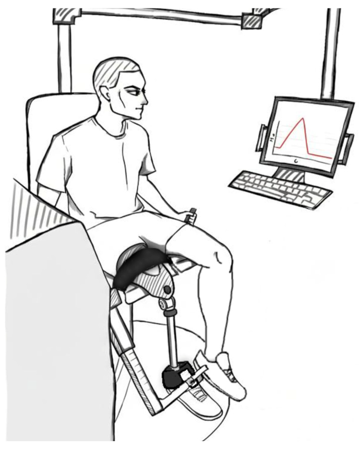
Participants were seated with the hip at ~90° and stabilised with standard straps (
Figure 1). The dynamometer axis was aligned with the lateral femoral condyle. Gravity correction was applied using the standard IsoMed procedure (passive limb weight recorded at 45° knee flexion).
For the amputated side, testing was performed with the prosthesis worn. The dynamometer lever arm was positioned according to each athlete’s prosthetic configuration: for athletes with a conventional prosthesis incorporating a defined tibial shank segment, the padded lever arm was secured to the distal aspect of the prosthetic shank; for athletes with a pylon-type configuration (pylonic rod and prosthetic foot, without an accessible shank), the lever arm was placed at the proximal residual limb above the prosthetic socket. In all cases, lever arm length was recorded individually and used for torque calculations, and coaxial alignment of the dynamometer axis with the lateral femoral condyle of the residual limb was maintained. Because torque on the amputated side is transmitted through the prosthetic socket–residual limb interface, measured peak torque reflects both the biological knee muscle output and the mechanical coupling characteristics of the individual prosthetic configuration; this is acknowledged as a limitation (
Section 4.4).
The intact limb was tested first. Angular velocities were tested in descending order (300, 240, 180, 60°·s−1). Each velocity block included brief familiarisation, then maximal efforts (10 reps at 300/240°·s−1; 5 reps at 180/60°·s−1) with 2 min rest between blocks. Range of motion was individualised (~10–100°) and kept constant across velocities and limbs.
2.3. Data Reduction and Outcome Measures
The primary outcome was gravity-corrected peak torque (PT), defined as the single highest torque value recorded across all valid repetitions within each velocity × movement direction block. PT was exported from IsoMed 2000 proprietary software and normalised to body mass (measured without prosthesis), yielding values in Nm·kg−1. All subsequent calculations were performed in Microsoft Excel Version 16.110.3 (Microsoft Corporation, Redmond, WA, USA).
Four outcome variables were derived. (1) Residual-limb PT deficit was quantified using a ratio-of-medians approach: Deficit (%) = [1 − median(PT pros·kg−1)/median(PT intact·kg−1)] × 100, calculated separately at each velocity and movement direction. (2) Individual inter-limb asymmetry (ILA) was computed for each athlete i as ILAi (%) = (1 − PT pros,i/PT intact,i) × 100, and the median [IQR] across athletes is reported alongside individual values. Note that the ratio-of-medians (outcome 1) and the median-of-individual ILAs (outcome 2) are arithmetically distinct and may differ, particularly at small n; both are reported to allow comparison with prior literature. (3) The conventional H/Q ratio was calculated as 100 × PT flex/PT ext (both concentric, Nm·kg−1) and expressed as a percentage (%), separately for each limb at each test velocity. The prosthetic-to-intact H/Q shift is expressed in percentage-point (pp) units as the within-subject difference (H/Q pros − H/Q intact); the group median [IQR] is reported. (4) The intact-limb PT reference is presented as median [IQR] across all eight athletes (n = 8) and is intended as an internal cohort benchmark only.
2.4. Statistical Analysis
Given the pilot and descriptive nature of the study, only descriptive statistics were calculated; no inferential hypothesis tests were applied. Participant characteristics are reported as mean ± SD. PT and H/Q outcomes are reported as median [IQR] to reflect the non-normal distribution expected in a small heterogeneous sample. Individual-level data are presented alongside group summaries throughout to preserve visibility of between-athlete variability—a deliberate choice given the pronounced inter-individual differences in residual limb length, prosthetic configuration, and training volume that characterise this population. The study is explicitly positioned as hypothesis-generating; effect sizes and confidence intervals will require a larger controlled sample in future work.
4. Discussion
This pilot study provides a multi-velocity (60–300°·s−1) isokinetic knee strength profile in competitive male para-athletes with unilateral TTA and documents four descriptive findings: substantial residual-limb PT deficits, velocity dependence of inter-limb asymmetry, an amputated-side H/Q upward shift at low-to-moderate velocities, and a monotonic intact-limb PT–velocity decline serving as an internal cohort reference. Overall, the results are consistent with a predominant extensor-deficit pattern alongside relative preservation of knee flexor torque on the prosthetic side. The discussion below contextualises these findings within the existing literature while acknowledging the descriptive and hypothesis-generating nature of the study.
4.1. Residual Limb Deficits and Velocity Specificity
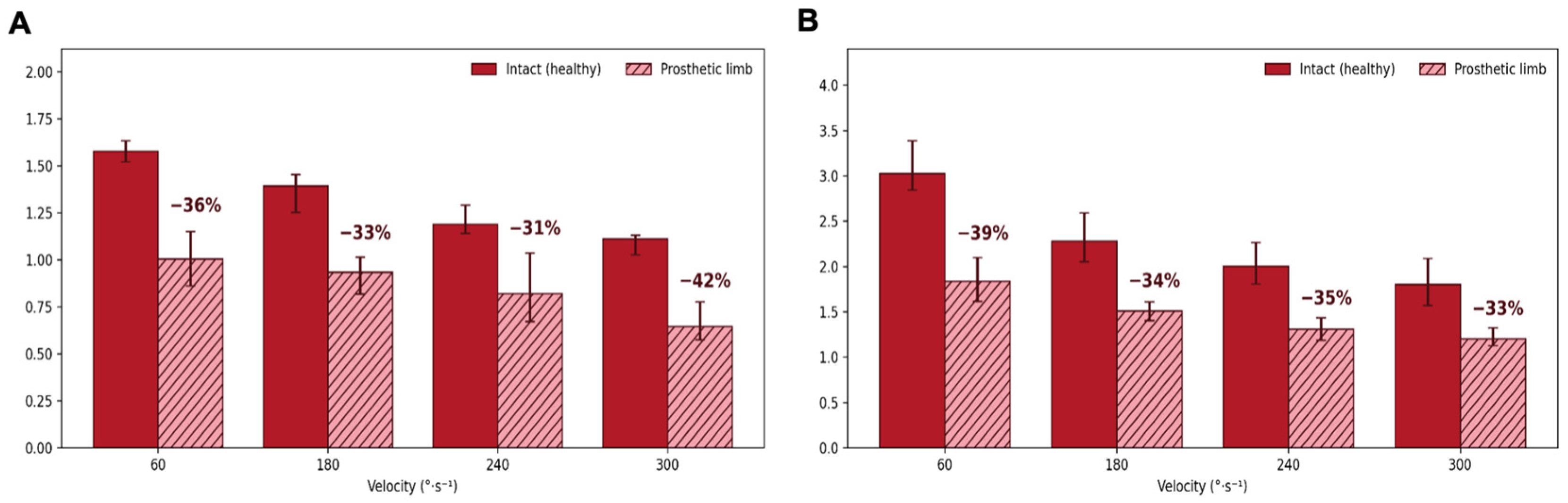
Amputated-side PT was lower than intact-limb PT across all velocities (flexion 31–42% lower; extension 33–39% lower), in the same direction as prior reports in unilateral TT amputees, where residual-limb extension and flexion are lower than the intact side [
1,
2]. Because cohorts, normalisation, and test velocities differ, we avoid direct numerical comparisons. Nolan [
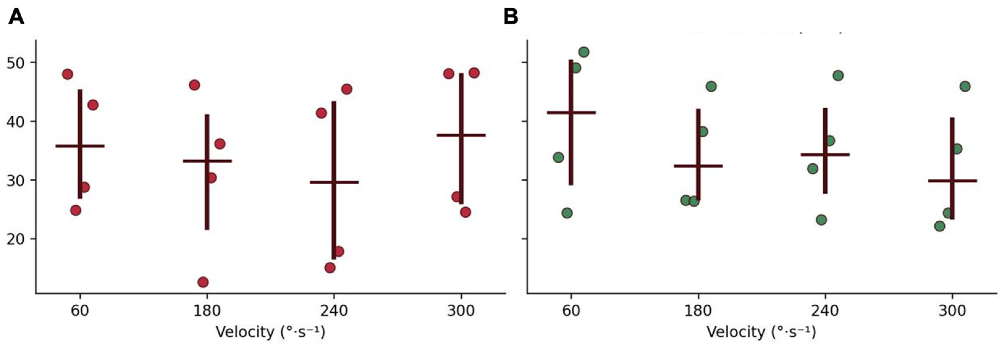
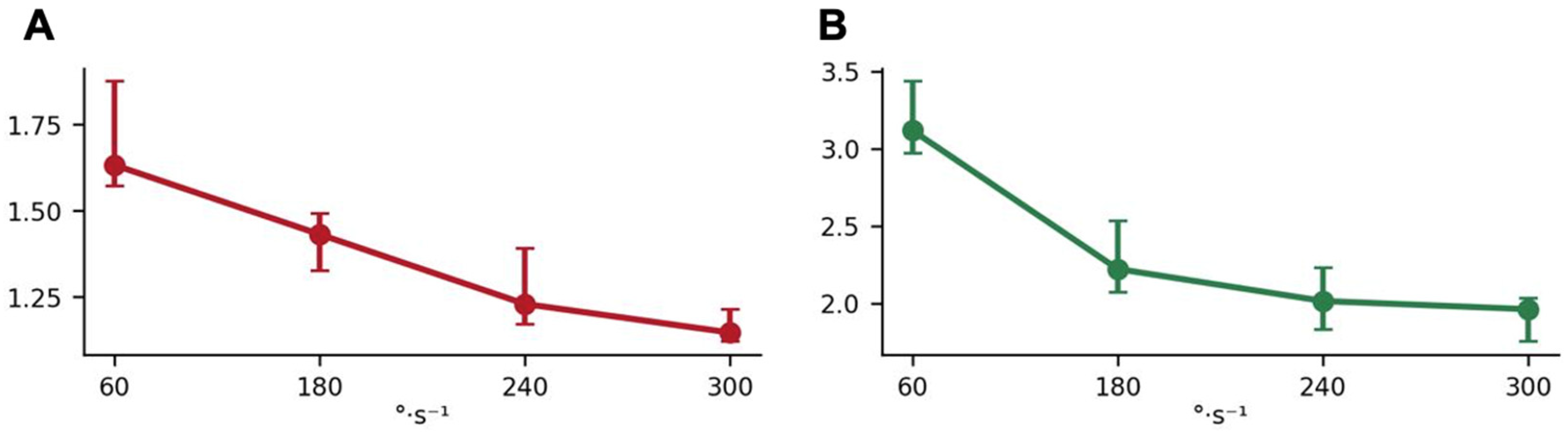
7] reported much smaller inter-limb asymmetry in sports-active TT amputees at the hip (8–14%) than in sedentary individuals (up to 49%); however, these are hip data and are not directly comparable to knee outcomes. In our knee data, ILA was velocity-dependent, with minima at 240°·s
−1 (flexion) and 300°·s
−1 (extension), highlighting information that would be missed by single-velocity testing.
4.2. H/Q Ratio Shift and Extensor-Dominant Deficit
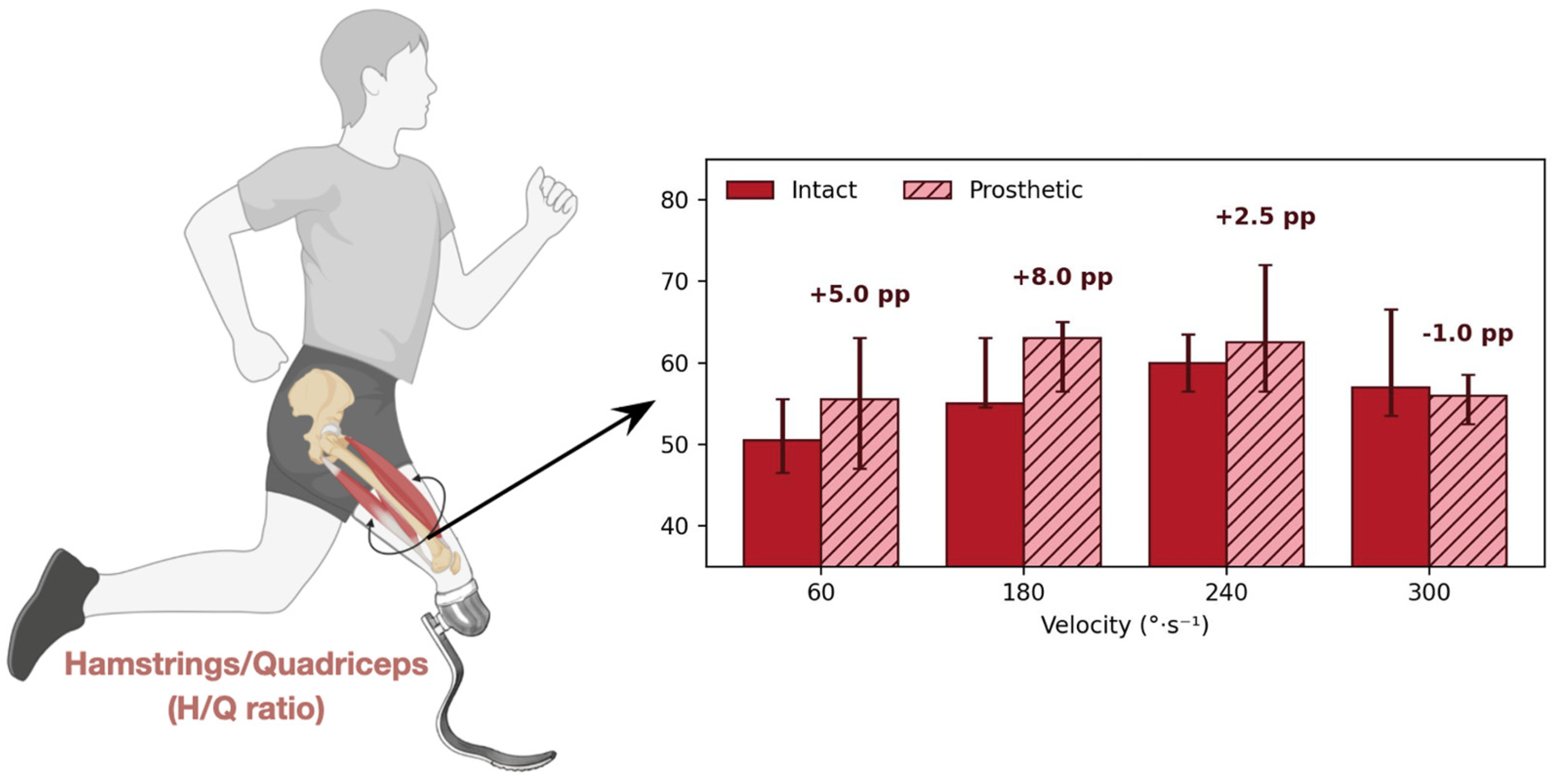
H/Q was modestly higher on the amputated side at 60–240°·s
−1 (+2.5 to +8.0 pp), a descriptive pattern compatible with relatively preserved knee flexor torque compared with extensor torque at low-to-moderate velocities. This direction is descriptively consistent with an extensor-dominant impairment reported in TT amputees [
1]. In able-bodied populations, a conventional concentric H/Q of approximately 0.60 is commonly cited [
8], while Aagaard et al. [
9] have argued that functional ratios incorporating eccentric contractions may better reflect dynamic joint stability. These considerations are relevant when interpreting the amputated-side H/Q shift: a ratio-level upward shift may appear favourable while reflecting a disproportionate extensor deficit and altered flexor–extensor balance. The reversal of the H/Q shift at 300°·s
−1 (−1.0 pp) is consistent with both flexor and extensor torque declining steeply at the highest velocity on the prosthetic side, partially equalising the ratio. The observed velocity dependence further supports the utility of multi-velocity profiling when characterising knee muscular balance in this population. Specifically, the asymmetry minima identified at 240°·s
−1 for flexion and 300°·s
−1 for extension may help define velocity-specific targets for future intervention studies. Such studies should test whether individualised, velocity-matched strengthening can reduce inter-limb imbalance and improve functional outcomes in para-athletes with TTA.
4.3. Intact-Limb Reference and Individual Variability
The intact-limb PT values provide an internal cohort reference that is more contextually appropriate for this para-athlete population than able-bodied normative datasets derived from different training backgrounds and body compositions. The monotonic PT decline with increasing velocity in the intact limb is consistent with the expected force–velocity profile [
6] and supports protocol fidelity. Individual ILA values showed substantial between-athlete variability, emphasising that group summaries may not capture the full spread of amputated-side impairment. Such variability likely reflects heterogeneity in residual-limb characteristics, prosthetic configuration, training exposure, and time since amputation. Accordingly, individual-level profiling is essential before translating group-level patterns into athlete-specific monitoring or future intervention targets.
4.4. Limitations
Several limitations should be acknowledged. First, the paired bilateral sub-sample was limited to n = 4. As this was a purely descriptive pilot study rather than a confirmatory hypothesis-testing study, no inferential statistical tests were performed or implied. All reported effect magnitudes should therefore be interpreted as preliminary exploratory estimates intended to guide future hypothesis-driven research. Second, the cross-sectional design provides no information about the trajectory of asymmetry over time or in response to training; no causal inferences about rehabilitation timing or programme efficacy are made or implied. Third, all athletes were recruited from a single training camp of one federation, limiting generalisability to other para-sport disciplines, classifications, and amputation aetiologies. Fourth, isokinetic concentric testing does not capture eccentric or isometric strength profiles, which may carry additional clinical relevance for joint loading and injury risk. Fifth, amputated-side testing was performed with the prosthesis in situ; measured peak torque on this side therefore reflects both the biological knee muscle output and the mechanical transmission characteristics of the individual prosthetic socket–residual limb interface. Prosthetic device characteristics (socket type, shank design, foot category, alignment) were not standardised across athletes, and this coupling effect may introduce additional between-athlete variability not present on the intact side.




 ,
,