
Skip to Content
You are currently on the new version of our website. Access the old version .

Medical Sciences Forum

Medical Sciences Forum is an open access journal dedicated to publishing findings resulting from academic conferences, workshops and similar events in all areas of medical sciences, for advances in basic, translational and clinical research and related disciplines. The conference organizers and proceedings editors are responsible for managing the peer-review process and selecting papers for conference proceedings.

Get Alerted

Add your email address to receive forthcoming volumes of this journal.

All Articles (865)

  • Proceeding Paper
  • Open Access

Tumor progression is frequently accompanied by intratumoral hypoxia caused by rapid proliferation and poor vascularization. Here, we investigated the effect of short-term (4 h) hypoxia induced by a nitrogen-enriched gas mixture on nanoparticle–cell association in highly metastatic MDA-MB-231, low-metastatic MCF-7, and non-tumorigenic MCF-10A breast cells. Short-term hypoxia significantly reduced nanoparticle uptake in all cell lines, with the strongest reduction observed in MCF-10A cells. These findings indicate that brief hypoxic exposure can differentially alter nanoparticle–cell interactions depending on cellular malignancy.

Med. Sci. Forum

10 November 2026
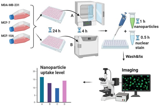
Experimental design. High-metastatic breast cancer MDA-MB-231 cells, low-metastatic breast cancer MCF-7 cells, and non-tumorigenic breast epithelial MCF-10A cells were cultured under normoxic conditions (21% O2, 5% CO2, 74% N2) and seeded into 384-well plates overnight. Subsequently, the plates were transferred either to a standard incubator (labeled A) or to a specialized chamber with a hypoxic nitrogen-enriched gas mixture (labeled B) containing 5% O2, 5% CO2, and 90% N2 for 4 h. Next, green fluorescent 200 nm nanoparticles were added for a total of 1 h, with a nuclear dye added after the first 30 min. Cells were then washed, fixed and processed for fluorescence microscopy. Nanoparticle encapsulation efficiency was quantified by calculating Pearson’s colocalization coefficient upon overlay of DAPI and GFP fluorescence channel images.
  • Proceeding Paper
  • Open Access

Bioinformatic Identification of ACTN4 as a Candidate Master Regulator of Local Invasion in Glioblastoma

  • Ekaterina Lindunen,
  • Margarita Pustovalova and
  • Yulia Merkher
  • + 1 author

Glioblastoma (GBM) is the most lethal brain tumor due to its highly infiltrative nature. Invasion requires mechanobiological processes including cytoskeletal remodeling and altered adhesion. We evaluated an 11-gene mechanobiological panel to distinguish GBM from low-grade gliomas (LGGs) and identify invasion drivers. Using TCGA mRNA expression data, we performed differential expression and ROC-AUC analyses. Of the four genes, FSCN1, TAGLN, and ACTN4 were significantly upregulated, whereas MYH9 was significantly downregulated in GBM (p < 0.01). ACTN4 alone achieved an AUC of 0.830, outperforming the full panel (AUC = 0.642) and the four-gene composite (AUC = 0.762). ACTN4 emerges as a promising diagnostic biomarker and potential master regulator of invasion, with its role in actin crosslinking supporting biological plausibility.

Med. Sci. Forum

26 October 2026

  • Proceeding Paper
  • Open Access

Assessing metastatic potential (MP) based on morphological parameters of cancer cells is a promising direction in experimental oncology. The shape of a cell often correlates with an invasive phenotype, characterized by greater elongation and decreased roundness. An analytical module based on the Omnipose neural network has been developed for automated morphometric analysis of cancer cells. The module performs cell segmentation on brightfield images and calculates eight morphological parameters (area, perimeter, axes, circularity, aspect ratio, roundness) without requiring special preprocessing or staining. Validation on 65 cells from breast cancer cell lines with high and low metastatic potential (MP) demonstrated high accuracy: the deviation from manual measurements did not exceed 5% for dimensional parameters and 11% for shape parameters. The module revealed statistically significant morphological differences between the high- and low-MP cells. The developed solution can serve as a foundation for the quantitative assessment of metastatic potential in experimental oncology.

Med. Sci. Forum

8 October 2026

  • Proceeding Paper
  • Open Access

Mpox can cause severe and fatal disease in people with advanced human immunodeficiency virus (HIV) disease, particularly when complicated by other infections. We report three hospitalized patients with advanced HIV disease, severe mpox, and tuberculosis diagnosed by positive urine lateral-flow lipoarabinomannan (LF-LAM) testing in Nigeria. A 44-year-old man with newly diagnosed HIV presented with extensive necrotizing cutaneous mpox, clinically suspected sepsis, and Staphylococcus aureus bacteraemia and died before antiretroviral therapy (ART) or tuberculosis treatment could be initiated. A 22-year-old woman with perinatally acquired HIV was receiving regular ART and had an HIV ribonucleic acid (RNA) level below 30 copies/mL. She developed severe nodulopustular mpox with genital involvement and improved following tuberculosis treatment while ART was continued. A 28-year-old woman who had interrupted ART for more than one year developed widespread mpox and deteriorated seven days after ART reinitiation, raising concern for suspected unmasking immune reconstitution inflammatory syndrome (IRIS). All three patients had positive urine LF-LAM results and PCR-confirmed mpox from vesiculopustular lesions and/or crusts. Molecular tuberculosis (TB) testing was not performed, and no culture or imaging was available to establish the anatomical extent of TB. These cases highlight the diagnostic complexity of severe mpox in advanced HIV disease and the importance of evaluating for concurrent opportunistic and bacterial infections.

Med. Sci. Forum

8 October 2026

Highly Accessed Articles

News & Conferences

Latest Volumes

Open for Submission

Journal Sections

XFacebookLinkedIn
Med. Sci. Forum - ISSN 2673-9992