Review

Interactions Between the Human Microbiome and Surgical Practice

by
Julia Kalinowski
1,2,
Nada Nasr
1,2,
Angelina Colamarino
1,2 and
Michelle Marie Esposito
1,2,3,*
1
Department of Biology, College of Staten Island, City University of New York, 2800 Victory Blvd., New York, NY 10314, USA
2
Macaulay Honors College, City University of New York, New York, NY 10023, USA
3
PhD Program in Biology, The Graduate Center, City University of New York, New York, NY 10016, USA
*
Author to whom correspondence should be addressed.
Hygiene 2026, 6(1), 10; https://doi.org/10.3390/hygiene6010010
Submission received: 26 December 2025 / Revised: 5 February 2026 / Accepted: 6 February 2026 / Published: 12 February 2026

Abstract

The human microbiome has emerged as a critical factor influencing surgical outcomes across a broad number of specialties. This paper explores the evolving interactions between the body’s microbiome and surgical practice, especially focusing on wound healing, post-operative recovery, and infection risk. We analyze current evidence from multiple surgical disciplines, including transplantation, reconstructive, bariatric, orthopedic, cardiothoracic, and oral surgery. Recognizing different microbial communities and their influence on surgical success opens doors to personalized medicine and targeted preventions. The studies discussed in this review provide valuable interdisciplinary insights that could ultimately uncover the microbiome’s fullest potential in optimizing future surgical protocols.

1. Introduction

The human microbiome encompasses a dynamic community of microorganisms that inhabit multiple organ systems and other mucosal tissues within the body, along with their genomes, metabolites, and environmental conditions [1]. The microbial communities (microbiota) within the microbiome include bacteria, fungi, viruses, and protozoa, with an estimated 38 trillion bacteria alone, indicating the vastness of this ecosystem [1,2]. Due to the complexity of the microbiome, any small disturbance can bring about catastrophic consequences. For example, a decrease in gut Bacteroides, a mutualistic bacterium responsible for digesting fibers into metabolites, can induce conditions such as Crohn’s Disease, and an increase can cause diabetes type 1, cardiovascular disease, and even autism [3,4]. Surgeries represent one of the most significant imbalances to these microbial networks by altering pathways, exposing to foreign objects, and administering peri/post-operative antibiotics. Radical distal gastrectomy, a treatment for gastric cancer, elevates the previously mentioned Bacteroides and decreases important short-chain fatty acids, disrupting nutrient absorption and causing greater mucosal permeability [5]. A recent genomic study based around spinal surgery introduced the idea that surgical site infections (SSIs) can arise from a patient’s own microbiota, with Escherichia coli (E. coli) being at low abundance preoperatively, then later dominating the infected wound [6]. Similarly, E. coli found at higher levels correlated with the incidence of post-pancreatectomy hemorrhage following pancreatic surgery [7]. A large number of these bacterial species have developed resistance toward antibiotics, with about 40–50% of today’s SSIs becoming resistant to prophylactic agents [8]. An important factor that influences this trend is the overprescription of antibiotics, unveiling a long-standing issue dominating the clinical world [8]. Anesthesia also has a measurable impact on microbial stability, as the loss of biodiversity and beneficial taxa leads to further dysbiosis [9]. All these examples combined display that surgical interventions have the capacity to influence long-term health outcomes by altering microbial community composition.
This review aims to focus on microbiome changes across several surgical disciplines, including reconstructive, transplantation, cardiothoracic, oral, bariatric, and orthopedic surgery. Our goal is to highlight how operative interventions could reshape microbiomes and how such patterns may guide future approaches in surgical care.

2. Materials and Methods

The process of creating this descriptive narrative literature review involved using the search engines Google Scholar and PubMed to evaluate relevant peer-reviewed research related to the topic at hand. The articles were selected based on relevance to microbiome changes following surgery, and preference was given to those published within the date range of January 2015 through December 2025 to emphasize studies published within the past ten years. Search terms included surgical microbiome, post-operative biofilms, and surgical site microbiota. Boolean operators (and/or) were used to refine the search and ensure coverage across relevant disciplines. Studies were included if they were peer-reviewed original articles or reviews, investigated microbiome composition or changes in relation to surgical procedures, surgical sites, or post-operative outcomes, and were published in English. Exclusion criteria included conference abstracts, editorials, non-peer-reviewed articles, studies deemed too outdated (outside the date range), studies unrelated to surgery, and publications lacking microbiome-related outcomes.
Article screening was conducted in a two-stage process, in which the titles and abstracts were reviewed first for relevance. Next, the full text was assessed to confirm eligibility based on the inclusion and exclusion criteria. Ultimately, analysis was performed as a qualitative narrative synthesis rather than a meta-analysis. Figures incorporated into this manuscript were created through Adobe Express and Canva.

3. Plastic, Reconstructive, and Cosmetic Surgery

As the body’s primary defense mechanism, the skin hosts a diverse microbiome that acts as a barrier from pathogens and plays a crucial role in regulating immune responses [10]. It is immensely disrupted within the context of surgery, specifically in procedures involving skin grafts, implants, and dermatological treatments. Such vulnerability can compromise the skin’s integrity and cause complications, such as delayed wound healing or surgical site infections [10]. Specifically, burn injuries cause continuous exposure to opportunistic microorganisms, leading to a heightened immune response and susceptibility to infections [11]. In affected areas, increased amounts of Staphylococcus aureus cause the emergence of host antimicrobial molecules and reduced levels of beneficial bacteria [11]. Propionibacterium acnes, normally responsible for suppressing pro-inflammatory cytokine expression, becomes impaired, causing diminished immune regulation and overgrowth of infectious species that can lead to pneumonia or sepsis [12]. The vulnerable skin also provides an excellent environment for thermophile bacteria, such as Aeribacillus, Caldalkalibacilus, and Nesterenkonia, which can delay wound healing [13]. Skin grafts, the primary technique used to reconstruct damaged tissue, rely heavily on a balanced microbial environment and epidermal homeostasis; therefore, the donor’s microbiome significantly influences the success of graft integration and recovery [12]. Given the importance of microbes in post-surgical healing, growing interest has emerged in using probiotics to support skin recovery [12]. Preliminary human studies suggest the use of probiotic-infused creams, such as those containing Streptococcus thermophilus, a facultative thermophile most known for its use in yogurt, can enhance the skin barrier by increasing ceramide levels in the stratum corneum that maintain flexibility and microbial defense [14]. Another potential solution that has been explored is a skin microbiome transplant, where the healthy skin of another individual is transferred to a patient who requires improvement in their skin’s microbiome [15].
Breast implant surgery, one of the most frequently performed reconstructive and cosmetic procedures, involves placing either silicone or saline implants beneath breast tissue and chest muscles. In the United States, around 450,000 breast implant operations are performed each year [16]. However, a major challenge presented in breast augmentation is the development of bacterial biofilms, specifically by bacteria such as Cutibacterium acnes and Staphylococcus epidermidis (Figure 1) [17]. Once established, these biofilms are difficult to eradicate and have been known to induce complications including capsular contracture, subclinical infection, and, in rare cases, breast-implant-associated large cell lymphoma (BIA-ALCL) (Figure 1) [17]. Around 48% of patients who undergo breast implant removal surgeries develop a subclinical infection [17]. Additionally, a higher level of biofilms could lead to breast implant illness and increased abundance of oxylipin 10-HOME [17]. Oxylipin 10-HOME, an immunogenic fatty-acid metabolite, is able to alter immune activity through activation of inflammation and impairment of normal tissue repair processes [18]. While the aforementioned Gram-positive organisms dominate the biofilm, Gram-negative bacteria may also play a role in the pathogenesis of BIA-ALCL. Through their lipopolysaccharides (LPS), they can activate toll-like receptor 4 (TLR-4), a pathway linked to chronic inflammation and cancer progression in other lymphomas (Figure 1) [19]. The signaling cascade leads to the release of IL-13 by mast cells, which has also been detected in BIA-ALCL tumor specimens, suggesting bacterial stimulation drives the immune environment for lymphoma development (Figure 1) [19].
Considered a cosmetic procedure, blepharoplasty is the removal of excess skin around the eyelids due to the weakening of the muscles with age [20]. Results from a study conducted noted that Proteobacteria were highly enriched in post-operative samples [20]. While not all Proteobacteria are pathogenic, they are most likely to cause diseases such as conjunctivitis or dacryocystitis, as well as systemic effects through connective tissue involvement [20]. Additionally, a decrease in Firmicutes and Bacteroidota exacerbates symptoms of dry eye in surgical patients [20]. In order to prevent disruptions in the ocular microbiome, certain measures should be adopted. For instance, in-depth patient histories and full medical examinations should be performed to understand risk factors that may predispose patients to postoperative diseases [21]. Proper surgical techniques are crucial, including adequate lubrication to minimize corneal abrasion or ulcerations, decreasing the inflammatory response by administration of dexamethasone or other topical steroids, and precise resection during surgery to prevent ectropion, where the inner eyelid is exposed, which can lead to excessive dryness [21].
Similarly, surgeries involving the sinuses introduce significant changes in microbial communities, particularly in patients undergoing functional endoscopic sinus surgery (FESS), an intervention for refractory chronic rhinosinusitis (CRS) to open nasal passages [22]. A study focusing on the response to FESS treatment highlighted the difference between “responders”, or those who saw significant improvement in their symptoms, versus “non-responders”, who gained little benefit [22]. It has been seen that elevated intra-nasal IL-5 and nasal polyps in patients with CRS are associated with symptomatic improvement after surgery [22]. This is due to the fact that their disease is IL-5 dependent, meaning IL-5 is the primary driver of eosinophilic inflammation, and FESS is more effective when the underlying inflammation is dominated by a targetable pathway [22,23]. Non-responders also have a higher abundance of S. aureus, which plays a significant role in the pathogenesis of CRS [22,24]. In CRS with nasal polyps, S. aureus exacerbates type 2 (Th2) inflammation, and its enterotoxins increase IgE concentration, which is a central antibody in allergic reactions [24]. Staphylococcus aureus has a major impact on the surgical outcome of non-responder groups, contributing to higher rates of post-operative inflammation [22]. After surgery, there is an increased amount of Acinetobacter, which has been seen to provide benefits as a commensal bacterium through its activation of IL-10, an anti-inflammatory cytokine aimed at reducing excessive immune response [22,25,26].
Vaginoplasty is a procedure performed for genital reconstruction and vaginal restoration due to congenital anomalies or trauma [27,28]. In gender-affirming vaginoplasty, a “neovagina” is typically constructed from penile inversion or intestinal tissue, also used in women with vaginal aplasia, to create a functional and aesthetically similar vaginal canal [27,29]. The microbiome of a neovagina often differs significantly based on the tissue source. Penile inversion vaginoplasty typically exhibits low amounts of Lactobacillus due to a lack of glycogen production and retained cornification [28,30]. Lactobacillus plays a crucial role in maintaining a healthy vaginal ecosystem, as it is responsible for lowering the vaginal pH and combating potential opportunistic pathogens by creating an acidic environment [31]. The neovagina constructed from a penile inversion has a greater resemblance to the foreskin fold of an uncircumcised penis rather than a cisgender vagina and therefore supports microbiota composed predominantly of Prevotella, Porphyromonas, and Peptoniphilus [30]. In a cisgender uncircumcised penis, these anaerobes are often linked to increased susceptibility to sexually transmitted infections, which should encourage studies to see if the same is true for a neovagina [30]. Similarly, an intestinal vaginoplasty presents an interesting dilemma, as the neovagina adopts the bacterial populations existing in the colon that are determined by diet [28]. Generally, the pH and original microbiota, with more facultative anaerobes, are retained from the colon after surgery, but the slight differences in composition and decreased bacterial counts suggest that dietary patterns influence microbial diversity [28]. Future research should be aimed at understanding long-term dietary habits and their effect on post-operative neovaginal microbial communities. Both require extensive post-operative care, such as oral probiotic supplementation to improve Lactobacillus levels and repeated rinsing to reduce the overgrowth of unwanted bacterial communities [31,32].

4. Transplantations

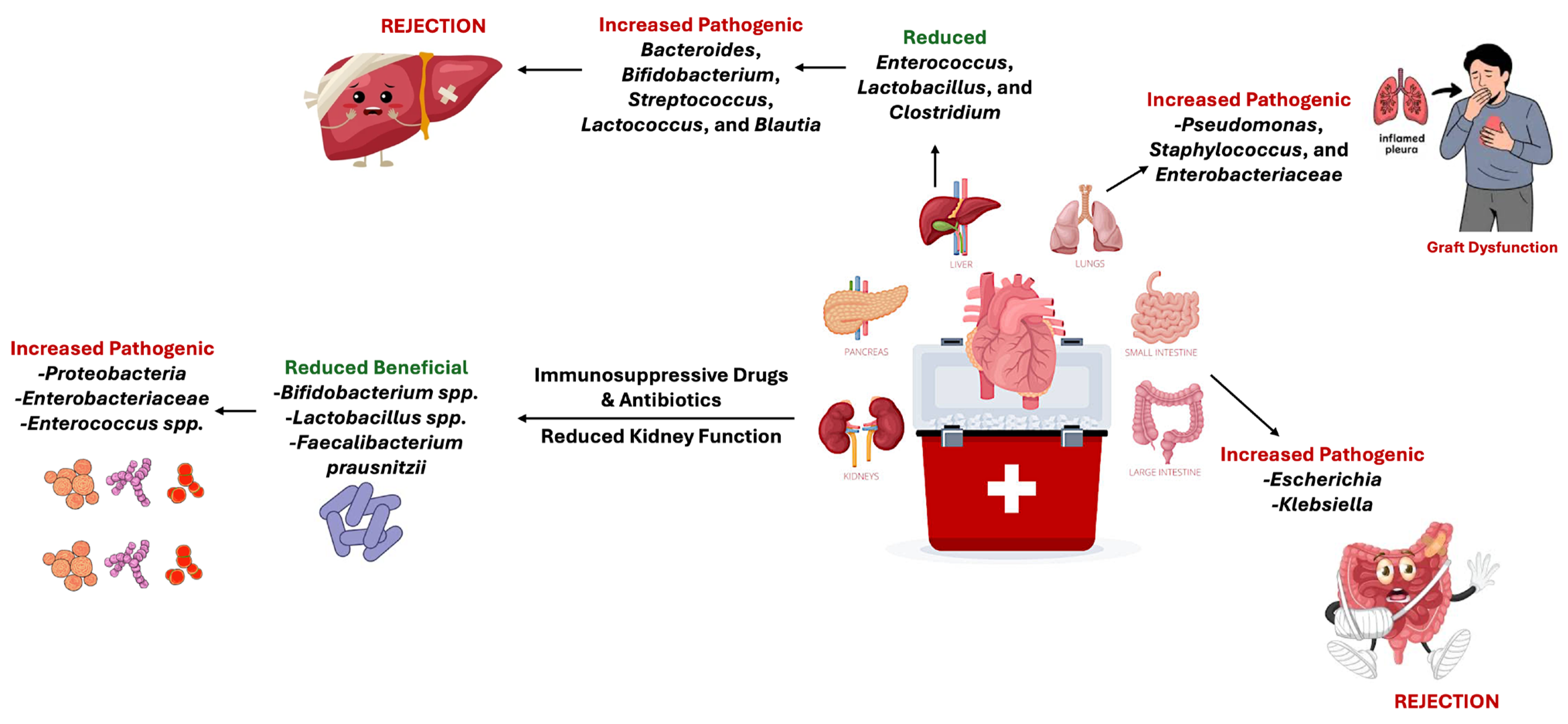
Organ transplantation offers one of the clearest examples of how the microbiome influences surgical success, since every graft triggers a complex interaction between donor tissue, immune defenses, and resident microbes that governs inflammation, healing, and immune tolerance [33]. Renal transplantation is the primary treatment for end-stage renal disease (ESRD), and it introduces significant immune and microbial challenges. Following surgery, the human microbiome undergoes rapid disruption due to immunosuppressive drugs, antibiotic exposure, and reduced kidney function [33]. These alterations in the oral, gut, and urinary microbiomes increase susceptibility to infection, inflammation, and graft injury. Major changes include declines in beneficial genera, such as Bifidobacterium and Lactobacillus, and expansion of pathogenic taxa, such as Enterobacteriaceae and Enterococcus spp., which are associated with post-transplant diarrhea and immune activation [34]. Similar findings have been observed in other transplant models [35]. A large-scale liver transplant study reported reductions in beneficial bacteria, such as Bifidobacterium spp., Lactobacillus spp., and Faecalibacterium prausnitzii, and increased levels of aforementioned pathogenic taxa across patients receiving cyclosporine, tacrolimus, and mycophenolate mofetil (Figure 2) [34,35]. Consistent trends across transplant studies include decreased microbial diversity and increased relative abundance of Proteobacteria, indicating a decline in microbial stability after transplantation [35]. Although immunosuppressive regimens are indispensable for preventing rejection, they may compromise long-term microbiota resilience and diversity [33].
Restoring microbial balance after transplantation remains an evolving therapeutic challenge, as several studies report persistent dysbiosis, an imbalance of the body’s microbiota, even after successful graft implantation [33]. Although fecal microbiota transplantation is well established for the treatment of Clostridioides difficile infection, its application in transplant recipients is still not fully understood [34]. A study comparing the urinary microbiome of 21 kidney transplant recipients and 8 healthy controls revealed a marked decrease in microbial diversity among transplant patients, along with a higher abundance of opportunistic pathogens, particularly Escherichia coli and Enterococcus faecalis (Figure 2) [36]. These alterations are clinically significant because they are associated with increased risk of urinary tract infections, which then lead to treatment practices that may result in species that have increased antibiotic resistance [36]. Elevated urinary urea concentrations following transplantation may also promote the growth of urealytic pathogens, further exacerbating infection risk [36]. Consequently, longitudinal monitoring of the urinary microbiome has been proposed as a potential non-invasive biomarker for early detection of graft dysfunction or rejection [36].
Distinct microbial signatures also correlate with tolerance and rejection across specific organ systems [33]. In liver transplant recipients, Bacteroides, Bifidobacterium, Streptococcus, Lactococcus, and Blautia are present in higher abundance among patients experiencing acute cellular rejection, whereas Enterococcus, Lactobacillus, and Clostridium are reduced in non-rejecting recipients (Figure 2) [33]. These findings align with a growing body of literature linking postoperative dysbiosis to graft outcomes [37,38]. Specifically, clinical studies on liver transplant recipients report that shifts in gut-microbiota composition, such as increased abundance of genera like Bacteroides, Streptococcus, Bifidobacterium, and Blautia, are associated with episodes of acute cellular rejection, whereas maintenance of a more stable and diverse community, often enriched in taxa like Lactobacillus or Clostridium, correlates with graft tolerance and better long-term outcomes (Figure 2) [38]. This pattern supports the hypothesis that microbiome disruption may promote alloimmune activation through altered metabolite production and compromised barrier function, indicating the gut microbiota as a potential biomarker and therapeutic target in transplant recipients [39].
Intestinal transplant recipients show elevated levels of Escherichia and Klebsiella in pre-rejecting and actively rejecting patients, while kidney recipients with spontaneous tolerance demonstrate enrichment of Janthinobacterium, Clostridia, Bacilli, and Lactobacillales (Figure 2) [8]. Members of genera like Escherichia and Klebsiella (Proteobacteria) often expand when the normal gut microbiome is disrupted (Figure 2) [39]. In transplant patients, the overgrowth of these potentially pathogenic bacteria is considered a hallmark of dysbiosis because it is associated with reduced microbial diversity and weakened colonization resistance [39,40]. This shift increases the likelihood of inflammation and translocation of bacterial components such as LPS, which can activate host immune pathways and contribute to graft rejection. In contrast, enrichment of commensal groups, such as Clostridia, Bacilli, and Lactobacillales, has been shown to support production of short-chain fatty acids, promote epithelial barrier integrity, and regulate immune responses, which are features associated with immune tolerance and improved graft outcomes [40].
Beneficial Lachnospiraceae correlate negatively with serum creatinine and blood urea nitrogen, indicating improved renal function [40]. Commensal-derived metabolites, such as short-chain fatty acids and inosine, have also been linked to prolonged graft survival in experimental models [41]. Microbial composition and metabolite activity play key roles in immune tolerance and post-transplant recovery across organ systems [41].
Further evidence of microbiome-mediated recovery is observed in liver transplantation, where graft implantation restores hepatic function and improves gut microbial balance [42]. Following surgery, reductions in pathogenic Enterobacteriaceae, such as Escherichia, Shigella, and Salmonella, were accompanied by increased abundance of beneficial Ruminococcaceae and Lachnospiraceae taxa [42]. These microbial shifts are associated with reduced endotoxin levels, higher bile acid activity, and lower venous ammonia concentration, reflecting restoration of gut–liver axis function following transplantation [42].
Microbiome disruption is also evident in pulmonary transplantation, a setting uniquely vulnerable to environmental and immunosuppressive stress [43]. After lung transplantation, reduced microbial diversity and expansion of opportunistic pathogens, including Pseudomonas, Staphylococcus, and Enterobacteriaceae spp., are often accompanied by heightened inflammation and immune activation, which contribute to chronic lung allograft dysfunction (CLAD), the leading cause of long-term graft failure (Figure 2) [43]. Conversely, recipients who maintain higher proportions of oral commensals, such as Streptococcus and Veillonella, tend to exhibit lower cytokine levels and improved graft stability, suggesting that microbial resilience supports immune tolerance [43].
The influence of microbial and viral disturbances on graft outcomes extends beyond allogeneic transplants, as shown in xenotransplantation studies, which involve transplanting organs across species, such as from pigs to baboons [44]. Porcine cytomegalovirus (PCMV) infection during pig-to-baboon cardiac xenotransplantation significantly shortened graft survival by increasing inflammatory cytokines like IL-6 and TNF-α and inducing coagulation dysfunction and endothelial injury [44,45]. In contrast, PCMV-free hearts functioned for nearly 200 days, demonstrating that even undetected viral presence can accelerate rejection [46]. These concerns closely translate to human clinical settings because porcine endogenous retroviruses (PERVs) are integrated into the pig genome and would be transplanted with the graft, raising the possibility of infection in recipients and potentially household contacts. Although the risk appears low due to genetically engineered pigs and pathogen-free breeding strategies, the potential for cross-species viral transmission remains a central regulatory barrier to human use [45]. Multiple studies cited in this review stress that careful monitoring and continued development of gene-editing tools such as CRISPR-Cas9, which may eliminate PERV sequences, are necessary before wider clinical adoption [46,47]. Additionally, research shows that systemic inflammation can precede coagulation abnormalities and graft deterioration in primate recipients, even when major immune barriers are genetically controlled, indicating that microbial or viral triggers may still provoke damaging immune cascades [48]. Viral contamination and infection-triggered inflammation continue to shape xenograft outcomes and highlight the critical role of microbial control in improving long-term surgical success [49].
A comparable disruption of microbial–immune balance is evident in allogeneic hematopoietic cell transplantation (HCT), one of the most established cellular therapies for hematologic cancers. Intensive pre-transplant therapies, including chemotherapy, radiation, and antibiotic use, have been shown to damage the intestinal barrier and disturb microbial balance, creating a dysbiotic environment dominated by taxa such as Enterococcus, Klebsiella, E. faecium, and Streptococcus [50]. This imbalance weakens colonization resistance and allows bacterial translocation, leading to complications such as bacteremia and graft-versus-host disease (GvHD), which manifests as injury to the skin, gastrointestinal mucosa, and liver due to an exaggerated immune response from donor cells against host tissues [50,51]. Patients with low intestinal diversity after transplantation have significantly higher transplant-related mortality and greater susceptibility to GvHD, with three-year survival rates of only 36% compared to 67% among those with higher diversity [52].
In contrast to pathogenic microbes, beneficial anaerobes such as Bacteroides, Clostridium, Bifidobacterium, and Blautia produce short-chain fatty acids, such as butyrate and propionate, which strengthen epithelial integrity, regulate immune signaling, and promote mucosal repair [50]. These microbial metabolites are essential for restoring intestinal homeostasis and reducing cytokine-mediated tissue injury caused by TNF-α, IL-1, and IL-6 [50,51]. Therapeutic strategies that preserve or restore microbiome stability, including selective antibiotic use, dietary interventions, prebiotic and probiotic supplementation, and fecal microbiota transplantation, have been linked to re-establishing microbial diversity and enhancing immune tolerance in experimental and clinical studies [50].
Across transplant disciplines, the microbiome functions as a key regulator of immune balance. It influences both innate and adaptive immune pathways through interactions with pattern-recognition receptors such as Toll-like receptors (TLRs) and NOD-like receptors (NLRs), which modulate cytokine release and guide T-cell differentiation toward either regulatory or effector responses [53]. Beneficial microbial metabolites, including short-chain fatty acids like butyrate, promote T-regulatory cell activity and epithelial integrity, fostering immune tolerance and recovery. In contrast, dysbiosis caused by antibiotics, chemotherapy, or surgical stress drives inflammatory cascades mediated by TNF-α, IL-1β, and IL-6, increasing the risk of graft rejection and delayed healing [53]. Collectively, the microbiome is not merely altered as a consequence of surgery, but actively drives surgical outcomes by shaping inflammation, immune tolerance, and long-term graft survival across diverse transplant settings [53].

5. Cardiothoracic Surgery

Cardiovascular disease has been on the rise in recent years, resulting in the growing demand for surgical intervention [54]. Approximately 11,000 cases were recorded in 2014, with an anticipated 47% increase in the number of surgeries by the year 2040 [55]. The gut microbiome plays a crucial role in determining the success of cardiothoracic surgery, as the gut microbiome has been linked to postoperative complications and prognosis [56]. Major surgeries like these put greater stress on the physiology of the body, disrupting the gut and immune systems and making them more susceptible to postoperative bacterial infections [57]. The reduction in immune efficiency, or postoperative immune suppression, can be caused by intraoperative factors, including anesthesia, ischemia, and cardiopulmonary bypass, all of which have the capability of leading to a systemic inflammatory response [56].
In cardiothoracic surgery, cardiopulmonary bypass (CPB), the respiratory support, results in widespread inflammation of the body, known as systemic inflammatory response syndrome (SIRS) [58]. Systemic inflammatory response syndrome is caused by trauma during the operation, ischemia, and the interaction between blood and the surface of the bypass machine [58]. Other factors that contribute to this syndrome include genetics, smoking, and high stress levels [58]. This inflammatory response causes post-surgical complications to arise, including respiratory failure, multi-organ failure, and even neurological debilitation. It has also been observed that patients with SIRS require longer intensive care unit stays, a greater need for extended ventilation, and inotropic support [58].
In a study involving 67 patients receiving cardiovascular surgery in Tongji Hospital in China, systemic inflammatory responses were observed preoperatively on days one and seven [56]. During postoperative day one, Bacteroidetes and Actinobacteria were prevalent, and Lachospiraceae, Ruminococcaceae, and Lactobacilliaceae were present on both the preoperative and postoperative day one [56]. A prevalence of Bacteroidetes and some non-pathogenic Actinobacteria was observed, and these are considered generally beneficial for the decomposition of food and for making vitamins for the body [59]. However, an imbalance of these gut microbiota leads to infections, inflammation, and digestive issues, including irritable bowel syndrome [60]. In addition, Enterococcus was increased after surgery, up until week one [56]. Some species of Enterococcus can be used as probiotics; however, too much of the gut microbiota can result in urinary tract infections and even heart failure [61,62]. This trend correlates with gut dysbiosis and increased systemic inflammation. It commonly exhibits antibiotic resistance and is frequently implicated in hospital-acquired infections [56]. Postoperative inflammatory responses are correlated to respiratory failure, multiple organ failure, and myocardial dysfunction [56]. This study demonstrated that cardiovascular surgery was accompanied by significant gut microbiome dysregulation, contributing to heightened systemic inflammation [56].
While systemic inflammation plays a crucial role in postoperative complications, localized surgical site infections (SSIs) result from microbiome-related influences in cardiothoracic surgeries [63]. Surgical site infection occurs 30 to 90 days following a cardiothoracic operation [63]. The most common infection after cardiac surgery is mediastinitis, which is diagnosed with a positive microbiological test of mediastinal tissue [64,65]. The microorganisms found in affected patients are Gram-positive bacteria, such as coagulase-negative Staphylococcus and S. aureus [63]. Pathogenic Staphylococci are present on the skin and can severely affect a patient’s life expectancy, as they can enter the bloodstream and infect the heart valves [63]. Symptoms of SSI include redness, chest pain, fever, and purulent drainage. Several pre-existing comorbidities increase the risk of postoperative infection, including obesity, chronic obstructive pulmonary disease, smoking, diabetes, larger breast size, and female sex [63]. Preventive strategies focus on reducing these risk factors prior to surgery through nutritional support in patients with hypoalbuminemia, smoking cessation, and targeted weight management approaches [63].
Other postoperative cardiothoracic surgical infections are associated with medical devices and environmental contamination, such as severe infections from Mycobacterium chimaera [66]. M. chimaera infections are associated with high infection and mortality rates due to late diagnosis [66,67]. This pathogen is primarily associated with pulmonary infections and is classified as a subspecies of Mycobacterium intracellulare [66]. Clinical manifestations commonly include chorioretinitis, fatigue, fever, and night sweats, and the infection has also been linked to auto-inflammatory disease. This infection has been linked to surgical site infections from heater-cooler units (HCUs) manufactured by LivaNova 3T HCDS [66]. It was reported that infectious aerosols, particles containing pathogens in the air, contaminated operating rooms and violated sterile fields, exposing patients to infections [68]. To combat this issue, HCUs were removed from operating rooms. M. chimaera is also present in other medical devices, such as thermoregulatory devices, that are not associated with HCUs [66]. Treating M. chimaera disease poses many challenges, as the bacteria form biofilms on artificial materials used during surgery, which resist antibiotics [66,69]. In response, the International Society of Cardiovascular Infectious Diseases (2020) issued guidelines for diagnosing and preventing HCU infections, recommending combination therapy with rifamycin and ethambutol and reoperation to remove prosthetic material when indicated [66].
To understand the severity of M. chimaera infection, data were extracted from microbiological specimens, antimycobacterial therapy, observation time, mortality, and HCU-intervened cardiac surgeries from the onset of first symptoms [66]. Among 180 patients from 54 different publications, the majority underwent aortic valve replacement (67.0%), aortic valve and root replacement (15.3%), and ascending aorta replacement (9.7%) [66]. Overall, 92.2% of patients received Nontuberculous Mycobacteria therapy consistent with guidelines [66]. This study highlighted a fatality rate of 45.5% and a median survival time of 24 months from the start of antimycobacterial therapy [66]. Reoperation also resulted in higher survival rates. The delayed onset of infection and late initiation of intervention and therapy also contributed to mortality. Therefore, this study emphasizes the need for surgeons to be more aware of the infection and the timing of treatment [66].
Aside from localized M. chimaera infections, broader post-surgical complications in cardiac surgeries, such as thoracic aortic dissections (TAAD), are very common [66]. There are approximately 100,000 TAAD cases every year [66]. Studies have shown that this surgery correlates with a 70% increase in gastrointestinal complications, with high levels of Oscillibacter, Anaerotruncus, Clostridium, and Alistipes [66]. These microorganisms are associated with cardiovascular disease and aging [66]. This also leads to systemic inflammation through pathways like mTOR, the mechanistic target of rapamycin [70]. MTORs control cell proliferation, survival, and metabolism, and can increase inflammation by increasing the pro-inflammatory response of cytokines [71]. Simultaneously, Eubacterium impairs immune regulation and intestinal barriers by increasing inflammatory markers, like white blood cells, cytokines, and neutrophils [66,72], which also delay recovery and alter liver function. Understanding the relationship between the gut microbiome and cardiac surgery can help mitigate the risks of post-surgical complications and optimize patient recovery and quality of life.

6. Oral Surgery

Oral surgery encompasses a variety of procedures aimed at treating diseases and structural abnormalities affecting the mouth, jaw, and facial structures. Both functional and aesthetic concerns are addressed, including correction of cleft palates or lips and management of oral malignancies, such as cancer [73,74].
Orofacial cleft surgeries are responsible for repairing congenital facial anomalies and are the cornerstone of oral and maxillofacial reconstruction [74]. An orofacial cleft occurs when the tissues of the upper lip and/or palate fail to fuse properly during fetal development, resulting in an opening that affects speech, dental health, hearing, and more [75]. Preoperatively, children are more susceptible to opportunistic pathogens, as a study presents 76.6% of infants exhibit colonization by Streptococcus mitis, Streptococcus aureus, and Klebsiella pneumoniae, which are particularly harmful [76]. S. aureus is a strain that is known for its antibiotic resistance, becoming difficult to eradicate [77]. S. mitis has been associated with severe infections such as bacteremia and infective endocarditis while also causing macrophage and epithelial cell death [76]. K. pneumoniae, the most recovered bacterial species in surgical site infections, has also been detected in inflammatory responses affecting the urinary tract, bloodstream, and respiratory tract [76,78]. Cleft palate surgery has the ability to produce an oro-nasal fistula (ONF) after the procedure, which has been seen to occur in 60% of pediatric cases [79]. An oro-nasal fistula is an abnormal opening that forms between the nasal passage and oral cavity, often as a result of surgical infection, that gives rise to the colonization of different pathogenic taxa, such as E. faecalis, Staphylococcus lentus, and Staphylococcus xylosus [79,80]. These organisms are implicated in persistent inflammation, periodontitis, and caries formation, bringing detrimental effects to the delicate balance of the oral microbiome [79]. Specifically, E. faecalis is of particular concern, as it creates biofilms that are highly resistant to antibiotics and has the capacity to withstand harsh conditions [81]. Its persistence in the mouth induces adherence to extracellular matrix proteins, enabling it to infiltrate deep mucosa and, in turn, makes E. faecalis hard to eliminate [81]. Despite these challenges, patients have seen a decrease in harmful bacteria 12 months after surgery, indicating that the restoration of normal anatomical barriers has the capacity to improve oral function [74]. In addition to the oral microbiome, the intestinal microbiome is also heavily altered by palatoplasty, as the use of cefazolin disrupts gut microbial diversity [82]. Cefazolin is one of the most commonly prescribed prophylactic antibiotics that is aimed at reducing the risk of post-surgical infection following palatoplasty [82,83]. The reduction of the genera Bifidobacterium sp., Lactobacillus sp., and Bacteroides sp. in fecal samples after cefazolin administration corresponds to a loss of commensal gut bacteria that are essential in maintaining metabolism and preventing pathogen colonization [82]. However, other factors following palatoplasty must also be considered, such as surgical stress and diet changes, in order to fully understand microbial disturbances and their effect on gut homeostasis [82].
Similar shifts in microbiota can be observed in surgery for oral cancers, where the removal of cancerous tissues has the potential to alter the environment [73]. In squamous cell carcinomas, an aggressive malignant cancer, affected areas of the mouth require excision, including part of the mandible [73,84]. In head and neck carcinomas, the trend of microbial abundance shifts toward those that resemble healthy oral cavities, such as Streptococcus, Rothia, and Gamella [85]. Certain species in these three genera are predominantly present in a normal oral cavity and contribute to maintaining a balanced ecosystem through colonization resistance from pathogens and mucosal stability [85,86]. Their return post-treatment suggests partial restoration after the excision of diseased areas [85]. However, complications following such a procedure can affect up to 60% of patients, and several beneficial bacteria could decrease as a result [73]. Neiserria, Porphyromonas, and Haemophilus, crucial nitrate-reducing bacteria, tend to reach levels lower than observed in normal controls [85]. The bacteria are involved in the nitrate–nitrite–nitric oxide pathway that produces NO2 and NO, two molecules that play key roles in antimicrobial defense and oral disease prevention [87]. With their decrease, the oral microbiome is compromised and can introduce serious post-operative consequences. Alternatively, patients can also receive therapeutic methods of cancer control; however, treatments such as chemotherapy are often an additional challenge [86]. Chemotherapeutic agents can increase vulnerability to opportunistic infections such as fungal overgrowth, bacterial invasion, and mucositis, which is inflammation of the mucosal tissue [86]. An emerging area of interest is the use of Lactobacillus brevis as a supportive therapy in the form of a lozenge to help promote healing of the oral mucosa [88]. The clinical use of L. brevis CD2 lozenges shows promising anti-inflammatory results and therefore opens doors for probiotic-based interventions in the context of oral health [88].
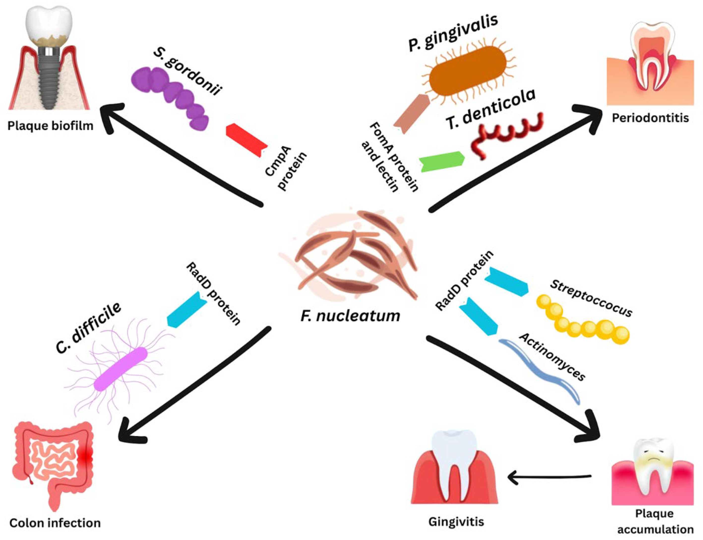
Implant-based surgeries introduce their own microbiological problems, particularly in the development of a condition known as peri-implantitis. Dental implants are intended to replace missing teeth using a titanium (or titanium alloy) fixture that integrates with the jawbone [89]. However, the successful placement of an implant depends heavily on the stability of the surrounding microbiome. Peri-implantitis results from the accumulation of biofilm, specifically a buildup of pathogenic species such as Fusobacterium nucleatum, around the area of the implant and induces severe inflammation, leading to significant bone loss [90,91]. F. nucleatum is of particular concern, as it acts as a “bridging organism”, using proteins to adhere to early and late colonizers of tooth surfaces that organize multi-species biofilms (Figure 3) [92]. F. nucleatum uses outer membrane protein RadD to bind to early colonizers Actinomyces and Streptococci, which are known to contribute to dental plaque and cause gingivitis [92,93]. Interestingly, RadD also allows F. nucleatum to become closely associated with Clostridioides difficile, a common species responsible for colon infection, through flagellal interaction (Figure 3) [94]. FomA and lectin, two groups of proteins, aid F. nucleatum in attaching to late colonizers Porphyromonas gingivalis and Treponema denticola, both implicated heavily as the masterminds behind periodontitis (Figure 3) [92,95]. Lastly, an important coaggregation protein, CmpA, links F. nucleatum to Streptococcus gordonii, a bacterium known to shape the initial biofilm community for later pathogen recruitment (Figure 3) [92,96]. This interspecies attachment accelerates the transition from a stable environment to a destructive inflammatory state. Unfortunately, peri-implantitis is difficult to eradicate, as F. nucleatum deeply penetrates the mucosal tissue, making both surgical and non-surgical methods only partially effective [97]. The most successful treatments are the removal of affected peri-implant pockets, ozone therapy, and implantoplasty, all of which showed some improvements by reducing inflammation but still fall short of fully eliminating tissue-invasive species [97]. Given these limitations, there is a clear need for the development of new strategies to manage pathogens involved in peri-implantitis more reliably.

7. Bariatric Surgery

Bariatric surgery has become one of the most effective interventions for severe obesity, producing outcomes far beyond what lifestyle treatment alone can achieve [98]. Large clinical reviews report long-term excess-weight loss between 47% and 70%, along with major reductions in obesity-related diseases and mortality [99]. These powerful benefits stem not only from anatomical change, but also from profound biological shifts, one of the most important being the transformation of the gut microbiome [100].
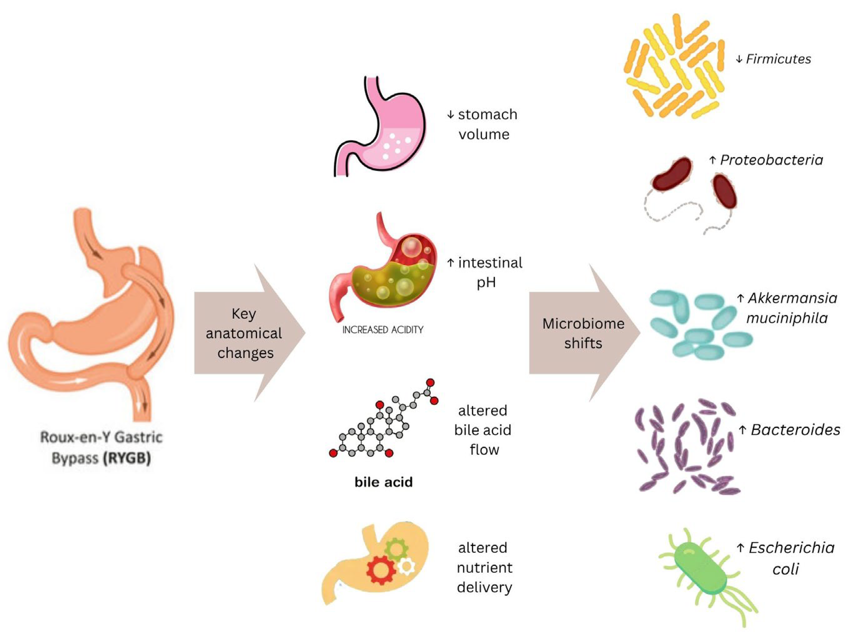
Bariatric surgery produces significant anatomical and metabolic changes that reshape the intestinal environment [101]. Procedures such as Roux-en-Y gastric bypass and sleeve gastrectomy alter stomach volume, gastric pH, bile acid flow, and nutrient exposure, all of which drive measurable microbial shifts [102,103]. Post-operative patients often show decreased Bacteroidetes, altered Firmicutes and Actinobacteria populations, and increases in Proteobacteria and species such as Akkermansia muciniphila, a mucin-decomposing bacterium of the phylum Verrucomicrobia. Higher levels of these bacteria are associated with a reduced likelihood of several disease conditions [103]. Akkermansia muciniphila resides in the mucus layer of the large intestine, where it is involved in maintaining intestinal integrity [104]. These shifts are further influenced by changes in bile acid metabolism and higher intestinal pH [104]. These microbial shifts have important implications for nutrient absorption, energy balance, inflammatory pathways, and the overall trajectory of postoperative weight-loss outcomes [101].
Postoperative bariatric surgery leads to rapid and measurable shifts in gut bacteria that appear to support weight loss and metabolic improvement [105]. For example, after Roux-en-Y gastric bypass, microbial richness increases significantly within 3 months, and this effect remains stable for 12 months and even up to 9 years in some patients [105,106]. Specific patterns repeatedly appear across studies: Bacteroides and Proteobacteria rise after surgery, while Firmicutes, which are elevated in obesity, consistently decrease [106]. These changes also link to patient outcomes (Figure 4) [106]. An increase in Faecalibacterium prausnitzii was associated with a reduction in low-grade inflammation in individuals with type 2 diabetes, while higher levels of Gammaproteobacteria correlated with greater weight loss [107]. The review also notes that increases in Escherichia coli after surgery may help the body harvest energy more efficiently during the “starvation-like” postoperative state [106]. Similarly, sleeve gastrectomy patients showed increases in Bacteroidetes and reductions in Firmicutes, with some species like Bacteroides thetaiotaomicron linked to decreases in body mass index and circulating glutamate levels [108]. Overall, these specific microbial changes suggest that the gut microbiome plays an active role in the metabolic benefits observed after bariatric surgery, beyond the mechanical effects of the procedure [108].
In a human cohort and mouse transfer model, microbiome alterations produced by sleeve gastrectomy reduced glucose-dependent insulinotropic polypeptide (GIP) release and protected against weight gain and non-alcoholic fatty liver disease [109]. Post-surgery microbial communities showed higher levels of Akkermansia, shifts in Bacteroides, and changes in tryptophan-related metabolites such as indolepropionate, which was negatively associated with GIP [110]. When stools collected after surgery were transplanted into antibiotic-treated mice, these animals developed less fat mass, lower hepatic triglyceride content, and reduced steatosis compared with mice colonized with pre-surgical microbiota, despite similar caloric intake [109,110]. This provides direct causal evidence that post-operative changes in specific microbes and their metabolites actively modify hormone signaling and liver pathology, contributing to the metabolic improvements seen after sleeve gastrectomy [110].
Bariatric surgery has also emerged as a therapeutic tool for improving non-alcoholic fatty liver disease (NAFLD) through microbiome-dependent pathways [111]. Post-surgical microbial patterns shift toward a healthier profile characterized by increased abundance of Fusobacteria, Veillonella, Streptococcus, Clostridium, Dorea, and Akkermansia, along with decreases in Blautia, Clostridiaceae, and Dorea, which are associated with dysbiosis in NAFLD patients [112]. These shifts are associated with a reduction in hepatic steatosis and inflammation and increasingly resemble the microbial signatures found in lean, metabolically healthy individuals. Increased microbial diversity and altered metabolic outputs, such as short-chain fatty acids and bile acids, play a critical role in restoring gut–liver axis function [110,111]. These changes enhance intestinal barrier integrity, improve insulin sensitivity, and promote favorable bile acid signaling, which together support NAFLD remission independent of weight loss [113]. Hence, bariatric surgery extends its impact beyond anatomical modification by transforming metabolic and hepatic signaling through the microbiome [113].
Despite these metabolic advantages, the postoperative microbiome does not always shift toward a uniformly beneficial state [114]. Post-operative microbial alterations can also give rise to several detrimental outcomes. Disruption of normal digestive physiology may lead to nutrient malabsorption, particularly of fats and micronutrients, and this shift in substrate availability can promote colonization by bacteria that metabolize unabsorbed nutrients, fostering intestinal inflammation and dysbiosis [115]. Clinical evidence shows that the loss of key protective genera such as Bacteroides, Lactobacillus, Bifidobacterium, and Akkermansia is associated with intestinal barrier dysfunction and increased susceptibility to gastrointestinal disease and pathogen colonization, while elevated Firmicutes and other opportunistic taxa contribute to inflammation and diarrheal infection [116]. Declines in these protective taxa weaken gut barrier integrity, reduce microbial gene richness, and promote the expansion of pro-inflammatory bacterial groups [116]. Destabilizing the gut ecosystem can impair immune homeostasis and promote infection [115,116]. In some cases, metabolic instability and weight regain can occur when surgery-induced microbial changes alter fermentation pathways and energy extraction, creating an unfavorable metabolic profile [114,115]. Microbial shifts have also been linked to reduced production of beneficial metabolites, including key short-chain fatty acids, and an increase in proteolytic fermentation that negatively affects gut health [116]. Although bariatric surgery improves obesity-related disorders, microbiome recovery after surgery is not uniform. Some patients experience only minimal or inconsistent increases in microbial diversity, and postoperative microbial patterns may remain unstable over time, introducing risks that require continued monitoring to maintain metabolic benefits [117].

8. Orthopedic Surgery

Orthopedic surgery entails the reconstruction of the musculoskeletal system after traumatic injury [118]. Types of surgeries include hip and knee arthroplasties, joint replacements, shoulder surgeries, fracture repairs, and spinal surgeries. Despite orthopedic surgeries focusing on diagnosis, treatment, and recovery, post-surgical complications can arise due to surgical site infections [119]. Risk factors, including pre-existing comorbidities such as poor nutrition, obesity, and diabetes, play a crucial role in the occurrence of SSIs. Surgical site infections include an increased risk of death, longer hospital stays, and higher healthcare costs [120]. More than 50% of patients with SSIs were five times more likely to be readmitted to the hospital following discharge and were two times more prone to death than those without post-surgical infections [121]. Other factors, including the duration of the surgery and blood loss, impact the presence and severity of SSIs [121].
A retrospective study was conducted in a teaching hospital in China from 2010 to 2012 to assess the efficiency of preoperative antibiotic prophylaxis and SSI risk factors [122]. A total of 2061 patients who underwent orthopedic surgery were included in this study. Forty-five patients developed a surgical site infection (2.18%) [122]. Additionally, 68.6% of the isolated bacteria showed resistance to cefuroxime, a drug used to treat bacterial infections [122]. Other factors that lead to post-orthopedic surgery SSIs include smoking, surgeries lasting over 3 h, and diabetes mellitus [121,123]. Studies reported that SSIs were more common in tumor orthopedic surgeries [122]. Out of 2752 patients, 14 (0.5%) had infections after oncologic surgeries, including soft-tissue and bone sarcoma. More SSIs were present in this surgical population from Enterococci and Pseudomonas aeruginosa, which are Gram-negative pathogens [122]. These are both opportunistic pathogens, as they resist ample antibiotics and are prevalent in hospital environments [124]. It was found that 10% of surgical patients develop Gram-negative SSIs from postoperative contaminations as well as poor personal hygiene [125].
Beyond SSIs, fracture-related infections (FRIs) also pose significant treatment challenges. Fracture-related infections after orthopedic surgeries have led to an increase in death rates and amputations, affecting up to 30% of all orthopedic surgical patients [125]. The most common microorganisms in SSIs include S. aureus (27.1%), Staphylococcus epidermidis (20.6%), and E. faecalis (13.6%) [125]. Approximately 30% of these bacteria are resistant to multiple drugs and beta-lactam antibiotic agents. The CDC notes that antibiotic resistance is due to medications being overly prescribed to patients [125]. With the significant rise in SSIs caused by S. aureus, great efforts have been made to find therapeutic and preventative methods to fight against this bacterium [126]. Topical antiseptics and specific systemic antibiotics have been used in an attempt to combat and decrease its severity [125]. However, the treatment of S. aureus remains challenging due to its rapid mutation rate and evolving resistance mechanisms.
In addition to fracture-related infections, periprosthetic joint infections (PJIs) are prevalent after orthopedic procedures, resulting in decreased bone strength [127]. After a total joint arthroplasty, PJI is the most common post-surgical infection, often leading to revision total knee arthroplasty surgeries [127]. This infection is primarily caused by S. aureus, which has prompted surgeons to adopt preventative measures, such as antibacterial strategies and reducing toxic effects on local tissues, to minimize bacterial presence during surgeries [127,128]. Despite these precautionary measures, sterile techniques, robotic surgery, and infection prevention protocols cannot fully eliminate microbiome contamination in the operating room. Unidirectional airflow systems have been shown to reduce intraoperative bacterial contamination [125]. Outside of the operative setting, several patient-related comorbidities increase the risk of PJIs, including smoking, malnutrition, diabetes, obesity, and hypertension [127]. A study performed on mice indicated that they were 50% more susceptible to developing PJIs when they already had a disrupted gut microbiome [127], resulting from a decreased host immune response to bacteria.
Microbiomes and infection rates have led to further studies in examining and understanding microbial populations after joint replacement surgeries. A study conducted between 2017 and 2019 examined the microbiomes present in the body after total knee arthroplasties [123]. A total of 113 patients were recruited for the study, and DNA sequencing technologies were utilized to examine the microbiomes in the joints of the osteoarthritis patients [123]. The results of this study concluded that the synovial fluid and tissue contained an array of different bacteria. Cutibacterium, Staphylococcus, Acinetobacter, Escherichia, and Pseudomonas were among the most prominent bacteria [123]. The findings indicate that prior administration of injections, as well as environmental cleanliness, influences the microbiome presence in the joint post-surgery [123,129]. Additionally, preclinical studies have highlighted how decreasing gut microbiomes has prevented bone loss after surgery resulting from estrogen depletion and glucocorticoid treatment [127]. Both estrogen, the hormone responsible for female reproductive and sexual function, and glucocorticoids, steroid hormones that suppress the immune system and regulate metabolic processes, play a crucial role in osteoporosis [130,131]. Collectively, these findings suggest that microbiome balance is crucial in enhancing outcomes after joint surgeries.
Disruption to microbial communities can result in the formation of opportunistic bacteria, forming biofilms and weakening the immune system [132]. Opportunistic pathogens, including S. aureus, Pseudomonas aeruginosa, and C. acnes, are the most common bacterial infections associated with orthopedic infections following implant replacements, like knee arthroplasties [132]. Pseudomonas aeruginosa forms biofilms, which impede skin healing by producing elastase and proteases at the surgical sites, delaying recovery and degrading tissue [133]. Additionally, S. aureus becomes pathogenic, impacting the skin barrier and allowing for the persistence of infection, weakening the human immune system and putting the patient at risk of serious infections [134]. Another factor that exposes patients to these bacteria is prolonged hospital stays. Extended hospital stays increase exposure to drug-resistant organisms, such as S. epidermidis and C. acnes [134]. Furthermore, overuse of antibiotics reduces normal skin microbiota, enabling the presence of antibiotic-resistant pathogens at infection sites. To help minimize bacterial contamination in orthopedic surgeries, metagenomics can assist in creating a personalized care plan to reduce a patient’s risk for surgical site infections [135]. Opportunistic pathogens and microbial balance play a major role in the risk for postoperative infections, highlighting the need for personalized strategies to prevent them in orthopedic patients.

9. Conclusions

The relationship between surgical practices and microbial profiles is an essential interaction worth exploring. Understanding the mechanisms in which these communities respond to operative disturbance provides crucial insight into improving post-surgical patient outcomes. The main sources of procedural infections are antibiotic-resistant strains, such as S. aureus, which has been mentioned in several disciplines throughout this review, that cause many issues related to surgical site infections [136]. As a Gram-positive bacterium, S. aureus forms dense biofilms and complicates treatment through resistance to multiple drugs [137]. Interestingly, a host’s own genetics can influence S. aureus pathogenicity, as toll-like receptors share the same domain as cytokine receptor IL-1RI, which, at low levels, has shown greater susceptibility to S. aureus infection in mouse models [137]. Both microbial adaptations and patient-specific factors shape infection risk, highlighting the urgent need for better preventative methods.
Many treatments have been recently explored in order to combat the issues involving surgical infections, such as targeted antimicrobial approaches, proper administration of prophylactic agents, and the development of strategies to reduce pathogen colonization. With regards to the gut microbiome, prebiotics, probiotics, and symbiotics are essential to the maintenance of beneficial genera while also suppressing opportunistic bacteria by strengthening the intestinal barrier, along with reducing overall inflammation through the reduction of IL-6 and CRP, two key markers indicated in post-operative stress [138]. Other methods involve optimal sterilization methods during surgery, such as minimizing operative time and adhering to aseptic techniques to limit microbial exposure [139]. Even the smallest changes, such as the use of triclosan-impregnated sutures, can induce measurable reductions in bacterial colonization [139]. Additional emerging studies have identified the connection between genetic variation and susceptibility to infection, suggesting that inherited differences in inflammatory pathways may influence individual risk [140]. New diagnostic measures might be the next step in addressing this vulnerability, as synovial fluid, for example, has been used as an indicator of periprosthetic joint infection (PJI) through biomarker identification [140]. This type of genetic identification should be further developed to assess genetic predispositions in surgical patients. Promising results can also be seen in phage therapies, where viruses infecting bacteria reduce bacterial load and reduce multidrug-resistant infections through the inhibition of quorum sensing and targeted bacterial lysis [141].
Ultimately, the human microbiome is an intricate network of many variables that can have significant implications for health and well-being, especially when people are vulnerable during and after surgical procedures. With the strategies explored in our review, we have demonstrated that continued research at the intersection of surgery, microbiology, and host genetics will be crucial for improving post-surgical recovery and limiting the burden of pathogenic microbes in clinical settings.

Author Contributions

All authors (J.K., N.N., A.C. and M.M.E.) contributed to writing. J.K and M.M.E. contributed to editing and reviewing. J.K. contributed to figure development. M.M.E. was responsible for project management and supervision. All authors have read and agreed to the published version of the manuscript.

Funding

This research received no external funding.

Institutional Review Board Statement

Not applicable.

Informed Consent Statement

Not applicable.

Data Availability Statement

Not applicable.

Acknowledgments

We would like to thank the CUNY College of Staten Island, the CUNY Graduate Center, and the CUNY Macaulay Honors College for their support in our research endeavors and professional development. We would also like to thank Charlie Esposito Kalinowski for his support throughout this manuscript’s development.

Conflicts of Interest

The authors declare no conflicts of interest.

References

  1. Dekaboruah, E.; Suryavanshi, M.V.; Chettri, D.; Verma, A.K. Human Microbiome: An Academic Update on Human Body Site Specific Surveillance and Its Possible Role. Arch. Microbiol. 2020, 202, 2147–2167. [Google Scholar] [CrossRef] [PubMed]
  2. Sender, R.; Fuchs, S.; Milo, R. Revised Estimates for the Number of Human and Bacteria Cells in the Body. PLoS Biol. 2016, 14, e1002533. [Google Scholar] [CrossRef] [PubMed]
  3. Madhogaria, B.; Bhowmik, P.; Kundu, A. Correlation between Human Gut Microbiome and Diseases. Infect. Med. 2022, 1, 180–191. [Google Scholar] [CrossRef] [PubMed]
  4. Zafar, H.; Saier, M.H. Gut Bacteroides Species in Health and Disease. Gut Microbes 2021, 13, 1848158. [Google Scholar] [CrossRef]
  5. Liang, W.; Yang, Y.; Wang, H.; Wang, H.; Yu, X.; Lu, Y.; Shen, S.; Teng, L. Gut Microbiota Shifts in Patients with Gastric Cancer in Perioperative Period. Medicine 2019, 98, e16626. [Google Scholar] [CrossRef]
  6. Long, D.R.; Bryson-Cahn, C.; Waalkes, A.; Holmes, E.A.; Penewit, K.; Tavolaro, C.; Bellabarba, C.; Zhang, F.; Chan, J.D.; Fang, F.C.; et al. Contribution of the Patient Microbiome to Surgical Site Infection and Antibiotic Prophylaxis Failure in Spine Surgery. Sci. Transl. Med. 2024, 16, eadk8222. [Google Scholar] [CrossRef]
  7. Biesel, E.A.; Sundheimer, J.; Badr, M.T.; Posadas-Cantera, S.; Chikhladze, S.; Fichtner-Feigl, S.; Wittel, U.A. Microbiological Colonization of the Pancreatic Tumor Affects Postoperative Complications and Outcome after Pancreatic Surgery. Front. Cell. Infect. Microbiol. 2025, 15, 1521952. [Google Scholar] [CrossRef]
  8. Long, D.R.; Alverdy, J.C.; Vavilala, M.S. Emerging Paradigms in the Prevention of Surgical Site Infection: The Patient Microbiome and Antimicrobial Resistance. Anesthesiology 2022, 137, 252–262. [Google Scholar] [CrossRef]
  9. Serbanescu, M.; Lee, S.; Li, F.; Boppana, S.H.; Elebasy, M.; White, J.R.; Mintz, C.D. Effects of Perioperative Exposure on the Microbiome and Outcomes from an Immune Challenge in C57Bl/6 Adult Mice. Anesth. Analg. 2025, 142, 171–180. [Google Scholar] [CrossRef]
  10. Gupta, S.; Poret, A.J.; Hashemi, D.; Eseonu, A.; Yu, S.H.; D’Gama, J.; Neel, V.A.; Lieberman, T.D. Cutaneous Surgical Wounds Have Distinct Microbiomes from Intact Skin. Microbiol. Spectr. 2023, 11, e03300-22. [Google Scholar] [CrossRef]
  11. Teymouri, S.; Pourhajibagher, M.; Bahador, A. The Relationship between the Skin Microbiome and Probiotics in the Healing of Burn Injuries. Folia Microbiol. 2025, 70, 535–544. [Google Scholar] [CrossRef]
  12. Plichta, J.K.; Gao, X.; Lin, H.; Dong, Q.; Toh, E.; Nelson, D.E.; Gamelli, R.L.; Grice, E.A.; Radek, K.A. Cutaneous Burn Injury Promotes Shifts in the Bacterial Microbiome in Autologous Donor Skin: Implications for Skin Grafting Outcomes. Shock 2017, 48, 441–448. [Google Scholar] [CrossRef] [PubMed]
  13. Yang, Y.; Huang, J.; Zeng, A.; Long, X.; Yu, N.; Wang, X. The Role of the Skin Microbiome in Wound Healing. Burn. Trauma 2024, 12, tkad059. [Google Scholar] [CrossRef] [PubMed]
  14. Bekiaridou, A.; Karlafti, E.; Oikonomou, I.M.; Ioannidis, A.; Papavramidis, T.S. Probiotics and Their Effect on Surgical Wound Healing: A Systematic Review and New Insights into the Role of Nanotechnology. Nutrients 2021, 13, 4265. [Google Scholar] [CrossRef] [PubMed]
  15. Callewaert, C.; Knödlseder, N.; Karoglan, A.; Güell, M.; Paetzold, B. Skin Microbiome Transplantation and Manipulation: Current State of the Art. Comput. Struct. Biotechnol. J. 2021, 19, 624–631. [Google Scholar] [CrossRef]
  16. Quesada, A.E.; Medeiros, L.J.; Clemens, M.W.; Ferrufino-Schmidt, M.C.; Pina-Oviedo, S.; Miranda, R.N. Breast Implant-Associated Anaplastic Large Cell Lymphoma: A Review. Mod. Pathol. 2019, 32, 166–188. [Google Scholar] [CrossRef]
  17. Whitfield, R.; Tipton, C.D.; Diaz, N.; Ancira, J.; Landry, K.S. Clinical Evaluation of Microbial Communities and Associated Biofilms with Breast Augmentation Failure. Microorganisms 2024, 12, 1830. [Google Scholar] [CrossRef]
  18. Bauer, T.M.; Gallagher, K.A. Biofilm-Derived Oxylipin 10-HOME Mediated Immune Response in Women with Breast Implants. J. Clin. Investig. 2024, 134, e176547. [Google Scholar] [CrossRef]
  19. Walker, J.N.; Hanson, B.M.; Pinkner, C.L.; Simar, S.R.; Pinkner, J.S.; Parikh, R.; Clemens, M.W.; Hultgren, S.J.; Myckatyn, T.M. Insights into the Microbiome of Breast Implants and Periprosthetic Tissue in Breast Implant-Associated Anaplastic Large Cell Lymphoma. Sci. Rep. 2019, 9, 10393. [Google Scholar] [CrossRef]
  20. Zhao, H.; Chen, Y.; Mao, H.; Ye, J.; Fu, M.; Qiu, J.; Zhang, Y.; Pan, E.; Xiong, K. Clinical Case-Control Study of Postoperative Ocular Microbiota Colonization Using Microbial Analysis in Patients Undergoing Blepharoplasty. Plast. Reconstr. Surg.-Glob. Open 2025, 13, e6876. [Google Scholar] [CrossRef]
  21. Zhang, S.-Y.; Yan, Y.; Fu, Y. Cosmetic Blepharoplasty and Dry Eye Disease: A Review of the Incidence, Clinical Manifestations, Mechanisms and Prevention. Int. J. Ophthalmol. 2020, 13, 488–492. [Google Scholar] [CrossRef]
  22. Hoying, D.; Sangwan, N.; Chaaban, M.R. Investigating the Role of the Sinus Microbiome and Cytokine Profile in the SNOT-22 Response After Functional Endoscopic Sinus Surgery in Chronic Rhinosinusitis Patients. J. Clin. Med. 2025, 14, 4446. [Google Scholar] [CrossRef] [PubMed]
  23. Gevaert, P.; Lang-Loidolt, D.; Lackner, A.; Stammberger, H.; Staudinger, H.; Van Zele, T.; Holtappels, G.; Tavernier, J.; Van Cauwenberge, P.; Bachert, C. Nasal IL-5 Levels Determine the Response to Anti–IL-5 Treatment in Patients with Nasal Polyps. J. Allergy Clin. Immunol. 2006, 118, 1133–1141. [Google Scholar] [CrossRef] [PubMed]
  24. Poddighe, D.; Vangelista, L. Staphylococcus aureus Infection and Persistence in Chronic Rhinosinusitis: Focus on Leukocidin ED. Toxins 2020, 12, 678. [Google Scholar] [CrossRef] [PubMed]
  25. Sivasubramaniam, R.; Douglas, R. The Microbiome and Chronic Rhinosinusitis. World J. Otorhinolaryngol.—Head Neck Surg. 2018, 4, 216–221. [Google Scholar] [CrossRef]
  26. Glocker, E.; Kotlarz, D.; Klein, C.; Shah, N.; Grimbacher, B. IL-10 and IL-10 Receptor Defects in Humans. Ann. N. Y. Acad. Sci. 2011, 1246, 102–107. [Google Scholar] [CrossRef]
  27. Li, J.S.; Crane, C.N.; Santucci, R.A. Vaginoplasty Tips and Tricks. Int. Braz. J. Urol. 2021, 47, 263–273. [Google Scholar] [CrossRef]
  28. Stoehr, J.R.; Moss, C.; Hamidian Jahromi, A. The Microbiome of the Neovagina: A Systematic Review and Comparison of Surgical Techniques. Int. J. Transgender Health 2024, 25, 623–633. [Google Scholar] [CrossRef]
  29. Faehnle-Schiegg, I.; Christmann-Schmid, C. Surgical Techniques for the Prolapse of Neovagina in Women: Case Report and Review of Literature. Int. J. Womens Health 2021, 13, 81–86. [Google Scholar] [CrossRef]
  30. Krakowsky, Y.; Potter, E.; Hallarn, J.; Monari, B.; Wilcox, H.; Bauer, G.; Ravel, J.; Prodger, J.L. The Effect of Gender-Affirming Medical Care on the Vaginal and Neovaginal Microbiomes of Transgender and Gender-Diverse People. Front. Cell. Infect. Microbiol. 2022, 11, 769950. [Google Scholar] [CrossRef]
  31. Liu, P.; Lu, Y.; Li, R.; Chen, X. Use of Probiotic Lactobacilli in the Treatment of Vaginal Infections: In Vitro and In Vivo Investigations. Front. Cell. Infect. Microbiol. 2023, 13, 1153894. [Google Scholar] [CrossRef] [PubMed]
  32. van der Sluis, W.B.; de Haseth, K.B.; Elfering, L.; Özer, M.; Smit, J.M.; Budding, A.E.; van Bodegraven, A.A.; Buncamper, M.E.; de Boer, N.K.H.; Mullender, M.G.; et al. Neovaginal Discharge in Transgender Women after Vaginoplasty: A Diagnostic and Treatment Algorithm. Int. J. Transgender Health 2020, 21, 367–372. [Google Scholar] [CrossRef] [PubMed]
  33. Campbell, P.M.; Humphreys, G.J.; Summers, A.M.; Konkel, J.E.; Knight, C.G.; Augustine, T.; McBain, A.J. Does the Microbiome Affect the Outcome of Renal Transplantation? Front. Cell. Infect. Microbiol. 2020, 10, 558644. [Google Scholar] [CrossRef]
  34. Wu, J.F.; Muthusamy, A.; Al-Ghalith, G.A.; Knights, D.; Guo, B.; Wu, B.; Remmel, R.P.; Schladt, D.P.; Alegre, M.; Oetting, W.S.; et al. Urinary Microbiome Associated with Chronic Allograft Dysfunction in Kidney Transplant Recipients. Clin. Transplant. 2018, 32, e13436. [Google Scholar] [CrossRef]
  35. Lee, J.R.; Magruder, M.; Zhang, L.; Westblade, L.F.; Satlin, M.J.; Robertson, A.; Edusei, E.; Crawford, C.; Ling, L.; Taur, Y.; et al. Gut Microbiota Dysbiosis and Diarrhea in Kidney Transplant Recipients. Am. J. Transplant. 2019, 19, 488–500. [Google Scholar] [CrossRef]
  36. Rani, A.; Ranjan, R.; McGee, H.S.; Andropolis, K.E.; Panchal, D.V.; Hajjiri, Z.; Brennan, D.C.; Finn, P.W.; Perkins, D.L. Urinary Microbiome of Kidney Transplant Patients Reveals Dysbiosis with Potential for Antibiotic Resistance. Transl. Res. 2017, 181, 59–70. [Google Scholar] [CrossRef]
  37. Abenavoli, L.; Scarlata, G.G.M.; Paravati, M.R.; Boccuto, L.; Luzza, F.; Scarpellini, E. Gut Microbiota and Liver Transplantation: Immune Mechanisms behind the Rejection. Biomedicines 2023, 11, 1792. [Google Scholar] [CrossRef]
  38. Sucu, S.; Basarir, K.E.; Mihaylov, P.; Balik, E.; Lee, J.T.C.; Fridell, J.A.; Emamaullee, J.A.; Ekser, B. Impact of Gut Microbiota on Liver Transplantation. Am. J. Transplant. 2023, 23, 1485–1495. [Google Scholar] [CrossRef]
  39. Duong, N.; Bajaj, J.S. The Impact of the Gut Microbiome on Liver Transplantation. Curr. Opin. Organ Transplant. 2021, 26, 587–594. [Google Scholar] [CrossRef]
  40. Medina, C.K.; Aykut, B. Gut Microbial Dysbiosis and Implications in Solid Organ Transplantation. Biomedicines 2024, 12, 2792. [Google Scholar] [CrossRef]
  41. Pirozzolo, I.; Li, Z.; Sepulveda, M.; Alegre, M.-L. Influence of the Microbiome on Solid Organ Transplant Survival. J. Heart Lung Transplant. 2021, 40, 745–753. [Google Scholar] [CrossRef] [PubMed]
  42. Bajaj, J.S.; Kakiyama, G.; Cox, I.J.; Nittono, H.; Takei, H.; White, M.; Fagan, A.; Gavis, E.A.; Heuman, D.M.; Gilles, H.C.; et al. Alterations in Gut Microbial Function Following Liver Transplant. Liver Transpl. 2018, 24, 752–761. [Google Scholar] [CrossRef] [PubMed]
  43. McGinniss, J.E.; Whiteside, S.A.; Simon-Soro, A.; Diamond, J.M.; Christie, J.D.; Bushman, F.D.; Collman, R.G. The Lung Microbiome in Lung Transplantation. J. Heart Lung Transplant. 2021, 40, 733–744. [Google Scholar] [CrossRef] [PubMed]
  44. Denner, J.; Längin, M.; Reichart, B.; Krüger, L.; Fiebig, U.; Mokelke, M.; Radan, J.; Mayr, T.; Milusev, A.; Luther, F.; et al. Impact of Porcine Cytomegalovirus on Long-Term Orthotopic Cardiac Xenotransplant Survival. Sci. Rep. 2020, 10, 17531. [Google Scholar] [CrossRef]
  45. Cooper, D.K.C.; Gaston, R.; Eckhoff, D.; Ladowski, J.; Yamamoto, T.; Wang, L.; Iwase, H.; Hara, H.; Tector, M.; Tector, A.J. Xenotransplantation—The Current Status and Prospects. Br. Med. Bull. 2018, 125, 5–14. [Google Scholar] [CrossRef]
  46. Denner, J. Porcine Endogenous Retroviruses and Xenotransplantation, 2021. Viruses 2021, 13, 2156. [Google Scholar] [CrossRef]
  47. Cowan, P.J.; Tector, A.J. The Resurgence of Xenotransplantation. Am. J. Transplant. 2017, 17, 2531–2536. [Google Scholar] [CrossRef]
  48. Iwase, H.; Ekser, B.; Zhou, H.; Liu, H.; Satyananda, V.; Humar, R.; Humar, P.; Hara, H.; Long, C.; Bhama, J.K.; et al. Further Evidence for Sustained Systemic Inflammation in Xenograft Recipients (SIXR). Xenotransplantation 2015, 22, 399–405. [Google Scholar] [CrossRef]
  49. Ezzelarab, M.B.; Ekser, B.; Azimzadeh, A.; Lin, C.C.; Zhao, Y.; Rodriguez, R.; Echeverri, G.J.; Iwase, H.; Long, C.; Hara, H.; et al. Systemic Inflammation in Xenograft Recipients Precedes Activation of Coagulation. Xenotransplantation 2015, 22, 32–47. [Google Scholar] [CrossRef]
  50. Van Lier, Y.F.; Van Den Brink, M.R.M.; Hazenberg, M.D.; Markey, K.A. The Post-Hematopoietic Cell Transplantation Microbiome: Relationships with Transplant Outcome and Potential Therapeutic Targets. Haematologica 2021, 106, 2042–2053. [Google Scholar] [CrossRef]
  51. Matsukuma, K.E.; Wei, D.; Sun, K.; Ramsamooj, R.; Chen, M. Diagnosis and Differential Diagnosis of Hepatic Graft versus Host Disease (GVHD). J. Gastrointest. Oncol. 2016, 7, S21–S31. [Google Scholar] [CrossRef] [PubMed]
  52. Chong, P.P.; Koh, A.Y. The Gut Microbiota in Transplant Patients. Blood Rev. 2020, 39, 100614. [Google Scholar] [CrossRef] [PubMed]
  53. Tabibian, J.H.; Kenderian, S.S. The Microbiome and Immune Regulation After Transplantation. Transplantation 2017, 101, 56–62. [Google Scholar] [CrossRef] [PubMed]
  54. Zilla, P.; Yacoub, M.; Zühlke, L.; Beyersdorf, F.; Sliwa, K.; Khubulava, G.; Bouzid, A.; Mocumbi, A.O.; Velayoudam, D.; Shetty, D.; et al. Global Unmet Needs in Cardiac Surgery. Glob. Heart 2018, 13, 293–303. [Google Scholar] [CrossRef]
  55. Lee, J.J.; Park, N.H.; Lee, K.S.; Chee, H.K.; Sim, S.B.; Kim, M.J.; Choi, J.S.; Kim, M.; Park, C.S. Projections of Demand for Cardiovascular Surgery and Supply of Surgeons. J. Chest Surg. 2016, 49, 37–43. [Google Scholar] [CrossRef]
  56. Xia, X.; Ni, J.; Yin, S.; Yang, Z.; Jiang, H.; Wang, C.; Peng, J.; Wei, H.; Wang, X. Elevated Systemic and Intestinal Inflammatory Response Are Associated with Gut Microbiome Disorder After Cardiovascular Surgery. Front. Microbiol. 2021, 12, 686648. [Google Scholar] [CrossRef]
  57. Giannoudis, P.V.; Dinopoulos, H.; Chalidis, B.; Hall, G.M. Surgical Stress Response. Injury 2006, 37, S3–S9. [Google Scholar] [CrossRef]
  58. Mc Loughlin, J.; Hinchion, J. The Gut Microbiome and Cardiac Surgery an Unusual Symphony. Perfusion 2023, 38, 1330–1339. [Google Scholar] [CrossRef]
  59. Miao, V.; Davies, J. Actinobacteria: The Good, the Bad, and the Ugly. Antonie Leeuwenhoek 2010, 98, 143–150. [Google Scholar] [CrossRef]
  60. Zhao, Y.; Zhu, S.; Dong, Y.; Xie, T.; Chai, Z.; Gao, X.; Dai, Y.; Wang, X. The Role of Gut Microbiome in Irritable Bowel Syndrome: Implications for Clinical Therapeutics. Biomolecules 2024, 14, 1643. [Google Scholar] [CrossRef]
  61. Im, E.J.; Lee, H.H.-Y.; Kim, M.; Kim, M.-K. Evaluation of Enterococcal Probiotic Usage and Review of Potential Health Benefits, Safety, and Risk of Antibiotic-Resistant Strain Emergence. Antibiotics 2023, 12, 1327. [Google Scholar] [CrossRef] [PubMed]
  62. Murray, B.E. The Life and Times of the Enterococcus. Clin. Microbiol. Rev. 1990, 3, 46–65. [Google Scholar] [CrossRef] [PubMed]
  63. Zukowska, A.; Zukowski, M. Surgical Site Infection in Cardiac Surgery. J. Clin. Med. 2022, 11, 6991. [Google Scholar] [CrossRef] [PubMed]
  64. Bor, D.H.; Rose, R.M.; Modlin, J.F.; Weintraub, R.; Friedland, G.H. Mediastinitis After Cardiovascular Surgery. Clin. Infect. Dis. 1983, 5, 885–897. [Google Scholar] [CrossRef]
  65. Lepelletier, D.; Bourigault, C.; Roussel, J.C.; Lasserre, C.; Leclère, B.; Corvec, S.; Pattier, S.; Lepoivre, T.; Baron, O.; Despins, P. Epidemiology and Prevention of Surgical Site Infections after Cardiac Surgery. Med. Mal. Infect. 2013, 43, 403–409. [Google Scholar] [CrossRef]
  66. Wetzstein, N.; Kohl, T.A.; Diricks, M.; Mas-Peiro, S.; Holubec, T.; Kessel, J.; Graf, C.; Koch, B.; Herrmann, E.; Vehreschild, M.J.G.T.; et al. Clinical Characteristics and Outcome of Mycobacterium Chimaera Infections after Cardiac Surgery: Systematic Review and Meta-Analysis of 180 Heater-Cooler Unit-Associated Cases. Clin. Microbiol. Infect. 2023, 29, 1008–1014. [Google Scholar] [CrossRef]
  67. Ditommaso, S.; Memoli, G.; Anselmi, F.; Molineris, I.; Zotti, C.M.; Giacomuzzi, M. The Global Outbreak of M. Chimaera Infection Following Cardiac Surgery: Another Piece of the Puzzle. Pathogens 2025, 14, 964. [Google Scholar] [CrossRef]
  68. Tang, J.W.; Li, Y.; Eames, I.; Chan, P.K.S.; Ridgway, G.L. Factors Involved in the Aerosol Transmission of Infection and Control of Ventilation in Healthcare Premises. J. Hosp. Infect. 2006, 64, 100–114. [Google Scholar] [CrossRef]
  69. Siddam, A.D.; Zaslow, S.J.; Wang, Y.; Phillips, K.S.; Silverman, M.D.; Regan, P.M.; Amarasinghe, J.J. Characterization of Biofilm Formation by Mycobacterium Chimaera on Medical Device Materials. Front. Microbiol. 2021, 11, 586657. [Google Scholar] [CrossRef]
  70. Perl, A. Activation of mTOR (Mechanistic Target of Rapamycin) in Rheumatic Diseases. Nat. Rev. Rheumatol. 2016, 12, 169–182. [Google Scholar] [CrossRef]
  71. Hwang, S.-K.; Kim, H.-H. The Functions of mTOR in Ischemic Diseases. BMB Rep. 2011, 44, 506–511. [Google Scholar] [CrossRef] [PubMed]
  72. Fu, Y.; Lyu, J.; Wang, S. The Role of Intestinal Microbes on Intestinal Barrier Function and Host Immunity from a Metabolite Perspective. Front. Immunol. 2023, 14, 1277102. [Google Scholar] [CrossRef] [PubMed]
  73. Gallant, J.-N.; Vivek, N.; McKeon, M.G.; Sharma, R.K.; Kim, Y.J.; Rosenthal, E.L.; Das, S.R.; Thomas, C.M. Establishing a Role for the Oral Microbiome in Infectious Complications Following Major Oral Cavity Cancer Surgery. Oral Oncol. 2024, 156, 106926. [Google Scholar] [CrossRef] [PubMed]
  74. Cocco, J.F.; Antonetti, J.W.; Burns, J.L.; Heggers, J.P.; Blackwell, S.J. Characterization of the Nasal, Sublingual, and Oropharyngeal Mucosa Microbiota in Cleft Lip and Palate Individuals before and after Surgical Repair. Cleft Palate Craniofacial J. 2010, 47, 151–155. [Google Scholar] [CrossRef]
  75. Kanmodi, K.K.; Atteya, S.M.; Elwan, A.H.; Adewole, I.; Akinsolu, F.T.; Abodunrin, O.R.; Olagunju, M.T.; Nwafor, J.N.; Aly, N.M.; Salami, A.A.; et al. Nutrition and Diet in Children with Orofacial Clefts in Africa: A Scoping Review. BMC Oral Health 2024, 24, 1341. [Google Scholar] [CrossRef]
  76. Chandrasekaran, S.; Rao, S.S.; Shetty, V. Association Between Oral Microbiome in Children Undergoing Palatoplasty and Surgical Site Infection. Cleft Palate Craniofacial J. 2025, 10556656251335181. [Google Scholar] [CrossRef]
  77. Oliveira, M.C.; Boriollo, M.F.G.; de Souza, A.C.; da Silva, T.A.; da Silva, J.J.; Magalhães-Guedes, K.T.; Dias, C.T.D.S.; Bernardo, W.L.d.C.; Höfling, J.F.; de Sousa, C.P. Oral Staphylococcus Species and MRSA Strains in Patients with Orofacial Clefts Undergoing Surgical Rehabilitation Diagnosed by MALDI-TOF MS. Pathogens 2024, 13, 763. [Google Scholar] [CrossRef]
  78. Abbas, R.; Chakkour, M.; Zein El Dine, H.; Obaseki, E.F.; Obeid, S.T.; Jezzini, A.; Ghssein, G.; Ezzeddine, Z. General Overview of Klebsiella Pneumonia: Epidemiology and the Role of Siderophores in Its Pathogenicity. Biology 2024, 13, 78. [Google Scholar] [CrossRef]
  79. Goudy, S.L.; Bradley, H.L.; Gacasan, C.A.; Toma, A.; Sekar, K.P.C.; Wuest, W.M.; Tomov, M.; Serpooshan, V.; Coskun, A.; Jones, R.M. Microbial Changes Occurring During Oronasal Fistula Wound Healing. Microorganisms 2025, 13, 327. [Google Scholar] [CrossRef]
  80. Sadhu, P. Oronasal Fistula in Cleft Palate Surgery. Indian J. Plast. Surg. Off. Publ. Assoc. Plast. Surg. India 2009, 42, S123–S128. [Google Scholar] [CrossRef]
  81. Kouidhi, B.; Zmantar, T.; Mahdouani, K.; Hentati, H.; Bakhrouf, A. Antibiotic Resistance and Adhesion Properties of Oral Enterococci Associated to Dental Caries. BMC Microbiol. 2011, 11, 155. [Google Scholar] [CrossRef] [PubMed]
  82. Vieira, N.A.; Borgo, H.C.; da Silva Dalben, G.; Bachega, M.I.; Pereira, P.C.M. Evaluation of Fecal Microorganisms of Children with Cleft Palate before and after Palatoplasty. Braz. J. Microbiol. Publ. Braz. Soc. Microbiol. 2013, 44, 835–838. [Google Scholar] [CrossRef] [PubMed][Green Version]
  83. Piccillo, E.M.; Farsar, C.J.; Holmes, D.M. Prophylactic Antibiotics After Cleft Lip and Palate Reconstruction: A Review From a Global Health Perspective. Cureus 2023, 15, e36371. [Google Scholar] [CrossRef] [PubMed]
  84. Imbesi Bellantoni, M.; Picciolo, G.; Pirrotta, I.; Irrera, N.; Vaccaro, M.; Vaccaro, F.; Squadrito, F.; Pallio, G. Oral Cavity Squamous Cell Carcinoma: An Update of the Pharmacological Treatment. Biomedicines 2023, 11, 1112. [Google Scholar] [CrossRef]
  85. Chan, J.Y.K.; Ng, C.W.K.; Lan, L.; Fung, S.; Li, J.-W.; Cai, L.; Lei, P.; Mou, Q.; Meehan, K.; Lau, E.H.L.; et al. Restoration of the Oral Microbiota After Surgery for Head and Neck Squamous Cell Carcinoma Is Associated with Patient Outcomes. Front. Oncol. 2021, 11, 737843. [Google Scholar] [CrossRef]
  86. Abranches, J.; Zeng, L.; Kajfasz, J.K.; Palmer, S.R.; Chakraborty, B.; Wen, Z.T.; Richards, V.P.; Brady, L.J.; Lemos, J.A. Biology of Oral Streptococci. Microbiol. Spectr. 2018, 6, 6.5.11. [Google Scholar] [CrossRef]
  87. Moran, S.P.; Rosier, B.T.; Henriquez, F.L.; Burleigh, M.C. The Effects of Nitrate on the Oral Microbiome: A Systematic Review Investigating Prebiotic Potential. J. Oral Microbiol. 2024, 16, 2322228. [Google Scholar] [CrossRef]
  88. Sharma, A.; Rath, G.K.; Chaudhary, S.P.; Thakar, A.; Mohanti, B.K.; Bahadur, S. Lactobacillus Brevis CD2 Lozenges Reduce Radiation- and Chemotherapy-Induced Mucositis in Patients with Head and Neck Cancer: A Randomized Double-Blind Placebo-Controlled Study. Eur. J. Cancer 2012, 48, 875–881. [Google Scholar] [CrossRef]
  89. Pokrowiecki, R.; Mielczarek, A.; Zaręba, T.; Tyski, S. Oral Microbiome and Peri-Implant Diseases: Where Are We Now? Ther. Clin. Risk Manag. 2017, 13, 1529–1542. [Google Scholar] [CrossRef]
  90. Ghensi, P.; Manghi, P.; Zolfo, M.; Armanini, F.; Pasolli, E.; Bolzan, M.; Bertelle, A.; Dell’Acqua, F.; Dellasega, E.; Waldner, R.; et al. Strong Oral Plaque Microbiome Signatures for Dental Implant Diseases Identified by Strain-Resolution Metagenomics. npj Biofilms Microbiomes 2020, 6, 47. [Google Scholar] [CrossRef]
  91. Espinoza-Arrue, J.; Arce, M.; Endo, N.; Hoare, A.; Dutzan, N.; Abusleme, L. Profiling the Bacterial Microbiome Across Peri-Implant Conditions. J. Clin. Periodontol. 2025, 52, 1662–1672. [Google Scholar] [CrossRef] [PubMed]
  92. Chen, Y.; Shi, T.; Li, Y.; Huang, L.; Yin, D. Fusobacterium Nucleatum: The Opportunistic Pathogen of Periodontal and Peri-Implant Diseases. Front. Microbiol. 2022, 13, 860149. [Google Scholar] [CrossRef] [PubMed]
  93. Ye, P.; Harty, D.W.S.; Chapple, C.C.; Nadkarni, M.A.; Carlo, A.A.D.E.; Hunter, N. Streptococci and Actinomyces Induce Antibodies Which Cross React with Epithelial Antigens in Periodontitis. Clin. Exp. Immunol. 2003, 131, 468–476. [Google Scholar] [CrossRef] [PubMed]
  94. Engevik, M.A.; Danhof, H.A.; Auchtung, J.; Endres, B.T.; Ruan, W.; Bassères, E.; Engevik, A.C.; Wu, Q.; Nicholson, M.; Luna, R.A.; et al. Fusobacterium Nucleatum Adheres to Clostridioides Difficile via the RadD Adhesin to Enhance Biofilm Formation in Intestinal Mucus. Gastroenterology 2021, 160, 1301–1314.e8. [Google Scholar] [CrossRef]
  95. Tan, K.H.; Seers, C.A.; Dashper, S.G.; Mitchell, H.L.; Pyke, J.S.; Meuric, V.; Slakeski, N.; Cleal, S.M.; Chambers, J.L.; McConville, M.J.; et al. Porphyromonas Gingivalis and Treponema Denticola Exhibit Metabolic Symbioses. PLoS Pathog. 2014, 10, e1003955. [Google Scholar] [CrossRef]
  96. Aravindraja, C.; Jeepipalli, S.; Duncan, W.D.; Vekariya, K.M.; Rahaman, S.O.; Chan, E.K.L.; Kesavalu, L. Streptococcus Gordonii Supragingival Bacterium Oral Infection-Induced Periodontitis and Robust miRNA Expression Kinetics. Int. J. Mol. Sci. 2024, 25, 6217. [Google Scholar] [CrossRef]
  97. Di Palo, M.P. Peri-Implantitis Treatment on Microbial Decontamination. Microorganisms 2025, 13, 1681. [Google Scholar] [CrossRef]
  98. Lupoli, R.; Lembo, E.; Saldalamacchia, G.; Avola, C.K.; Angrisani, L.; Capaldo, B. Bariatric Surgery and Long-Term Nutritional Issues. World J. Diabetes 2017, 8, 464. [Google Scholar] [CrossRef]
  99. Santo, M.A.; Riccioppo, D.; Pajecki, D.; Kawamoto, F.; De Cleva, R.; Antonangelo, L.; Marçal, L.; Cecconello, I. Weight Regain After Gastric Bypass: Influence of Gut Hormones. Obes. Surg. 2016, 26, 919–925. [Google Scholar] [CrossRef]
  100. Schiavo, L.; Scalera, G.; Pilone, V.; De Sena, G.; Capuozzo, V.; Barbarisi, A. Micronutrient Deficiencies in Patients Candidate for Bariatric Surgery: A Prospective, Preoperative Trial of Screening, Diagnosis, and Treatment. Int. J. Vitam. Nutr. Res. 2015, 85, 340–347. [Google Scholar] [CrossRef]
  101. Ulker, İ.; Yildiran, H. The Effects of Bariatric Surgery on Gut Microbiota in Patients with Obesity: A Review of the Literature. Biosci. Microbiota Food Health 2019, 38, 3–9. [Google Scholar] [CrossRef] [PubMed]
  102. Duranti, S.; Ferrario, C.; Van Sinderen, D.; Ventura, M.; Turroni, F. Obesity and Microbiota: An Example of an Intricate Relationship. Genes Nutr. 2017, 12, 18. [Google Scholar] [CrossRef] [PubMed]
  103. Perin, J.; Prokopowicz, G.; Furtado, M.; Papas, K.; Steele, K.E. A Randomized Trial of a Novel Chewable Multivitamin and Mineral Supplement Following Roux-En-Y Gastric Bypass. Obes. Surg. 2018, 28, 2406–2420. [Google Scholar] [CrossRef] [PubMed]
  104. Cani, P.D.; Depommier, C.; Derrien, M.; Everard, A.; De Vos, W.M. Akkermansia Muciniphila: Paradigm for next-Generation Beneficial Microorganisms. Nat. Rev. Gastroenterol. Hepatol. 2022, 19, 625–637. [Google Scholar] [CrossRef]
  105. Federico, A.; Dallio, M.; Tolone, S.; Gravina, A.G.; Patrone, V.; Romano, M.; Tuccillo, C.; Mozzillo, A.L.; Amoroso, V.; Misso, G.; et al. Gastrointestinal Hormones, Intestinal Microbiota and Metabolic Homeostasis in Obese Patients: Effect of Bariatric Surgery. In Vivo 2016, 30, 321–330. [Google Scholar]
  106. Coimbra, V.O.R.; Crovesy, L.; Ribeiro-Alves, M.; Faller, A.L.K.; Mattos, F.; Rosado, E.L. Gut Microbiota Profile in Adults Undergoing Bariatric Surgery: A Systematic Review. Nutrients 2022, 14, 4979. [Google Scholar] [CrossRef]
  107. Dixon, J.B.; Le Roux, C.W.; Rubino, F.; Zimmet, P. Bariatric Surgery for Type 2 Diabetes. Lancet 2012, 379, 2300–2311. [Google Scholar] [CrossRef]
  108. Luijten, J.C.H.B.M.; Vugts, G.; Nieuwenhuijzen, G.A.P.; Luyer, M.D.P. The Importance of the Microbiome in Bariatric Surgery: A Systematic Review. Obes. Surg. 2019, 29, 2338–2349. [Google Scholar] [CrossRef]
  109. Laferrère, B. Bariatric Surgery and Obesity: Influence on the Incretins. Int. J. Obes. Suppl. 2016, 6, S32–S36. [Google Scholar] [CrossRef]
  110. Dong, T.S.; Katzka, W.; Yang, J.C.; Chang, C.; Arias-Jayo, N.; Lagishetty, V.; Balioukova, A.; Chen, Y.; Dutson, E.; Li, Z.; et al. Microbial Changes from Bariatric Surgery Alters Glucose-Dependent Insulinotropic Polypeptide and Prevents Fatty Liver Disease. Gut Microbes 2023, 15, 2167170. [Google Scholar] [CrossRef]
  111. Pérez-Rubio, Á.; Soluyanova, P.; Moro, E.; Quintás, G.; Rienda, I.; Periañez, M.D.; Painel, A.; Vizuete, J.; Pérez-Rojas, J.; Castell, J.V.; et al. Gut Microbiota and Plasma Bile Acids Associated with Non-Alcoholic Fatty Liver Disease Resolution in Bariatric Surgery Patients. Nutrients 2023, 15, 3187. [Google Scholar] [CrossRef]
  112. Fakhry, T.K.; Mhaskar, R.; Schwitalla, T.; Muradova, E.; Gonzalvo, J.P.; Murr, M.M. Bariatric Surgery Improves Nonalcoholic Fatty Liver Disease: A Contemporary Systematic Review and Meta-Analysis. Surg. Obes. Relat. Dis. 2019, 15, 502–511. [Google Scholar] [CrossRef] [PubMed]
  113. Xia, Y.; Ren, M.; Yang, J.; Cai, C.; Cheng, W.; Zhou, X.; Lu, D.; Ji, F. Gut Microbiome and Microbial Metabolites in NAFLD and after Bariatric Surgery: Correlation and Causality. Front. Microbiol. 2022, 13, 1003755. [Google Scholar] [CrossRef] [PubMed]
  114. Chen, M.; Tang, T.-C.; Chen, Y.-Y.; Zheng, H. A Meta-Analysis of Changes in Gut Microbiota Structure Following Bariatric Surgery. Int. J. Surg. 2025, 111, 8480–8492. [Google Scholar] [CrossRef] [PubMed]
  115. Oh, C.A. Postoperative Gut Dysbiosis and Its Clinical Implications, with an Emphasis on Probiotic Strategies in Gastric Cancer Patients Undergoing Gastrectomy: A Narrative Review. Ann. Clin. Nutr. Metab. 2025, 17, 114–124. [Google Scholar] [CrossRef]
  116. Debédat, J.; Clément, K.; Aron-Wisnewsky, J. Gut Microbiota Dysbiosis in Human Obesity: Impact of Bariatric Surgery. Curr. Obes. Rep. 2019, 8, 229–242. [Google Scholar] [CrossRef]
  117. Mohammadzadeh, N.; Razavi, S.; Shahriari, M.; Ebrahimipour, G. Impact of Bariatric Surgery on Gut Microbiota in Obese Patients: A Systematic Review. Indian J. Gastroenterol. 2025, 44, 457–477. [Google Scholar] [CrossRef]
  118. Stinner, D.J.; Edwards, D. Surgical Management of Musculoskeletal Trauma. Surg. Clin. N. Am. 2017, 97, 1119–1131. [Google Scholar] [CrossRef]
  119. Liu, H.; Xing, H.; Zhang, G.; Wei, A.; Chang, Z. Risk Factors for Surgical Site Infections after Orthopaedic Surgery: A Meta-analysis and Systematic Review. Int. Wound J. 2025, 22, e70068. [Google Scholar] [CrossRef]
  120. Badia, J.M.; Casey, A.L.; Petrosillo, N.; Hudson, P.M.; Mitchell, S.A.; Crosby, C. Impact of Surgical Site Infection on Healthcare Costs and Patient Outcomes: A Systematic Review in Six European Countries. J. Hosp. Infect. 2017, 96, 1–15. [Google Scholar] [CrossRef]
  121. Li, G.; Guo, F.; Ou, Y.; Dong, G.; Zhou, W. Epidemiology and Outcomes of Surgical Site Infections Following Orthopedic Surgery. Am. J. Infect. Control 2013, 41, 1268–1271. [Google Scholar] [CrossRef] [PubMed]
  122. Peel, T.; May, D.; Buising, K.; Thursky, K.; Slavin, M.; Choong, P. Infective Complications Following Tumour Endoprosthesis Surgery for Bone and Soft Tissue Tumours. Eur. J. Surg. Oncol. EJSO 2014, 40, 1087–1094. [Google Scholar] [CrossRef] [PubMed]
  123. Goswami, K.; Clarkson, S.; Tipton, C.; Phillips, C.D.; Dennis, D.A.; Klatt, B.A.; O’Malley, M.; Smith, E.L.; Gililland, J.; Pelt, C.E.; et al. The Microbiome of Osteoarthritic Hip and Knee Joints: A Prospective Multicenter Investigation. J. Bone Jt. Surg. 2023, 105, 821–829. [Google Scholar] [CrossRef] [PubMed]
  124. El-Saed, A.; Balkhy, H.H.; Alshamrani, M.M.; Aljohani, S.; Alsaedi, A.; Al Nasser, W.; El Gammal, A.; Almohrij, S.A.; Alyousef, Z.; Almunif, S.; et al. High Contribution and Impact of Resistant Gram Negative Pathogens Causing Surgical Site Infections at a Multi-Hospital Healthcare System in Saudi Arabia, 2007–2016. BMC Infect. Dis. 2020, 20, 275. [Google Scholar] [CrossRef]
  125. Ali, K.M.; Al-Jaff, B.M.A. Source and Antibiotic Susceptibility of Gram-Negative Bacteria Causing Superficial Incisional Surgical Site Infections. Int. J. Surg. Open 2021, 30, 100318. [Google Scholar] [CrossRef]
  126. Pal, S.; Sayana, A.; Joshi, A.; Juyal, D. Staphylococcus aureus: A Predominant Cause of Surgical Site Infections in a Rural Healthcare Setup of Uttarakhand. J. Fam. Med. Prim. Care 2019, 8, 3600–3606. [Google Scholar] [CrossRef]
  127. Hernandez, C.J. Musculoskeletal Microbiology: The Utility of the Microbiome in Orthopedics. J. Orthop. Res. 2021, 39, 251–257. [Google Scholar] [CrossRef]
  128. Perl, T.M. Prevention of Staphylococcus aureus Infections among Surgical Patients: Beyond Traditional Perioperative Prophylaxis. Surgery 2003, 134, S10–S17. [Google Scholar] [CrossRef]
  129. Robilotti, E.V.; Henry, M.W.; Miller, A.O. Osteoarthritis and the Microbiome: Knock, Knock, Anybody Home?: Commentary on an Article by Karan Goswami, MD; et al.: “The Microbiome of Osteoarthritic Hip and Knee Joints. A Prospective Multicenter Investigation”. J. Bone Jt. Surg. 2023, 105, e26. [Google Scholar] [CrossRef]
  130. Ramasamy, B.; Sharma, D.K.; Callary, S.A.; Ramadass, B.; Solomon, L.B.; Atkins, G.J. Role of Gut Microbiota Disruption in Prosthetic Joint Infection: A Scoping Review. Lancet Microbe 2025, 6, 101193. [Google Scholar] [CrossRef]
  131. Chen, M.; Wang, J.; Yang, Y.; He, Y.; Li, L. The Interplay of Estrogen, Gut Microbiome, and Bone Immunity in Osteoporosis. Cell Commun. Signal. 2025, 23, 516. [Google Scholar] [CrossRef] [PubMed]
  132. Montanari, E.; Bernardo, G.; Le Noci, V.; Anselmi, M.; Pupa, S.M.; Tagliabue, E.; Sommariva, M.; Sfondrini, L. Biofilm Formation by the Host Microbiota: A Protective Shield against Immunity and Its Implication in Cancer. Mol. Cancer 2025, 24, 148. [Google Scholar] [CrossRef] [PubMed]
  133. Thi, M.T.T.; Wibowo, D.; Rehm, B.H.A. Pseudomonas Aeruginosa Biofilms. Int. J. Mol. Sci. 2020, 21, 8671. [Google Scholar] [CrossRef] [PubMed]
  134. Gehrke, A.-K.E.; Giai, C.; Gómez, M.I. Staphylococcus aureus Adaptation to the Skin in Health and Persistent/Recurrent Infections. Antibiotics 2023, 12, 1520. [Google Scholar] [CrossRef]
  135. Nadeem-Tariq, A.; Kazemeini, S.; Michelberger, M.; Fang, C.; Maitra, S.; Nelson, K. The Role of Gut Microbiota in Orthopedic Surgery: A Systematic Review. Microorganisms 2025, 13, 1048. [Google Scholar] [CrossRef]
  136. Arzilli, G.; De Vita, E.; Pasquale, M.; Carloni, L.M.; Pellegrini, M.; Di Giacomo, M.; Esposito, E.; Porretta, A.D.; Rizzo, C. Innovative Techniques for Infection Control and Surveillance in Hospital Settings and Long-Term Care Facilities: A Scoping Review. Antibiotics 2024, 13, 77. [Google Scholar] [CrossRef]
  137. Saadatian-Elahi, M.; Teyssou, R.; Vanhems, P. Staphylococcus aureus, the Major Pathogen in Orthopaedic and Cardiac Surgical Site Infections: A Literature Review. Int. J. Surg. 2008, 6, 238–245. [Google Scholar] [CrossRef]
  138. Ruiz, N.I.; Herrera Giron, C.G.; Arragan Lezama, C.A.; Frias Redroban, S.J.; Ventura Herrera, M.O.; Sanic Coj, G.A. Timing and Protocols for Microbiome Intervention in Surgical Patients: A Literature Review of Current Evidence. Cureus 2025, 17, e86104. [Google Scholar] [CrossRef]
  139. Abedi, A.O.; Abedi, A.A.; Parvizi, J. Prevention of Surgical Site Infection: Need to Focus on the Enemy Within. J. Jt. Surg. Res. 2025, 3, 73–77. [Google Scholar] [CrossRef]
  140. Lizcano, J.D.; Visperas, A.; Piuzzi, N.S.; Abdelbary, H.; Higuera-Rueda, C.A. Genomic Insights into Host Susceptibility to Periprosthetic Joint Infections: A Comprehensive Literature Review. Microorganisms 2024, 12, 2486. [Google Scholar] [CrossRef]
  141. Moghadam, M.T.; Mojtahedi, A.; Salamy, S.; Shahbazi, R.; Satarzadeh, N.; Delavar, M.; Ashoobi, M.T. Phage Therapy as a Glimmer of Hope in the Fight against the Recurrence or Emergence of Surgical Site Bacterial Infections. Infection 2024, 52, 385–402. [Google Scholar] [CrossRef]
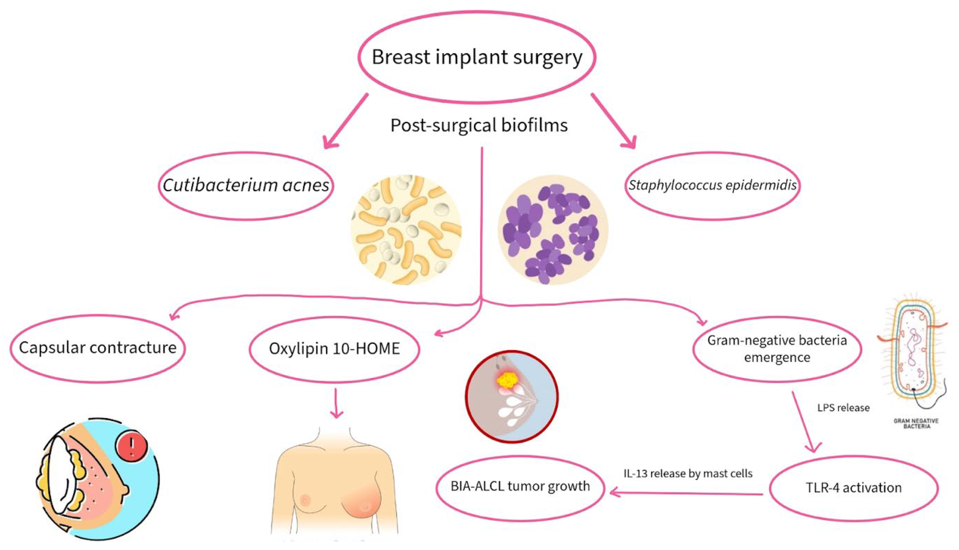
Figure 1. Bacterial biofilms commonly form on breast implant surfaces, predominantly involving Cutibacterium acnes and Staphylococcus epidermis. Such implant-associated bacteria contribute to conditions including capsular contracture, tumor growth, and inflammation. Their capacity to exacerbate post-surgical complications is linked to the promotion of immune dysregulation.
Hygiene 06 00010 g001
Figure 2. Organ transplantation can have a significant impact on the microbiome, leading to dysbiosis with an increase in potentially pathogenic species and a reduction of beneficial microbiota. Ultimately, these changes yield an increase in immune activation, inflammation, and increased incidence of rejection or organ dysfunction.
Hygiene 06 00010 g002
Figure 3. Fusobacterium nucleatum as a central bridging organism using specific adhesive proteins that promote various disease progression. The interspecies interaction promotes microbial dysbiosis, driving the creation of a pro-inflammatory environment. Its central position within the biofilm contributes to pathogen persistence and reduced treatment efficacy.
Hygiene 06 00010 g003
Figure 4. Roux-en-Y gastric bypass fundamentally reshapes the gastrointestinal environment, driving physiological shifts and subsequent microbial reconfiguration. The new selective pressures favor taxa that are better adapted to a new gastrointestinal environment and play an active role in the surgery’s long-term outcomes. Arrows represent key anatomical changes and microbiome shifts.
Hygiene 06 00010 g004
Disclaimer/Publisher’s Note: The statements, opinions and data contained in all publications are solely those of the individual author(s) and contributor(s) and not of MDPI and/or the editor(s). MDPI and/or the editor(s) disclaim responsibility for any injury to people or property resulting from any ideas, methods, instructions or products referred to in the content.