
Article

Establishing Reference Metrics for Respiratory Exercises Through Wearable Sensors: A Comparative Study

by
Federico Caramia
1,2,
Emanuele D’Angelantonio
1,2,3,
Leandro Lucangeli
1,2,3 and
Valentina Camomilla
1,2,*
1
Department of Movement, Human and Health Sciences, University of Rome “Foro Italico”, Piazza Lauro de Bosis 6, 00135 Rome, Italy
2
Interuniversity Centre of Bioengineering of the Human Neuromusculoskeletal System, University of Rome “Foro Italico”, Piazza Lauro de Bosis 6, 00135 Rome, Italy
3
Technoscience PST, Via Enrico Toti, 15, 04100 Latina, Italy
*
Author to whom correspondence should be addressed.
Biomechanics 2025, 5(4), 90; https://doi.org/10.3390/biomechanics5040090
Submission received: 16 August 2025 / Revised: 15 October 2025 / Accepted: 22 October 2025 / Published: 5 November 2025

Abstract

Background: Respiratory exercises play a key role in rehabilitation programs, especially for older adults and individuals with chronic pulmonary conditions. Despite growing interest in wearable sensors for home-based care, structured reference metrics to quantitatively characterize respiratory exercises are still limited. This study aimed to provide a quantitative characterization of respiratory exercises and evaluate the level of agreement between a low-cost prototypical sensor and a commercial one. Methods: Eleven older adults (9 females; age = 72.6 ± 5.0 years; height = 1.66 ± 0.09 m; mass = 68 ± 10 kg) performed a structured respiratory exercises protocol. Algorithms were developed to identify respiratory cycles, their execution time, and parameters related to respiratory capacity, using accelerometer signals from the two wearable sensors placed on the rib cage. Results: The average respiratory cycle duration ranged from 2.8 to 4.3 s, with normalized inspiratory and expiratory peaks. Tidal volume variability was minimal, confirming consistency in breathing patterns across exercises. User comfort was high (mean VAS = 8.7). Sensor comparison confirmed strong agreement between the two sensors in detecting respiratory cycles, though some variability was observed in timing and tidal volume estimation. Conclusions: These findings suggest that even simple accelerometers can reliably capture key respiratory parameters, supporting the feasibility of using wearable sensors to monitor structured respiratory exercises performed in home-based settings.

1. Introduction

Monitoring respiratory function and performance during exercises is fundamental in rehabilitation, especially for the elderly population and individuals with chronic pulmonary conditions. Structured breathing exercises have been shown to improve ventilatory efficiency, postural control, and overall functional capacity [1,2,3]. In particular, pursed-lips breathing exercises have been shown to reduce respiratory rate and increase tidal volume, inspiratory time, and total respiratory cycle duration, thereby helping to decrease the load on respiratory muscles and potentially alleviate dyspnea [4,5]. Diaphragmatic breathing has also demonstrated a significant reduction in respiratory rate, with positive effects on oxygen consumption and autonomic control, suggesting a potential role in the management of dyspnea [6,7,8]. Finally, the combination of ventilatory feedback techniques (training approaches that use real-time visual or auditory cues to guide and optimize breathing patterns) with progressive physical exercise has shown benefits in improving ventilatory parameters such as inspiratory capacity [9], supporting monitoring of respiratory performance during structured exercises within rehabilitation programs.
Traditional methods for monitoring respiratory activity (spirometry, optoelectronic plethysmography, or respiratory inductance plethysmography) provide accurate assessments, but are typically restricted to clinical settings due to their cost, complexity, or invasiveness [10,11]. In contrast, wearable sensors have emerged as a low-cost, portable, and scalable alternative for monitoring chest wall and rib cage movements during respiration, allowing continuous monitoring [12]. Recent literature has increasingly highlighted the potential of wearable technologies for respiratory monitoring during daily life activities, especially in the context of chronic disease management, telehealth, and post-COVID-19 recovery [12]. In particular, these solutions are used in different static postures, including standing, lying and sitting [12,13], and during more complex physical activities such as cycling or running [13,14]. Among wearable devices, inertial measurement units (IMUs) have gained attention for their potential in estimating key respiratory parameters such as respiratory rate (RR), tidal volume (TV), and breathing variability [15,16,17]. In addition, the integration of machine learning and deep learning approaches has reinforced their use as an affordable solution [13,16].
Despite the potential offered by monitoring these exercises to significantly improve the effectiveness of rehabilitation protocols, particularly in remote rehabilitation settings, reference metrics to objectively assess respiratory exercises remain scarce, especially in structured rehabilitation programs. Moreover, the development of low-cost instruments capable of supporting this type of assessment is essential to ensure access to continuous respiratory monitoring in long-term rehabilitation settings. Balancing measurement reliability and affordability remains a crucial factor in delivering effective telerehabilitation services to individuals with diverse health conditions. Building on our previous validation of a low-cost IMU system for general rehabilitation movements within a telerehabilitation framework (REHACT) [18], this study focuses on respiratory-specific exercises, aiming to establish targeted metrics and assess their reliability using a simplified sensor configuration.
The aims of this study are twofold: (1) to provide IMU-based quantitative metrics to characterize selected respiratory exercises; and (2) to evaluate the agreement between a low-cost IMU prototype, already developed [18], and a reference IMU in extracting these metrics. To achieve these goals, a structured respiratory exercise protocol was developed and tested, involving the combined use of an inertial reference sensor and a low-cost prototype to detect and characterize breathing activity.
We hypothesized that the prototype IMU would achieve Bland–Altman agreement with the reference device within clinically acceptable limits, namely a bias < 0.1 s and limits of agreement (LoA) within ±1.0 s for respiratory cycle duration (TimeRR), across the four tested postures. This criterion aligns with acceptable differences reported in respiratory rate estimation literature, where errors within ±2–3 breaths per minute have been considered acceptable in monitoring applications [19]. This hypothesis reflects a practical benchmark for functional equivalence in unsupervised monitoring contexts.
This step is a prerequisite for future validation against clinical gold standards, in view of integrating wearable technologies into monitoring systems, enabling personalized and remote respiratory rehabilitation. Establishing reference metrics and verifying the prototype’s consistency with an established inertial system is part of a construct validity approach. Given the current development stage of the prototype (Technology Readiness Level 4—TRL4—corresponding to early-stage laboratory testing), this methodological choice enables a functional assessment of its measurement capabilities.

2. Materials and Methods

2.1. Respiratory Exercises

The REHACT telerehabilitation protocol was developed based on literature reviews [20,21] and, as regards respiratory exercises, in accordance with specific guidelines on respiration [1,2,3]. It includes sixteen exercises for improving strength and mobility of the lower limbs, upper limbs, and trunk selected following the designed “EASE” principles: Easy to perform, Adaptable to different levels of functional ability, Safe, and Effective. Its main innovation lies in the integration of wearable inertial sensors, which allow for real-time, quantitative monitoring and remote feedback. For instance, so-called “open chain” exercises [20,21] are easy to perform and low-impact, yet still provide significant rehabilitative benefits. At the same time, they require only a minimal sensor setup, unlike more complex movements that demand more elaborate instrumentation.
To accommodate different functional levels, exercises can be performed in a variety of positions: lying down, seated with or without back support, or standing with support.
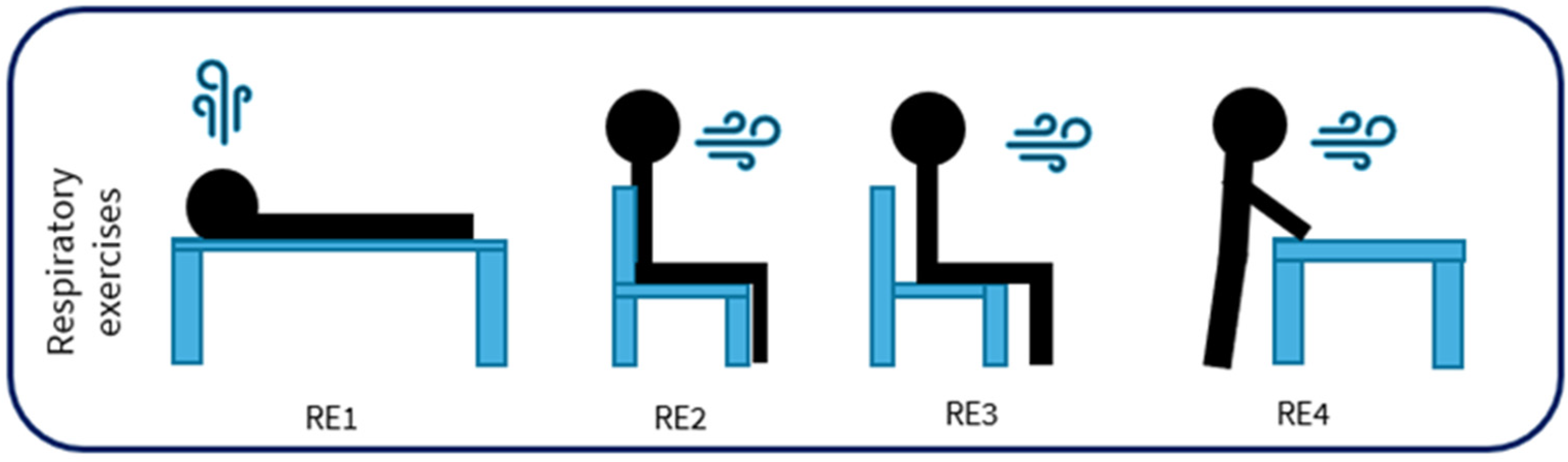
In this study, we focused specifically on four respiratory exercises (Figure 1), among the sixteen available in the REHACT protocol [18]. These exercises are essential for improving respiratory function and are particularly suitable for remote monitoring through inertial sensors.
These exercises were selected to train respiratory muscles by voluntary inspiration and expiration. Every cycle is repeated for 8 repetitions in 2 different series, with a one-minute rest between series to prevent ventilatory discomfort in participants. The respiration exercises required participants to perform conscious and voluntary breathing, distinct from spontaneous breathing at rest, likely involving increased rib cage excursion and intercostal muscle activation.
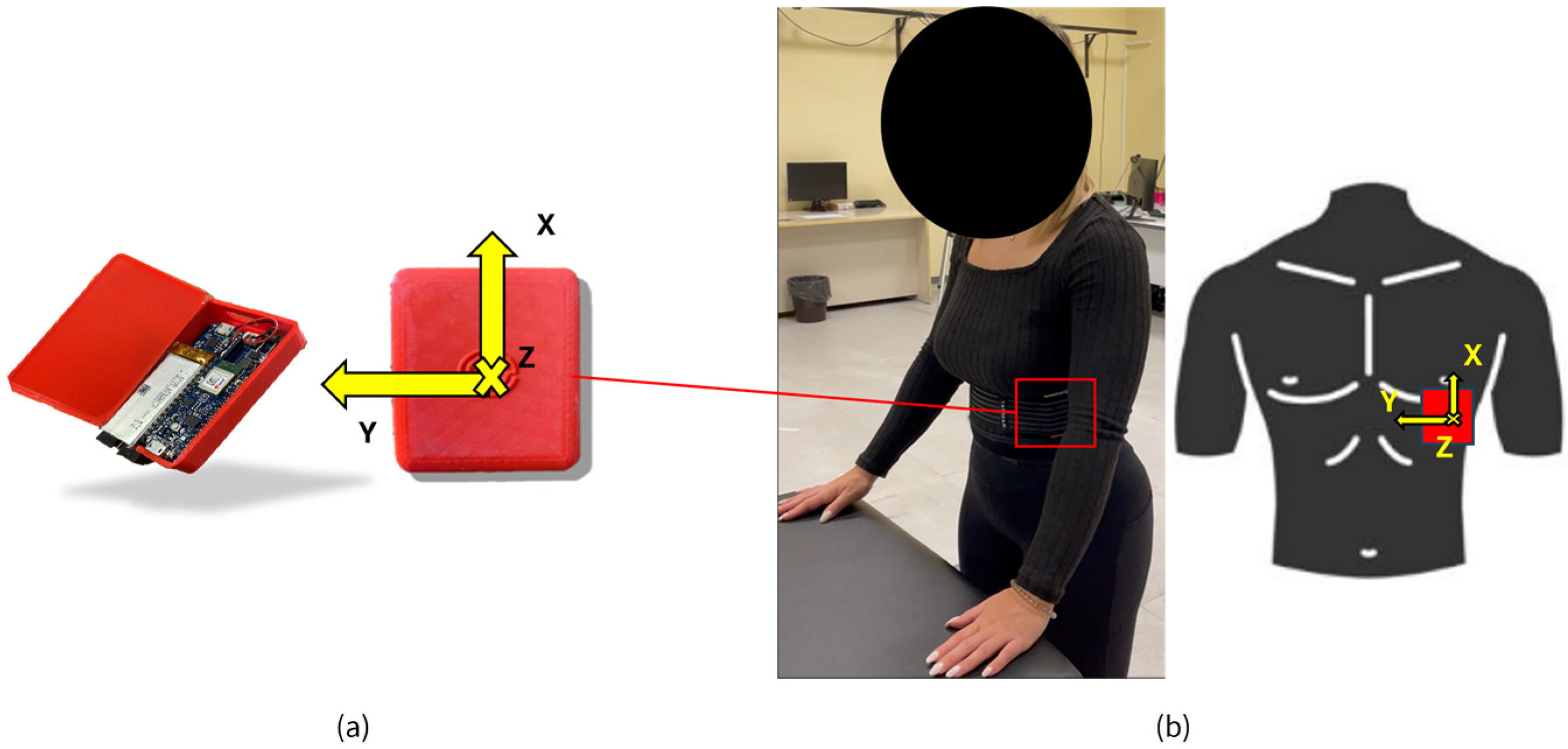
The study, approved by the University Research Committee (CAR code 158/2023), was conducted in accordance with the Helsinki Declaration as revised in 2024. Written informed consent was obtained from the participants of the study. A total of 11 medically stable older adults (9 females and 2 males; age = 72.6 ± 5.0 years; height = 1.66 ± 0.09 m; mass = 68 ± 10 kg) participated in the study, performing the four respiratory exercises included in the REHACT protocol (Figure 1). This sample was selected to allow for a representative characterization of the respiratory component of the protocol. To monitor respiratory motion during exercise, a single IMU was placed ~2–3 cm above the left last rib, where thoracic motion is most detectable during structured respiratory exercises, and secured with an elastic band (Figure 2b). The sensor was placed above the left lower rib, as determined in a prior feasibility study [22], which identified this location as the most reliable for detecting respiratory movements during structured exercises. This placement is also consistent with established literature on respiratory monitoring using wearable IMUs [14,15]. A prototypical 9-axis IMU (LSM9DS1, STMicroelectronics, Plan-les-Ouates, Switzerland; full scale ranges: ±16 g accelerometer, ±2000 deg/s gyroscope and ±16 Gauss magnetometer; sampling frequency: 30–35 samples/s) (Figure 2a) was used to monitor the different breathing exercises. This technology represents the central part of the telerehabilitation infrastructure used in the REHACT project [18]. The sensor was placed above the left last rib using an elastic band. A 0–10 Visual Analog Scale (VAS) was used to assess the comfort of this configuration.
Reference values for respiratory exercise parameters were also established using one laboratory sensor (OPAL, APDM, Portland, OR, USA, full-scale ranges: ±200 g accelerometer, ±2000 deg/s gyroscope and ±8 Gauss magnetometer; sampling frequency: 128 samples/s), placed above the prototypical one.
The two sensors were stacked directly one over the other, taking advantage of their flat surfaces, and secured with an elastic band above the last rib. A calibration pipeline for each IMU and the gravity-related correction were applied.

2.2. Respiratory Pattern Characterization

Different parameters were identified and used to monitor the respiratory cycles during the breathing exercises of the REHACT protocol, using only the accelerometer following relevant literature [14,15]. The antero-posterior axis (Z-axis), which is perpendicular to the ribs, was selected as the most suitable direction for detecting respiratory movements, as it captures the expansion and contraction of the rib cage during breathing. Gravity was removed from the signal to isolate the respiratory component. Specifically, for the supine exercise (RE1), the gravitational component was subtracted from the vertical axis, which was aligned with gravity in this posture. For the seated and standing exercises (RE2, RE3, and RE4), gravity compensation was applied using the orientation correction method described by [23].
Raw accelerometric signal was first filtered using a fourth-order Butterworth low-pass filter. The optimal cut-off frequency was determined based on the method proposed by [24], and was set at 0.6 Hz as a compromise value across subjects, values commonly accepted for the frequency range of normal breathing [25].
After filtering, respiratory cycles were identified by applying a custom algorithm developed in MATLAB R2021b (MathWorks Inc., Natick, MA, USA). This algorithm, adapted from previous applications used for the analysis of motor tasks [18], was specifically optimized for respiratory signal characteristics. For each cycle, using MATLAB’s “findpeaks “function, acceleration peaks on the filtered Z axis were identified by an iterative optimization for stable detection, with various cutoff values applied, defining the following function parameters: minimum peak height (0.01 g), minimum distance between peaks (20 samples), minimum peak width (0.2) and minimum peak prominence (0.2).
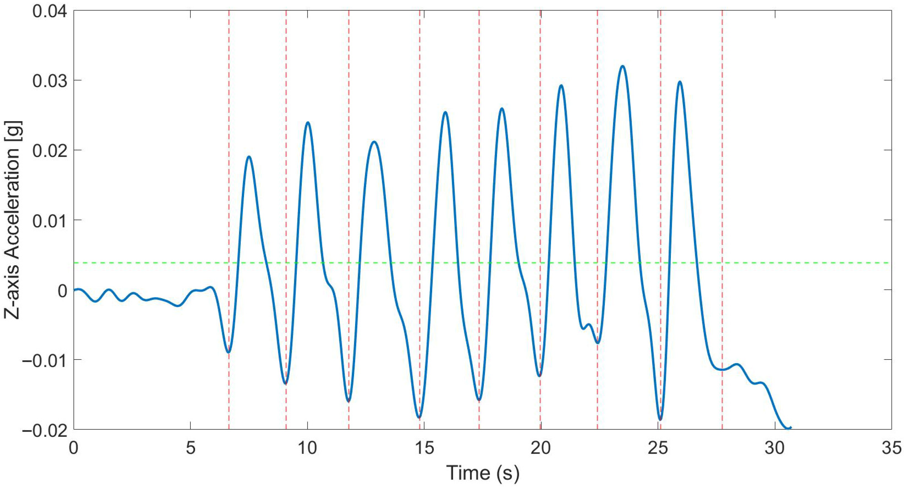
To ensure stable and consistent identification of respiratory cycles, regardless of inter-individual variability, a threshold of 30% of the maximum peak was found through an iterative process and applied to the identified peaks. With this threshold, the algorithm searched for local minima before and after each peak: the preceding minimum was considered the beginning of the respiratory cycle, while the following minimum marked its end. In cases of continuous repetitions, the end of one cycle coincided with the beginning of the next, ensuring temporal continuity in the identification of respiratory cycles [25] (Figure 3).
Each respiratory cycle, defined as a complete sequence of inspiration followed by expiration, was thus automatically segmented. By applying the segmentation algorithm used to identify respiratory cycles, peaks (p), and valleys (v). Subsequently, the signal of each cycle was normalized by scaling its values between 0 and 1 based on its minimum and maximum values [25]. These combined procedures, segmentation and normalization, enabled the extraction of respiratory pattern indicators for each cycle:
  • number of respiratory cycles (Resp): the number of breaths performed;
  • respiratory cycle duration (TimeRR): time duration of the inspiration and expiration cycle;
  • inspiration phase duration (TimeInsp) [26]: time duration of the inspiration phase;
  • expiration phase duration (TimeExp) [26]: time duration of the expiration phase;
  • peak of normalized acceleration of inspiration phase (PeakInsp): descriptive of the inspiration depth. A normalized signal near one suggests deep inspiration;
  • peak of normalized acceleration of expiration phase (PeakExp): indicates the maximum deceleration reached during expiration. A value closer to zero suggests a prolonged or smoother expiration;
  • normalized Tidal Volume (TVolume): estimate of the volume of air inside the lungs during each respiratory cycle [25,26,27]. Subsequently, the volume of each i-th breath is estimated as follows [25]:
T V o l u m e i = p i ( v i + v i + 1 ) 2
where TVolumei is the volume of the ith breath derived from the normalized signals, pi and vi are the ith peak and valley, respectively.
  • Tidal Volume Variability (TVar): represents the trend of TVolume oscillations over time. The variations in TVolume are analyzed using a linear fitting applied to moving windows of variable length. The analysis window corresponds to the number of consecutive breaths. TVar is obtained based on the slope of the linear fits; and is calculated using the following formula [25]:
T V a r i = t i = 1 t n i T V o l i ( i = 1 t n i ) ( i = 1 t T V o l i ) t i = 1 t n i 2 ( i = 1 t n i ) 2
where n is the breath number. The window length ( t ) was set at 2 [25].
These parameters represent an essential set to describe the respiratory exercises within the developed protocol. In the future, additional metrics or specific exercise-related measures may be incorporated to further enhance the understanding of respiratory rehabilitation exercises.

2.3. Sensors Comparison

To achieve the second objective of the study, the prototype was compared to a commercially available accelerometer, serving as a validated reference device. This comparison was conceived as a construct validity-based functional assessment, appropriate to the current Technology Readiness Level (TRL4). Respiratory parameters were selected for analysis among those involving wearable technologies and already validated [25,26,27]. The goal was to assess the feasibility of the system and of the experimental setup and to support future validation studies using clinical gold standards, such as spirometry.
Reference values were reported as mean ± standard deviation and 95% confidence intervals (CI), computed using the standard error of the mean assuming normal distribution (CI = mean ± 1.96 × SD/ n ).
For the comparison between the two sensors, a Bland–Altman (BA) analysis [28] was carried out for each respiratory parameter. Prior to the analysis, outliers (>2 SD from the mean) were inspected and excluded, to ensure reliable agreement assessment outliers. To assess the consistency of the measurements, the presence of heteroscedasticity in the data was evaluated using Kendall’s Tau test [29]. This test compares the distribution of the average values with the absolute differences between the two devices [30]. A Tau value (τ) below 0.1 indicates homoscedastic data, while values equal to or greater than 0.1 suggest heteroscedasticity. The limits of agreement were calculated using the standard Bland–Altman method (Upper Limits (UL) = BIAS + 1.96 × SD; Lower limits (LL) = BIAS − 1.96 × SD), where BIAS represents the mean difference between the prototype and reference sensor data, and SD is the standard deviation of these differences. Additionally, 95% confidence intervals (CIs) for BIAS, UL, and LL were calculated as described by [31], using the t-value, sample size (n), and standard error of the BIAS (seBIAS).

3. Results

Participants reported a high value of comfort, measured by the VAS, during the respiratory exercises (Table 1).
Reference values, of both reference and prototype sensors, for a total of 704 respiratory cycles across all exercises are given in Table 2.
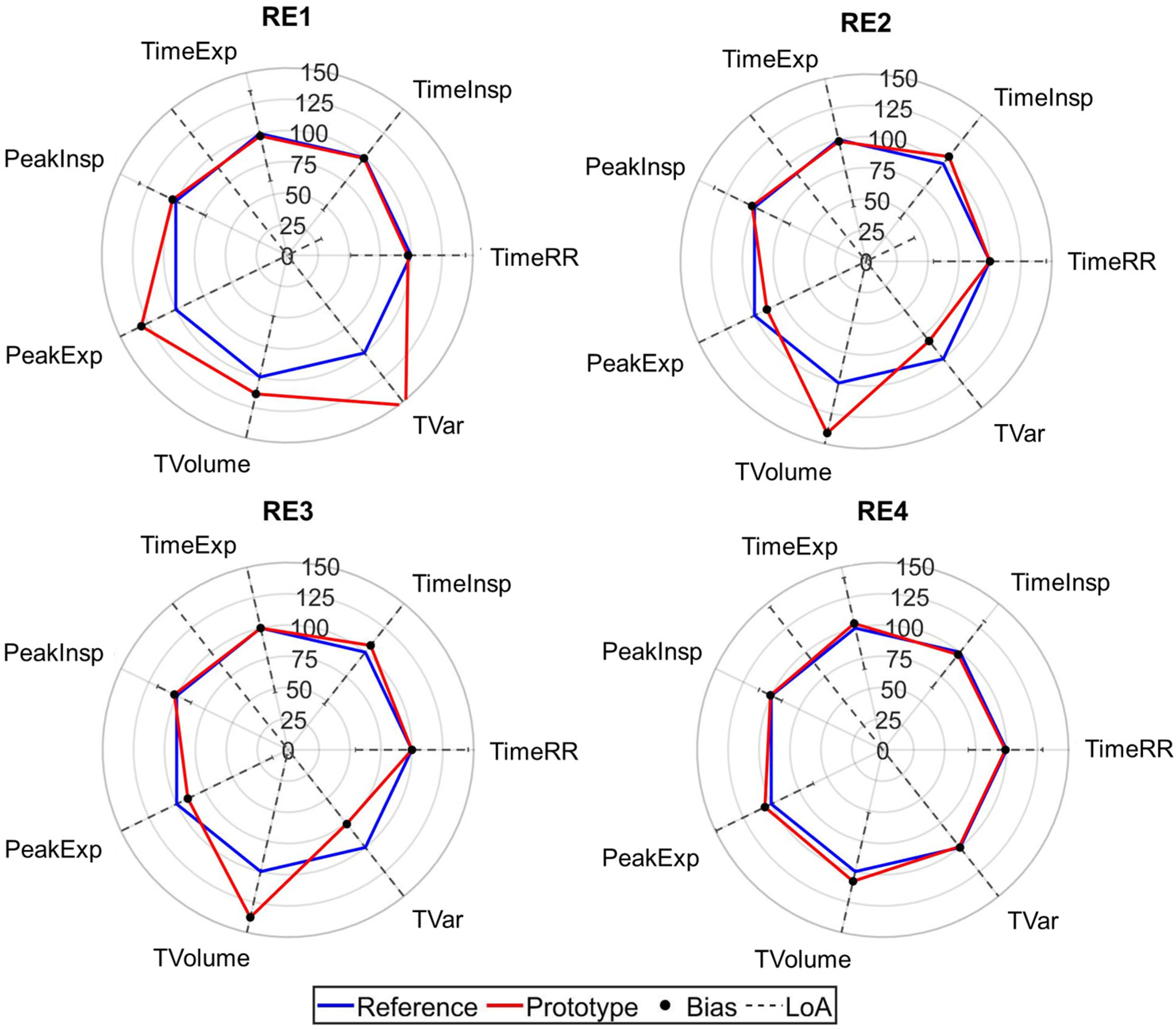
Regarding the number of respiratory cycles (Resp), only few cycles (17 out of the 704 total respiratory cycles across all exercises) were not correctly counted by both sensors while sitting with back support, while sensors were capable of correctly detecting all cycles for the other exercises. Figure 4 presents radar plots that illustrate the comparative analysis between reference and prototype IMU sensors of the other parameters, following the format presented in [18]. Each axis represents the mean ratio between prototype and reference parameters (100% meaning prototype value = reference value). Bland–Altman plots and analysis for each parameter and posture are provided in the Supplementary Material.

4. Discussion

The results of this construct validity study support the functional plausibility of using a single low-cost inertial sensor to monitor respiratory exercises in older adults, with consistent detection of respiratory cycles and high comfort scores across conditions. The following sections explore both the performance of the prototype (Section 4.1) and its consistency with the reference device (Section 4.2), offering a comprehensive view of its potential for remote respiratory monitoring.

4.1. Values for Respiratory Exercises

To the authors’ knowledge, this is the first study providing a quantitative characterization of selected respiratory exercises part of a telerehabilitation protocol (REHACT [18]) using an inertial sensor placed on the rib. First, the number of respiratory cycles (Resp) were consistently detected, confirming the robustness of the segmentation algorithm. Temporal parameters (TimeRR, TimeInsp, TimeExp) showed, in general, very close mean values between the two devices, with small standard deviations, suggesting that the prototype can reliably detect the timing of respiratory phases (Table 2). Similarly, peak normalized acceleration values (PeakInsp, PeakExp), indicative of inspiratory and expiratory movements, were consistent across exercises and devices. Normalized tidal volume (TVolume) and its variability (TVar) showed low intra-exercise variability and stable 95% CI, proving that the breathing pattern remained consistent in the tested participants across tasks. This consistency is essential for long-term remote monitoring and may support the integration of such metrics in adaptive rehabilitation platforms. Overall, these results may contribute to the establishment of reference values for proposed respiratory exercises in clinical and home environments.
In addition, high level of comfort reported by the participants, as evidenced by the scores reported on the VAS, with mean values above 8 out of 10 in all conditions tested (Table 1), confirms that the proposed configuration is well tolerated by users.

4.2. Comparison Between Prototypical and Reference Sensor

Both sensors effectively detected the number of respiratory cycles, confirming the robustness of the segmentation algorithm. Respiratory cycle duration (TimeRR), among the temporal parameters, and the inspiratory acceleration peak (PeakInsp) confirmed the initial hypothesis, showing Bland–Altman agreement within clinically acceptable limits. Conversely, inspiration and expiration phase durations (TimeInsp, TimeExp), as well as expiratory acceleration peaks (PeakExp) and volume-related parameters (TVolume, TVar), exhibited wider Limits of Agreement (LoA), although the biases remained small and stable across all conditions.
These differences are likely related to variations in signal morphology, particularly the more pronounced plateau phase observed in the reference sensor. Therefore, these metrics should be interpreted with caution, especially in applications requiring high measurement precision [19]. Nevertheless, the prototype demonstrated good overall performance in detecting respiratory cycles and capturing temporal trends, which may be sufficient for home-based or unsupervised rehabilitation contexts, where relative changes are more relevant than absolute values.

4.3. Limitations and Future Development

Several limitations must be acknowledged when interpreting the results of this study. Firstly, the data collected are representative of an elderly population and may not be directly generalizable to individuals with chronic obstructive pulmonary disease (COPD) [1]. Participants who completed standing respiratory exercises likely exhibited higher functional capacity than individuals restricted to supine positions. Therefore, the respiratory values reported here should be interpreted as upper-bound references, rather than representative benchmarks for subjects with lower functional capacity. The small sample size (n = 11), selected for exploratory validation, further limits the statistical robustness and generalizability of the reference values.
Secondly, sensor positioning selectively affected data from specific exercise postures. Cycle detection errors were mainly observed in the seated position with back support (RE2), likely due to reduced rib cage excursion and consequent lower peak Z-axis amplitude, possibly due to the mechanical support or changes in the breathing pattern. To improve robustness, future protocols may explore alternative sensor positions (e.g., lateral rib cage or abdominal wall), and enhanced fixation systems. Additionally, anthropometric characteristics—such as adipose tissue in the pectoral area—may have influenced signal amplitude and should be considered in sensor design.
Thirdly, data processing choices did not entail adaptive segmentation or cut-off frequency, due to the limited sample size and the exploration nature of this study. The fixed threshold (30% of the maximum peak) used for respiratory cycle segmentation was selected through iterative empirical testing and led to a stable performance across all tested postures and participants. Larger studies employing posture-specific adaptative threshold for peak detection and filtering optimization are needed to better accommodate inter-individual variability and signal differences across conditions, thereby enabling the establishment of normative values applicable across different clinical and demographic populations.
Regarding the second objective, differences in hardware between the prototype and the reference device determined measurement discrepancies, despite the use of a validated signal processing method [25,26,27]. Moreover, the use of a secondary accelerometer rather than a clinical gold standard limits the construct validity of the comparison. Future validation should therefore incorporate clinical reference instruments to confirm the ability of low-cost IMUs to provide reliable respiratory metrics.
Finally, embedding the sensor in textile-based garments could improve comfort, positioning repeatability, and long-term usability. These technologies have already shown potential in clinical and sports settings [32,33]. Their extension to monitoring voluntary respiratory exercises, particularly in individuals with COPD, offers a valuable direction for future development.

5. Conclusions

This study highlights the utility of characterizing respiratory exercises through wearable inertial sensors, demonstrating that it is feasible and effective to monitor respiratory exercises using a single sensor placed on the rib.
The first aim of this work was to provide a quantitative characterization of specific respiratory exercises, using data derived from wearable inertial sensors. The ability to detect respiratory cycles automatically and accurately, with minimal inter-trial variability, along with the extraction of relevant temporal and kinematic parameters, confirms the validity of this approach and contributes to the definition of normative reference values for respiratory rehabilitation. The high comfort scores reported by participants further strengthen the suitability of this approach, particularly for home-based applications.
The second aim was to evaluate the consistency between a low-cost prototype IMU and a commercial reference device. Despite differences in certain parameters, the agreement in respiratory cycle counting could support the potential of the low-cost sensor as a reliable and accessible tool for respiratory monitoring, by virtue of future hardware upgrades and validation against gold standard instruments
Overall, the results suggest that a single, low-cost IMU placed on the rib cage can provide meaningful, repeatable respiratory data during structured exercises. This supports the development of practical tools for home-based respiratory training, especially in contexts where clinical supervision is limited but ongoing monitoring is needed to support adherence and safety.

Supplementary Materials

The following supporting information can be downloaded at https://www.mdpi.com/article/10.3390/biomechanics5040090/s1.

Author Contributions

Conceptualization, F.C. and V.C.; methodology, F.C. and V.C.; software, F.C.; validation, F.C., E.D. and L.L.; formal analysis, F.C.; investigation, F.C., E.D. and L.L.; data curation, F.C.; writing—original draft preparation, F.C. and V.C.; writing—review and editing, F.C. and V.C.; visualization, F.C. and V.C.; supervision, V.C.; project administration, E.D., L.L. and V.C.; funding acquisition, V.C. All authors have read and agreed to the published version of the manuscript.

Funding

This research project “teleREHabilitation for respiratory and motor reACTivation exercises” (REHACT) was funded by the Italian National Institute of Social Security (INPS), Call: Industry 4.0, 2020-21.

Institutional Review Board Statement

The study was conducted in accordance with the Declaration of Helsinki and approved by the Institutional Review Board of University of Rome Foro Italico (protocol code CAR158/2023 Rev, date of approval 4 September 2024).

Informed Consent Statement

Informed consent was obtained from all subjects involved in the study.

Data Availability Statement

The raw data supporting the conclusions of this article will be made available by the authors on request.

Conflicts of Interest

The authors declare the following conflicts of interest: Emanuele D’Angelantonio and Leandro Lucangeli are employed by Technoscience, producing the prototype assessed in the study. The company funded their PhD research, but was not involved in the study design, data collection, analysis, or manuscript preparation. All these aspects were managed by Valentina Camomilla and Federico Caramia, who do not have any conflicts of interest associated with this publication.

Abbreviations

The following abbreviations are used in this manuscript:
IMUInertial measurement unit
RRRespiratory rate
TVTidal volume
RE1Supine respiratory exercise
RE2Seated respiratory exercise with back support
RE3Seated respiratory exercise without back support
RE4Standing respiratory exercise with support
VASVisual analog scale
RespNumber of respiratory cycles
TimeRRTime duration of one respiratory cycle
TimeInspTime duration of inspiration phase
TimeExpTime duration of expiration phase
PeakInspPeak of normalized acceleration of inspiration phase
PeakExpPeak of normalized acceleration of expiration phase
TVolumeNormalized Tidal Volume
TVarTidal Volume Variability
TRLTechnology Readiness Level
BIASDifference between the mean reference and the prototype sensor values
LoALimit of agreement
BABland and Altman analysis
CIConfidence intervals
seBIASStandard error for the BIAS
COPDChronic obstructive pulmonary diseases

References

  1. Ubolnuar, N.; Tantisuwat, A.; Thaveeratitham, P.; Lertmaharit, S.; Kruapanich, C.; Mathiyakom, W. Effects of Breathing Exercises in Patients with Chronic Obstructive Pulmonary Disease: Systematic Review and Meta-Analysis. Ann. Rehabil. Med. 2019, 43, 509–523. [Google Scholar] [CrossRef]
  2. Yeh, T.-T.; Ho, Y.-C. Immediate effects of structured and natural deep breathing on heart rate variability and blood pressure in community-dwelling older adults. Exp. Gerontol. 2024, 198, 112644. [Google Scholar] [CrossRef]
  3. Vaz Fragoso, C.A.; Beavers, D.P.; Anton, S.D.; Liu, C.K.; McDermott, M.M.; Newman, A.B.; Pahor, M.; Stafford, R.S.; Gill, T.M. Effect of Structured Physical Activity on Respiratory Outcomes in Sedentary Elderly Adults with Mobility Limitations. J. Am. Geriatr. Soc. 2016, 64, 501–509. [Google Scholar] [CrossRef]
  4. Holland, A.E.; Hill, C.; McDonald, C. Breathing exercises for chronic obstructive pulmonary disease. Cochrane Database Syst. Rev. 2010, 17, 1–18. [Google Scholar] [CrossRef]
  5. Roberts, S.E.; Stern, M.; Schreuder, F.M.; Watson, T. The use of pursed lips breathing in stable chronic obstructive pulmonary disease: A systematic review of the evidence. Phys. Ther. Rev. 2009, 14, 240–246. [Google Scholar] [CrossRef][Green Version]
  6. Ambrosino, N.; Paggiaro, P.L.; Macchi, M.; Filieri, M.; Toma, G.; Lombardi, A.; Del Cesta, F.; Parlanti, A.; Loi, A.M.; Baschieri, L. A Study of Short-Term Effect of Rehabilitative Therapy in Chronic Obstructive Pulmonary Disease. Respiration 2009, 41, 40–44. [Google Scholar] [CrossRef] [PubMed]
  7. Jones, A.; Jones, G.; Greig, N.; Bower, P.; Brown, J.; Hind, K.; Francis, P. Epidemiology of injury in English Professional Football players: A cohort study. Phys. Ther. Sport. 2019, 35, 18–22. [Google Scholar] [CrossRef] [PubMed]
  8. Borge, C.R.; Mengshoel, A.M.; Omenaas, E.; Moum, T.; Ekman, I.; Lein, M.P.; Mack, U.; Wahl, A.K. Effects of guided deep breathing on breathlessness and the breathing pattern in chronic obstructive pulmonary disease: A double-blind randomized control study. Patient Educ. Couns. 2015, 98, 182–190. [Google Scholar] [CrossRef]
  9. Collins, E.G.; Langbein, W.E.; Fehr, L.; O’Connell, S.; Jelinek, C.; Hagarty, E.; Edwards, L.; Reda, D.; Tobin, M.J.; Laghi, F. Can Ventilation–Feedback Training Augment Exercise Tolerance in Patients with Chronic Obstructive Pulmonary Disease? Am. J. Respir. Crit. Care Med. 2008, 177, 844–852. [Google Scholar] [CrossRef]
  10. Pierce, R. Spirometry: An essential clinical measurement. Aust. Fam. Physician 2005, 34, 535. [Google Scholar] [PubMed]
  11. Stubbe, L.; Houel, N.; Cottin, F. Accuracy and reliability of the optoelectronic plethysmography and the heart rate systems for measuring breathing rates compared with the spirometer. Sci. Rep. 2022, 12, 19255. [Google Scholar] [CrossRef]
  12. Aliverti, A. Wearable technology: Role in respiratory health and disease. Breathe 2017, 13, e27–e36. [Google Scholar] [CrossRef]
  13. Massaroni, C.; Di Tocco, J.; Bravi, M.; Carnevale, A.; Presti, D.L.; Sabbadini, R.; Miccinilli, S.; Sterzi, S.; Formica, D.; Schena, E. Respiratory Monitoring During Physical Activities with a Multi-Sensor Smart Garment and Related Algorithms. IEEE Sens. J. 2020, 20, 2173–2180. [Google Scholar] [CrossRef]
  14. Nicolò, A.; Massaroni, C.; Schena, E.; Sacchetti, M. The Importance of Respiratory Rate Monitoring: From Healthcare to Sport and Exercise. Sensors 2020, 20, 6396. [Google Scholar] [CrossRef] [PubMed]
  15. Massaroni, C.; Nicolò, A.; Lo Presti, D.; Sacchetti, M.; Silvestri, S.; Schena, E. Contact-Based Methods for Measuring Respiratory Rate. Sensors 2019, 19, 908. [Google Scholar] [CrossRef] [PubMed]
  16. Vitazkova, D.; Foltan, E.; Kosnacova, H.; Micjan, M.; Donoval, M.; Kuzma, A.; Kopani, M.; Vavrinsky, E. Advances in Respiratory Monitoring: A Comprehensive Review of Wearable and Remote Technologies. Biosensors 2024, 14, 90. [Google Scholar] [CrossRef]
  17. Hussain, T.; Ullah, S.; Fernández-García, R.; Gil, I. Wearable Sensors for Respiration Monitoring: A Review. Sensors 2023, 23, 7518. [Google Scholar] [CrossRef]
  18. Caramia, F.; D’Angelantonio, E.; Lucangeli, L.; Camomilla, V. Validation of Low-Cost IMUs for Telerehabilitation Exercises. Sensors 2025, 25, 3129. [Google Scholar] [CrossRef]
  19. Nizami, S.; Bekele, A.; Hozayen, M.; Greenwood, K.J.; Harrold, J.; Green, J.R. Green Measuring Uncertainty During Respiratory Rate Estimation Using Pressure-Sensitive Mats. IEEE Trans. Instrum. Meas. 2018, 67, 1535–1542. [Google Scholar] [CrossRef]
  20. Izquierdo, M.; Merchant, R.A.; Morley, J.E.; Anker, S.D.; Aprahamian, I.; Arai, H.; Aubertin-Leheudre, M.; Bernabei, R.; Cadore, E.L.; Cesari, M.; et al. International Exercise Recommendations in Older Adults (ICFSR): Expert Consensus Guidelines. J. Nutr. Health Aging 2021, 25, 824–853. [Google Scholar] [CrossRef] [PubMed]
  21. Nikitas, C.; Kikidis, D.; Bibas, A.; Pavlou, M.; Zachou, Z.; Bamiou, D.E. Recommendations for physical activity in the elderly population: A scoping review of guidelines. J. Frailty Sarcopenia Falls 2022, 7, 18–28. [Google Scholar] [CrossRef]
  22. Caramia, F.; D’Angelantonio, E.; De Angelis, F.; Lucangeli, L.; Campanini, I.; Pallotti, A.; Camomilla, V. Design of a care pathway in telerehabilitation on respiratory exercise and physical reactivation of patients suffering from respiratory diseases or COVID-19. In Proceedings of the 17th International Symposium of 3D Analysis of Human Movement (3D-AHM 2022), Tokyo, Japan, 16–19 July 2022. [Google Scholar]
  23. Picerno, P.; Camomilla, V.; Capranica, L. Countermovement jump performance assessment using a wearable 3D inertial measurement unit. J. Sports Sci. 2011, 29, 139–146. [Google Scholar] [CrossRef]
  24. Winter, D.A. Signal Processing. In Biomechanics and Motor Control of Human Movement; John Wiley & Sons: Hoboken, NJ, USA, 2009; pp. 14–44. ISBN 978-0-470-54914-8. [Google Scholar]
  25. Fekr, A.R.; Radecka, K.; Zilic, Z. Tidal volume variability and respiration rate estimation using a wearable accelerometer sensor. In Proceedings of the 2014 4th International Conference on Wireless Mobile Communication and Healthcare—Transforming Healthcare Through Innovations in Mobile and Wireless Technologies (MOBIHEALTH), Athens, Greece, 3–5 November 2014; pp. 1–6. [Google Scholar]
  26. Van Diest, I.; Verstappen, K.; Aubert, A.E.; Widjaja, D.; Vansteenwegen, D.; Vlemincx, E. Inhalation/Exhalation Ratio Modulates the Effect of Slow Breathing on Heart Rate Variability and Relaxation. Appl. Psychophysiol. Biofeedback 2014, 39, 171–180. [Google Scholar] [CrossRef]
  27. Zhu, X.; Chen, W.; Nemoto, T.; Kanemitsu, Y.; Kitamura, K.; Yamakoshi, K.; Wei, D. Real-Time Monitoring of Respiration Rhythm and Pulse Rate During Sleep. IEEE Trans. Biomed. Eng. 2006, 53, 2553–2563. [Google Scholar] [CrossRef] [PubMed]
  28. Bland, J.M.; Altman, D.G. Statistics Notes: Measurement error proportional to the mean. BMJ 1996, 313, 106. [Google Scholar] [CrossRef] [PubMed]
  29. Kendall, M.G. A New Measure of Rank Correlation. Biometrika 1938, 30, 81–93. [Google Scholar] [CrossRef]
  30. Brehm, M.-A.; Scholtes, V.A.; Dallmeijer, A.J.; Twisk, J.W.; Harlaar, J. The importance of addressing heteroscedasticity in the reliability analysis of ratio-scaled variables: An example based on walking energy-cost measurements. Dev. Med. Child. Neurol. 2012, 54, 267–273. [Google Scholar] [CrossRef]
  31. Giavarina, D. Understanding Bland Altman analysis. Biochem. Med. 2015, 25, 141–151. [Google Scholar] [CrossRef] [PubMed]
  32. Massaroni, C.; Venanzi, C.; Silvatti, A.P.; Lo Presti, D.; Saccomandi, P.; Formica, D.; Giurazza, F.; Caponero, M.A.; Schena, E. Smart textile for respiratory monitoring and thoraco-abdominal motion pattern evaluation. J. Biophotonics 2018, 11, e201700263. [Google Scholar] [CrossRef]
  33. Ciocchetti, M.; Massaroni, C.; Saccomandi, P.; Caponero, M.A.; Polimadei, A.; Formica, D.; Schena, E. Smart Textile Based on Fiber Bragg Grating Sensors for Respiratory Monitoring: Design and Preliminary Trials. Biosensors 2015, 5, 602–615. [Google Scholar] [CrossRef] [PubMed]
Figure 1. Respiratory exercises of the REHACT protocol. RE1: supine respiratory exercise, RE2: seated respiratory exercise with back support, RE3: seated respiratory exercise without back support, RE4: standing respiratory exercise with support.
Biomechanics 05 00090 g001
Figure 2. (a) Prototypical IMU, (b) positioning of prototypical sensor during a standing respiratory exercise with support (RE4).
Biomechanics 05 00090 g002
Figure 3. Identification of respiratory cycles. Filtered Z-axis (main axis) signal in the RE1 exercises. The red dotted lines segment the different respiratory cycles; the green line represents the threshold used to identify the end of repetitions.
Biomechanics 05 00090 g003
Figure 4. Percentage values of prototype parameters (red line) are reported compared to the reference values (100% line reported in blue). The black dot represents the mean of the differences between the two devices (Bias), in percentage of the reference value. The dashed line highlights the amplitude of the limits of agreement (LoA), in percentage the reference value TimeRR: respiratory cycle duration. TimeInsp: inspiration phase duration. TimeExp: expiration phase duration. PeakInsp: normalized peak acceleration during inspiratory phase. PeakExp: normalized peak acceleration during expiration phase. TVolume: normalized Tidal volume. TVar: Tidal volume variability.
Biomechanics 05 00090 g004
Table 1. Comfort level during the respiratory exercises.
ExerciseLevel of Comfort (0–10)
RE18.68 ± 1.35
RE28.77 ± 1.4
RE38.68 ± 1.35
RE48.77 ± 1.33
Table 2. Parameter values (mean and standard deviation) as assessed using the reference IMUs and the prototype IMU are reported for all exercises grouped in categories. CI: Confidence interval. TimeRR: respiratory cycle duration. TimeInsp: inspiration phase duration. TimeExp: expiration phase duration. PeakInsp: normalized peak acceleration during inspiratory phase. PeakExp: normalized peak acceleration during expiration phase. Tvolume: normalized Tidal volume. Tvar: Tidal volume variability.
Reference Sensor
ExercisesResp.TimeRR
[s]
TimeInsp
[s]
TimeExp
[s]
PeakInsp
[a.u.]
PeakExp
[a.u.]
TVolume
[a.u.]
TVar
[a.u.]
RE1Mean 8.04.32.71.70.70.10.70.0
Std0.01.91.30.60.10.00.20.0
CI0.01.120.770.350.060.000.120.0
RE2Mean 7.83.11.61.50.80.20.40.1
Std0.31.30.70.70.10.10.30.0
CI0.180.770.410.410.060.060.180.0
RE3Mean 8.02.91.51.30.80.20.50.0
Std0.01.10.60.60.10.10.30.0
CI0.00.650.350.350.060.060.180.0
RE4Mean 8.04.32.61.60.80.20.50.0
Std0.01.60.10.60.10.10.30.0
CI0.00.950.060.350.060.060.180.0
Prototypical Sensor
ExercisesResp.TimeRR
[s]
TimeInsp
[s]
TimeExp
[s]
PeakInsp
[g]
PeakExp
[g]
TVolume
[a.u.]
TVar
[a.u.]
RE1Mean 8.04.22.61.60.80.10.70.0
Std0.01.81.20.60.10.10.10.0
CI0.01.060.710.350.060.060.060.0
RE2Mean 7.73.11.71.40.80.20.60.2
Std0.51.30.70.70.10.10.30.4
CI0.30.770.410.410.060.060.180.24
RE3Mean 8.02.81.61.30.80.20.60.0
Std0.01.00.70.50.10.10.30.0
CI0.00.590.410.30.00.00.330.0
RE4Mean 8.04.32.51.60.80.20.60.0
Std0.01.51.00.60.10.20.20.0
CI0.00.890.590.350.060.120.120.0
Disclaimer/Publisher’s Note: The statements, opinions and data contained in all publications are solely those of the individual author(s) and contributor(s) and not of MDPI and/or the editor(s). MDPI and/or the editor(s) disclaim responsibility for any injury to people or property resulting from any ideas, methods, instructions or products referred to in the content.