
Proceeding Paper

Alginate/Carboxymethyl Cellulose Nanoparticles for Enhanced Delivery of Vitexin: Physicochemical Characterization, Anti-Oxidant, and Multitarget Antidiabetic Potential †
1
Institute of Biology, Vietnam Academy of Science and Technology, Hanoi 100000, Vietnam
2
Graduate University of Science and Technology, Vietnam Academy of Science and Technology, Hanoi 100000, Vietnam
3
Institute of Materials Science, Vietnam Academy of Science and Technology, Hanoi 100000, Vietnam
4
Institute of Chemistry, Vietnam Academy of Science and Technology, Hanoi 100000, Vietnam
5
Research and Development Department, Hemotek Joint Stock Company, Hanoi 100000, Vietnam
*
Author to whom correspondence should be addressed.
Presented at the 9th Mechanical Engineering, Science and Technology International Conference (MEST 2025), Samarinda, Indonesia, 11–12 December 2025.
Eng. Proc. 2026, 137(1), 10; https://doi.org/10.3390/engproc2026137010
Published: 22 May 2026

Abstract

Vitexin is a potent C-glycosyl flavone from mung bean coats with significant antioxidant properties, constrained by poor solubility and bioavailability. In this study, Nanovitexin (NV) was encapsulated within a biocompatible Alginate/Carboxymethyl Cellulose (Alg/CMC) matrix via a modified solvent evaporation technique assisted by chemical cross-linking. The optimized NV exhibited a mean dry particle size of 50–70 nm, high concentration (0.05–0.25 mg/mL), and stability (Zeta potential >30 mV). FT-IR analysis confirmed the successful entrapment via intermolecular interactions. Notably, NV exhibited enhanced activities compared to free vitexin (FV), showing superior DPPH scavenging (IC50 of 115.38 μg/mL) versus FV (IC50 of 226.06 μg/mL). Furthermore, NV demonstrated significantly enhanced in vitro antidiabetic potential, displayed no cytotoxicity towards HepG2 cells, and effectively protected against H2O2-induced oxidative stress. The Alg/CMC nanomatrix effectively improves vitexin bioactivity, suggesting promising potential for pharmaceutical and nutraceutical applications.

1. Introduction

Vitexin (apigenin-8-C-glucoside) is a C-glycosylated flavonoid widely extracted from numerous edible and medicinal plants, prominently including the mung bean seed coat (Vigna radiata L.) [1]. Its chemical structure, featuring a flavonoid core, underpins a broad spectrum of potent pharmacological activities. Extensive preclinical research confirms vitexin’s efficacy as a powerful antioxidant capable of scavenging reactive oxygen species (ROS) and upregulating endogenous defense mechanisms (Nrf2/SOD) [2,3]. Furthermore, it exhibits significant anti-diabetic effects by enhancing glucose uptake via GLUT-4 translocation and modulating key metabolic regulators like PPAR-γ [4,5]. Its utility extends to combating chronic conditions through demonstrated anti-inflammatory properties (e.g., inhibiting NF-κB, MAPKs) [6,7], neuroprotective capacity (e.g., against Alzheimer’s or Parkinson’s disease) [1,8], and cancer cell lines such as hepatocellular carcinoma and lung cancer [1].
Despite its remarkable bioactivities in vitro and promising molecular targets, the clinical and functional application of free vitexin is severely hampered by major physicochemical limitations. Vitexin suffers from extremely low water solubility due to its rigid molecular structure and high hydrophobicity. This results in poor dissolution kinetics [2,9]. Consequently, the oral bioavailability and subsequent therapeutic efficacy in vivo are significantly compromised [2,5]. Several studies confirm the rapid elimination and degradation of unconjugated vitexin in the gastrointestinal tract [2]. Achieving effective plasma concentration often requires a prohibitively high dosage for practical therapeutic use [2]. This limited absorption profile significantly decreases the overall in vivo effects of this otherwise potent phytochemical. The significant pharmacological potential of vitexin is currently challenged by its poor physicochemical profile, necessitating advanced formulation strategies to improve its translational outcome.
Nanotechnology has emerged as the most promising solution, and current strategies largely fall into two categories as nanosuspension technology and polymeric nanoparticles [10]. The drug nanosuspensions is a universal formulation approach for improved drug delivery of hydrophobic drugs and one the most promising approaches for increasing the biopharmaceutical performance of poorly water-soluble drug substances, especially for nature products [11]. Initial research focused on size reduction to enhance dissolution kinetics, a fundamental barrier for this highly hydrophobic flavonoid [9]. Gu et al. (2017) successfully produced vitexin nanocrystals by employing a hybrid technique combining Antisolvent Precipitation (ASP) with High-Pressure Homogenization (HPH) [9]. This dual approach achieved particles with reduced size (approximately 217 nm) and demonstrably improved the in vitro dissolution rate, validating the core principle that particle size reduction is an effective pathway to increase vitexin’s solubility and bioavailability [9].
More advanced strategies utilize polymeric matrices to confer protective and targeting functionalities. This approach addresses not only poor solubility but also the rapid degradation and elimination of the compound in the harsh gastrointestinal (GI) tract [2,5]. While these pioneering studies demonstrate successful nano-construction, a critical research gap remains in the optimal utilization of cost-effective, non-toxic, and biocompatible polysaccharide matrices. Specifically, previous works employed Alginate or chemically modified Chitosan systems separately. There is a notable absence of studies exploring the synergistic potential of combining the pH-sensitive and gelling properties of Alginate with the mucoadhesive and protective capabilities of Carboxymethyl Cellulose (CMC). This combination of natural biopolymers holds immense promise as a resilient, single-step fabricated scaffold for nanovitexin, offering simplified production and enhanced stability, which is crucial for scalable pharmaceutical application.
The overall research aims to overcome the critical bioavailability challenges of the potent flavonoid as vitexin. This is achieved by systematically developing and characterizing a novel nanovitexin formulation using a biopolymeric Alginate/CMC scaffold. The study encompasses two specific objectives as Formulation and Characterization: To optimize the fabrication parameters for producing stable nanovitexin particles. We will perform a comprehensive physicochemical characterization of the resulting nanovitexin formulation, including morphology, size distribution, zeta potential, and encapsulation efficiency. Comparative Biological Assessment: To conduct a detailed quantitative comparison of the biological activities between the synthesized nanovitexin and the free vitexin counterpart. This assessment will focus on antioxidant capacity (using DPPH assays), antidiabetic potential (α-glucosidase and α-amylase inhibition) and neuroprotective potential (cholinesterase inhibition, AChE) and cell viability assays and protective in HepG2 cells.

2. Materials and Methods

2.1. Materials

Free vitexin (FV) with a purity of 91.73% was obtained from the isolation and extraction process of mung bean coats (Vigna radiata L.) as described in our previous study [12]. Sodium alginate was purchased from Merck (Rahway, NJ, USA), and Sodium carboxymethyl cellulose (CMC) was obtained from Sigma-Aldrich (Singapore). Activation agents included N-hydroxysuccinimide (NHS) and N-(3-Dimethylaminopropyl)-N′-ethylcarbodiimide hydrochloride (EDC) (Sigma-Aldrich, Singapore). Solvents such as Ethanol, Ethyl acetate, Potassium dihydrophosphate, and Acetic acid were supplied by Sigma-Aldrich (Singapore) and were of analytical or HPLC grade. HepG2 cells were kindly provided by Dr. Nguyen Van Tru (Institute of Biology, VAST, Ho Chi Minh City, Vietnam) and cultured in Dulbecco’s minimum essential medium (DMEM)/high glucose supplemented with 10% (v/v) fetal bovine serum (FBS) and 1% (v/v) penicillin/streptomycin under 5% CO2 at 37 °C. Reagents for biological assays, including DPPH (2,2-diphenyl-1-picrylhydrazyl, 95%), acetylthiocholine iodide (ACTI), 3-(4,5-dimethylthiazol-2-yl)-2,5-diphenyltetrazolium bromide (MTT), H2O2 (34.5–36.5%), and galantamine hydrobromide, were purchased from Sigma-Aldrich (Singapore) or Alfa Aesar (Tokyo, Japan).

2.2. Extraction and Purification of Free Vitexin

Free vitexin (FV) was isolated and purified from mung bean coats (Vigna radiata L.) following established procedures [12]. Briefly, dried mung bean coat powder was subjected to ultrasonic-assisted extraction with 80% ethanol (1:3 w/v ratio). The extract was concentrated, partitioned with ethyl acetate, and purified via recrystallization in hot 80% ethanol. The final FV product was obtained as a yellow powder (purity 91.73%), confirmed by HPLC and NMR spectroscopy (1H-NMR and 13C-NMR) consistent with the vitexin reference standard (>99.5%, Sigma-Aldrich, Singapore).

2.3. Preparation of the Nanovitexin Drug Delivery System Based on Alginate/CMC Carrier

The nanovitexin (NV) system was prepared using a modified solvent evaporation technique assisted by chemical cross-linking. First, sodium alginate solution (1 mg/mL) was activated by adding 3-aminopropyl triethoxysilane (APTES) (10% alcoholic acid solution) and stirring at 400 rpm for 2 h at 80 °C to introduce amino groups (-NH2). Simultaneously, CMC was activated by slowly adding 25 mg of NHS and 100 mg of EDC to a solution containing 150 mg CMC in 20 mL distilled water (pH adjusted to 8.5 with triethylamine), followed by stirring at 55 °C for 4 h. The activated alginate and CMC were mixed and reacted for 24 h at room temperature to form the Alginate/CMC carrier matrix. Subsequently, 100 mL of an ethanol solution containing 40 mg of FV was added dropwise to the carrier solution under continuous magnetic stirring (350 rpm) for 24 h. The solvent was evaporated, and the mixture was ultrasonicated for 10 min, centrifuged at 5600 rpm for 10 min, and lyophilized to obtain the nanovitexin powder.

2.4. Physicochemical Characterization

The structural interactions were evaluated using Fourier-Transform Infrared (FT-IR) spectroscopy based on the absorption of specific functional groups [13]. Morphology was examined using Field-Emission Scanning Electron Microscopy (FE-SEM). The vitexin content and encapsulation efficiency (EE) were determined using High-Performance Liquid Chromatography (HPLC) (Agilent 1100 Series, Santa Clara, CA, USA, Eclipse XDB C18 column, 4.6 × 250 mm, 5 μm). The mobile phase consisted of 0.5% ortho-phosphoric acid (A) and acetonitrile (B) with a gradient program, detected at 336 nm. The encapsulation efficiency (EE) was calculated using Equation (1):
E E   ( % )   =   T o t a l     V i t e x i n   E n c a p s u l a t e d T o t a l   I n i t i a l     V i t e x i n   ×   100 %

2.5. In Vitro Biological Activity Evaluation

2.5.1. DPPH Radical Scavenging Assay

The antioxidant activity was determined using the DPPH method. Samples (FV or NV, 20–200 μg/mL) were mixed with DPPH (0.2 μM) and incubated in the dark for 30 min. Absorbance was measured at 517 nm. Ascorbic acid served as the positive control. The IC50 value was calculated from the linear regression curve.

2.5.2. α-Glucosidase and α-Amylase Inhibition Assays

α-Glucosidase inhibitory activity was evaluated colorimetrically using p-nitrophenyl-α-D-glucopyranoside (pNPG) as the substrate [14]. Absorbance was measured at 405 nm. For α-amylase inhibition, the DNSA method was employed using starch as the substrate, measuring absorbance at 540 nm [15]. Acarbose was used as the positive control for both assays.

2.5.3. AChE Inhibitory Activity Assay

Acetylcholinesterase (AChE) inhibitory activity was determined using Ellman’s method [16]. The reaction mixture containing sample, phosphate buffer, and AChE was incubated, followed by the addition of DTNB and ACTI. Absorbance was measured at 412 nm with Galantamine as the positive control.

2.6. Cytotoxicity and Cell Protection Assays

Cell viability was assessed using the MTT assay HepG2 cells (0.5 × 10⁵ cells/mL) were treated with FV or NV (0.04–100 μg/mL) for 24 h. For oxidative stress protection, cells were pre-incubated with samples (50 μg/mL) or ascorbic acid (20 μg/mL) for 24 h prior to exposure to 10 mM H2O2 for 1 h. Cell survival was calculated based on absorbance at 570 nm [17].

2.7. Statistical Analysis

Data were analyzed using Microsoft Excel 2013 and presented as Mean ± SD. One-way ANOVA was utilized to determine statistical differences, with p < 0.05 considered significant.

3. Results

3.1. Fabrication and Physicochemical Characterization of Nanovitexin

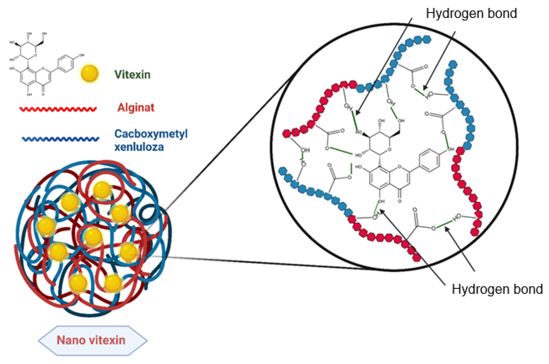
The nanovitexin system was successfully fabricated via the solvent evaporation method using a chemically cross-linked Alginate/CMC carrier. The schematic structure is illustrated in Figure 1 and Table 1.
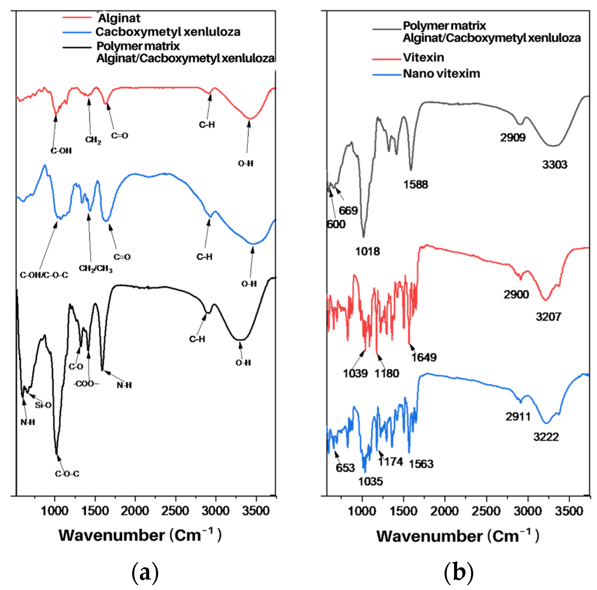
Figure 2a shows the FT-IR spectra of Alginate, Carboxymethyl cellulose (CMC), and the Alginate/CMC carrier system. Figure 2b illustrates the FT-IR spectra of the Alginate/CMC carrier, free vitexin, and the synthesized vitexin nanoparticles. Successful nano-encapsulation is evidenced by distinct shifts in characteristic absorption bands, indicating strong intermolecular interactions between the drug and the polymeric matrix. Specifically, the O-H stretching vibrations of the carrier (3303 cm−1) and vitexin (3207 cm−1) shifted to 3222 cm−1 in the nanoparticle spectrum. Additionally, the C=O peak of vitexin (1649 cm−1) and the N-H band of the carrier (1588 cm−1) merged and shifted to 1563 cm−1. Further structural confirmation is provided by the shift of the C-OH band (1180–1174 cm−1) and the presence of Si-O symmetric stretching at 653 cm−1. The slight variation in C-O-C vibration (1035 cm−1) compared to the starting materials also corroborates the formation of a stable nanostructure. These spectral shifts confirm the successful encapsulation and strong intermolecular interactions (hydrogen bonding) between vitexin and the polymeric matrix.
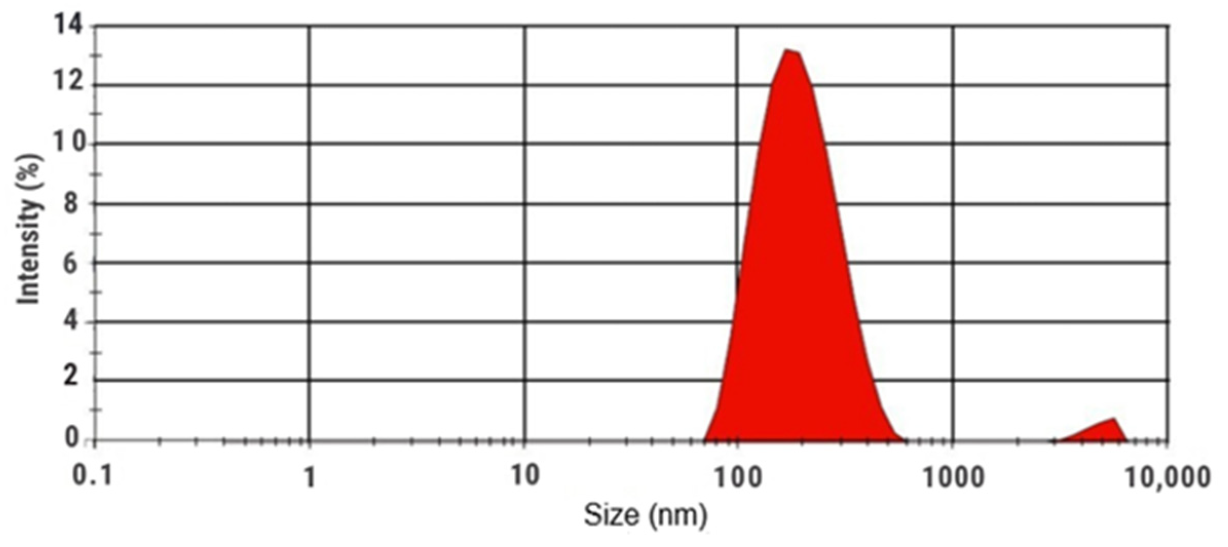
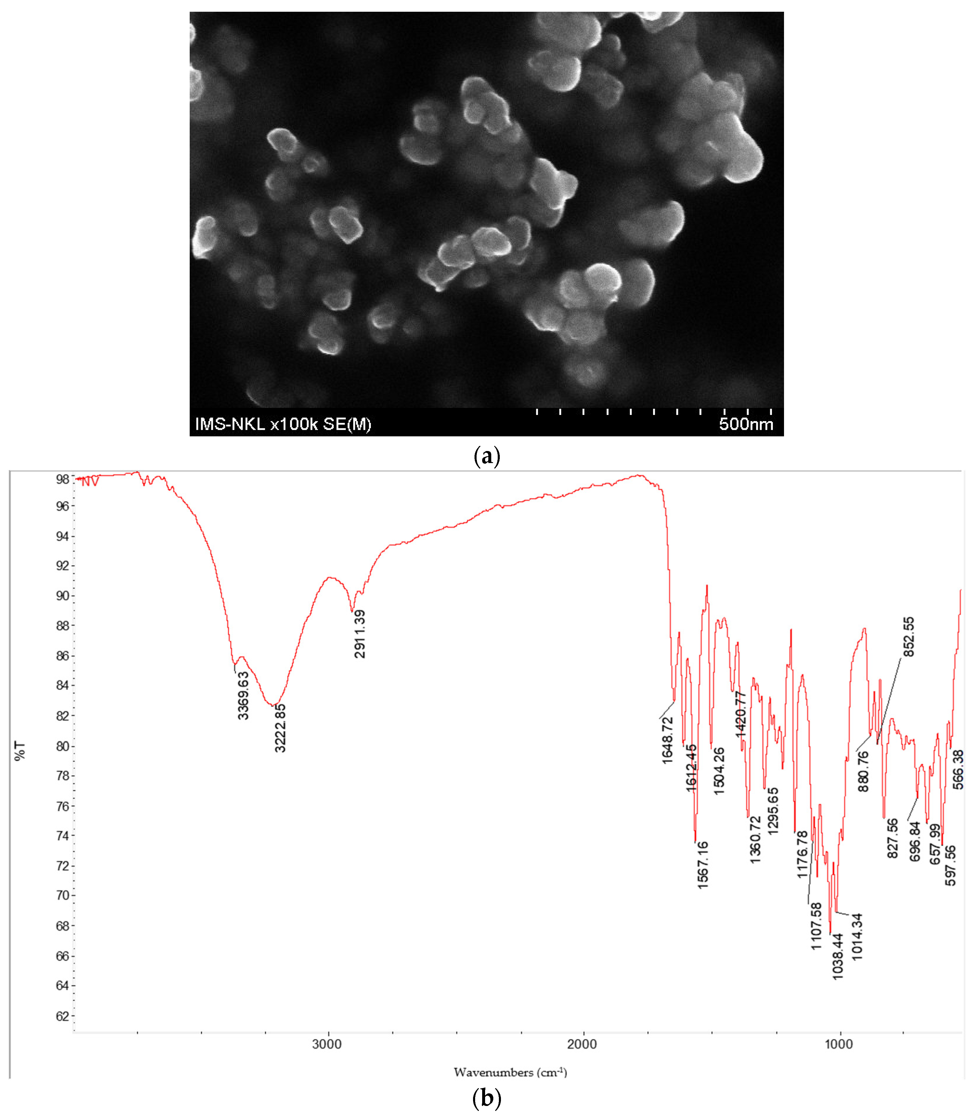
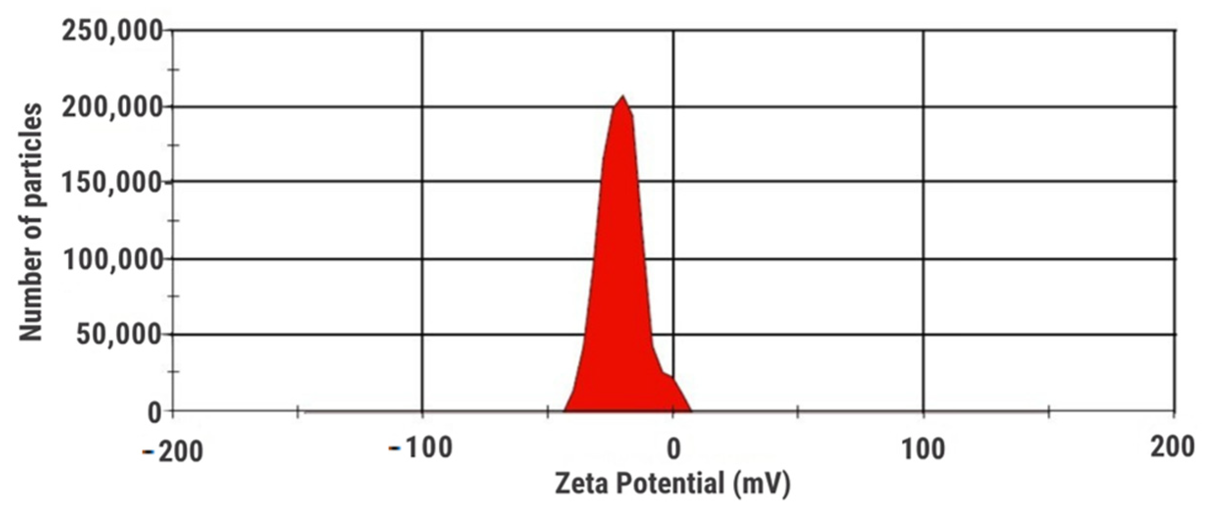
Morphological analysis via FE-SEM (Figure 3) revealed that the nanoparticles are spherical and uniform, with a dry diameter ranging from 50 to 70 nm. However, Dynamic Light Scattering (DLS) measurements indicated a larger average hydrodynamic diameter of 178.6 nm in aqueous solution (Figure 4). The nanovitexin system demonstrated excellent stability, evidenced by a zeta potential of −33.5 mV (Figure 5).

3.2. Nanoencapsulation Efficiency (EE)

The encapsulation efficiency was determined using HPLC-MS. The chromatograms showed a single peak for nanovitexin at a retention time of 20.473 min (m/z 431.500), consistent with the vitexin standard. Based on the linear regression equation (y = 52.22756x + 40.08303, R2 = 0.9986), the encapsulation efficiency was calculated to be 93.67%, as detailed in Table 2.

3.3. Enhanced Antioxidant Activity

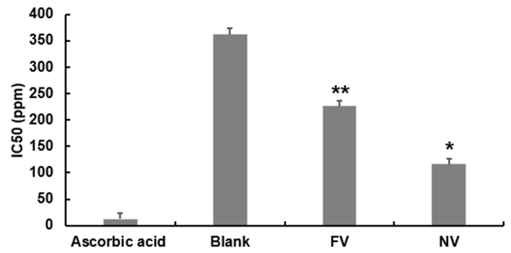
The DPPH radical scavenging assay results in Figure 6 showed that the Alginate/CMC blank carrier had negligible antioxidant activity (IC50 > 500 μg/mL). In contrast, NV exhibited significantly superior antioxidant capacity compared to FV. The IC50 value of NV was 115.38 μg/mL, nearly half that of FV (226.06 μg/mL), representing a 51.04% improvement in activity (p < 0.05).

3.4. In Vitro Antidiabetic Potential

  • α-Glucosidase Inhibition
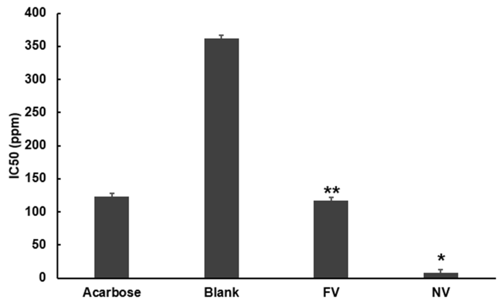
As shown in Figure 7, NV demonstrated remarkable potency with an IC50 of 2.03±0.14 μg/mL. This represents a 57.71 fold enhancement compared to FV (117.15 ± 5.40 μg/mL) and is significantly more potent than the positive control, Acarbose (123.81 ± 3.58 μg/mL).
  • α-Amylase Inhibition
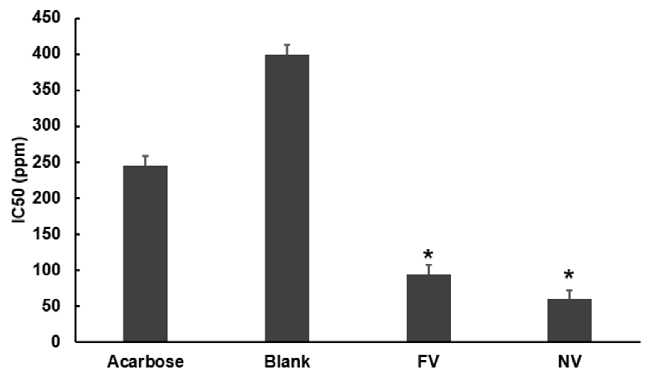
The results in Figure 8 indicate that NV inhibited α-amylase with an IC50 of 60.06 ± 0.42 μg/mL, which was 1.57 fold more effective than FV (95.02 ± 4.60 μg/mL) and superior to Acarbose (245.85 ± 7.71 μg/mL).

3.5. Cytotoxicity and Hepatoprotective Effects

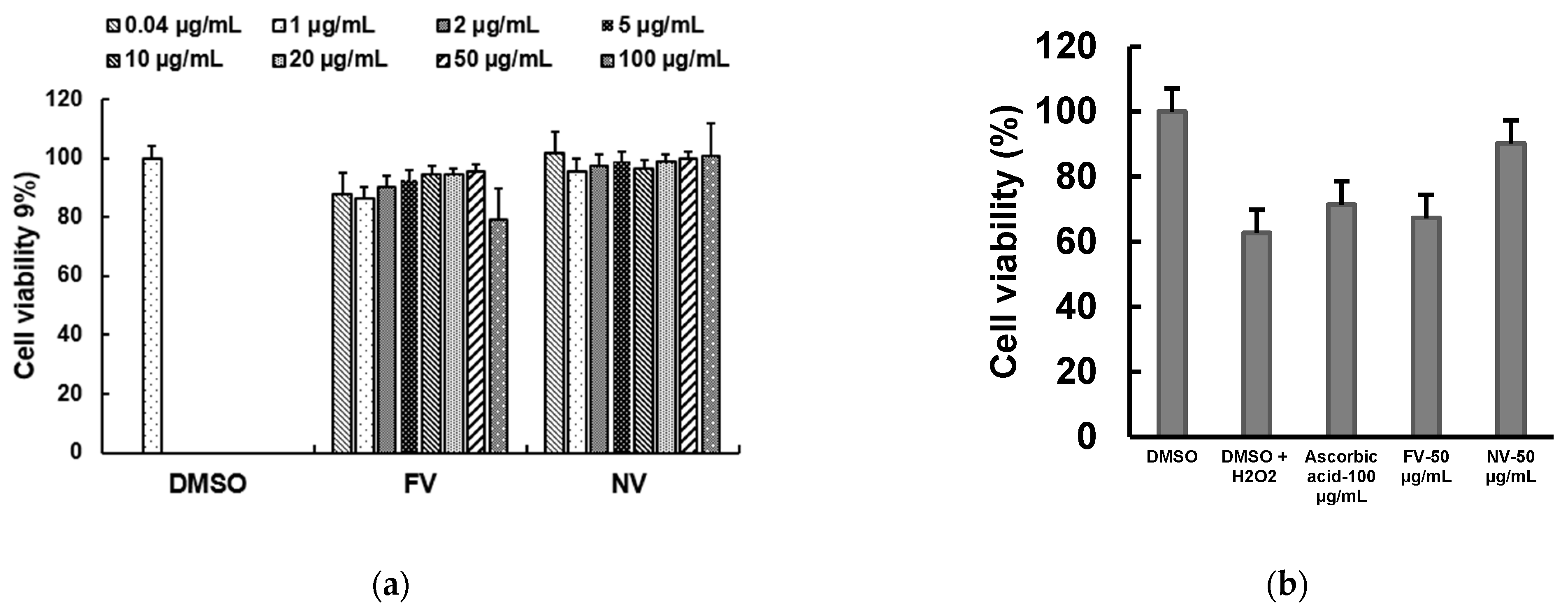
In the cytotoxicity assay (Figure 9a), both FV and NV showed no significant toxicity towards HepG2 cells at concentrations up to 100 μg/mL, maintaining cell viability above 86%. Under H2O2-induced oxidative stress (Figure 9b), the model group viability dropped to 62.67%. Treatment with NV (50 μg/mL) effectively restored cell viability to 90.24 ± 2.45%, which was significantly higher than both the FV group (74.29 ± 3.33%) and the Ascorbic acid positive control (78.42 ± 4.20%) (p < 0.05).

4. Discussion

The clinical application of vitexin is currently limited by its poor solubility and rapid metabolic elimination. In this study, we successfully developed a nanovitexin delivery system using a novel Alginate/CMC matrix. The physicochemical characterization revealed a discrepancy between the dry size observed in SEM (50–70 nm) and the hydrodynamic size in DLS (178.6 nm). This swelling behavior is advantageous for hydrophilic polymer systems, as the hydrated matrix facilitates bioadhesion and prolongs retention time [18]. Furthermore, the DLS size of <200 nm is optimal for avoiding rapid renal clearance while enabling effective tissue permeation [18]. FT-IR analysis provided molecular evidence of encapsulation. The shift in the O–H stretching vibration to 3222 cm−1 suggests strong intermolecular hydrogen bonding between vitexin and the carrier, stabilizing the drug in an amorphous state [2]. This difference is attributed to the swelling capability of the hydrophilic Alg/CMC matrix in the aqueous environment, creating a hydration shell around the particles.
Importantly, the validity of the nano-formulation is further confirmed by the particle size distribution analysis (Figure 4). The DLS profile displays a single, narrow peak, indicating a low polydispersity index (PDI value of 0.22). This unimodal distribution suggests that the synthesized nanovitexin is highly uniform and monodisperse, devoid of significant aggregation. Furthermore, the high absolute Zeta potential value of −33.5 mV corroborates the physical stability of the system, preventing particle coalescence through electrostatic repulsion. Therefore, combining the morphological evidence from FESEM (50–70 nm) with the uniformity data from DLS, we can confirm the successful fabrication of vitexin nanoparticles with a consistent size range suitable for biomedical applications.
A remarkable finding of this study is the high encapsulation efficiency of 93.67%, which surpasses previous reports for Zein/Pectin systems (67%) [18] or Alginate-Chitosan microspheres (68.92%) [19]. This efficiency likely results from the high compatibility between the polar functional groups of vitexin and the hydrophilic Alg/CMC network. Pharmacologically, the nano-formulation drastically improved the therapeutic potential of vitexin. The antioxidant activity of NV was nearly double that of FV. This aligns with Li et al. [2], who noted that nanostructuring increases the specific surface area available for redox reactions.
Regarding antidiabetic potential, NV exhibited a 57.71-fold increase in α-glucosidase inhibition compared to FV. While vitexin is known to bind to the hydrophobic pocket of α-glucosidase, its bulk form has limited affinity due to steric hindrance [20]. The reduction of particle size to the nanoscale likely facilitates rapid dissolution and molecular interaction according to the Ostwald–Freundlich principle, thereby enhancing inhibitory efficacy [3]. Interestingly, NV showed a strong but balanced inhibition of α-amylase (IC50 of 60.06 μg/mL). This profile—strong α-glucosidase inhibition coupled with moderate α-amylase inhibition—is clinically favorable for minimizing the gastrointestinal side effects often associated with synthetic inhibitors like acarbose. Finally, the study demonstrated the superior hepatoprotective effect of NV. While oxidative stress is a key driver of metabolic disorders, NV maintained HepG2 cell viability at 90.24 ± 2.45% under H2O2 challenge, outperforming even ascorbic acid. This enhanced protection is attributed to the improved intracellular uptake of the nanoparticles compared to the crystalline free drug [9,21].

5. Conclusions

This study presents the first successful fabrication of vitexin-loaded nanoparticles using a biocompatible Alginate/Carboxymethyl Cellulose matrix via chemical cross-linking and solvent evaporation. The optimized nanovitexin system exhibited uniform particle size, high stability, and exceptional encapsulation efficiency (>93%). Compared to free vitexin, the nano-formulation demonstrated significantly enhanced antioxidant activity, superior inhibition of diabetes-related enzymes (α-glucosidase and α-amylase), and remarkable cytoprotective effects against oxidative stress in HepG2 cells. These results confirm that the Alg/CMC nanocarrier effectively overcomes the biopharmaceutical limitations of vitexin. Consequently, nanovitexin emerges as a promising candidate for the development of novel functional foods or pharmaceuticals aimed at managing diabetes and oxidative stress-related disorders. Future in vivo pharmacokinetic and pharmacodynamic studies are warranted to fully translate these findings into clinical applications.

Author Contributions

Conceptualization, N.T.H.T. and H.P.T.; Methodology, N.T.H.T. and H.P.T.; Validation, N.T.M.H.; Formal Analysis, P.K.S. and N.C.H.; Investigation, N.T.H.T., H.P.T., L.T.T. and N.V.T.; Resources, P.N.L.; Writing—Original Draft Preparation, N.T.H.T., H.P.T. and P.N.L.; Writing—Review & Editing, N.T.H.T. and H.P.T.; Supervision, N.T.H.T. All authors have read and agreed to the published version of the manuscript.

Funding

This research was funded by the Vietnam Academy of Science and Technology (VAST) under grant number UDPTCN.03/22-24.

Institutional Review Board Statement

Not applicable.

Informed Consent Statement

Not applicable.

Data Availability Statement

Data is contained within the article.

Acknowledgments

The authors would like to thank the Institute of Biology and the Institute of Materials Science, Vietnam Academy of Science and Technology, for providing the necessary facilities for this research.

Conflicts of Interest

The authors declare no conflict of interest. Hemotek Joint Stock Company has no commercial conflict of interest with this work.

References

  1. Lin, J.; Ran, H.; Feng, Q.; Shen, Q.; Zhou, S.; Sun, Y.; Hou, D. Unveiling the differences between vitexin and isovitexin: From the perspective of sources, green advanced extraction technologies, biological activities, and safety. Food Chem. 2025, 485, 144600. [Google Scholar] [CrossRef] [Scilit] [PubMed]
  2. Li, S.; Lv, H.; Chen, Y.; Song, H.; Zhang, Y.; Wang, S.; Luo, L.; Guan, X. N-trimethyl chitosan coated targeting nanoparticles improve the oral bioavailability and antioxidant activity of vitexin. Carbohydr. Polym. 2022, 286, 119273. [Google Scholar] [CrossRef] [Scilit]
  3. Ranjan, R.; Kishore, K.; Ranjan, R.; Sheikh, T.J.; Jha, A.K.; Ojha, B.K.; Kumar, S.; Kumar, R. Nutraceutical Potential of Vitexin: A Flavone Glycoside. J. Phytopharmacol. 2023, 12, 44–50. [Google Scholar] [CrossRef] [Scilit]
  4. Gayathri, G.A.; Kavya, P.; Ashwini, D.; Chakraborty, E.; Ahmed, I.A.; Mahalingam, G. Vitexin isolated from Acanthus ilicifolius L. leaf enhances GLUT-4 translocation in experimental diabetic rats. Aquacult. Int. 2023, 31, 3159–3187. [Google Scholar] [CrossRef] [Scilit]
  5. Shaedi, N.; Naharudin, I.; Choo, C.Y.; Wong, T.W. Design of oral intestinal-specific alginate-vitexin nanoparticulate system to modulate blood glucose level of diabetic rats. Carbohydr. Polym. 2021, 254, 117312. [Google Scholar]
  6. Chen, P.C.; Chang, Y.C.; Tsai, K.L.; Shen, C.H.; Lee, S.D. Vitexin Suppresses High-Glucose-upregulated Adhesion Molecule Expression in Endothelial Cells through Inhibiting NF-κB Signaling Pathway. ACS Omega 2024, 9, 32727–32734. [Google Scholar]
  7. Chen, D.; Chen, Y.; Huang, F.; Zhang, X.; Zhou, Y.; Xu, L. The underlying regulatory mechanisms of colorectal carcinoma by combining Vitexin and Aspirin: Based on systems biology, molecular docking, molecular dynamics simulation, and in vitro study. Front. Endocrinol. 2023, 14, 1147132. [Google Scholar] [CrossRef] [Scilit] [PubMed]
  8. Mustapha, M.; Taib, C.N.M. Beneficial Role of Vitexin in Parkinson’s Disease. Malays. J. Med. Sci. 2023, 30, 8–25. [Google Scholar]
  9. Gu, C.; Liu, Z.; Yuan, X.; Li, W.; Zu, Y.; Fu, Y. Preparation of Vitexin Nanoparticles by Combining the Antisolvent Precipitation and High Pressure Homogenization Approaches Followed by Lyophilization for Dissolution Rate Enhancement. Molecules 2017, 22, 2038. [Google Scholar] [CrossRef] [Scilit]
  10. Eltaib, L. Polymeric Nanoparticles in Targeted Drug Delivery: Unveiling the Impact of Polymer Characterization and Fabrication. Polymers 2025, 17, 833. [Google Scholar] [CrossRef] [Scilit] [PubMed]
  11. Ma, Y.; Cong, Z.; Gao, P.; Wang, Y. Nanosuspensions technology as a master key for nature products drug delivery and in vivo fate. Eur. J. Pharm. Sci. 2023, 185, 106425. [Google Scholar] [CrossRef] [Scilit]
  12. Ngo, T.H.T.; Nguyen, T.M.H.; Ha, P.T.; Phan, K.S.; Nguyen, V.T. Characterization and Neuroprotective Potential of Vitexin Isolated from Mung Bean coats (Vigna radiata L.). J. Environ. 2025, 3, 85–92. (In Vietnamese) [Google Scholar]
  13. Zhang, G.; Li, D.; Chen, H.; Zhang, J.; Jin, X. Vitexin induces G2/M phase arrest and apoptosis via Akt/mTOR signaling pathway in human glioblastoma cells. Mol. Med. Rep. 2018, 17, 4599–4604. [Google Scholar] [CrossRef] [Scilit]
  14. Pistia-Brueggeman, G.; Hollingsworth, R.I. A preparation and screening strategy for glycosidase inhibitors. Tetrahedron 2001, 57, 8773–8778. [Google Scholar] [CrossRef] [Scilit]
  15. Wickramaratne, M.N.; Punchihewa, J.C.; Wickramaratne, D.B. In-vitro alpha amylase inhibitory activity of the leaf extracts of Adenanthera pavonina. BMC Complement. Altern. Med. 2016, 16, 466. [Google Scholar] [CrossRef] [Scilit]
  16. Ellman, G.L.; Courtney, K.D.; Andres, J.V.; Featherstone, R.M. A new and rapid colorimetric determination of acetylcholinesterase activity. Biochem. Pharmacol. 1961, 7, 88–95. [Google Scholar] [CrossRef] [Scilit]
  17. Hoang, T.M.H.; Ho, T.O.; Quach, T.Q.; Ngo, T.H.T.; Nguyen, V.H.; Dang, D.H.; Mai, H.H. Astaxanthin-loaded nanoparticles enhance cell uptake, antioxidant and hypolipidemic activities in multiple cell lines. J. Drug Deliv. Sci. Technol. 2023, 80, 104133. [Google Scholar] [CrossRef] [Scilit]
  18. Huang, X.; Li, T.; Li, S. Encapsulation of vitexin-rhamnoside based on zein/pectin nanoparticles improved its stability and bioavailability. Curr. Res. Food Sci. 2023, 6, 100419. [Google Scholar] [CrossRef] [Scilit]
  19. Huynh, M.N.; Do, H.H.; Tran, P.H.Y.; Nguyen, C.P.; Nguyen, V.H.; Nguyen, N.P.N.; Ngo, K.D.; Nguyen, D.T.; Le, M.Q. Optimization of Controlled-Release Microspheres Containing Vitexin and Isovitexin Through Experimental Design and Evaluation of Their Hypoglycemic Effects. Pharmaceutics 2025, 17, 819. [Google Scholar] [CrossRef] [Scilit] [PubMed]
  20. Yang, J.P.; He, H.; Lu, Y.H. Four flavonoid compounds from Phyllostachys edulis leaf extract retard the digestion of starch and its working mechanisms. J. Agric. Food Chem. 2014, 62, 7760–7770. [Google Scholar] [CrossRef] [Scilit] [PubMed]
  21. Marinho, L.F.; Sganzerla, W.G.; Ferreira, V.C.; Moreno, J.A.J.; Rostagno, M.A.; Forster-Carneiro, T. Advances in green extraction methods, biological properties, and applications of betanin and vitexin: An updated review and bibliometric analysis. Biocatal. Agric. Biotechnol. 2023, 51, 102744. [Google Scholar] [CrossRef] [Scilit]
Figure 1. Schematic diagram of the structure of the nanovitexin system.
Engproc 137 00010 g001
Figure 2. FT-IR Spectroscopic Analysis: (a) FT-IR spectra of Alginate, Carboxymethylcellulose (CMC), and the Alginate/CMC carrier system; (b) FT-IR spectra of the Alginate/CMC carrier system, vitexin, and nanovitexin system.
Engproc 137 00010 g002
Figure 3. (a) Morphology of the vitexin nano system by Field Emission Scanning Electron Microscopy (FESEM); (b) FT-IR spectrum of nanovitexin system.
Engproc 137 00010 g003
Figure 4. Size distribution of the nanovitexin system.
Engproc 137 00010 g004
Figure 5. Zeta potential of the nanovitexin system.
Engproc 137 00010 g005
Figure 6. Antioxidant activity of free vitexin and nanovitexin by DPPH free radical scavenging. Data are expressed as Mean ± SD (n = 3). Values with different superscript letters (*, **) in the same column indicate significant differences (p < 0.05).
Engproc 137 00010 g006
Figure 7. α-Glucosidase inhibitory activity of free vitexin, nanovitexin, Blank and acarbose. Data are expressed as Mean ± SD (n = 3). Values with different superscript letters (*, **) in the same column indicate significant differences (p < 0.05).
Engproc 137 00010 g007
Figure 8. α-Amylase inhibitory activity of free vitexin, nanovitexin, Blank and acarbose. Data are expressed as Mean ± SD (n = 3). Values with different superscript letters (*) in the same column indicate significant differences (p < 0.05).
Engproc 137 00010 g008
Figure 9. (a) Effect of free vitexin and nanovitexin on the survival of HepG2 cell lines. DMSO was used as the control group. (b) Protective effects of free vitexin and nanovitexin against H2O2-induced oxidative stress in HepG2 cell lines. Cells were pre-incubated with free vitexin or nanovitexin (50 µg/mL) or ascorbic acid (20 µg/mL) at the indicated concentrations for 24 h prior to 10 mM H2O2 exposure for 1 h. Cell viability was assessed by a MTT assay. The data are expressed as the mean ± SD (n = 3). Significant differences in the cell damage induced by H2O2 are denoted by p < 0.05.; H2O2: hydrogen peroxide.
Engproc 137 00010 g009
Table 1. Characteristic peaks on the FT-IR spectrum (cm−1).
FunctionalPeaks (cm−1)
AlginateCarboxymetyl CelluloseAlginate/Carboxymetyl Cellulose Carrier
Si-O 659
C-O-C101410801018
-C-O 13401323
-COO- 14401413
N-H 1588 and 600
-C=O16311650
C-H sp3292329402909
The -OH group in the trans isomer 3303
-OH34213470
Table 2. Vitexin content in the nanovitexin system.
SamplePeak Area (mAu·s)Vitexin Concentration in Diluted Sample (µg/mL)Vitexin Content in Nanovitexin Powder (g)Average Content (g)
1388673.636368.18374.67 ± 5.65
2398275.474377.37
3399375.696378.48
Disclaimer/Publisher’s Note: The statements, opinions and data contained in all publications are solely those of the individual author(s) and contributor(s) and not of MDPI and/or the editor(s). MDPI and/or the editor(s) disclaim responsibility for any injury to people or property resulting from any ideas, methods, instructions or products referred to in the content.