Previous Issue
Volume 5, March


Livers, Volume 5, Issue 2 (June 2025) – 7 articles

  • Issues are regarded as officially published after their release is announced to the table of contents alert mailing list.
  • You may sign up for e-mail alerts to receive table of contents of newly released issues.
  • PDF is the official format for papers published in both, html and pdf forms. To view the papers in pdf format, click on the "PDF Full-text" link, and use the free Adobe Reader to open them.
Order results
Result details
Select all
Export citation of selected articles as:
28 pages, 2657 KiB  
Review
Exploring the Classic and Novel Pathogenetic Insights of Plastic Exposure in the Genesis and Progression of Metabolic Dysfunction-Associated Steatotic Liver Disease (MASLD)
by Mario Romeo, Marcello Dallio, Fiammetta Di Nardo, Giuseppina Martinelli, Claudio Basile, Alessia Silvestrin, Giusy Senese, Annachiara Coppola, Carmine Napolitano, Angela Amoresano, Carlo Altucci and Alessandro Federico
Livers 2025, 5(2), 21; https://doi.org/10.3390/livers5020021 - 2 May 2025
Viewed by 264
Abstract
The term “plastics” is an umbrella term generally referring to any material containing a high level of polymer content as an essential ingredient. Micro(nano)plastics (MNPs) are derived from the degradation of plastics, representing exogenous substances whose exposure can potentially interfere with different physiological [...] Read more.
The term “plastics” is an umbrella term generally referring to any material containing a high level of polymer content as an essential ingredient. Micro(nano)plastics (MNPs) are derived from the degradation of plastics, representing exogenous substances whose exposure can potentially interfere with different physiological processes. In this scenario, even considering the relative paramount detoxification role, the liver emerges as a key active organ in the relationship between plastic exposure and human disease. 
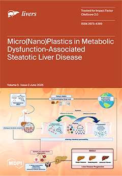
In industrialized countries, where plastics constitute largely diffused components of objects routinely adopted in daily/social life, including food packaging, Metabolic dysfunction-associated Steatotic Liver Disease (MASLD) represents the predominant hepatopathy and is progressively becoming the leading cause of cirrhosis and liver cancer, with an incompletely elucidated multifactorial pathogenesis. Notably, oral exposure to MNPs has been revealed to impact the gut–liver axis by influencing gut microbiota composition, gastrointestinal absorption, and, ultimately, determining hepatic accumulation. At the hepatic level, MNPs can contribute to the onset and worsening of steatosis by inducing metabolic dysfunction and inflammation. Plastics can also serve as vectors for different potentially toxic additives, with specific MNPs constituting a persistent source of release of bisphenol A (BPA), a well-recognized exogenous etiological factor contributing to MASLD genesis and worsening. Recently, exposure to MNPs and additives has demonstrated significant impacts on the immune system, oxidative stress, and metabolism. In particular, polystyrene-derived MNPs impair the mechanisms regulating hepatic lipid metabolism, simultaneously acting as antigens abnormally triggering the innate immune response. At the same time, environmental BPA exposure has been revealed to trigger trained immunity-related pathways, configuring novel pathogenetic drivers potentially promoting the progression of MASLD. The present review, after rapidly overviewing the main sources and toxicological properties of MNPs and related additives, explores plastic-related exposure’s potential implications in the genesis and progression of hepatic steatosis, highlighting the urgent need for further clarification of relative pathogenetic mechanisms. Full article
Show Figures

Figure 1

19 pages, 4546 KiB  
Review
Acetylcysteine Treatment of Acetaminophen Overdose: Foundational and Clinical Development
by Barry H. Rumack
Livers 2025, 5(2), 20; https://doi.org/10.3390/livers5020020 - 25 Apr 2025
Viewed by 351
Abstract
N-acetyl para-aminophenol was suggested as a safer alternative to other drugs on the market for pain and fever in 1948. It was given the generic name “acetaminophen” in 1951 and the trade name “Tylenol” when it was put on the market in the [...] Read more.
N-acetyl para-aminophenol was suggested as a safer alternative to other drugs on the market for pain and fever in 1948. It was given the generic name “acetaminophen” in 1951 and the trade name “Tylenol” when it was put on the market in the USA in 1955 as a prescription drug to treat pediatric fever. It also received the generic name “paracetamol” in the UK where it was initially marketed in 1956 under the name “Panadol.” Toxicity from overdose of acetaminophen was reported in 1966. Research at the US National Institutes of Health uncovered the mechanisms of toxicity and proposed a treatment in a foundational series of papers in 1973 and 1974. A nomogram was developed in 1973 and published in 1975 to guide estimation of patient risk of hepatic toxicity. Rapid development followed utilizing acetylcysteine given both orally and intravenously. Various protocols and methods of administration have been employed over time with the primary use today of acetylcysteine intravenously as the therapeutic method. The nomogram has been revised over time to the current version, published in 2023, which allows stratification of patients to a high-risk group over 300 mg/L at 4 h and standard risk above 150 mg/L at 4 h, except in the UK where the standard risk is defined very conservatively with a line above 100 mg/L at 4 h. Adjunct therapy with fomepizole in patients with massive ingestions, delay until arrival in a health care facility or renal injury has been proposed. The mortality rate with treatment has been substantially reduced and recovery from hepatic injury is achieved in almost all patients. Full article
(This article belongs to the Special Issue Recent Advances in Acetaminophen Hepatotoxicity)
Show Figures

Figure 1

10 pages, 2145 KiB  
Communication
RANTES, IP-10 and MCP-1 Profiles in Patients with Autoimmune Hepatitis (AIH) at Baseline and During Immunosuppressive Treatment
by Giuseppe Colucci, Enrico Sguazzini, Sara Uceda Renteria, Riccardo Perbellini, Ferruccio Ceriotti, Clara Dibenedetto, Maria Francesca Donato and Pietro Lampertico
Livers 2025, 5(2), 19; https://doi.org/10.3390/livers5020019 - 23 Apr 2025
Viewed by 224
Abstract
Background and aims: The CC5, CXC3, and CC2 chemokines (CK) are known to play a role in the pathogenesis of autoimmune hepatitis (AIH). However, no data are available on their potential utility as markers of disease progression or response to treatment. Material and [...] Read more.
Background and aims: The CC5, CXC3, and CC2 chemokines (CK) are known to play a role in the pathogenesis of autoimmune hepatitis (AIH). However, no data are available on their potential utility as markers of disease progression or response to treatment. Material and methods: We analyzed their role as markers of remission in a population of patients with AIH. We retrospectively investigated the kinetics of RANTES (CCL5), IP-10 (CXCL10), and MCP-1 (CCL2) in 48 patients with AIH at the time of treatment initiation and also in 32 at biochemical, clinical and histological remission. Forty-nine healthy donors (HDs) served as controls. Results: At baseline, IP-10 and MCP-1 levels were higher in AIH patients than in HDs (261 vs. 101 pg/mL and 689 vs. 330 pg/mL, p < 0.01), and RANTES levels showed no differences. Correlations were observed between RANTES and IgG concentrations (r = 0.36 p = 0.04) and between IP-10 and Ishak’s grade (r = 0.52 p = 0.02). At remission, in 32 patients, while IP-10 and MCP-1 values showed a significant decrease from baseline reaching HD levels (261 vs. 106 pg/mL and 689 vs. 387 pg/mL, p < 0.01), RANTES did not. However, two kinetics patterns emerged, with 20 patients showing lower and 12 higher baseline RANTES values compared to HDs (29,450 pg/mL and 70,960 pg/mL vs. 52,010 pg/mL, p < 0.01). The former required longer treatment to reach remission and had higher Ishak’s grades than the latter (p < 0.01). Conclusions: RANTES, IP-10, and MCP-1 may help in predicting response to treatment and stable remission and in supporting the decision if and when to discontinue immune suppressive therapy. Full article
Show Figures

Figure 1

13 pages, 3901 KiB  
Article
Comparison of Differentially Expressed Genes in Human Versus in Chimeric Mouse Livers Following HBV Infection
by Huarui Bao, Masataka Tsuge, Serami Murakami, Yasutoshi Fujii, Shinsuke Uchikawa, Hatsue Fujino, Atsushi Ono, Eisuke Murakami, Tomokazu Kawaoka, Daiki Miki, Clair Nelson Hayes and Shiro Oka
Livers 2025, 5(2), 18; https://doi.org/10.3390/livers5020018 - 14 Apr 2025
Viewed by 205
Abstract
Background/Objectives: Hepatitis B virus (HBV) infection is a worldwide health problem responsible for chronic liver disease and hepatocellular carcinoma. Both innate immunity and the adaptive immune response play central roles in the development of chronic hepatitis and liver cancer. We previously performed a [...] Read more.
Background/Objectives: Hepatitis B virus (HBV) infection is a worldwide health problem responsible for chronic liver disease and hepatocellular carcinoma. Both innate immunity and the adaptive immune response play central roles in the development of chronic hepatitis and liver cancer. We previously performed a comprehensive analysis of gene expression in the livers of HBV-infected chimeric mice and found that several genes associated with cell growth or carcinogenesis via hypoxia and KRAS signaling were upregulated by HBV infection. However, due to the absence of adaptive immunity in uPA/SCID chimeric mice, we were unable to analyze the effect of the host immune response. Methods: In this study, we compared gene expression profiles in the livers obtained from HBV-infected chimeric mice with those of HBV carriers. Results: After HBV infection, the expression of genes associated with inflammation and immune response, especially involving the Th1 and Th2 activation pathways, was altered as HBV-specific intracellular immune responses both in vivo and in clinical samples. Interestingly, the proinflammatory gene IL12A was induced by HBV infection in the chimeric mouse livers but not in the human livers, and associated genes, such as SRDA5A2, AR, and CCR3, showed differential alteration by HBV infection between the chimeric mouse and human livers. Conclusions: These results suggest that hepatocarcinogenesis may be suppressed by host immunity in HBV carriers. This study highlights potential new implications for inhibiting the progression of HBV-related liver diseases, including hepatocarcinogenesis. Full article
Show Figures

Figure 1

30 pages, 2589 KiB  
Review
The Intestinal Thread of Fate: How the Microbiota Shapes the Story of Liver Disease
by Carlo Acierno, Riccardo Nevola, Luca Rinaldi, Ferdinando Carlo Sasso, Luigi Elio Adinolfi and Alfredo Caturano
Livers 2025, 5(2), 17; https://doi.org/10.3390/livers5020017 - 10 Apr 2025
Viewed by 251
Abstract
Metabolic dysfunction–associated steatotic liver disease (MASLD) is a multifactorial condition linked to liver injury, insulin resistance, and disrupted gut–liver interactions. A key aspect of MASLD pathogenesis is the dysfunction of intestinal barriers, including mechanical, immunological, and microbial alterations that amplify liver damage. The [...] Read more.
Metabolic dysfunction–associated steatotic liver disease (MASLD) is a multifactorial condition linked to liver injury, insulin resistance, and disrupted gut–liver interactions. A key aspect of MASLD pathogenesis is the dysfunction of intestinal barriers, including mechanical, immunological, and microbial alterations that amplify liver damage. The disruption of tight junctions and increased intestinal permeability allow microbial products, such as lipopolysaccharides, to enter the bloodstream, triggering liver inflammation via Kupffer cell activation. In MASLD, the gut vascular barrier is also compromised, marked by increased expression of PV-1. Additionally, dysbiosis, driven by high-fat, high-sugar diets, shifts the gut microbiota toward pro-inflammatory species, exacerbating systemic inflammation and intestinal permeability. This imbalance activates Toll-like receptor signaling, which promotes endotoxin-induced liver injury. Gut dysbiosis further impairs lipid metabolism, contributing to hepatic steatosis and MASLD progression. The gut–liver axis plays a critical role, with factors like altered bile acid metabolism and toxic metabolites such as hydrogen sulfide worsening intestinal barrier function and fueling chronic inflammation. This review aims to explore the complex role of the gut–liver axis in MASLD progression, highlighting the mechanisms of intestinal barrier dysfunction, dysbiosis, and microbial contributions to liver injury. It also discusses therapeutic strategies targeting intestinal barriers, including dietary and microbiota-based interventions, while acknowledging the challenges of personalized treatment approaches. Future research should focus on multi-omics technologies and the safety and efficacy of microbiota-targeted therapies in MASLD management. Full article
Show Figures

Figure 1

24 pages, 1632 KiB  
Article
Comparative Analysis of the Human Proteome Profile in Visceral Adipose and Liver Tissue in Individuals with Obesity with and Without MASLD and MASH
by Julie S. Pedersen, Lili Niu, Nicolai J. Wewer Albrechtsen, Viggo B. Kristiansen, Inge Marie Poulsen, Reza R. Serizawa, Torben Hansen, Lise Lotte Gluud, Sten Madsbad and Flemming Bendtsen
Livers 2025, 5(2), 16; https://doi.org/10.3390/livers5020016 - 10 Apr 2025
Viewed by 353
Abstract
Background/Objectives: Visceral adipose tissue (VAT) may play a direct role in the development of metabolic dysfunction-associated steatotic liver disease (MASLD) and its progression to metabolic dysfunction-associated steatohepatitis (MASH). In this study, we employed untargeted proteomics analyses on paired biopsies from VAT and liver [...] Read more.
Background/Objectives: Visceral adipose tissue (VAT) may play a direct role in the development of metabolic dysfunction-associated steatotic liver disease (MASLD) and its progression to metabolic dysfunction-associated steatohepatitis (MASH). In this study, we employed untargeted proteomics analyses on paired biopsies from VAT and liver tissues of patients with obesity, MASLD, and MASH. Our objective was to investigate tissue-specific protein expression patterns in search of a potential proteomic signature associated with MASH in both VAT and liver tissue. Methods: VAT and liver tissue were collected from 70 subjects with severe obesity (SWOs) and nine control study subjects without obesity (CON). SWOs were stratified on the basis of liver histology into LS− (no liver steatosis), LS+ (liver steatosis), and MASH. Peptides were extracted from frozen tissue and were analyzed by liquid chromatography coupled to tandem mass spectrometry (LC-MS/MS). Raw files were analyzed with Spectronaut, proteins were searched against the human FASTA Uniprot database, and the significantly expressed proteins in the two tissues were analyzed. The p-values were false discovery rate (FDR) corrected. Results: A total of 59 VAT and 42 liver proteins were significantly differentially expressed between the four groups: LS−, LS+, MASH, and CON. The majority were upregulated, and many were related to lipid metabolism. In VAT, only one protein, the mitochondrial sulfide:quinone oxidoreductase (SQOR), was significantly downregulated in the MASH group only. In liver tissue from patients with MASH, six proteins were significantly altered compared with the three other groups. Correlation analyses between the top 10 positive VAT and liver proteins were dominated by inflammatory and detoxification proteins. Conclusions: The presence of MASH was not reflected in the VAT proteome, and both the VAT and the liver proteome were generally affected more by the presence of obesity than by MASLD severity. Several immunomodulating proteins correlated significantly between VAT and liver tissue and could reflect common pathophysiological characteristics. Full article
(This article belongs to the Special Issue Liver Fibrosis: Mechanisms, Targets, Assessment and Treatment)
Show Figures

Figure 1

42 pages, 1309 KiB  
Review
An Integrated Pathogenetic Model of Primary Biliary Cholangitis
by Elias Kouroumalis, Ioannis Tsomidis and Argyro Voumvouraki
Livers 2025, 5(2), 15; https://doi.org/10.3390/livers5020015 - 28 Mar 2025
Viewed by 722
Abstract
The pathogenesis of primary biliary cholangitis (PBC) is not fully understood. Despite recent progress, many aspects require further clarification. Thus, PBC is regarded as an autoimmune disease, but immunosuppressive treatment, which is effective in other autoimmune diseases, is not working in the case [...] Read more.
The pathogenesis of primary biliary cholangitis (PBC) is not fully understood. Despite recent progress, many aspects require further clarification. Thus, PBC is regarded as an autoimmune disease, but immunosuppressive treatment, which is effective in other autoimmune diseases, is not working in the case of PBC. Moreover, there are controversies over the pathogenetic role of anti-mitochondrial antibodies as mitochondria are present in all cells but only cholangiocytes are damaged. In this review, all the proposed models and factors that have been involved in the pathogenesis of PBC are presented. They include mechanisms such as dysregulated autophagy, senescence, apoptosis, impairment of the protective bicarbonate umbrella, immunological abnormalities, the dysbiosis of gut microbiota, and the role of bile acids. Genetics of PBC and epigenetic transcriptional modifications are also presented. Data supporting molecular mimicry and the viral etiology of PBC are analyzed. Finally, an integrated model is proposed based on interactions of the factors that may participate in PBC pathogenesis. Therefore, the purpose of this review is to provide a unifying presentation of the various aspects of PBC pathophysiology, which will allow for a better understanding of this multifaceted disease. New treatment targets may also be identified in such a holistic model. Full article
Show Figures

Figure 1

Previous Issue
Back to TopTop