Review

Exploring the Classic and Novel Pathogenetic Insights of Plastic Exposure in the Genesis and Progression of Metabolic Dysfunction-Associated Steatotic Liver Disease (MASLD)

1
Hepatogastroenterology Division, Department of Precision Medicine, University of Campania Luigi Vanvitelli, Piazza Miraglia 2, 80138 Naples, Italy
2
Department of Chemical Sciences, University of Naples Federico II, Strada Comunale Cinthia 26, 80126 Naples, Italy
3
Department of Advanced Biomedical Sciences, University of Naples Federico II, Via Sergio Pansini 5, 80131 Naples, Italy
*
Author to whom correspondence should be addressed.
Livers 2025, 5(2), 21; https://doi.org/10.3390/livers5020021
Submission received: 10 March 2025 / Revised: 16 April 2025 / Accepted: 25 April 2025 / Published: 2 May 2025

Abstract

:
The term “plastics” is an umbrella term generally referring to any material containing a high level of polymer content as an essential ingredient. Micro(nano)plastics (MNPs) are derived from the degradation of plastics, representing exogenous substances whose exposure can potentially interfere with different physiological processes. In this scenario, even considering the relative paramount detoxification role, the liver emerges as a key active organ in the relationship between plastic exposure and human disease. In industrialized countries, where plastics constitute largely diffused components of objects routinely adopted in daily/social life, including food packaging, Metabolic dysfunction-associated Steatotic Liver Disease (MASLD) represents the predominant hepatopathy and is progressively becoming the leading cause of cirrhosis and liver cancer, with an incompletely elucidated multifactorial pathogenesis. Notably, oral exposure to MNPs has been revealed to impact the gut–liver axis by influencing gut microbiota composition, gastrointestinal absorption, and, ultimately, determining hepatic accumulation. At the hepatic level, MNPs can contribute to the onset and worsening of steatosis by inducing metabolic dysfunction and inflammation. Plastics can also serve as vectors for different potentially toxic additives, with specific MNPs constituting a persistent source of release of bisphenol A (BPA), a well-recognized exogenous etiological factor contributing to MASLD genesis and worsening. Recently, exposure to MNPs and additives has demonstrated significant impacts on the immune system, oxidative stress, and metabolism. In particular, polystyrene-derived MNPs impair the mechanisms regulating hepatic lipid metabolism, simultaneously acting as antigens abnormally triggering the innate immune response. At the same time, environmental BPA exposure has been revealed to trigger trained immunity-related pathways, configuring novel pathogenetic drivers potentially promoting the progression of MASLD. The present review, after rapidly overviewing the main sources and toxicological properties of MNPs and related additives, explores plastic-related exposure’s potential implications in the genesis and progression of hepatic steatosis, highlighting the urgent need for further clarification of relative pathogenetic mechanisms.

1. Background

1.1. Introducing the “World” of Plastics

1.1.1. Principal Definitions

In recent years, the growing scientific focus on polymeric waste, particularly plastics, is primarily due to the recognized adverse effects of these substances on human health [1]. The term “plastics” refers to any material containing a high level of polymer (i.e., a type of macromolecule composed of many repeated subunits) as an essential ingredient. Plastics consist of an assembly of polymers [polyethylene (PE), polypropylene (PP), polystyrene (PS), polyvinyl chloride (PVC), polyethylene terephthalate (PET), polycarbonate (PC), poly methyl-methacrylate (PMMA), polyurethane (PU), etc.] and additives (stabilizers, flame retardants, fillers, pigments, and plasticizers, including bisphenols) to increase their performance [1].
The massive production of plastics, together with their poor biodegradability and insufficient recycling has led to widespread environmental contamination by this “plague”, with serious consequent repercussions for animal and human health [1]. In particular, the degradation of plastics (photodegradation, oxidation, hydrolytic degradation, and biodegradation) produces different forms and sizes of debris: nanoplastics (NPs) (≤0.1 μm), microplastics (MPs) (<5 mm), mesoplastics (0.5–5 cm), macroplastics (5–50 cm), and megaplastics (>50 cm). Among the vast array of substances, MPs and NPs represent a significant focus of study due to their ability to induce toxic damage to the organism [2].
MPs are solid synthetic particles that are insoluble in water or polymeric matrices of primary or secondary origin with regular or irregular shapes and linear size ranging from 1 μm to 5 mm [1]. Contrariwise, the precise definition of NPs remains a subject of debate. Some researchers define them as particles with sizes in the 1 nm–1 μm range, while others align with the European Commission’s description of engineered nanomaterials (ENMs), defining them as particles measuring between 1 nm and 100 nm in at least one dimension [3]. NPs can originate from various degradation processes of larger plastics, including the fragmentation of plastic materials in waste and the degradation of larger plastic products through exposure to environmental factors such as sunlight, temperature, and humidity. They can also result from industrial production, where plastic particles are intentionally used in microscopic forms, such as additives or compounds in certain products [3].
Looking beyond micro(nano)plastics (MNPs) and focusing on the related additives, relevantly, plastic particles can serve as transportation vectors for different potentially toxic compounds, particularly referring to plasticizers, including bisphenols [4]. As an example, recently, low-density PE- and PC-MP particles have been proven to be a persistent source of releasing bisphenol A (BPA) [4].
BPA is a synthetic organic Endocrine-Disrupting Chemical (EDC) with a molecular weight of 22,829 Dalton (Da), which is included in the group of diphenylmethane and bisphenol derivatives primarily used in the production of polycarbonate plastics and epoxy resins. It has been widely used in the manufacturing of products such as food and beverage packaging, water bottles, medical devices, and coatings for food cans [5,6,7]. BPA has gained significant attention due to its potential endocrine-disrupting effects, leading to concerns about its safety and impact on human health, mainly regarding several chronic degenerative diseases such as cancer, diabetes, infertility, and hepatic and cardiovascular diseases [6,8,9,10].
Thus, to propose effective prevention strategies at a global level, the definition of all the possible sources of exposure, both for plastics and additives, represents an urgent need.

1.1.2. Main Social Sources of Exposure, Absorption, and Accumulation of Plastics

Human exposure to MPs and NPs can occur through three main routes: ingestion (via the digestive system), inhalation (via the lungs), and, potentially, direct skin contact [11]. Consistently in humans, MPs have been detected in different sites, such as lungs [12], hair, skin [13], blood, and the gastrointestinal tract [14].
The potential toxicity of MPs and NPs in humans and the subsequent associated issues can be attributed to several socio-epidemiological factors. First and foremost, the continuous and alarming increase in the production and emission of plastics into the surrounding environment, simultaneously with their long persistence in the environment, represents a constant source of respiratory exposure [1]. Regarding this, the increased consumption of disposable face masks during the Severe Acute Respiratory Syndrome (SARS-CoV-2) pandemic dramatically increased human contact via inhalation of these particles [7].
Even though inhalation constitutes a relevant issue, oral exposure continues to represent the leading source of exposure in Western industrialized countries [1]. In this area, the consumption of contaminated food and water is one of the main routes through which exposure to MPs and NPs occurs [1]. In particular, “packaged” food represents a critical source of exposure, including milk, honey, sugar, salt, and fish [15,16,17,18], since these particles are commonly present and ingested by aquatic species even in the marine environment [11]. Moreover, MPs and additives are also released into beverages through contact with plastic containers. About this, a recent study detected MPs by FTIR spectroscopy in drinking water from various sources in a metropolis, alarmingly revealing the presence of BPA in a relevant proportion (9.74%) of analyzed particles [19].
Aiming to explore absorption after ingestion, in a recent pilot study, Hartmann C. et al. [20] investigated the influence of different plastic use and food consumption scenarios on MPs ‘content in stool reflecting oral intake by performing an interventional pilot study. In all samples, MP particles were detected with median concentrations of up to 3.5 particles/g in stool in the MP size fraction of 50–500 μm. However, the use of plastics for food packaging/preparation and the consumption of highly processed food were statistically significantly associated with MP content in the stool [20]. Therefore, despite providing novel findings, the small sample size, the limited possibility of being able to control the regimen diet of the participants, and the chance that the presence of MPs in the feces could also be due to other sources of contamination (i.e., air- or environment-derived) represent the relevant limitations of this research [20].
Once absorbed, plastics can induce damage in the human body through accumulation and various toxicity-related mechanisms [21]. Although numerous studies have been conducted on the damage induced by MPs in both in vitro and in animal models, there is still a limitation in terms of determining the presence of MPs in human tissues through various analytical techniques [22].
Future research should focus on the quantification of MPs in human tissues, the combined effects of MPs with other contaminants, and their impact on pre-existing diseases. From this perspective, it is crucial to prioritize research in this area to gain a deeper understanding of the potential repercussions of MNPs on human health, simultaneously clarifying toxicological pathogenetic-related mechanisms.

1.1.3. General Toxicological Properties of MNPs Contributing to Human Disorders

MNPs represent one of the most relevant environmental and health challenges of our time, since growing concerns regarding their presence in the environment and food have sparked increasing interest within the scientific community, whose research efforts to assess the risks to human health have been massive.
Focusing on toxicological properties, the differentiation between MPs and NPs is essential, as strong evidence demonstrates that the specific type of particle, along with factors such as particle size and shape, can lead to distinct toxicological impacts on the organism [23].
A study conducted by Bing Wu et al. compared the cytotoxic effects of 0.1 μm and 5 μm polystyrene microplastics (PS-MPs) on human colon adenocarcinoma Caco-2 cells [24]. Specifically, it was observed that the cellular damage induced by MPs was size-dependent. PS-MPs of 0.1 μm and 5 μm induced low toxicity, as determined by cell viability, intracellular reactive oxygen species (ROS) levels, and membrane integrity, while 5 μm PS-MPs caused mitochondrial depolarization and alteration of ATP synthesis [24].
Numerous in vitro studies have shown that both NPs and MPs can induce various types of cellular damage, including apoptosis, ROS production, and modulation of pro-inflammatory cytokines [25,26]. Poma et al. demonstrated that in Human Fibroblast Hs27 cells, the induced stimulus by poly(methyl-methacrylate)-based plastic nanoparticles (PNP-type NPs) is attributable to the increased production of ROS [25]. In another study, Mattioda et al. assessed the cytotoxicity, oxidative stress, inflammation, and associated genetic modulation following exposure to PS-MPs in the epithelial cell lines HRT-18 and CMT-9 [26], revealing an increase in superoxide dismutase (SOD) activity in the CMT-93 cell line and an upregulation of the interleukin (IL-)-8 gene in the HRT-18 cell line [26].
W. A. da Silva Brito et al. reported particle accumulation in rodents exposed to MP and NP in several tissues, finding local inflammation, oxidative stress, and metabolic disruption, leading to gastrointestinal toxicity and hepatotoxicity [27].
In addition to MNPs, there is a substantial body of literature supporting BPA exposure implications in the pathogenesis of various liver diseases, particularly hepatic steatosis, commonly known as fatty liver disease and characterized by the accumulation of fat within liver cells [28]. In this regard, a significant role in the worsening of hepatic disease has been associated with the endocrine-disrupting properties of this plastic additive [29]. The actions of BPA as an endocrine disruptor compound occur at several levels, including the liver, and are reported in Table 1.
Table 1 summarizes the main toxicological properties of plastics and plastic additives and reports the leading pathogenetic mechanisms.
Altogether, this evidence suggests the relevance of investigating the implications of exposure to plastic and related additives, particularly in relation to hepatic human disorders.

1.2. Plastics and Liver: A Consolidated Physiological and Pathological Binomial

MNPs with related additives are “xenobiotics”, intended as exogenous substances that the human body cannot produce, whose exposure can potentially interfere with various physiological processes in different human organs [31,32].
In this scenario, the liver emerges undoubtedly as a key organ in the relationship between xenobiotics and disease [33]. Its role has been well established for years in pharmacology, as it serves as an important first site of interaction in the pharmacokinetics of drugs, both in terms of biotransformation through enzymatic reactions catalyzed by microsomal enzymes (e.g., cytochrome P450) and in the hepatobiliary excretion of these substances [33].
Relevantly, in industrialized countries, where plastics constitute largely diffused components of objects routinely adopted in daily social life (including food packaging) [34], the manifestations of Steatotic Liver Disease in association with metabolic dysfunction (i.e., obesity, type 2 diabetes, arterial hypertension, and dyslipidemia) constitute Metabolic dysfunction-associated Steatotic Liver Disease (MASLD), representing the predominant hepatopathy and progressively becoming the leading cause of cirrhosis and liver cancer, with an incompletely elucidated pathogenesis [35].
While the concept of MASLD has only recently been introduced [35] to characterize what may be regarded as an epiphenomenon of metabolic syndrome, thus situating liver disease within the broader framework of a systemic disorder, it has long been recognized that metabolic liver disease arises from a multifactorial pathogenic process, which includes genetic background and environmental factors, including unhealthy lifestyle habits and exposure to routine Western-used pollutants [28,36,37,38,39,40].
Among the environmental contaminants and xenobiotics proposed as potential etiological factors in SLD onset and progression, BPA has been the subject of extensive investigation in the hepatological research field [28]. BPA undergoes hepatic metabolism to form bisphenol A glucuronide, which is primarily excreted in the urine in this conjugated form [28].
The above-mentioned background has prompted extensive research to further investigate the role of BPA, particularly in the context of liver diseases, and, in line with the above-reported toxicological properties (disruption of endocrine pathways, pro-oxidative role, and pro-inflammatory response promoter; Table 1), the scientific literature currently recognizes BPA as an environmental pathogenetic factor in MASLD [28].
In contrast, the role of MNPs in (MA)SLD remains surrounded by uncertainty and debate, representing an incompletely explored scenario with an urgent need to be further investigated, even considering the close constitutional relationship between plastic particles and additives, including bisphenols [1]. Nevertheless, a substantial body of literature exists concerning the potential of MNPs to impact the intestinal microbiota, modulate the immune system, and induce insulin resistance (IR)—pathways that are also crucial pathogenetic moments implicated in the genesis and progression of MASLD.
In light of these considerations, this research aims to critically examine the current knowledge regarding how MNPs (and related additives) may interfere with metabolic processes and the associated pathways that could contribute to the onset and progression of SLD in humans. For this purpose, the present review presents the state-of-the-art evidence supporting the potential implications of MNP exposure in the most crucial MASLD physiopathological moments, from the potential “direct” effects of these particles on lipidic hepatic metabolism to the consequences on liver fat accumulation mediated by altered gut microbiota composition and functioning with impaired intestinal permeability (gut–liver axis), as well as by the well-known “MASLD pathogenetic triangle” [inflammation, oxidative stress, and immune dysfunction] [35].

2. Plastic Exposure Influences Hepatic Lipid Metabolism Driving Steatosis Progression

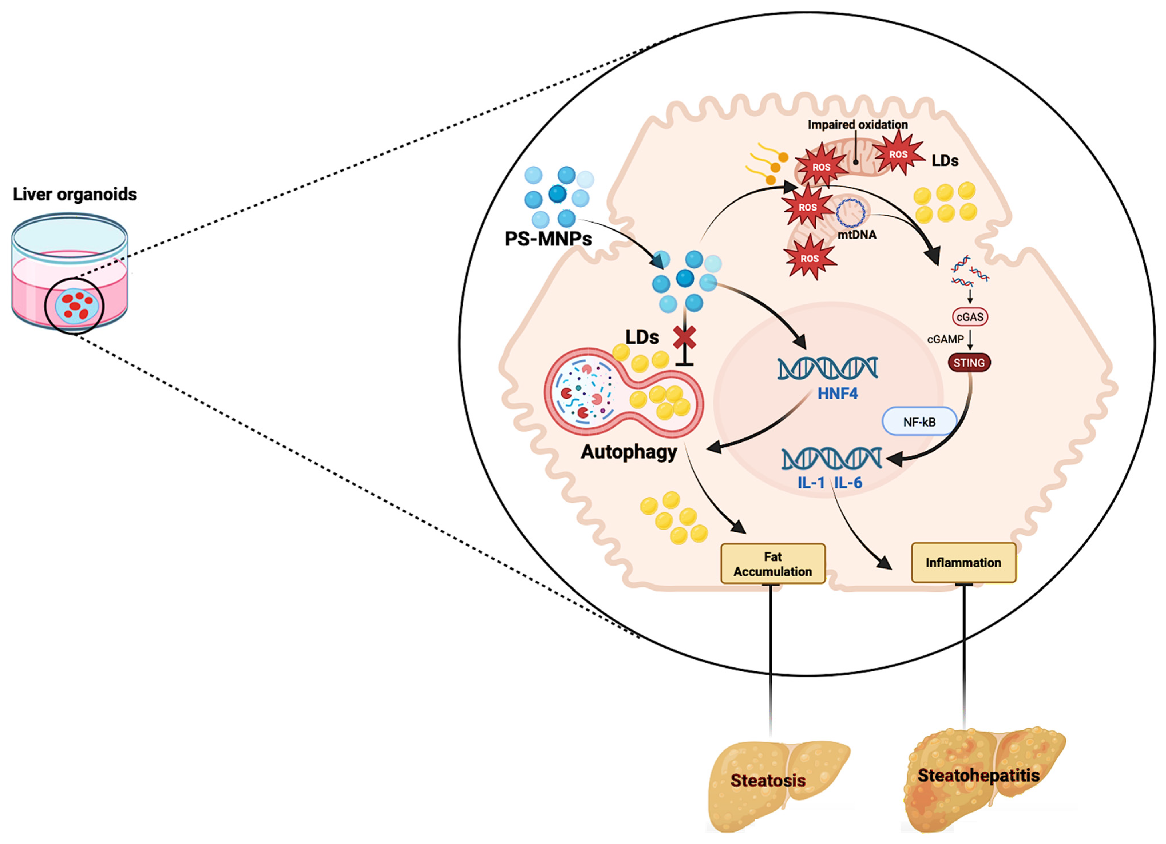
2.1. Plastic Exposure Contributes to Hepatic Fat Accumulation via Impacting Lipophagy

Hepatic fat accumulation represents the consequence of disrupted glycolipid metabolism and the crucial pathogenetic moment in the genesis of steatosis [35]. Interestingly, emerging preclinical evidence suggests the potential role of MNP exposure in influencing the physiopathological mechanisms contributing to this event [35].
In support of this, Zhao Y et al. orally exposed adult male zebrafish to two different PS-MNP concentrations (20 or 100 μg/L) for 21 days, aiming to assess the hepatic effects related to glycolipid metabolism at the biochemical and transcriptomic levels [41]. The authors reported a significant decrease in major glycolipid-related metabolism genes in the liver, as well as in the levels of major biochemical parameters (including pyruvic acid, α-ketoglutaric acid, and IDH), especially in the higher concentration (100 μg/L) PS-MP-treated group [41]. In addition, the transcriptomic liver data revealed that crucial metabolic genes (related to fatty acid metabolism, amino acid metabolism, and carbon metabolism) tended to be decreased, confirming that PS-MP can induce hepatic glycolipid metabolism disorder at the physiological, biochemical, and transcriptomic levels [41].
Consistent with this, modern in vitro research has supported the potential role of PS-MNP in inducing hepatotoxicity and disrupting lipid metabolism, both in the case of repeated and non-static (i.e., non-continuative) exposure to PS-MP [42,43]. Concerning the first, Guraka et al. investigated long-term low repeated (three weeks) PS-MP exposure in a functionally active 3D liver microtissue model, composed of primary human hepatocytes, Kupffer cells, sinusoidal endothelial cells, and hepatic stellate cells [43]. The authors highlighted time-dependent (up to 504 h) aberrations in the tissue architecture, including dilated bile canaliculi and large lipid droplets inside the hepatic cells, simultaneously revealing the accumulation of the materials within the cells of microtissue, predominately in the organ macrophages [43].
On the other hand, regarding the second exposure pattern (i.e., non-continuative), Cheng et al. adopted a novel in vitro 3D model, generating liver organoids (LOs) from human pluripotent stem cells, and explored the adverse biological effect of PS-MP microbeads (1 μm), revealing the impact of non-static exposure to PS-MP on the expression of cytochrome P450 2E1 (CYP2E1), a key monooxygenase involved in fatty acid metabolism, as well as of hepatic hepatocyte nuclear factor 4 alpha (HNF4-alpha) [42]. HNF4-alpha is considered the master regulator of liver-specific gene expression involved in the basic metabolism and has been shown to attenuate fibrosis and cirrhosis in mouse models, as well as to regulate hepatic fat storage via inducing lipophagy, a form of autophagy that involves the fusion of lipid droplets (LDs) [44].
Along the same lines, a brilliant recent study by Fan Z et al., aiming to explore how PS-NPs influence lipid metabolism in hepatocytes via lipophagy, revealed that PS-NPs, after their internalization in human hepatocytes, promote the accumulation of LDs, with autophagy inhibition exacerbating this accumulation [45]. Interestingly, after this crucial pathogenetic moment, the authors also found that PS-NPs activate lipophagy by recruiting LDs into autophagosomes through the 5′AMP-activated protein kinase (AMPK/ULK1) pathway and blocking the lipophagic flux by impairing lysosomal function, inhibiting LD degradation [45]. Comprehensively, these findings elucidated crucial mechanisms concerning the role of plastics in hepatic fat accumulation, suggesting that PS-NPs can trigger lipophagy and block lipophagic flux via AMPK-mediated pathways [45].
In confirmation of this, PS-MP exposure has been shown to induce hepatic lipid metabolism and energy disorders by upregulating the nuclear receptor 4A1 (NR4A1)-AMPK-autophagy signaling pathway in mouse models, where the transcriptional profiles of the liver revealed a significant upregulation in NR4A1 gene expression after exposure to PS-MPs [46], and, consistently, in an in vitro study, NR4A1 knockdown in hepatocytes exposed to PS-MPs reduced the expression of AMPK and lipid metabolism-related proteins [46].

2.2. Plastic Exposure Contributes to Hepatic Fat Accumulation via Mitochondrial and ER Dysfunction

In hepatocytes, mitochondrial homeostasis represents a paramount requisite in the explication of several metabolic processes, including free fatty acids (FFAs) oxidation, and mitochondrial dysfunction has been pathogenetically associated with hepatic fat accumulation [47]. Interestingly, growing evidence supports the implication of plastic exposure in disrupting these physiological moments.
Concerning this, in a recent in vitro study investigating the effects of MNPs (both MPs and NPs) on mitochondrial functions and metabolic pathways in normal human hepatic (L02) cells, transmission electron microscopy analysis showed that the NPs could enter the cells and cause mitochondrial damage, determining overproduction of mitochondrial ROS, alterations in the mitochondrial membrane potential, and suppression of mitochondrial respiration, ultimately impairing beta-oxidation of FFAs, and thus contributing to hepatic lipid accumulation [48]. In support of this, in the same research, nontarget metabolomic analysis confirmed that the most significantly impacted processes were mitochondrial-related [48]. Moreover, Shen et al. revealed that MPs could enter hepatocytes from circulation and result in liver damage even at a low concentration, determining mitochondrial DNA (mtDNA damage), ultimately culminating in DNA fragment translocation into the cytoplasm where the DNA sensing adaptor stimulator of interferon genes (STING) is triggered [49]. Activation of the cGAS/STING pathway initiated the downstream cascade reaction, leading to NFκB translocated into the nucleus and, ultimately, to the upregulation of key genes involved in pro-inflammatory cytokine expression, thus facilitating hepatic fibrosis progression [49].
Besides mitochondria, the endoplasmic reticulum (ER) represents another relevant cellular compartment in the hepatocyte, as well as the cell site where determinant metabolism regulation functions are completed [50]. Relevantly, ER–mitochondria miscommunication has been demonstrated to contribute to hepatic steatosis worsening [50], and emerging findings support the involvement of MNPs in these mechanisms. Concerning this, a high-fat diet (HFD) containing PS-NPs induced the progression from simple steatosis to steatohepatitis in C57BL/6 J mice, triggering a domino effect, including the upregulation of fatty acid transport protein 2 (Fatp2), the facilitation of ER–mitochondria contacts (MAMs) assembly in the hepatocytes, resulting thus in mitochondrial Ca2+ overload and redox imbalance, and, ultimately, a decrease in redox-sensitive nuclear factor erythroid 2-related factor 2 (NrF2) activity [51]. In turn, down-regulated NRf2 determines the decreased expression of miR26a, culminating in the regulation of target genes involved in lipid uptake, and MAMs’formation, creating thus a vicious circle, as well as in inflammation and fibrosis, promoting steatosis progression [51].
Figure 1 summarizes the potential pathogenetic involvement of MNPs in contributing to hepatic fat accumulation, illustrating the main related molecular mechanisms.
Altogether, the above-presented evidence supports the potential role of MNP exposure as a pleiotropic disruptor of multiple crucial mechanisms contributing to fat accumulation at the hepatic level. Anyway, reflecting the multifactorial nature of SLD pathogenesis, the MNP-mediated hepatic fat accumulation also seems to be simultaneously impacted by various exogenous factors, influencing the related worsening or, at least, its onset [voce]. About this, relevantly, Du J. et al. demonstrated increased levels of hepatic lipids in zebrafish subjected to PS-MP exposure and HFD treatment in comparison to the group exposed exclusively to PS-MPs [voce], proposing relevant translational repercussions in real-life experience, considering the large spread of improper dietary habits among MASLD patients [35].
Taken together, these preliminary preclinical findings open the way to further study in humans, as well as enlarge the frontiers of this research field to investigate the impact of these particles also on the other extra-hepatic moments contributing to SLD onset.

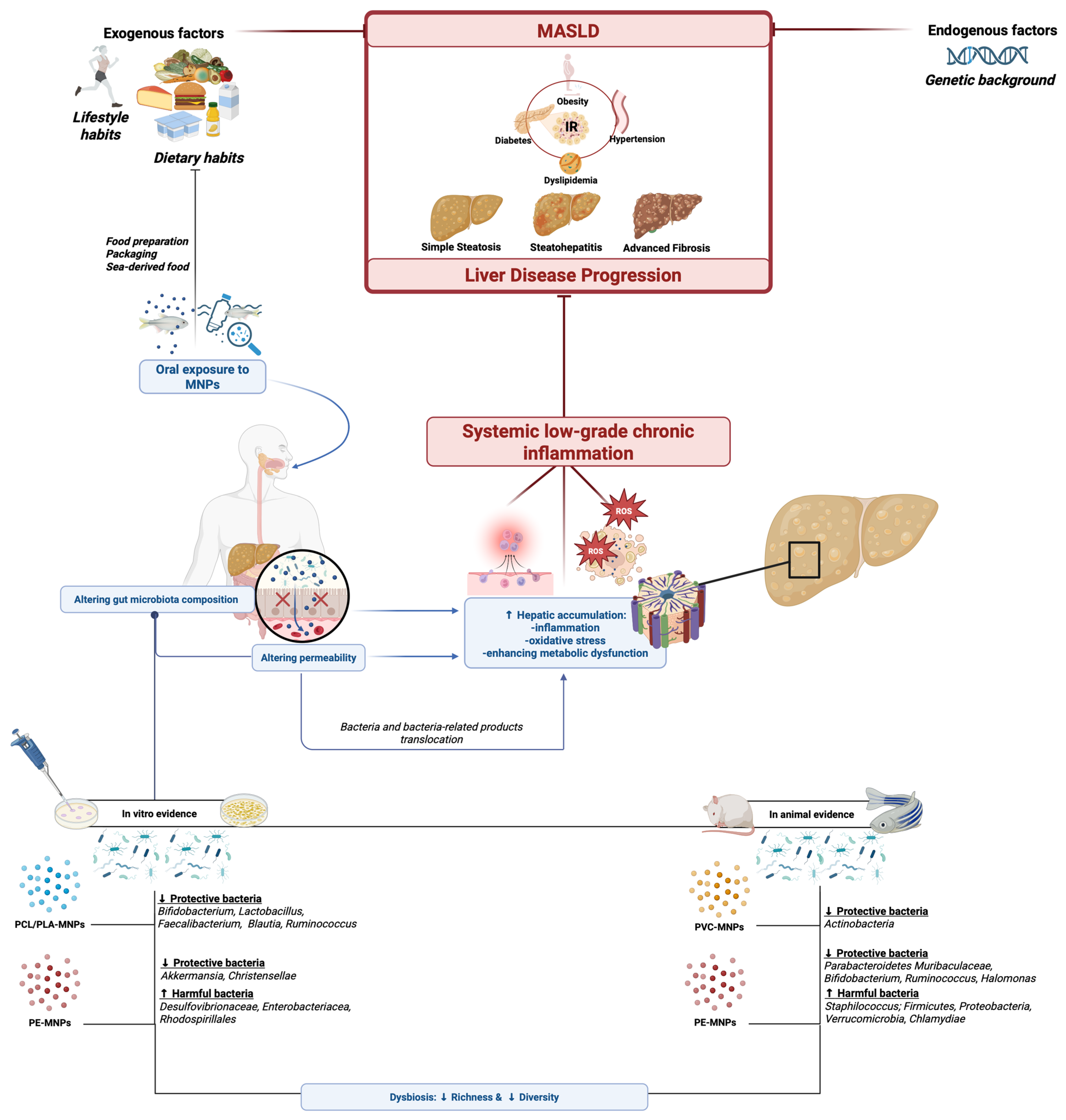
3. Plastic Exposure and the Gut–Liver Axis in the Pathogenesis of Steatotic Liver Disease

3.1. Gut Microbiota and Intestinal Permeability Status in Hepatic Steatosis: An Overview

The human intestinal microbiota is a key factor in host metabolism and a potential source of novel therapies [51]. In healthy individuals, the gastrointestinal tract harbors a different array of bacterial species, with Bacteroidetes and Firmicutes being the most predominant [51]. In particular, a relative abundance of Proteobacteria, Verrucomicrobia, Actinobacteria, Fusobacteria, and Cyanobacteria has been reported [51,52].
Alterations in the composition and function of the intestinal microbiota are associated with various pathological conditions, including IR-related manifestations, such as obesity, type 2 diabetes, and MASLD [51,52]. Relevantly, realizing the bidirectional relationship between “fatty diet” and “microbiota functioning”, dietary lipids can influence metabolic health through microbiota-mediated mechanisms; however, the potential role of the lipid–microbiota relationship in hepatic steatosis is still not well defined [53].
Schoeler et al., in a study conducted on mice, investigated the effects of dietary lipids on the composition of the human gut microbiota and the role of these interactions in hepatic steatosis [53]. They highlighted how different types of fatty acids influence both gut microbiota composition and lipid metabolism, with significant implications for liver health [53]. This research showed that certain diets, particularly those enriched with stearic acid, altered the gut microbiota diversity by promoting a favorable microbial profile characterized by a reduction in the abundance of specific butyrate-producing species. such as Roseburia, Oscillibacter, Anaerotruncus, and Intestinimonas, alongside an increase in the presence of propionate-producing species, such as Akkermansia muciniphila, a bacterium known to be associated with metabolic health, and Bacteroides [53]. These changes in the microbiota were associated with improvements in host metabolic characteristics, including a significant reduction in hepatic fat accumulation, enhanced glucose tolerance, and an increase in the saturation of FFAs, which may help reduce inflammation and the risk of hepatic steatosis [53]. Thus, the microbiota of mice fed with stearic acid-enriched diets differed significantly from that of mice fed with a high-fat diet (HFD), which was characterized by reduced microbiota richness and diversity, leading to dysbiosis [53]. Furthermore, in Schoeler et al.’s study, it was observed that transferring the microbiota from mice fed with the stearic acid-enriched diet to mice on the HFD diet protected against obesity, glucose homeostasis disorders, and hepatic steatosis [53]. In addition, several murine models have demonstrated that variations in microbiota composition influence the development of MASLD following an HFD [54]. Specifically, this influence is associated with an increased abundance of the phylum Firmicutes, along with the genera Barnesiella and Roseburia, as well as the Lachnospiraceae family and Barnesiella intestine hominis [54,55].
Moreover, along the same lines, several studies in humans have also demonstrated the role of dysbiosis in the development and progression of MASLD [54]. Notable changes include an increase in the phylum Proteobacteria [56], an elevation in Enterobacteriaceae, and a reduction in Rikinellaceae and Rummunoccaceae at the family level [57], while at the genus level, an increase in Escherichia and Dorea was observed, alongside a decrease in Coprococcus, Faecalibacterium, and Prevotella [54,58]. In patients with MASLD-related advanced fibrosis, specific alterations have been identified, including a general increase in gram-negative bacteria and Proteobacteria, coupled with a decrease in Firmicutes [54,57]. Indeed, as simple steatosis progresses to advanced hepatic fibrosis, the number of gram-negative bacteria, particularly Proteus species, increases [59,60,61].
Intestinal dysbiosis, referring to translocation, may increase intestinal permeability, leading to enhanced absorption of fatty acids [59], as well as to bacterial migration across the epithelial barrier, ultimately resulting in the concomitant release of bacterial products, lipopolysaccharides (LPS), and pro-inflammatory cytokines sustaining inflammation [59].
Finally, certain microbial metabolites and endotoxins appear to play a role in the pathophysiology of MASLD, including increased circulation of bile acids, acetate, and short-chain fatty acids (SCFAs) such as butyrate, as well as ethanol and choline-related metabolites [54], determining increased intestinal permeability, inducing inflammation or promoting hepatic lipogenesis [55].

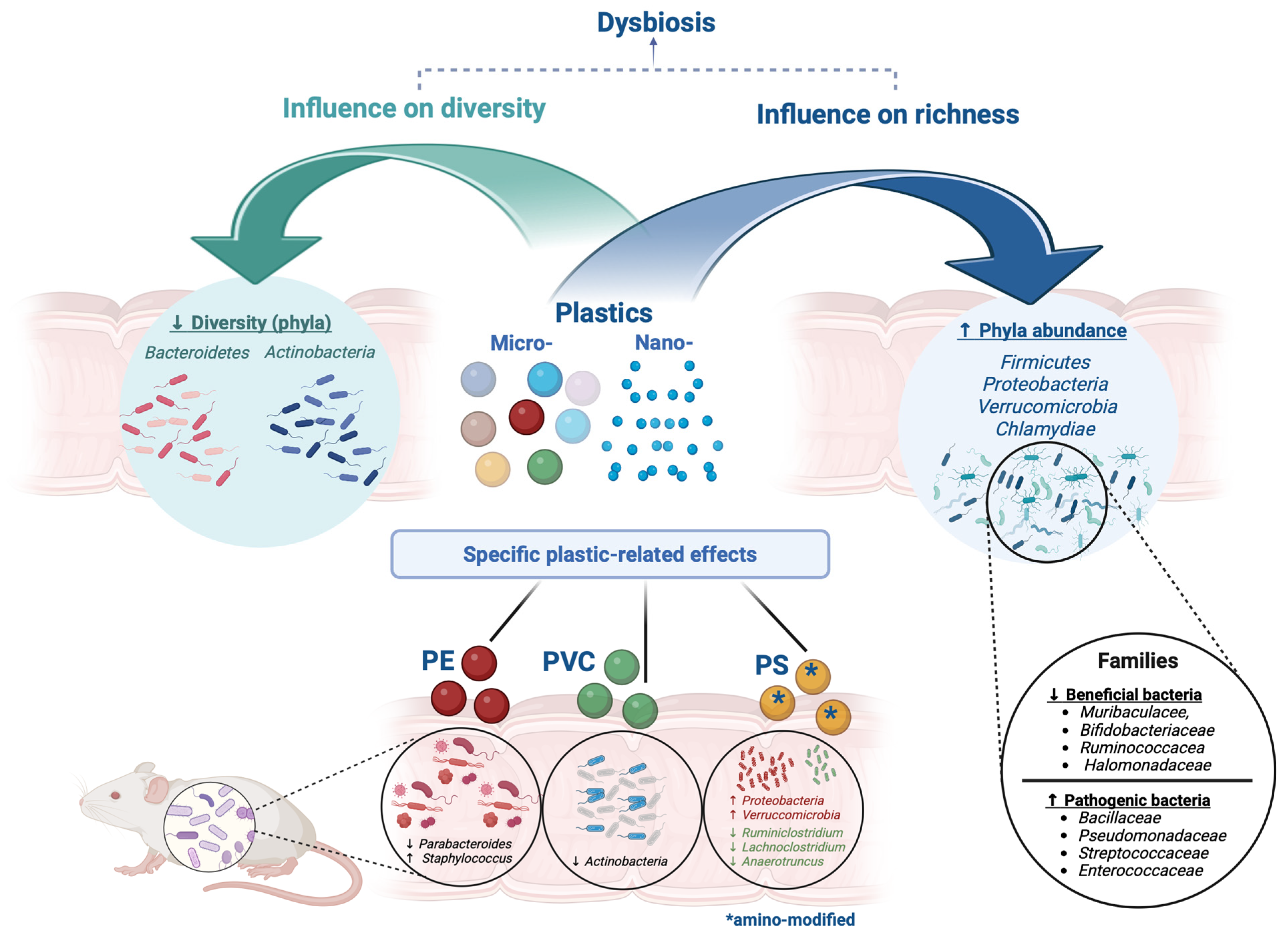
3.2. Micro(nano)plastic Exposure Impacts the Gut–Liver Axis by Influencing Gut Microbiota Composition and Functioning

The gut microbiota represents a highly complex ecosystem of microorganisms with a crucial role in human health [62]. Indeed, the gut microbiota plays evolutionarily conserved roles in host metabolism, immunity, development, and behavior [63,64,65]. The significant roles of intestinal microbes in maintaining host health have attracted considerable attention over the past decade, and several investigations have reported the modulatory effects of MNPs on the composition of the gut microbiota [66].
With the increasing focus on the impacts of MNPs on the intestine, an expanding array of studies has recently addressed this issue [67], demonstrating that oral exposure to these particles results in dysbiosis of the gut microbiome, gastrointestinal absorption, immune activation, and accumulation in various tissues [68]. Indeed, MNPs can accumulate in the intestine [67,69,70] and disrupt the intestinal barrier, leading to inflammation, bacterial translocation, and dysbiosis, with potentially adverse effects on the immune system and greater susceptibility to chronic disease [71,72].
Numerous studies presented below that investigated the impact of MNPs on the gut microbiota have been conducted in vitro and using animal models, particularly murine and zebrafish models, whereas clinical studies and epidemiological investigations in humans remain relatively limited.

3.2.1. Micro(nano)plastic Exposure Alters Gut Microbiota Composition: In Vitro Evidence

In vitro models have shown that exposure to MNPs alters the intestinal microbial composition, resulting in an increased relative abundance of harmful pathogenic bacteria [73].
Furthermore, it has been discovered that MNPs and the phthalates released from them may exert synergistic effects on the composition of the human gut microbiota [73]. In support of this, in an in vitro study, Peng et al. identified that MPs and their oligomeric forms disrupt the intestinal microbiota [74].
Their study revealed that MNPs derived from polycaprolactone (PCL) and polylactic acid (PLA) significantly impacted the microbial composition, leading to a decrease in alpha diversity within the gut microbiota [74]. Furthermore, these PCL and PLA MNPs were found to exert inhibitory effects on several beneficial probiotic species, including Bifidobacterium, Lactobacillus, Faecalibacterium, Limosilactobacillus, Blautia, Romboutsia, and Ruminococcus, underscoring the potential human health hazards posed by these materials [74].
Along the same lines, Fornier et al. conducted an in vitro study to investigate the effects of repeated exposure to PE-MNPs on human gut microbiota and intestinal barrier integrity, utilizing the Mucosal Artificial Colon (M-ARCOL) model in conjunction with a co-culture of intestinal epithelial and mucus-secreting cells [75]. The results demonstrated that there was an increase in the relative abundance of potentially harmful pathobionts, such as Desulfovibrionaceae and Enterobacteriaceae, alongside a decrease in beneficial bacteria like Christensenellaceae and Akkermansiaceae [75]. Furthermore, exposure to the PE microsphere mixture resulted in a reduction in protective Lactobacillales [75]. The PE mixture also increased the frequency of Rhodospirillales, which has been associated with various pathological conditions [75]. Interestingly, it appears that the effects of PE- and PS-MNPs on the microbiota are size-dependent [75]. Regarding the potential impact of MPs on the intestinal microbiota, however, the results have been heterogeneous, likely due to the considerable variability among the models employed, as well as the heterogeneity regarding composition, shape, dose, and duration of exposure [76]. Table 2 summarizes the main in vitro evidence supporting the impact of MNPs on gut microbiota composition.

3.2.2. Micro(nano)plastics Exposure Alters Gut Microbiota Composition: In-Animal Evidence

Several animal studies have revealed how exposure to MNPs may alter the gut microbiota composition in murine and zebrafish models, contributing to disturbances in the intestinal microenvironment [67,77,78]. It is plausible that ingested MPs frequently interact with the intestine before excretion, thereby triggering potential intestinal toxicity [73]. When exposed to MNPs, mice exhibited dysbiosis of the intestinal microbiota, including alterations in diversity indices, a reduced relative abundance of probiotics, and an increased abundance of pathogenic bacteria [73]. Many studies in animals highlight that exposure to MNPs has resulted in a reduction in species richness and diversity in gut microbiota [79,80,81,82,83]; however, in contrast, some research suggests that higher doses of MPs might positively affect species richness, suggesting the need to further characterize the correspondence between specific plastic patterns exposure with bacterial species representation impairment, contributing overall to altering intestinal microbiota composition [84,85].
Souza-Silva et al. have generically suggested, based on current in-animal model data, that the ingestion of MNPs may trigger intestinal dysbiosis, promoting a significant increase overall of bacteria belonging to the phyla Firmicutes, Proteobacteria, Verrucomicrobia, and Chlamydiae [76].
In addition to variations in the relative abundance, a reduction in the diversity of the phyla Bacteroidetes and Actinobacteria has also been observed, and, more deeply, a concurrent decrease in beneficial families (such as Muribaculaceae, Bifidobacteriaceae, Ruminococcaceae, and Halomonadaceae), in contrast with an enhanced representation of various pathogenic bacterial families (including, among others, Bacillaceae, Pseudomonadaceae, Aeromanadaceae, Rhodobacteraceae, Mycobacteriaceae, Xanthobacteraceae, Clostridiacea, Enterococcaceae, and Streptococcaceae), has been highlighted [76]. Notably, contrary to findings reported i in nvitro studies [74,75], exposure to MPs has been associated with an increase in Lactobacillus in animal models [76].
In parallel with commendable research efforts aiming to particularly characterize plastic-induced microbiota composition alterations at different taxonomic levels, other studies have evaluated the association between exposure to specific MNPs and potential consequential repercussions on dysbiosis, also exploring the burden of particle size on these effects.
On this topic, Li et al. specifically investigated the effects of PE-MP exposure in C57BL/6 murine models, observing an increase in intestinal microbial diversity and a qualitative shift in the bacterial composition, characterized by an increase in opportunistic pathogenic genera, such as Staphylococcus, and a decrease in others, such as Parabacteroides [71,85].
Subsequently, Chen et al. observed that exposure to PVC-MPs specifically alters the intestinal microbiota composition, particularly reducing Actinobacteria and compromising microbial homeostasis [71,86]. Similar results were observed by Djouina et al., who studied the effects of two sizes of PE-MPs (36 and 116 μm) in mice, showing that both MP sizes induced changes in the intestinal microenvironment [71,87]. On the other hand, Qiao et al., in a study on mice, found that chemically modified MNPs had a more relevant effect on gut bacteria than unmodified ones, with micro-sized particles showing a more significant impact than nano-sized ones [87]. Indeed, while Firmicutes and Bacteroidetes were largely unaffected by various PS-MNPs, exposure to amino-modified PS-MNPs increased Proteobacteria and Verrucomicrobia, simultaneously determining a reduction in SCFA producer species (Ruminiclostridium, Lachnoclostridium, and Anaerotruncus) [87].
Finally, in contrast to this evidence, other animal research has not reported intestinal dysbiosis or altered bacterial diversity following exposure to MNP particles [72]. In line with this, a study on European sea bass (Dicentrarchus labrax), conducted by Caruso et al., showed no intestinal dysbiosis [88], but methodological limitations, such as a small sample size and outdated sequencing techniques, may have influenced the results [72].
Another study on pond snails (Lymnaea stagnalis), conducted by Horton et al. [89], with a very small sample size and a non-specific analysis of the gut microbiome, encountered similar issues, as the entire snail biomass was used instead of isolating the digestive tract [72,89]. These contrasting findings suggest that further research, using more advanced methods, is needed to better understand the effects of MNP on the gut microbiota [72].
Figure 2 summarizes the most relevant plastic exposure-related repercussions on gut bacteria diversity and richness in animals.

3.2.3. Micro(nano)plastics Exposure Alters Gut Microbiota Composition: In-Human Evidence

Focusing on human studies, numerous embryonal studies have highlighted that exposure to MNPs potentially alters the composition of the gut microbiota, reducing alpha diversity (i.e., the richness species within a microbial community) and modifying beta diversity (i.e., the differential composition between microbial communities) [72,81,90,91,92].
Dandane et al. brilliantly investigated the association between MNPs and gut microbiota composition in preschool children in Xiamen, China [73]. The results showed that the children with low MNP exposure had an overrepresentation of protective bacteria with anti-inflammatory effects, such as Lactobacillales, Rikenellaceae, Alistipes, and Streptococcus. Notably, Alistipes was negatively correlated with PVC concentration [73].
On the other hand, the children with high MNP exposure exhibited an increase in Faecalibacterium, with higher levels of PVC and total MP, suggesting a potentially negative impact on the gut microbiota [73]. Additionally, the high-exposure group showed a decrease in Eubacterium, indicative of a possible effect on lipid metabolism. Other bacteria, such as Parabacteroides and Lachnospiraceae_NK4A136_group, were negatively correlated with the concentration of PE [73]. In both groups, the bacterial composition was dominated by Firmicutes and Bacteroides, with no significant differences between the low- and high-exposure groups at the phylum or genus level. However, the children with high exposure exhibited lower alpha diversity indices [73]. Altogether, in the human setting, the interesting emerging findings have to be considered preliminary results opening the door to further research investigating the impact of MNP exposure on gut microbiota composition.

3.2.4. Micro(nano)plastics Exposure Impacts the Gut–Liver Axis by Altering Gut Microbiota Functioning and Impairing Intestinal Permeability

In addition to altering the bacterial community structure, MNPs have been shown to impact also bacterial metabolism significantly [76]. Research has revealed that exposure to MNP leads to the down-regulation of pathways associated with secondary metabolite biosynthesis, sulfur metabolism, fluorobenzoate degradation, polycyclic aromatic hydrocarbons, and chlorocyclohexane degradation [93]. Conversely, pathways related to the biosynthesis of isoflavones and carotenoids, as well as mineral absorption, are upregulated following MP exposure [94], and also an increase in pathways involved in steroid and sesquiterpenoid biosynthesis, nitrogen metabolism, and antimicrobial resistance has been reported [93]. Furthermore, Jin et al. suggested that MP exposure can down-regulate metabolic pathways linked to pyruvate metabolism, tyrosine metabolism, vitamin B6 metabolism, and fatty acid biosynthesis [77]. Moreover, studies have shown that MNPs impact not only the intestinal microbiota but also the overall health of the intestinal tissue [76,95]. Once retained in the intestine, MNPs increase intestinal permeability, which is characterized by reduced expression of tight junction proteins, such as Zo-1 and claudin-1 [76,96], and decrease mucus secretion [77] via reducing the colic expression of mucin genes (Muc1, Muc2, and Muc3) [96], simultaneously enhancing vascularization, causing damage to villi and microvilli [95], and determining the thinning of the intestinal wall [76,81].
Besides, solid research has shown that MNP, in addition to promoting gut microbiota dysbiosis and damaging the intestinal barrier, can also compromise liver health by indirectly affecting the liver through the gut–liver axis [71]. This axis, which represents the interaction between the gut, liver, and microbiota, is essential for protecting the organism from harmful substances and maintaining immune system homeostasis [71,97].
By inducing intestinal barrier dysfunction through inflammation and dysbiosis, MNPs increase intestinal permeability [71,98], allowing pathogenic bacteria and harmful metabolites to enter the bloodstream via the portal vein and subsequently reach the liver, causing hepatic phlogosis, oxidative stress, and metabolic disorders, resulting in liver damage and low-chronic inflammation status-related dysmetabolic IR-associated comorbidities [71,99,100,101]. Therefore, the effect of MNPs on the alteration of gut microbiota and intestinal barrier appears to play a central role in the development of hepatic steatosis and dysmetabolic extra-hepatic manifestations, contributing to the complex picture of MASLD.
Conclusively, MNP exposure emerges as more than exposure to mere inert environmental pollutants, representing significant active potential risk factors impacting intestinal, hepatic, and systemic health, definitely suggesting the absolute need for further research in this field [71]. Figure 3 summarizes the most relevant pathogenetic involvements of MNP exposure in MASLD pathogenesis by impacting the gut–liver axis.

4. Inflammation, Oxidative Stress, and Innate Immune Dysfunction as Mutually Influenced Drivers in Steatotic Liver Disease: Is Micro(nano)plastic Exposure a Potential Pathogenetic Deus Ex Machina?

4.1. Inflammation and Oxidative Stress in Steatotic Liver Disease: A Consolidated Binomial

MASLD represents the hepatic manifestation of a multisystem disorder with multifactorial determinants, including genetic predisposition, environmental influences, and dysmetabolism, where IR plays a central role [28,36,37,38,39,40]. From a purely pathogenetic perspective, the primum movens of steatosis is represented by the overload of FFAs determining hepatic lipid accumulation, ultimately inducing mitochondrial dysfunction [102]. In particular, excess FFAs from diet, adipose tissue, or de novo lipogenesis overload the liver, causing the production of lipotoxic substances impairing mitochondria activity [102]. In response to this phenomenon, mitochondria increase the permeability of the inner membrane, thereby compromising their functionality [103]. Consequently, mitochondrial dysfunction occurs, leading to the accumulation of ROS, causing mitochondrial DNA mutations and the production of lipotoxic intermediates: both mutated mitochondrial DNA and oxidation-specific epitopes constitute damage-associated molecular patterns (DAMPs), ultimately inducing the release of pro-inflammatory cytokines [103]. In this sense, lipotoxic metabolites and DAMPs are sensed by pattern recognition receptors (PRRs), such as toll-like receptors (TLRs) and nucleotide-binding oligomerization domain-like receptors (NLRs), with the consequent pro-inflammatory cytokine release (consequent to the activation of NLRP3 inflammasome) and cell death, key drivers of liver damage [104]. These processes collectively exacerbate the inflammatory state characteristic of the disease [104]. Therefore, as the disease progresses from simple steatosis to steatohepatitis and fibrosis, inflammation combined with oxidative stress occurs in a vicious positive feedback loop [103,105].
Moreover, PRRs can recognize pathogen-associated molecular patterns (PAMPs) (e.g., bacterial products, and LPS) derived from intestinal dysbiosis, impaired permeability, and bacterial-product translocation, ultimately activating Kupffer cells (KCs) and hepatic stellate cells (HSCs) through TLRs, contributing thus to further increasing the hepatic inflammatory state [106].
Relevantly, in this complex imbricated scenario, these pathogenetic processes, by engaging hepatocytes, KCs, neutrophils, and HSCs, emphasize the crosstalk between oxidative stress, inflammation, and innate immune responses in the advancement of liver disease. In this sense, KCs are activated by ROS, becoming the main hepatic producers of ROS, cytokines (TNF-alpha, IL-1β, IL-6, IL-12, and IL-18), growth factors, and chemokines [104,107].
In support of this, reactive species have been shown to activate several transcription factors and receptors, including nuclear factor kappa B (NF-κB), activator protein-1 (AP-1), p53, hypoxia-inducible factor 1-alpha (HIF-1alpha), peroxisome proliferator-activated receptor (PPAR), β-catenin/Wnt, and nuclear factor erythroid 2–related factor 2 (Nrf2). These factors, in turn, regulate the expression of molecules directly involved in the inflammation processes [108].
In conclusion, inflammation and oxidative stress occur simultaneously in the liver, perpetuating each other in a vicious cycle: Persistent oxidative stress is more than a mere driver guiding the development of chronic inflammation, simultaneously acting as the cornerstone of MASLD progression by activating innate immunity to produce cytokines and further reactive species [103]. This, in turn, fuels innate immune cells, perpetuating inflammation and forming the common thread that links liver disease to its systemic complications [103].

4.2. Micro(nano)plastic Influences Hepatic Lipid Metabolism via Inflammation and Oxidative Stress

Exposure to MNPs has demonstrated significant impacts on both inflammation and oxidative stress, with clear adverse effects on hepatic metabolism [109]. This phenomenon primarily stems from their ability to alter immune system function, impacting both the intestine and other organs through various absorption mechanisms [1]. Ingested MNPs can accumulate in the intestinal tract, and while particles larger than 150 μm do not cross the mucosal barrier, those smaller than 150 μm can penetrate tissues, primarily through endocytosis, transcytosis, persorption, and paracellular absorption [1,110]. Once absorbed, MNPs can negatively affect the immune response, particularly in the intestinal mucosa, which constantly interacts with commensal microbiota and food antigens [1].
Chronic exposure to MNPs can impair immune function, as evidenced by studies on invertebrates (e.g., Daphnia magna and Mytilus spp.), which highlighted immune cell damage, increased ROS production, and reduced phagocytosis [1,111,112]. Studies on vertebrates (fish and mice) have shown significant alterations in immune response, including changes in cytokine levels and an increase in Th17 cells in mice exposed to MNPs [1,85].
Plastic-derived nanoparticles (NaPs), particularly inorganic and metallic types, can exhibit biocidal activity, altering the composition and metabolic functions of the microbiota, leading to intestinal dysbiosis and influencing immune responses [113]. At the same time, interactions of NaPs with the immune system may, in turn, modify the composition of gut microbiota [113]. Alterations in the microbiota–immune system axis are associated with chronic diseases, raising the concern that prolonged exposure to inorganic NaPs may contribute to their onset and progression [113].
Mancia et al., in a study on the Scyliorhinus canicular, observed that the presence of MNPs in the gastrointestinal tract correlated with a significant increase in the expression of T cell receptors beta and delta (TCRβ and TCRδ) and immunoglobulin M (IgM) in the spleen, indicating activation of the immune response [1,114]. On the other hand, exposure to PS and PE particles has been associated with reduced activity of certain immune enzymes and altered phagocytosis, suggesting an immunosuppressive effect, as well as, once again, the need to stratify the observation in this field according to particle size and dosage [1]. About this, Zha H. et al., in a study on mice exposed to MNPs, found that the level of eotaxin/CCL11 in serum was lower in mice exposed to 200 μg and 500 μg of NPs (groups 2NP and 5NP, respectively) compared to those exposed to 500 μg of MPs (group 5MP), while interleukins IL-2 and IL-4 were both higher in the 5NP group than in the 5MP group [115]. Moreover, it has been observed that smaller-sized MPs (e.g., 0.1 μm PS particles) induced hepatic steatosis and metabolic dysfunctions in fish (O. niloticus), triggering a series of biological responses altering the balance between inflammation, immunity, and lipid metabolism [109].
This leads to an inflammatory immune response, with an increase in the expression of the pro-inflammatory cytokines TNF-alpha and IL-1β, which are involved in the regulation of lipid metabolism [109]. Indeed, TNF-alpha is known to enhance hepatic lipogenesis and inhibit FFA oxidation, while IL-1β interferes with insulin homeostasis and lipase activity [109]. The elevated expression of these cytokines has therefore contributed to lipid accumulation in the liver, a hallmark of MNP-induced steatosis [109]. In contrast, Pei X. et al. highlighted in a study on zebrafish that after exposure to PS-MNPs with diameters of 50 μm and 100 nm and concentrations of 100 and 1000 μg/mL, the mRNA expression levels of most pro- and anti-inflammatory factors, including IL-8, NF-κB, and IL-10, were increased, while the mRNA expression of TNF-alpha, a pro-inflammatory factor, was decreased [116]. These findings suggest that PS-MNPs may represent a potential threat to oxidative status and innate immunity, with size- and concentration-dependent toxicity [116].
Oxidative stress represents another critical mechanism through which plastics affect liver health [109]. The interaction between inflammation and oxidative stress is a central aspect of how MNPs influence hepatic metabolism [109]. As previously mentioned, hepatic lipid accumulation, partly caused by the inflammatory response, generates ROS, which, in turn, exacerbates oxidative stress and leads to lipid peroxidation [109]. This vicious cycle of lipid accumulation and ROS generation creates a favorable environment for hepatic steatosis [109]. This has been observed in several studies, which have shown an increase in lipid peroxidation markers, such as malondialdehyde (MDA), following exposure to MNPs [1]. Exposure to 0.1 μm particles activates the Nrf2/Keap1 pathway, a cellular system with a protective role against free radicals and electrophiles, which, if unregulated, can damage cells and tissues [109]. Activation of the antioxidant Nrf2 system can be overwhelmed by high-level exposures, leading to cellular damage and a reduced ability to protect tissues from oxidative stress [1,110]. Indeed, under conditions of MNP exposure, an overregulation of Nrf2 and Keap1 protein has been observed, an indicator of oxidative stress, highlighting that the antioxidant system may be insufficient to protect tissues from oxidative damage [109].
In particular, the activity of SOD, a key enzyme in combating reactive oxygen species, was significantly reduced in the liver of fish exposed to PS, indicating an inability of the antioxidant system to adequately neutralize free radicals; lipid peroxidation induction was observed with an increase in malondialdehyde (MDA) content in the liver of fish exposed to higher concentrations of PS (1 and 100 μg/L) [109]. This process is indicative of cellular membrane damage and chronic inflammation, which may further exacerbate hepatic dysfunction [109].
In a study conducted on zebrafish, Li R. et al. observed a series of modifications in the levels of glutathione S-transferase (GST), GSH, catalase (CAT), and SOD following exposure to MNPs, highlighting the onset of oxidative damage in the gill and intestinal cells of zebrafish [117]. Consistently, in a study on the liver of Nile tilapia (Oreochromis niloticus), in response to PS exposure, a dose-dependent overregulation of both Nrf2 and Keap1 was observed, along with a reduction in SOD activity in the tilapia liver and specific activation of the PERK-eIF2α pathway in endoplasmic reticulum (ER) stress, which is implicated in the disruption of lipid metabolism through the initiation of a process that amplifies both inflammatory and oxidative damage in the liver [109].
Altogether, even though supported by research mainly conducted in animal models, the above-presented evidence proposes MNP exposure as a potential deus ex machina impacting the binomial relationship between inflammation and oxidative stress in the pathogenesis of hepatic steatosis, suggesting the absolute need for further investigations.

4.3. Micro(nano)plastics and Related Additives as Activators of the Innate Immune Response: Investigating the Novel Frontier of Steatotic Liver Disease Pathogenesis

Although further research is required, preliminary studies suggest the potential role of MNPs as immunotoxic agents, consequently disrupting intestinal homeostasis (both impairing intestinal barrier function and disturbing the gut microbiota functioning) and interfering with metabolic pathways, thus representing an emerging threat to human health and chronic liver disorders [1].
In this sense, MNPs, with particular reference to PS-NPs, in addition to altering the mechanisms regulating lipid metabolism, have been reported to simultaneously act as activators of a dysregulated innate immune response, ultimately exacerbating tissue-level damage [109,118].
An additional risk associated with MNPs is their ability to adsorb toxins and pathogens [1]. As previously reported, indeed, MNPs can transport contaminants such as phthalates and bisphenols [including BPA, polychlorinated biphenyls (PCBs)], and polycyclic aromatic hydrocarbons (PAHs), whose consequent release into the gastrointestinal tract may exacerbate damage to the local mucosa via dysregulating innate immune system activity [1]. Some studies have shown that the presence of MNPs exacerbates the toxicity of chemical contaminants such as chlorpyrifos and cadmium [1,119,120]. Moreover, MNPs can serve as a substrate for bacterial biofilms, hosting pathogens (Vibrio spp. and E. coli) that may abnormally amplify the innate immune response [1,121].
Interestingly, the concept of innate immune cell response has been recently revolutionized by the novel theory of “trained immunity” (TI), revealing an immunological memory even for innate immune cells [122]. Relevantly, innate immune cells can react to second not-specific antigenic (exogenous, endogenous, and metabolism-derived stimuli) contact, determining, via the activation of different signaling pathways, the epigenetic remodeling and the rewiring of various intracellular metabolic pathways, resulting in the acquisition of new functions, including the production of lipid mediators, cytokines, and tissue remodeling enzymes, with several physiopathological implications in human diseases, including hepatic steatosis and the related extra-hepatic manifestations [122]. Considering this, the term “immunometabolism” has been proposed with a progressively increasing interest in MASLD pathogenesis [122].
Notably, MNP-derived contaminants exposure also appears to impact this novel pathogenetic scenario. Concerning this, a recent study by Dallio et al. evaluated the ability of BPA to induce TI response in human primary monocytes in vitro [123].
In particular, monocytes were acutely stimulated in these cells with BPA, LPS as a positive control, and Roswell Park Memorial Institute Medium (RPMI+) as a negative control, subsequently assessing the pro- and anti-inflammatory cytokine (IL-1beta, TNF-alpha, IL-6, and IL-10) production after 24 h of acute stimulation and after LPS rechallenge. Interestingly, increased pro- and anti-inflammatory cytokine responses upon restimulation in monocytes primed with BPA were revealed. In particular, the data showed a slight increase in the pro-inflammatory cytokines TNF-alpha and IL-1beta, while IL-6 resulted in higher expression together with the anti-inflammatory cytokine IL-10, preliminarily showing how an environmental chemical factor can induce a TI response [123].
More recently, based on these encouraging preliminary results, the induction of TI mediated by BPA was investigated in MASLD patients, revealing an enhanced response determining higher levels of pro-inflammatory cytokines (TNF-alpha and IL-6) in individuals presenting steatohepatitis compared to subjects affected by simple steatosis, as well as a decrease in anti-inflammatory mediators (e.g., IL-10), probably due to a compensative phenomenon for the aberrant inflammatory process [124].
Comprehensively, the above-reported evidence supports the role of MNP-derived contaminants in altering innate immune response, simultaneously driving the progression of simple steatosis towards the more advanced stage of disease in MASLD, suggesting the absolute requirement to further investigate the relative pathogenetic implications to translationally apply these emerging findings in proposing adequate prevention and management strategies in steatotic liver disease.
Table 3 summarizes the main implications of MNPs and plastic-derived compounds in impacting immune response, intestinal homeostasis, and metabolic pathways in the pathogenesis of MASLD, reporting the related main sources and level of supporting evidence.

5. Micro(nano)plastic Exposure and Steatotic Liver Disease: From Basic Evidence to Clinical Bench-Side: An Urgent Need to Translate and Apply This Evidence. How Far Are We?

5.1. Main Research Challenges in the Field of Plastics and Hepatic Steatosis

MNP-derived pollutants represent an emerging threat to human health, with a particularly concerning impact on liver diseases, including MASLD. Recent research suggests that these particles can enter the human body, accumulate in tissues, and interfere with fundamental biological processes, causing liver damage [128]. Emerging evidence suggests an interaction between MNPs and the gut microbiota, as exposure to these contaminants can alter microbiota composition, promoting dysbiosis, which, in turn, may affect lipid metabolism, inflammation, and liver health [76]. Moreover, chronic exposure to the MNP-derived contaminant BPA has been shown to negatively impact metabolic health, thereby contributing to liver dysfunction and steatosis.
Specifically, the study by Dallio et al. highlighted that BPA can activate immune pathways, particularly trained immunity in monocytes, suggesting that repeated BPA exposure may sensitize the immune system, inducing a state of chronic systemic inflammation, potentially severely contributing to the development and progression of steatosis and extra-hepatic comorbidities of MASLD [123].
Although there is growing evidence supporting the potential involvement of MNP-derived pollutant exposure in the pathogenesis of hepatic steatosis by impacting the gut–liver axis and immune response, currently, translating these findings into concrete clinical applications remains a complex challenge [21].
The main difficulties are represented by the lack of effective translational models linking preclinical results to clinical practice, the limitations simultaneously with the relative heterogeneity (lack of standardized approaches) of current methodologies adopted in measuring exposure MNPs, and the unavailability of results concerning the consequences of long-term effects of exposure to these particles in humans [23,129]. Consistently, the main advance in this research field would be represented by the designation of longitudinal studies, as most available research on this topic is cross-sectional and lacks long-term data on chronic exposure to MNPs. In this sense, longitudinal studies in both animals and humans emerge as essential also to establish a causal link between the timing of exposures and steatotic liver disease [23,129].
Additionally, the specific molecular mechanisms through which these compounds influence inflammation and lipid metabolism remain partially unclear [129]. In this sense, the difficulty in detecting MNPs in the liver and microbiota limits our ability to correlate exposure with the development of steatosis, suggesting that the application of advanced imaging techniques, multi-omics analysis, and liver organoids could improve the understanding of how these pollutants mechanistically impact liver health [22,42,130].

5.2. Future Perspectives

Looking optimistically beyond the “scientific fog”, the success of future research in overcoming the above-mentioned obstacles and further clarifying all the sources of MNP exposure and how these particles influence MASLD pathogenesis would have important clinical repercussions, opening the availability of various objectives.
These embrace the implementation of early tailored diagnostic approaches (based on the timing and pattern of MNP exposure), as well as the realization of personalized treatment strategies, including specific geographical area-/social habit-based low-MNP dietary regimens (in parallel with the current clinical practice guidelines recommendations on lifestyle) [131], targeted modulation of the gut microbiota, and pharmacological therapies regulating the aberrant immune response and systemic inflammation, ultimately preventing MASLD onset and progression.
Furthermore, the optimization of these strategies could generically improve public health policies. In this sense, as highlighted also by Winiarska et al., it is essential for research to approach these issues from an interdisciplinary perspective, moving beyond the sectoral approach that limits a comprehensive understanding of the phenomenon [132].
Rather than treating fields such as toxicology, clinical medicine, and environmental sciences in isolation, it is crucial to foster closer dialogue and collaboration among them to fully understand the impact of MPs and NPs on human health [132]. In the future, this integrated approach may lead to the development of effective strategies for preventing and treating liver diseases, addressing one of the most pressing healthcare challenges of this era.

6. Conclusions

Exposure to MNPs and MNP-derived contaminants represents a global social health issue, potentially contributing to the MASLD “pandemic” in industrialized countries.
Growing evidence suggests the pathogenetic implications of MNPs and related additives in MASLD by impacting the gut–liver axis and influencing the inflammatory and oxidative stress balance via dysregulating immune-metabolic pathways.
Anyway, further investigations are required to precisely clarify the mechanisms and translate these findings from preclinical findings to clinical applications. In this context, it is essential for research to approach these issues from an interdisciplinary perspective, moving beyond the sectoral approach that limits a comprehensive understanding of the phenomenon to develop effective strategies for preventing and treating hepatic steatosis.

Author Contributions

M.R.: guarantor of the article, conceptualization, methodology, formal analysis, investigation, and writing the original draft; M.D.: conceptualization, methodology, formal analysis, investigation, and writing the original draft. F.D.N., G.M., C.B., A.S., G.S., A.C., C.N., A.A. and C.A.: investigation, resources, data curation, writing the original draft; A.F.: conceptualization, data curation, supervision, and project administration. All authors have read and agreed to the published version of the manuscript.

Funding

This research received no external funding.

Data Availability Statement

No new data were created or analyzed in this study. Data sharing is not applicable to this article.

Conflicts of Interest

The authors declare no conflicts of interest.

List of Abbreviations

The following abbreviations are used in this manuscript:
MPsmicroplastics
NPsnanoplastics
MNPsmicro(nano)plastics
PS-MPspolystyrene microplastics
PEpolyethylene
PSpolystyrene
PE-MPspolyethylene microplastics
PVCpolyvinyl chloride
PCBpolychlorinated biphenyl
PLApolylactic acid
PLCpolycaprolactone
ROSreactive oxygen species
SODsuperoxide dismutase
BPAbisphenol A
EDCsendocrine-disrupting chemicals
MASLDMetabolic associated Steatotic Liver Disease
EASLEuropean Association for the Study of Liver
GSHglutathione
TNF-αtumor necrosis factor alpha
IL-1βinterleukin-1 beta
IL-1interleukin-1
IL-6interleukin-6
IL-8interleukin-8
ARandrogen receptor
GPERG protein-coupled estrogen receptor
TItrained immunity
LPSlipopolysaccharide
HFDhigh-fat diet
PCLpolycaprolactone
PLApolylactic acid
M-ARCOLMucosal Artificial Colon
FFAsfree fatty acids
CYPcytochrome P450
PRRspattern recognition receptors
NLRsnucleotide-binding oligomerization domain-like receptors
TLRstoll-like receptors
PAMPspathogen-associated molecular patterns
DAMPsdamage-associated molecular patterns
KCsKupffer cells
HIF-1αhypoxia-inducible factor 1-alpha
PPARperoxisome proliferator-activated receptor
NaPsPlastic-derived nanoparticles
TCRβT cell receptor beta
TCRδT cell receptor delta

References

  1. Hirt, N.; Body-Malapel, M. Immunotoxicity and Intestinal Effects of Nano- and Microplastics: A Review of the Literature. Part. Fibre Toxicol. 2020, 17, 57. [Google Scholar] [CrossRef]
  2. Pelegrini, K.; Pereira, T.C.B.; Maraschin, T.G.; Teodoro, L.D.S.; Basso, N.R.D.S.; De Galland, G.L.B.; Ligabue, R.A.; Bogo, M.R. Micro- and Nanoplastic Toxicity: A Review on Size, Type, Source, and Test-Organism Implications. Sci. Total Environ. 2023, 878, 162954. [Google Scholar] [CrossRef] [PubMed]
  3. Gigault, J.; Halle, A.T.; Baudrimont, M.; Pascal, P.-Y.; Gauffre, F.; Phi, T.-L.; El Hadri, H.; Grassl, B.; Reynaud, S. Current Opinion: What Is a Nanoplastic? Environ. Pollut. 2018, 235, 1030–1034. [Google Scholar] [CrossRef] [PubMed]
  4. Liu, X.; Shi, H.; Xie, B.; Dionysiou, D.D.; Zhao, Y. Microplastics as Both a Sink and a Source of Bisphenol A in the Marine Environment. Environ. Sci. Technol. 2019, 53, 10188–10196. [Google Scholar] [CrossRef]
  5. Vandenberg, L.N.; Hauser, R.; Marcus, M.; Olea, N.; Welshons, W.V. Human Exposure to Bisphenol A (BPA). Reprod. Toxicol. 2007, 24, 139–177. [Google Scholar] [CrossRef] [PubMed]
  6. Rochester, J.R. Bisphenol A and Human Health: A Review of the Literature. Reprod. Toxicol. 2013, 42, 132–155. [Google Scholar] [CrossRef]
  7. Al-Tohamy, R.; Ali, S.S.; Zhang, M.; Elsamahy, T.; Abdelkarim, E.A.; Jiao, H.; Sun, S.; Sun, J. Environmental and Human Health Impact of Disposable Face Masks During the COVID-19 Pandemic: Wood-Feeding Termites as a Model for Plastic Biodegradation. Appl. Biochem. Biotechnol. 2023, 195, 2093–2113. [Google Scholar] [CrossRef]
  8. Cimmino, I.; Fiory, F.; Perruolo, G.; Miele, C.; Beguinot, F.; Formisano, P.; Oriente, F. Potential Mechanisms of Bisphenol A (BPA) Contributing to Human Disease. Int. J. Mol. Sci. 2020, 21, 5761. [Google Scholar] [CrossRef]
  9. Konieczna, A.; Rutkowska, A.; Rachoń, D. Health Risk of Exposure to Bisphenol A (BPA). Rocz. Panstw. Zakl. Hig. 2015, 66, 5–11. [Google Scholar]
  10. Melzer, D.; Rice, N.E.; Lewis, C.; Henley, W.E.; Galloway, T.S. Association of Urinary Bisphenol a Concentration with Heart Disease: Evidence from NHANES 2003/06. PLoS ONE 2010, 5, e8673. [Google Scholar] [CrossRef]
  11. Smith, M.; Love, D.C.; Rochman, C.M.; Neff, R.A. Microplastics in Seafood and the Implications for Human Health. Curr. Environ. Health Rep. 2018, 5, 375–386. [Google Scholar] [CrossRef] [PubMed]
  12. Jenner, L.C.; Rotchell, J.M.; Bennett, R.T.; Cowen, M.; Tentzeris, V.; Sadofsky, L.R. Detection of Microplastics in Human Lung Tissue Using μFTIR Spectroscopy. Sci. Total Environ. 2022, 831, 154907. [Google Scholar] [CrossRef]
  13. Abbasi, S.; Turner, A. Human Exposure to Microplastics: A Study in Iran. J. Hazard. Mater. 2021, 403, 123799. [Google Scholar] [CrossRef]
  14. Leslie, H.A.; Van Velzen, M.J.M.; Brandsma, S.H.; Vethaak, A.D.; Garcia-Vallejo, J.J.; Lamoree, M.H. Discovery and Quantification of Plastic Particle Pollution in Human Blood. Environ. Int. 2022, 163, 107199. [Google Scholar] [CrossRef] [PubMed]
  15. Kwon, J.-H.; Kim, J.-W.; Pham, T.D.; Tarafdar, A.; Hong, S.; Chun, S.-H.; Lee, S.-H.; Kang, D.-Y.; Kim, J.-Y.; Kim, S.-B.; et al. Microplastics in Food: A Review on Analytical Methods and Challenges. Int. J. Environ. Res. Public Health 2020, 17, 6710. [Google Scholar] [CrossRef] [PubMed]
  16. Kutralam-Muniasamy, G.; Pérez-Guevara, F.; Elizalde-Martínez, I.; Shruti, V.C. Branded Milks—Are They Immune from Microplastics Contamination? Sci. Total Environ. 2020, 714, 136823. [Google Scholar] [CrossRef]
  17. Liebezeit, G.; Liebezeit, E. Non-Pollen Particulates in Honey and Sugar. Food Addit. Contam. Part A 2013, 30, 2136–2140. [Google Scholar] [CrossRef]
  18. Mason, S.A.; Welch, V.G.; Neratko, J. Synthetic Polymer Contamination in Bottled Water. Front. Chem. 2018, 6, 407. [Google Scholar] [CrossRef]
  19. Buyukunal, S.K.; Koluman, A.; Muratoglu, K. Microplastic Pollution of Drinking Water in a Metropolis. J. Water Health 2023, 21, 687–701. [Google Scholar] [CrossRef]
  20. Hartmann, C.; Lomako, I.; Schachner, C.; El Said, E.; Abert, J.; Satrapa, V.; Kaiser, A.-M.; Walch, H.; Köppel, S. Assessment of Microplastics in Human Stool: A Pilot Study Investigating the Potential Impact of Diet-Associated Scenarios on Oral Microplastics Exposure. Sci. Total Environ. 2024, 951, 175825. [Google Scholar] [CrossRef]
  21. Bastyans, S.; Jackson, S.; Fejer, G. Micro and Nano-Plastics, a Threat to Human Health? Emerg. Top. Life Sci. 2022, 6, 411–422. [Google Scholar] [CrossRef] [PubMed]
  22. Mittal, N.; Tiwari, N.; Singh, D.; Tripathi, P.; Sharma, S. Toxicological Impacts of Microplastics on Human Health: A Bibliometric Analysis. Environ. Sci. Pollut. Res. Int. 2024, 31, 57417–57429. [Google Scholar] [CrossRef]
  23. Winiarska, E.; Jutel, M.; Zemelka-Wiacek, M. The Potential Impact of Nano- and Microplastics on Human Health: Understanding Human Health Risks. Environ. Res. 2024, 251, 118535. [Google Scholar] [CrossRef]
  24. Wu, B.; Wu, X.; Liu, S.; Wang, Z.; Chen, L. Size-Dependent Effects of Polystyrene Microplastics on Cytotoxicity and Efflux Pump Inhibition in Human Caco-2 cells. Chemosphere 2019, 221, 333–341. [Google Scholar] [CrossRef] [PubMed]
  25. Poma, A.; Vecchiotti, G.; Colafarina, S.; Zarivi, O.; Aloisi, M.; Arrizza, L.; Chichiriccò, G.; Di Carlo, P. In Vitro Genotoxicity of Polystyrene Nanoparticles on the Human Fibroblast Hs27 Cell Line. Nanomaterials 2019, 9, 1299. [Google Scholar] [CrossRef]
  26. Mattioda, V.; Benedetti, V.; Tessarolo, C.; Oberto, F.; Favole, A.; Gallo, M.; Martelli, W.; Crescio, M.I.; Berio, E.; Masoero, L.; et al. Pro-Inflammatory and Cytotoxic Effects of Polystyrene Microplastics on Human and Murine Intestinal Cell Lines. Biomolecules 2023, 13, 140. [Google Scholar] [CrossRef] [PubMed]
  27. Da Silva Brito, W.A.; Mutter, F.; Wende, K.; Cecchini, A.L.; Schmidt, A.; Bekeschus, S. Consequences of Nano and Microplastic Exposure in Rodent Models: The Known and Unknown. Part. Fibre Toxicol. 2022, 19, 28. [Google Scholar] [CrossRef]
  28. Dallio, M.; Diano, N.; Masarone, M.; Gravina, A.G.; Patanè, V.; Romeo, M.; Di Sarno, R.; Errico, S.; Nicolucci, C.; Abenavoli, L.; et al. Chemical Effect of Bisphenol A on Non-Alcoholic Fatty Liver Disease. Int. J. Environ. Res. Public Health 2019, 16, 3134. [Google Scholar] [CrossRef]
  29. Nicolucci, C.; Errico, S.; Federico, A.; Dallio, M.; Loguercio, C.; Diano, N. Human Exposure to Bisphenol A and Liver Health Status: Quantification of Urinary and Circulating Levels by LC-MS/MS. J. Pharm. Biomed. Anal. 2017, 140, 105–112. [Google Scholar] [CrossRef]
  30. Dallio, M.; Masarone, M.; Errico, S.; Gravina, A.G.; Nicolucci, C.; Di Sarno, R.; Gionti, L.; Tuccillo, C.; Persico, M.; Stiuso, P.; et al. Role of Bisphenol A as Environmental Factor in the Promotion of Non-Alcoholic Fatty Liver Disease: In Vitro and Clinical Study. Aliment. Pharmacol. Ther. 2018, 47, 826–837. [Google Scholar] [CrossRef]
  31. Savastano, S.; Tarantino, G.; D’Esposito, V.; Passaretti, F.; Cabaro, S.; Liotti, A.; Liguoro, D.; Perruolo, G.; Ariemma, F.; Finelli, C.; et al. Bisphenol-A Plasma Levels Are Related to Inflammatory Markers, Visceral Obesity and Insulin-Resistance: A Cross-Sectional Study on Adult Male Population. J. Transl. Med. 2015, 13, 169. [Google Scholar] [CrossRef]
  32. Croom, E. Metabolism of Xenobiotics of Human Environments. Prog. Mol. Biol. Transl. Sci. 2012, 112, 31–88. [Google Scholar] [CrossRef]
  33. Lu, K.; Song, Y.; Zeng, R. The Role of Cytochrome P450-Mediated Detoxification in Insect Adaptation to Xenobiotics. Curr. Opin. Insect Sci. 2021, 43, 103–107. [Google Scholar] [CrossRef] [PubMed]
  34. Landrigan, P.J.; Raps, H.; Cropper, M.; Bald, C.; Brunner, M.; Canonizado, E.M.; Charles, D.; Chiles, T.C.; Donohue, M.J.; Enck, J.; et al. The Minderoo-Monaco Commission on Plastics and Human Health. Ann. Glob. Health 2023, 89, 23. [Google Scholar] [CrossRef]
  35. Rinella, M.E.; Lazarus, J.V.; Ratziu, V.; Francque, S.M.; Sanyal, A.J.; Kanwal, F.; Romero, D.; Abdelmalek, M.F.; Anstee, Q.M.; Arab, J.P.; et al. A Multisociety Delphi Consensus Statement on New Fatty Liver Disease Nomenclature. J. Hepatol. 2023, 79, 1542–1556. [Google Scholar] [CrossRef] [PubMed]
  36. Dallio, M.; Romeo, M.; Gravina, A.G.; Masarone, M.; Larussa, T.; Abenavoli, L.; Persico, M.; Loguercio, C.; Federico, A. Nutrigenomics and Nutrigenetics in Metabolic- (Dysfunction) Associated Fatty Liver Disease: Novel Insights and Future Perspectives. Nutrients 2021, 13, 1679. [Google Scholar] [CrossRef] [PubMed]
  37. Dallio, M.; Sangineto, M.; Romeo, M.; Cipullo, M.; Coppola, A.; Mammone, S.; Di Gioia, G.; Masarone, M.; Persico, M.; Serviddio, G.; et al. The Influence of Acute Lifestyle Changes on NAFLD Evolution in a Multicentre Cohort: A Matter of Body Composition. Nutr. Diabetes 2024, 14, 33. [Google Scholar] [CrossRef]
  38. Meroni, M.; Longo, M.; Rustichelli, A.; Dongiovanni, P. Nutrition and Genetics in NAFLD: The Perfect Binomium. Int. J. Mol. Sci. 2020, 21, 2986. [Google Scholar] [CrossRef]
  39. Dallio, M.; Masarone, M.; Romeo, M.; Tuccillo, C.; Morisco, F.; Persico, M.; Loguercio, C.; Federico, A. PNPLA3, TM6SF2, and MBOAT7 Influence on Nutraceutical Therapy Response for Non-Alcoholic Fatty Liver Disease: A Randomized Controlled Trial. Front. Med. 2021, 8, 734847. [Google Scholar] [CrossRef]
  40. Gravina, A.G.; Romeo, M.; Pellegrino, R.; Tuccillo, C.; Federico, A.; Loguercio, C. Just Drink a Glass of Water? Effects of Bicarbonate-Sulfate-Calcium-Magnesium Water on the Gut-Liver Axis. Front. Pharmacol. 2022, 13, 869446. [Google Scholar] [CrossRef]
  41. Zhao, Y.; Bao, Z.; Wan, Z.; Fu, Z.; Jin, Y. Polystyrene Microplastic Exposure Disturbs Hepatic Glycolipid Metabolism at the Physiological, Biochemical, and Transcriptomic Levels in Adult Zebrafish. Sci. Total Environ. 2020, 710, 136279. [Google Scholar] [CrossRef] [PubMed]
  42. Cheng, W.; Li, X.; Zhou, Y.; Yu, H.; Xie, Y.; Guo, H.; Wang, H.; Li, Y.; Feng, Y.; Wang, Y. Polystyrene Microplastics Induce Hepatotoxicity and Disrupt Lipid Metabolism in the Liver Organoids. Sci. Total Environ. 2022, 806, 150328. [Google Scholar] [CrossRef] [PubMed]
  43. Guraka, A.; Souch, G.; Duff, R.; Brown, D.; Moritz, W.; Kermanizadeh, A. Microplastic-Induced Hepatic Adverse Effects Evaluated in Advanced Quadruple Cell Human Primary Models Following Three Weeks of Repeated Exposure. Chemosphere 2024, 364, 143032. [Google Scholar] [CrossRef]
  44. Yang, T.; Poenisch, M.; Khanal, R.; Hu, Q.; Dai, Z.; Li, R.; Song, G.; Yuan, Q.; Yao, Q.; Shen, X.; et al. Therapeutic HNF4A mRNA Attenuates Liver Fibrosis in a Preclinical Model. J. Hepatol. 2021, 75, 1420–1433. [Google Scholar] [CrossRef] [PubMed]
  45. Fan, Z.; Zhang, Y.; Fang, Y.; Zhong, H.; Wei, T.; Akhtar, H.; Zhang, J.; Yang, M.; Li, Y.; Zhou, X.; et al. Polystyrene Nanoplastics Induce Lipophagy via the AMPK/ULK1 Pathway and Block Lipophagic Flux Leading to Lipid Accumulation in Hepatocytes. J. Hazard. Mater. 2024, 476, 134878. [Google Scholar] [CrossRef]
  46. Chiu, H.-W.; Chu, C.-W.; Huang, C.-C.; Chia, Z.-C.; Wang, Y.-L.; Lee, Y.-H. Polystyrene Microplastics Induce Hepatic Lipid Metabolism and Energy Disorder by Upregulating the NR4A1-AMPK Signaling Pathway. Environ. Pollut. (Barking Essex 1987) 2025, 369, 125850. [Google Scholar] [CrossRef]
  47. Prasun, P.; Ginevic, I.; Oishi, K. Mitochondrial Dysfunction in Nonalcoholic Fatty Liver Disease and Alcohol Related Liver Disease. Transl. Gastroenterol. Hepatol. 2021, 6, 4. [Google Scholar] [CrossRef]
  48. Lin, S.; Zhang, H.; Wang, C.; Su, X.-L.; Song, Y.; Wu, P.; Yang, Z.; Wong, M.-H.; Cai, Z.; Zheng, C. Metabolomics Reveal Nanoplastic-Induced Mitochondrial Damage in Human Liver and Lung Cells. Environ. Sci. Technol. 2022, 56, 12483–12493. [Google Scholar] [CrossRef]
  49. Shen, R.; Yang, K.; Cheng, X.; Guo, C.; Xing, X.; Sun, H.; Liu, D.; Liu, X.; Wang, D. Accumulation of Polystyrene Microplastics Induces Liver Fibrosis by Activating cGAS/STING Pathway. Environ. Pollut. (Barking Essex 1987) 2022, 300, 118986. [Google Scholar] [CrossRef]
  50. Beaulant, A.; Dia, M.; Pillot, B.; Chauvin, M.-A.; Ji-Cao, J.; Durand, C.; Bendridi, N.; Chanon, S.; Vieille-Marchiset, A.; Da Silva, C.C.; et al. Endoplasmic Reticulum-Mitochondria Miscommunication Is an Early and Causal Trigger of Hepatic Insulin Resistance and Steatosis. J. Hepatol. 2022, 77, 710–722. [Google Scholar] [CrossRef]
  51. Wei, J.; Liu, J.; Wang, H.; Wen, K.; Ni, X.; Lin, Y.; Huang, J.; You, X.; Lei, Z.; Li, J.; et al. Nanoplastic Propels Diet-Induced NAFL to NASH via ER-Mitochondrial Tether-Controlled Redox Switch. J. Hazard. Mater. 2024, 465, 133142. [Google Scholar] [CrossRef] [PubMed]
  52. Jayachandran, M.; Qu, S. Non-Alcoholic Fatty Liver Disease and Gut Microbial Dysbiosis- Underlying Mechanisms and Gut Microbiota Mediated Treatment Strategies. Rev. Endocr. Metab. Disord. 2023, 24, 1189–1204. [Google Scholar] [CrossRef] [PubMed]
  53. Rinninella, E.; Raoul, P.; Cintoni, M.; Franceschi, F.; Miggiano, G.A.D.; Gasbarrini, A.; Mele, M.C. What Is the Healthy Gut Microbiota Composition? A Changing Ecosystem across Age, Environment, Diet, and Diseases. Microorganisms 2019, 7, 14. [Google Scholar] [CrossRef]
  54. Schoeler, M.; Ellero-Simatos, S.; Birkner, T.; Mayneris-Perxachs, J.; Olsson, L.; Brolin, H.; Loeber, U.; Kraft, J.D.; Polizzi, A.; Martí-Navas, M.; et al. The Interplay between Dietary Fatty Acids and Gut Microbiota Influences Host Metabolism and Hepatic Steatosis. Nat. Commun. 2023, 14, 5329. [Google Scholar] [CrossRef] [PubMed]
  55. Gómez-Pérez, A.M.; Ruiz-Limón, P.; Salas-Salvadó, J.; Vioque, J.; Corella, D.; Fitó, M.; Vidal, J.; Atzeni, A.; Torres-Collado, L.; Álvarez-Sala, A.; et al. Gut Microbiota in Nonalcoholic Fatty Liver Disease: A PREDIMED-Plus Trial Sub Analysis. Gut Microbes 2023, 15, 2223339. [Google Scholar] [CrossRef] [PubMed]
  56. Zhou, J.; Tripathi, M.; Sinha, R.A.; Singh, B.K.; Yen, P.M. Gut Microbiota and Their Metabolites in the Progression of Non-Alcoholic Fatty Liver Disease. Hepatoma Res. 2021, 7, 11. [Google Scholar] [CrossRef]
  57. Hoyles, L.; Fernández-Real, J.-M.; Federici, M.; Serino, M.; Abbott, J.; Charpentier, J.; Heymes, C.; Luque, J.L.; Anthony, E.; Barton, R.H.; et al. Molecular Phenomics and Metagenomics of Hepatic Steatosis in Non-Diabetic Obese Women. Nat. Med. 2018, 24, 1070–1080. [Google Scholar] [CrossRef]
  58. Loomba, R.; Seguritan, V.; Li, W.; Long, T.; Klitgord, N.; Bhatt, A.; Dulai, P.S.; Caussy, C.; Bettencourt, R.; Highlander, S.K.; et al. Gut Microbiome-Based Metagenomic Signature for Non-Invasive Detection of Advanced Fibrosis in Human Nonalcoholic Fatty Liver Disease. Cell Metab. 2017, 25, 1054–1062.e5. [Google Scholar] [CrossRef]
  59. Zhu, L.; Baker, S.S.; Gill, C.; Liu, W.; Alkhouri, R.; Baker, R.D.; Gill, S.R. Characterization of Gut Microbiomes in Nonalcoholic Steatohepatitis (NASH) Patients: A Connection between Endogenous Alcohol and NASH. Hepatology 2013, 57, 601–609. [Google Scholar] [CrossRef]
  60. Fang, J.; Yu, C.-H.; Li, X.-J.; Yao, J.-M.; Fang, Z.-Y.; Yoon, S.-H.; Yu, W.-Y. Gut Dysbiosis in Nonalcoholic Fatty Liver Disease: Pathogenesis, Diagnosis, and Therapeutic Implications. Front. Cell. Infect. Microbiol. 2022, 12, 997018. [Google Scholar] [CrossRef]
  61. Caussy, C.; Hsu, C.; Lo, M.-T.; Liu, A.; Bettencourt, R.; Ajmera, V.H.; Bassirian, S.; Hooker, J.; Sy, E.; Richards, L.; et al. Link between Gut-Microbiome Derived Metabolite and Shared Gene-Effects with Hepatic Steatosis and Fibrosis in NAFLD. Hepatology 2018, 68, 918–932. [Google Scholar] [CrossRef] [PubMed]
  62. Pettinelli, P.; Arendt, B.M.; Schwenger, K.J.P.; Sivaraj, S.; Bhat, M.; Comelli, E.M.; Lou, W.; Allard, J.P. Relationship Between Hepatic Gene Expression, Intestinal Microbiota, and Inferred Functional Metagenomic Analysis in NAFLD. Clin. Transl. Gastroenterol. 2022, 13, e00466. [Google Scholar] [CrossRef] [PubMed]
  63. Clemente, J.C.; Ursell, L.K.; Parfrey, L.W.; Knight, R. The Impact of the Gut Microbiota on Human Health: An Integrative View. Cell 2012, 148, 1258–1270. [Google Scholar] [CrossRef]
  64. Bäckhed, F.; Roswall, J.; Peng, Y.; Feng, Q.; Jia, H.; Kovatcheva-Datchary, P.; Li, Y.; Xia, Y.; Xie, H.; Zhong, H.; et al. Dynamics and Stabilization of the Human Gut Microbiome during the First Year of Life. Cell Host Microbe 2015, 17, 690–703. [Google Scholar] [CrossRef] [PubMed]
  65. Erkosar, B.; Storelli, G.; Defaye, A.; Leulier, F. Host-Intestinal Microbiota Mutualism: “Learning on the Fly”. Cell Host Microbe 2013, 13, 8–14. [Google Scholar] [CrossRef]
  66. Cabreiro, F.; Gems, D. Worms Need Microbes Too: Microbiota, Health and Aging in Caenorhabditis Elegans. EMBO Mol. Med. 2013, 5, 1300–1310. [Google Scholar] [CrossRef]
  67. Qiao, J.; Chen, R.; Wang, M.; Bai, R.; Cui, X.; Liu, Y.; Wu, C.; Chen, C. Perturbation of Gut Microbiota Plays an Important Role in Micro/Nanoplastics-Induced Gut Barrier Dysfunction. Nanoscale 2021, 13, 8806–8816. [Google Scholar] [CrossRef]
  68. Huang, Z.; Weng, Y.; Shen, Q.; Zhao, Y.; Jin, Y. Microplastic: A Potential Threat to Human and Animal Health by Interfering with the Intestinal Barrier Function and Changing the Intestinal Microenvironment. Sci. Total Environ. 2021, 785, 147365. [Google Scholar] [CrossRef]
  69. Sofield, C.E.; Anderton, R.S.; Gorecki, A.M. Mind over Microplastics: Exploring Microplastic-Induced Gut Disruption and Gut-Brain-Axis Consequences. Curr. Issues Mol. Biol. 2024, 46, 4186–4202. [Google Scholar] [CrossRef]
  70. Karami, A.; Groman, D.B.; Wilson, S.P.; Ismail, P.; Neela, V.K. Biomarker Responses in Zebrafish (Danio Rerio) Larvae Exposed to Pristine Low-Density Polyethylene Fragments. Environ. Pollut. (Barking Essex 1987) 2017, 223, 466–475. [Google Scholar] [CrossRef]
  71. Güven, O.; Gökdağ, K.; Jovanović, B.; Kıdeyş, A.E. Microplastic Litter Composition of the Turkish Territorial Waters of the Mediterranean Sea, and Its Occurrence in the Gastrointestinal Tract of Fish. Environ. Pollut. (Barking Essex 1987) 2017, 223, 286–294. [Google Scholar] [CrossRef] [PubMed]
  72. Wang, X.; Deng, K.; Zhang, P.; Chen, Q.; Magnuson, J.T.; Qiu, W.; Zhou, Y. Microplastic-Mediated New Mechanism of Liver Damage: From the Perspective of the Gut-Liver Axis. Sci. Total Environ. 2024, 919, 170962. [Google Scholar] [CrossRef]
  73. Fackelmann, G.; Sommer, S. Microplastics and the Gut Microbiome: How Chronically Exposed Species May Suffer from Gut Dysbiosis. Mar. Pollut. Bull. 2019, 143, 193–203. [Google Scholar] [CrossRef] [PubMed]
  74. Montenegro-Quiñonez, C.A.; Louis, V.R.; Horstick, O.; Velayudhan, R.; Dambach, P.; Runge-Ranzinger, S. Corrigendum to: Interventions against Aedes/Dengue at the Household Level: A Systematic Review and Meta-Analysis. eBioMedicine 2023, 93, 104660, corrigendum to eBioMedicine 2024, 107, 105292. https://doi.org/10.1016/j.ebiom.2024.105292. [Google Scholar] [CrossRef] [PubMed]
  75. Peng, Y.; Lu, J.; Fan, L.; Dong, W.; Jiang, M. Simulated Gastrointestinal Digestion of Two Different Sources of Biodegradable Microplastics and the Influence on Gut Microbiota. Food Chem. Toxicol. 2024, 185, 114474. [Google Scholar] [CrossRef]
  76. Fournier, E.; Leveque, M.; Ruiz, P.; Ratel, J.; Durif, C.; Chalancon, S.; Amiard, F.; Edely, M.; Bezirard, V.; Gaultier, E.; et al. Microplastics: What Happens in the Human Digestive Tract? First Evidences in Adults Using in Vitro Gut Models. J. Hazard. Mater. 2023, 442, 130010. [Google Scholar] [CrossRef]
  77. de Souza-Silva, T.G.; Oliveira, I.A.; da Silva, G.G.; Giusti, F.C.V.; Novaes, R.D.; de Almeida Paula, H.A. Impact of Microplastics on the Intestinal Microbiota: A Systematic Review of Preclinical Evidence. Life Sci. 2022, 294, 120366. [Google Scholar] [CrossRef]
  78. Jin, Y.; Lu, L.; Tu, W.; Luo, T.; Fu, Z. Impacts of Polystyrene Microplastic on the Gut Barrier, Microbiota and Metabolism of Mice. Sci. Total Environ. 2019, 649, 308–317. [Google Scholar] [CrossRef]
  79. Jin, Y.; Xia, J.; Pan, Z.; Yang, J.; Wang, W.; Fu, Z. Polystyrene Microplastics Induce Microbiota Dysbiosis and Inflammation in the Gut of Adult Zebrafish. Environ. Pollut. (Barking Essex 1987) 2018, 235, 322–329. [Google Scholar] [CrossRef]
  80. Zhao, Y.; Qin, Z.; Huang, Z.; Bao, Z.; Luo, T.; Jin, Y. Effects of Polyethylene Microplastics on the Microbiome and Metabolism in Larval Zebrafish. Environ. Pollut. (Barking Essex 1987) 2021, 282, 117039. [Google Scholar] [CrossRef]
  81. Wan, Z.; Wang, C.; Zhou, J.; Shen, M.; Wang, X.; Fu, Z.; Jin, Y. Effects of Polystyrene Microplastics on the Composition of the Microbiome and Metabolism in Larval Zebrafish. Chemosphere 2019, 217, 646–658. [Google Scholar] [CrossRef] [PubMed]
  82. Qiao, R.; Sheng, C.; Lu, Y.; Zhang, Y.; Ren, H.; Lemos, B. Microplastics Induce Intestinal Inflammation, Oxidative Stress, and Disorders of Metabolome and Microbiome in Zebrafish. Sci. Total Environ. 2019, 662, 246–253. [Google Scholar] [CrossRef]
  83. Bautista-Puig, N.; Barreiro-Gen, M.; Statulevičiūtė, G.; Stančiauskas, V.; Dikmener, G.; Akylbekova, D.; Lozano, R. Unraveling Public Perceptions of the Sustainable Development Goals for Better Policy Implementation. Sci. Total Environ. 2024, 912, 169114, corrigendum to Sci. Total Environ. 2024, 916, 170450. https://doi.org/10.1016/j.scitotenv.2024.170450. [Google Scholar] [CrossRef] [PubMed]
  84. Guo, X.; Lv, M.; Li, J.; Ding, J.; Wang, Y.; Fu, L.; Sun, X.; Han, X.; Chen, L. The Distinct Toxicity Effects between Commercial and Realistic Polystyrene Microplastics on Microbiome and Histopathology of Gut in Zebrafish. J. Hazard. Mater. 2022, 434, 128874. [Google Scholar] [CrossRef]
  85. Xu, R.; Cao, J.-W.; Lv, H.-L.; Geng, Y.; Guo, M.-Y. Polyethylene Microplastics Induced Gut Microbiota Dysbiosis Leading to Liver Injury via the TLR2/NF-κB/NLRP3 Pathway in Mice. Sci. Total Environ. 2024, 917, 170518. [Google Scholar] [CrossRef]
  86. Li, B.; Ding, Y.; Cheng, X.; Sheng, D.; Xu, Z.; Rong, Q.; Wu, Y.; Zhao, H.; Ji, X.; Zhang, Y. Polyethylene Microplastics Affect the Distribution of Gut Microbiota and Inflammation Development in Mice. Chemosphere 2020, 244, 125492. [Google Scholar] [CrossRef]
  87. Chen, X.; Zhuang, J.; Chen, Q.; Xu, L.; Yue, X.; Qiao, D. Polyvinyl Chloride Microplastics Induced Gut Barrier Dysfunction, Microbiota Dysbiosis and Metabolism Disorder in Adult Mice. Ecotoxicol. Environ. Saf. 2022, 241, 113809. [Google Scholar] [CrossRef] [PubMed]
  88. Djouina, M.; Vignal, C.; Dehaut, A.; Caboche, S.; Hirt, N.; Waxin, C.; Himber, C.; Beury, D.; Hot, D.; Dubuquoy, L.; et al. Oral Exposure to Polyethylene Microplastics Alters Gut Morphology, Immune Response, and Microbiota Composition in Mice. Environ. Res. 2022, 212, 113230. [Google Scholar] [CrossRef]
  89. Caruso, G.; Cristina, P.; Cappello, S.; Leonardi, M.; La Ferla, R.; Lo Giudice, A.; Maricchiolo, G.; Rizzo, C.; Maimone, G.; Rappazzo, A.C.; et al. Effects of Microplastics on Trophic Parameters, Abundance and Metabolic Activities of Seawater and Fish Gut Bacteria in Mesocosm Conditions. Environ. Sci. Pollut. Res. Int. 2018, 25, 30067–30083. [Google Scholar] [CrossRef]
  90. Horton, A.A.; Newbold, L.K.; Palacio-Cortés, A.M.; Spurgeon, D.J.; Pereira, M.G.; Carter, H.; Gweon, H.S.; Vijver, M.G.; van Bodegom, P.M.; Navarro da Silva, M.A.; et al. Accumulation of Polybrominated Diphenyl Ethers and Microbiome Response in the Great Pond Snail Lymnaea Stagnalis with Exposure to Nylon (Polyamide) Microplastics. Ecotoxicol. Environ. Saf. 2020, 188, 109882. [Google Scholar] [CrossRef]
  91. Ju, H.; Zhu, D.; Qiao, M. Effects of Polyethylene Microplastics on the Gut Microbial Community, Reproduction and Avoidance Behaviors of the Soil Springtail, Folsomia Candida. Environ. Pollut. (Barking Essex 1987) 2019, 247, 890–897. [Google Scholar] [CrossRef] [PubMed]
  92. Lu, L.; Wan, Z.; Luo, T.; Fu, Z.; Jin, Y. Polystyrene Microplastics Induce Gut Microbiota Dysbiosis and Hepatic Lipid Metabolism Disorder in Mice. Sci. Total Environ. 2018, 631–632, 449–458. [Google Scholar] [CrossRef] [PubMed]
  93. Zhu, B.-K.; Fang, Y.-M.; Zhu, D.; Christie, P.; Ke, X.; Zhu, Y.-G. Exposure to Nanoplastics Disturbs the Gut Microbiome in the Soil Oligochaete Enchytraeus Crypticus. Environ. Pollut. (Barking Essex 1987) 2018, 239, 408–415. [Google Scholar] [CrossRef]
  94. Li, L.-L.; Amara, R.; Souissi, S.; Dehaut, A.; Duflos, G.; Monchy, S. Impacts of Microplastics Exposure on Mussel (Mytilus Edulis) Gut Microbiota. Sci. Total Environ. 2020, 745, 141018. [Google Scholar] [CrossRef] [PubMed]
  95. Duan, Y.; Xiong, D.; Wang, Y.; Zhang, Z.; Li, H.; Dong, H.; Zhang, J. Toxicological Effects of Microplastics in Litopenaeus Vannamei as Indicated by an Integrated Microbiome, Proteomic and Metabolomic Approach. Sci. Total Environ. 2021, 761, 143311. [Google Scholar] [CrossRef]
  96. Qiao, R.; Deng, Y.; Zhang, S.; Wolosker, M.B.; Zhu, Q.; Ren, H.; Zhang, Y. Accumulation of Different Shapes of Microplastics Initiates Intestinal Injury and Gut Microbiota Dysbiosis in the Gut of Zebrafish. Chemosphere 2019, 236, 124334. [Google Scholar] [CrossRef]
  97. Luo, T.; Wang, C.; Pan, Z.; Jin, C.; Fu, Z.; Jin, Y. Maternal Polystyrene Microplastic Exposure during Gestation and Lactation Altered Metabolic Homeostasis in the Dams and Their F1 and F2 Offspring. Environ. Sci. Technol. 2019, 53, 10978–10992. [Google Scholar] [CrossRef]
  98. O’Hara, S.P.; Karlsen, T.H.; LaRusso, N.F. Cholangiocytes and the Environment in Primary Sclerosing Cholangitis: Where Is the Link? Gut 2017, 66, 1873–1877. [Google Scholar] [CrossRef]
  99. Hsu, C.L.; Schnabl, B. The Gut-Liver Axis and Gut Microbiota in Health and Liver Disease. Nat. Rev. Microbiol. 2023, 21, 719–733. [Google Scholar] [CrossRef]
  100. Tripathi, A.; Debelius, J.; Brenner, D.A.; Karin, M.; Loomba, R.; Schnabl, B.; Knight, R. The Gut-Liver Axis and the Intersection with the Microbiome. Nat. Rev. Gastroenterol. Hepatol. 2018, 15, 397–411. [Google Scholar] [CrossRef]
  101. Tilg, H.; Adolph, T.E.; Trauner, M. Gut-Liver Axis: Pathophysiological Concepts and Clinical Implications. Cell Metab. 2022, 34, 1700–1718. [Google Scholar] [CrossRef] [PubMed]
  102. Suk, K.T.; Kim, D.J. Gut Microbiota: Novel Therapeutic Target for Nonalcoholic Fatty Liver Disease. Expert Rev. Gastroenterol. Hepatol. 2019, 13, 193–204. [Google Scholar] [CrossRef] [PubMed]
  103. Palma, R.; Pronio, A.; Romeo, M.; Scognamiglio, F.; Ventriglia, L.; Ormando, V.M.; Lamazza, A.; Pontone, S.; Federico, A.; Dallio, M. The Role of Insulin Resistance in Fueling NAFLD Pathogenesis: From Molecular Mechanisms to Clinical Implications. J. Clin. Med. 2022, 11, 3649. [Google Scholar] [CrossRef]
  104. Dallio, M.; Sangineto, M.; Romeo, M.; Villani, R.; Romano, A.D.; Loguercio, C.; Serviddio, G.; Federico, A. Immunity as Cornerstone of Non-Alcoholic Fatty Liver Disease: The Contribution of Oxidative Stress in the Disease Progression. Int. J. Mol. Sci. 2021, 22, 436. [Google Scholar] [CrossRef] [PubMed]
  105. Ipsen, D.H.; Lykkesfeldt, J.; Tveden-Nyborg, P. Molecular Mechanisms of Hepatic Lipid Accumulation in Non-Alcoholic Fatty Liver Disease. Cell. Mol. Life Sci. 2018, 75, 3313–3327. [Google Scholar] [CrossRef]
  106. LeFort, K.R.; Rungratanawanich, W.; Song, B.-J. Contributing Roles of Mitochondrial Dysfunction and Hepatocyte Apoptosis in Liver Diseases through Oxidative Stress, Post-Translational Modifications, Inflammation, and Intestinal Barrier Dysfunction. Cell. Mol. Life Sci. 2024, 81, 34. [Google Scholar] [CrossRef]
  107. Cicchinelli, S.; Pignataro, G.; Gemma, S.; Piccioni, A.; Picozzi, D.; Ojetti, V.; Franceschi, F.; Candelli, M. PAMPs and DAMPs in Sepsis: A Review of Their Molecular Features and Potential Clinical Implications. Int. J. Mol. Sci. 2024, 25, 962. [Google Scholar] [CrossRef]
  108. Sharma, M.; Mitnala, S.; Vishnubhotla, R.K.; Mukherjee, R.; Reddy, D.N.; Rao, P.N. The Riddle of Nonalcoholic Fatty Liver Disease: Progression from Nonalcoholic Fatty Liver to Nonalcoholic Steatohepatitis. J. Clin. Exp. Hepatol. 2015, 5, 147–158. [Google Scholar] [CrossRef]
  109. Mittal, M.; Siddiqui, M.R.; Tran, K.; Reddy, S.P.; Malik, A.B. Reactive Oxygen Species in Inflammation and Tissue Injury. Antioxid. Redox Signal. 2014, 20, 1126–1167. [Google Scholar] [CrossRef]
  110. Wang, W.; Mao, X.; Zhang, R.; Zhou, X.-X.; Liu, Y.; Zhou, H.; Jia, J.; Yan, B. Nanoplastic Exposure at Environmental Concentrations Disrupts Hepatic Lipid Metabolism through Oxidative Stress Induction and Endoplasmic Reticulum Homeostasis Perturbation. Environ. Sci. Technol. 2023, 57, 14127–14137. [Google Scholar] [CrossRef]
  111. Powell, J.J.; Faria, N.; Thomas-McKay, E.; Pele, L.C. Origin and Fate of Dietary Nanoparticles and Microparticles in the Gastrointestinal Tract. J. Autoimmun. 2010, 34, J226–J233. [Google Scholar] [CrossRef] [PubMed]
  112. Brandts, I.; Teles, M.; Gonçalves, A.P.; Barreto, A.; Franco-Martinez, L.; Tvarijonaviciute, A.; Martins, M.A.; Soares, A.M.V.M.; Tort, L.; Oliveira, M. Effects of Nanoplastics on Mytilus Galloprovincialis after Individual and Combined Exposure with Carbamazepine. Sci. Total Environ. 2018, 643, 775–784. [Google Scholar] [CrossRef] [PubMed]
  113. Sadler, D.E.; Brunner, F.S.; Plaistow, S.J. Temperature and Clone-Dependent Effects of Microplastics on Immunity and Life History in Daphnia Magna. Environ. Pollut. (Barking Essex 1987) 2019, 255, 113178. [Google Scholar] [CrossRef] [PubMed]
  114. Lamas, B.; Martins Breyner, N.; Houdeau, E. Impacts of Foodborne Inorganic Nanoparticles on the Gut Microbiota-Immune Axis: Potential Consequences for Host Health. Part. Fibre Toxicol. 2020, 17, 19. [Google Scholar] [CrossRef]
  115. Mancia, A.; Chenet, T.; Bono, G.; Geraci, M.L.; Vaccaro, C.; Munari, C.; Mistri, M.; Cavazzini, A.; Pasti, L. Adverse Effects of Plastic Ingestion on the Mediterranean Small-Spotted Catshark (Scyliorhinus canicula). Mar. Environ. Res. 2020, 155, 104876. [Google Scholar] [CrossRef]
  116. Zha, H.; Tang, R.; Li, S.; Zhuge, A.; Xia, J.; Lv, J.; Wang, S.; Wang, K.; Zhang, H.; Li, L. Effects of Partial Reduction of Polystyrene Micro-Nanoplastics on the Immunity, Gut Microbiota and Metabolome of Mice. Chemosphere 2024, 349, 140940. [Google Scholar] [CrossRef]
  117. Pei, X.; Heng, X.; Chu, W. Polystyrene Nano/Microplastics Induce Microbiota Dysbiosis, Oxidative Damage, and Innate Immune Disruption in Zebrafish. Microb. Pathog. 2022, 163, 105387. [Google Scholar] [CrossRef]
  118. Li, R.; Nie, J.; Qiu, D.; Li, S.; Sun, Y.; Wang, C. Toxic Effect of Chronic Exposure to Polyethylene Nano/Microplastics on Oxidative Stress, Neurotoxicity and Gut Microbiota of Adult Zebrafish (Danio rerio). Chemosphere 2023, 339, 139774. [Google Scholar] [CrossRef]
  119. Burgos-Aceves, M.A.; Abo-Al-Ela, H.G.; Faggio, C. Physiological and Metabolic Approach of Plastic Additive Effects: Immune Cells Responses. J. Hazard. Mater. 2021, 404, 124114. [Google Scholar] [CrossRef]
  120. Banaee, M.; Soltanian, S.; Sureda, A.; Gholamhosseini, A.; Haghi, B.N.; Akhlaghi, M.; Derikvandy, A. Evaluation of Single and Combined Effects of Cadmium and Micro-Plastic Particles on Biochemical and Immunological Parameters of Common Carp (Cyprinus Carpio). Chemosphere 2019, 236, 124335. [Google Scholar] [CrossRef]
  121. Tang, Y.; Zhou, W.; Sun, S.; Du, X.; Han, Y.; Shi, W.; Liu, G. Immunotoxicity and Neurotoxicity of Bisphenol A and Microplastics Alone or in Combination to a Bivalve Species, Tegillarca granosa. Environ. Pollut. (Barking Essex 1987) 2020, 265, 115115. [Google Scholar] [CrossRef] [PubMed]
  122. Bowley, J.; Baker-Austin, C.; Porter, A.; Hartnell, R.; Lewis, C. Oceanic Hitchhikers—Assessing Pathogen Risks from Marine Microplastic. Trends Microbiol. 2021, 29, 107–116. [Google Scholar] [CrossRef]
  123. Bekkering, S.; Domínguez-Andrés, J.; Joosten, L.A.B.; Riksen, N.P.; Netea, M.G. Trained Immunity: Reprogramming Innate Immunity in Health and Disease. Annu. Rev. Immunol. 2021, 39, 667–693. [Google Scholar] [CrossRef]
  124. Dallio, M.; Ventriglia, L.; Romeo, M.; Scognamiglio, F.; Diano, N.; Moggio, M.; Cipullo, M.; Coppola, A.; Ziogas, A.; Netea, M.G.; et al. Environmental Bisphenol A Exposure Triggers Trained Immunity-Related Pathways in Monocytes. Front. Immunol. 2023, 14, 1270391. [Google Scholar] [CrossRef] [PubMed]
  125. Ventriglia, L.; Scognamiglio, F.; Romeo, M.; Cipullo, M.; Moggio, M.; Niosi, M.; Diano, N.; Dallio, M.; Federico, A. T.03.5 Effect of Environmental Bisphenol A Exposure on Trained Immunity-Related Pathways in Non-Alcoholic Fatty Liver Disease: A Preliminary Observation. Dig. Liver Dis. 2023, 55, S146–S147. [Google Scholar] [CrossRef]
  126. Hong, T.; Zou, J.; He, Y.; Zhang, H.; Liu, H.; Mai, H.; Yang, J.; Cao, Z.; Chen, X.; Yao, J.; et al. Bisphenol A Induced Hepatic Steatosis by Disturbing Bile Acid Metabolism and FXR/TGR5 Signaling Pathways via Remodeling the Gut Microbiota in CD-1 Mice. Sci. Total Environ. 2023, 889, 164307. [Google Scholar] [CrossRef]
  127. Liang, J.; Xu, C.; Xu, J.; Yang, C.; Kong, W.; Xiao, Z.; Chen, X.; Liu, Q.; Weng, Z.; Wang, J.; et al. PPARα Senses Bisphenol S to Trigger EP300-Mediated Autophagy Blockage and Hepatic Steatosis. Environ. Sci. Technol. 2023, 57, 21581–21592. [Google Scholar] [CrossRef]
  128. Kim, H.Y.; Park, C.H.; Park, J.B.; Ko, K.; Lee, M.H.; Chung, J.; Yoo, Y.H. Hepatic STAMP2 Alleviates Polychlorinated Biphenyl-induced Steatosis and Hepatic Iron Overload in NAFLD Models. Environ. Toxicol. 2022, 37, 2223–2234. [Google Scholar] [CrossRef]
  129. Zhang, Q.; He, Y.; Cheng, R.; Li, Q.; Qian, Z.; Lin, X. Recent Advances in Toxicological Research and Potential Health Impact of Microplastics and Nanoplastics in Vivo. Environ. Sci. Pollut. Res. 2022, 29, 40415–40448. [Google Scholar] [CrossRef]
  130. Liu, C.; Chen, S.; Chu, J.; Yang, Y.; Yuan, B.; Zhang, H. Multi-Omics Analysis Reveals the Toxicity of Polyvinyl Chloride Microplastics toward BEAS-2B Cells. Toxics 2024, 12, 399. [Google Scholar] [CrossRef]
  131. Zhang, K.; Yang, J.; Chen, L.; He, J.; Qu, D.; Zhang, Z.; Liu, Y.; Li, X.; Liu, J.; Li, J.; et al. Gut Microbiota Participates in Polystyrene Microplastics-Induced Hepatic Injuries by Modulating the Gut-Liver Axis. ACS Nano 2023, 17, 15125–15145. [Google Scholar] [CrossRef] [PubMed]
  132. European Association for the Study of the Liver (EASL); European Association for the Study of Diabetes (EASD); European Association for the Study of Obesity (EASO). EASL-EASD-EASO Clinical Practice Guidelines on the Management of Metabolic Dysfunction-Associated Steatotic Liver Disease (MASLD). J. Hepatol. 2024, 81, 492–542. [Google Scholar] [CrossRef] [PubMed]
Figure 1. The influence of micro(nano)plastic on hepatic lipidic metabolism (in vitro evidence). PS can disrupt mitochondrial functioning (including free fatty acids oxidation) and autophagy-related pathways, simultaneously regulating lipid-related gene expression, ultimately contributing to LD accumulation. After translocating in the cytosol, mtDNA can activate STING-related pathways and induce pro-inflammatory gene expression, mediating the worsening from simple steatosis to steatohepatitis. PS: polystyrene; MNPs: micro(nano)plastics; LD: lipid droplets; IL: interleukin; mtDNA: mitochondrial DNA; STING: stimulator of interferon genes; ROS: reactive oxygen species; HNF4: hepatocyte nuclear factor 4 alpha.
Livers 05 00021 g001
Figure 2. The influence of micro(nano)plastic exposure on gut microbiota composition (in animals). PVC: polyvinyl chloride; PE: polyethylene; PS: polystyrene.
Livers 05 00021 g002
Figure 3. Potential implications of MNP exposure in MASLD pathogenesis: alteration of the gut microbiota composition and functioning with impairment of the intestinal barrier permeability. The reduction of intestinal protective bacteria with an increase in harmful species has been proposed by large in vitro [63,64,65,66] and in-animal [79,80,81,82,83] evidence. IR: insulin resistance; MASLD: Metabolic dysfunction-associated Steatotic Liver Disease. MNP: micro(nano)plastic; PVC: polyvinyl chloride; PE: polyethylene; PCL: polycaprolactone; PLA: polylactic acid.
Livers 05 00021 g003
Table 1. Main MNPs’ and plastics/additives’ toxic effects and related leading pathogenetic mechanisms.
Type of MNPs/AdditiveToxic EffectsEvidencePathogenetic Mechanisms/TargetsReference
PS-MPs
(0.1 μm–5 μm)
Cellular damage/
Oxidative stress
In vitro
(Caco-2 cells)
Increased ROS production
Impaired membrane integrity
[24]
PS-MPs
(>5 μm)
Cellular damage/
Oxidative stress
In vitro
(Caco-2 cells)
Mitochondrial depolarization
Impaired ATP synthesis
[24]
PS-MPsInflammation/
Oxidative stress
In vitro
(HRT-18
CMT-93)
Upregulated IL-8 production
Altered SOD activity
[26]
PNP-NPsOxidative stressIn vitro
(Hs27 cells)
Increased ROS production[25]
BPAOxidative stressIn vitro and in humanInhibition of CYP450
Altered SOD expression
Reduction of GSH levels
Increased ROS production
[30]
BPAInflammation/
Activation of
endocrine pathways
In vitro and in human“Ligand mimicking”
(AR, ERα/β GPR30)
MAPK/PI3K pathways activation
TNF-alpha, IL-1, IL-6 production
[28]
PS-MPs: polystyrene microplastics; PNP-NPs: poly(methyl-methacrylate)-based plastic nanoparticles; ROS: reactive oxygen species; BPA: bisphenol A; SOD: superoxide dismutase; GSH: glutathione; GPR30: G protein-coupled receptor 30; ER: estrogen receptor; AR: androgen receptor; MAPK: mitogen-activated protein kinase; IL: interleukin.
Table 2. Principal in vitro evidence supporting the effects of MNPs on gut microbiota composition.
Type of MNPPrincipal Effects on Gut Microbiota CompositionIn Vitro ModelReference
PCL-MPs
PLA-MPs
↓ alpha diversity
↓ Protective bacteria abundance
(Lactobacillus, Faecalibacterium,
Blautia, Ruminococcus)
Stimulated digestion and fermentation models [74]
PE-MPs
(overall)
↓ Protective families
(Christensenellaceae, Akkermansiaceae)
↑ Harmful families
(Desulfovibrionaceae, Enterobacteriaceae)
Mucosal Artificial Colon (M-ARCOL) [75]
PE-MPs
(microspheres)
↓ Protective bacteria abundance
(Lactobacillus)
Mucosal Artificial
Colon (M-ARCOL)
[75]
PE
(mixture)
↑ Harmful bacteria abundance
(Rhodospirillales)
Mucosal Artificial
Colon (M-ARCOL)
[75]
MNPs: micro(nano)plastics; PCL: polycaprolactone; PLA: polylactic acid; PE: polyethylene.
Table 3. Main MNPs and plastic-derived compounds’ exposure contributing to SLD worsening.
Type of MNP/AdditiveMain Role in SLD PathogenesisLevel of EvidenceMost Common SourcesReferences
Bisphenol A (BPA)Metabolic and immune dysfunction
(TI response inducer)
Mouse, humanFood and beverage containers, receipts and tickets, water, toys, etc.[125]
Bisphenol-S (BPS)Metabolic dysfunctionHumanFood and beverage containers, receipts and tickets, water, etc.[126]
Polycaprolactone (PCL), Polylactic acid (PLA)Modulation of the gut microbiotaHumanOrthopedic fixation, sutures, tissue engineering scaffolds, food packaging, and compost bags.[74]
Polyethylene (PE), Polystyrene (PS)Modulation of the gut microbiota, immune dysfunctionHuman, mouse, fishPlastic bags, plastic bottles, food packaging, toys, and household items.[1,75,77,78,127]
Polyvinyl chloride
(PVC)
Modulation of the gut microbiotaHuman, mousePlastic bottles, shrink wrap and packaging films, toys, clothing, etc.[73,86]
Polychlorinated biphenyl
(PCB)
Immune dysfunction, damages to intestinal mucosa.HumanOld electrical equipment, building materials, and environment.[1,68]
MNPs: micro(nano)plastics; SLD: Steatotic Liver Disease; TI: trained immunity.
Disclaimer/Publisher’s Note: The statements, opinions and data contained in all publications are solely those of the individual author(s) and contributor(s) and not of MDPI and/or the editor(s). MDPI and/or the editor(s) disclaim responsibility for any injury to people or property resulting from any ideas, methods, instructions or products referred to in the content.

