Article

Semi-Synthesis of Chondroitin 6-Phosphate Assisted by Microwave Irradiation

1
Department of Chemical Sciences, University of Naples Federico II, Complesso Universitario Monte S. Angelo, via Cintia 4, I-80126 Naples, Italy
2
Department of Experimental Medicine, School of Medicine, University of Campania “Luigi Vanvitelli”, via L. De Crecchio 7, I-80138 Naples, Italy
3
Department of Environmental, Biological, and Pharmaceutical Sciences and Technologies, University of Campania “Luigi Vanvitelli”, via A. Vivaldi 43, I-81100 Caserta, Italy
*
Author to whom correspondence should be addressed.
Polysaccharides 2026, 7(1), 11; https://doi.org/10.3390/polysaccharides7010011
Submission received: 4 November 2025 / Revised: 11 December 2025 / Accepted: 13 January 2026 / Published: 19 January 2026
(This article belongs to the Collection Bioactive Polysaccharides)

Abstract

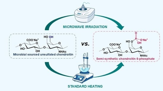
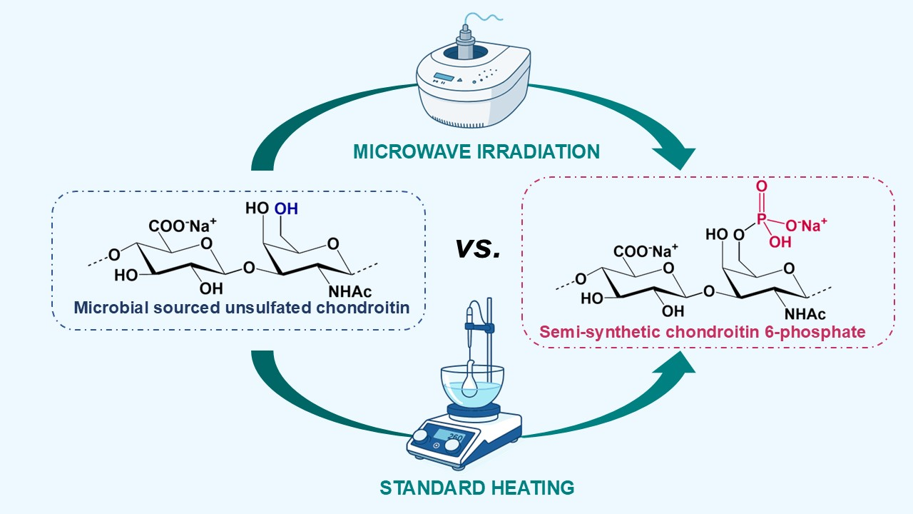
Chondroitin sulfate is a glycosaminoglycan polysaccharide, playing key roles in a plethora of physiopathological processes typical of higher animals. The position of sulfate groups within CS disaccharide subunits composing the polysaccharide chain is able to encode specific functional information. In order to expand such a “sulfation code”, access to non-natural CS variants and mimics thereof can be pursued. In this context, an interesting topic concerns phosphorylated analogs of CS polysaccharides, as the replacement of sulfate groups with phosphates can lead to unreported activities of phosphorylated CS. In light of this, the phosphorylation reaction of a microbial-sourced, unsulfated chondroitin polysaccharide with phosphoric acid is reported in the present study, testing different microwave irradiation conditions and comparing them with conventional heating procedures. The obtained products were subjected to a detailed characterization, in terms of chemical structure and hydrodynamic properties, by 1D- and 2D-NMR spectroscopy and HP-SEC-TDA analysis, respectively. The characterization study showed how different reaction conditions can not only influence the regioselectivity and degree of phosphorylation but also trigger the formation of phosphate diester functionalities acting as cross-linkers between polysaccharide chains. The results from the screening presented in this work could be interesting for any research devoted to the regioselective phosphorylation of a polysaccharide.

Graphical Abstract

1. Introduction

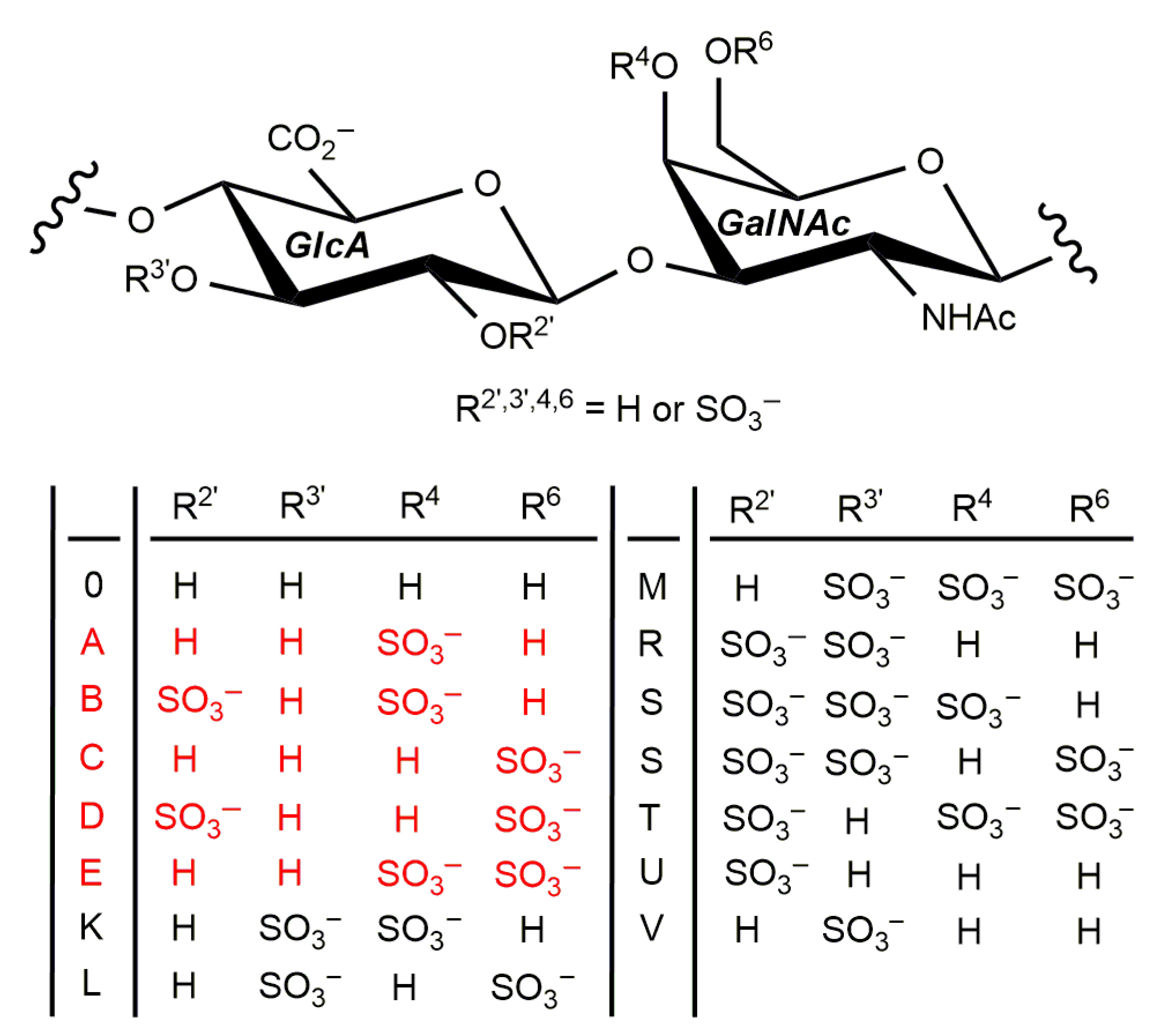
Chondroitin sulfate (CS) is a glycosaminoglycan (GAG) found in both vertebrates and invertebrates, consisting in a polysaccharide chain composed of disaccharide repeating units made by variously sulfated, alternating β-1⟶3-linked 2-acetamido-2-deoxy-D-galactose (N-acetyl-D-galactosamine, GalNAc) and β-1⟶4-linked D-glucuronic acid (GlcA) units [1]. Sulfate groups decorating the CS backbone seem to be a result of evolution, allowing for CS to play key roles in a plethora of physiopathological processes typical of higher animals [2]. It is important to note that their distribution on CS is not only animal- and tissue-specific, but it also depends on the physiopathological conditions of the single organism, i.e., age, inflammation, tumor formation, etc. [2,3]. The different distributions of sulfate groups within CS disaccharide repeating units are commonly indicated with a letter (Figure 1), and the resulting sequence of sulfated subunits seems to encode specific functional information. Different from other biological codes, such a “sulfation code” is far from being deciphered in detail [4]. Although for a few biological processes, the role of differently sulfated CS variants has been elucidated [5,6,7,8], cracking the sulfation code remains elusive due to several factors.
Natural CSs are very often composed of two or even more differently sulfated disaccharide subunits, and the identification of their sequence along the polysaccharide backbone is very difficult, having been achieved only in a few simple cases [9,10,11]. This also makes it very difficult to isolate structurally well-defined CS polysaccharides for structure–activity relationships investigations towards a full deciphering of the CS sulfation code. Nonetheless, to this aim, considerable efforts have been made in recent years through several approaches, spanning from the chemical [12], chemo-enzymatic [13,14], and microbial cell factory synthesis [15,16] of structurally homogeneous CS species to the development of increasingly powerful methods for highly efficient purification [17] and structural characterization of natural CSs [18]. Moreover, some research groups—including ours—are also directing their efforts to try to expand the “sulfation code”, i.e., by obtaining non-natural, structurally homogeneous CS variants [19,20,21] as well as mimics thereof [22]. In this frame, an interesting topic concerns the phosphorylated analogs of CS polysaccharides, because the replacement of sulfate groups with phosphates has been shown by a recent in silico study to lead to unreported activities in phosphorylated CS [23], triggered by the differences in size, polarity, acid–base, and chelation properties between sulfate and phosphate groups [24]. Nonetheless, the preparation of phosphorylated CS and, more generally, phosphorylated GAG species is still rather underdeveloped, with only a few, very recently reported examples [23,25,26,27]. One of the main reasons is that phosphorylation reactions on common polysaccharides (e.g., cellulose, dextran, alginate) are very often characterized by rather harsh conditions (i.e., heating the reaction mixture to temperatures up to 150 °C), poor yields and degree of phosphorylation (DP), and a lack of control over regiochemistry [28,29,30,31].
Surprisingly, to the best of our knowledge, only two examples of the derivatization of a polysaccharide with phosphorous-containing ester moieties have been reported to use microwave rather than conventional heating techniques, in order to provide a more efficient reaction. Indeed, microwave irradiation is well known in modern organic chemistry to provide a highly efficient form of heating, allowing for energy to be directly absorbed by the reactants, thus affording significantly higher reaction rates and product yields and selectivities [32]. The advantages of microwave irradiation have already been demonstrated for several reactions in the field of chemical modification of polysaccharide structures [33,34,35]. The two reports on microwave-assisted reactions of a polysaccharide with reacting phosphorous species regarded cellulose and levan. Cellulose was derivatized with phosphite groups with an almost quantitative DP, which was considerably higher and faster achieved with respect to the phosphite-derivatized cellulose obtained by conventional heating [36]. Similarly, levan was functionalized with phosphate monoester groups under microwave irradiation with a higher DP with respect to conventional heating [37].
Since we have very recently started to screen different phosphorylating reagents towards the semi-synthesis of regioselectively phosphorylated chondroitin polysaccharide [27], starting from a microbial-sourced, unsulfated chondroitin (CS-0, 42 ± 3 kDa, 1.34 dispersity) derived from the fed-batch fermentation of Escherichia coli O5:K4:H4 [38], we have now focused our efforts on the optimization of the DP for the semi-synthesis of chondroitin 6-phosphate (CP-C) by applying microwave irradiation for the phosphorylation reaction. All the obtained CP samples have been characterized in detail, from both a structural and hydrodynamic point of view, highlighting the differences detected between the products obtained by microwave irradiation and standard heating protocol.

2. Materials and Methods

2.1. General Methods

Commercial-grade reagents and solvents were used without further purification, except where differently indicated. The term “pure water” refers to water purified by a Millipore Milli-Q Gradient system (Millipore, Burlington, MA, USA). Microwave-irradiated reactions were performed with a CEM Discover 908010 instrument (CEM Corporation, Matthews, NC, USA). Dialyses were conducted on Spectra/Por 3.5 kDa cut-off membranes (Spectrum Labs, San Francisco, CA, USA) at 4 °C. Centrifugations were performed with an Eppendorf Centrifuge 5804R instrument (Eppendorf, Hamburg, Germany) at 4 °C (4600× g, 5 min). Sonications were performed with an Elmasonic S30H instrument (Elma Schmidbauer GmbH, Singen, Germany). Size-exclusion chromatography was performed on a Bio-Gel P4 column (0.75 × 67.5 cm, Bio-Rad, Hercules, CA, USA) using pure water as a buffer at a flow rate of 0.2 mL/min. Freeze-dryings were performed with a 5Pascal Lio 5P 4K freeze dryer (5Pascal, Milan, Italy). NMR spectra were recorded on a Bruker Avance-III HD (1H: 400 MHz, 13C: 100 MHz, 31P: 162 MHz, Bruker, Billerica, MA, USA) or on a Bruker Avance-III (1H: 600 MHz, 13C: 150 MHz, Bruker, Billerica, MA, USA) instrument—the latter equipped with a cryo-probe—in D2O (acetone as internal standard, 1H: (CH3)2CO at δ 2.22; 13C:(CH3)2CO at δ 31.5; H3PO4 as external standard, 31P: δ 0.0). A high-performance size-exclusion chromatography–triple detector array (HP-SEC-TDA) Viscotek TDA 305 instrument (Malvern Panalytical, Malvern, UK) was used for hydrodynamic analyses.

2.2. Typical Procedure for Phosphorylation Reaction

Polysaccharide 2 [39] (41.9 mg, 67.5 μmol) was mixed with urea (637 mg, 10.6 mmol) and then treated with dry N,N-dimethylformamide (DMF, 1.3 mL) and 85% H3PO4 (109 μL) under Ar atmosphere. The suspension was stirred at 80–150 °C under conventional heating or microwave irradiation for 1–3 h. The resulting yellowish suspension was cooled to room temperature, and then methanol (14 mL) was added to form a white precipitate. The mixture was cooled in a freezer, and then the precipitate was collected by centrifugation. The solid was dissolved in HPLC-grade H2O (4.0 mL; pH ~ 2) and treated under stirring with a few drops of aqueous 33% NaOH to neutral pH. The solution was dialyzed and freeze-dried to afford CP-C product (24.4 mg, 58% mass yield).

2.3. Enzymatic Depolymerization of CP-C-7

A suspension of CP-C-7 (20.4 mg) in a 0.15 M NaCl, 0.10 M NaOAc solution (1.0 mL) was buffered to pH = 5 by adding a drop of acetic acid. The mixture was incubated at 37 °C for 1 h, then a solution of bovine testicular hyaluronidase (BTH, 1.9 mg) in pure water (950 μL) was added. The production of oligosaccharide species was detected in progress by silica-gel thin-layer chromatography (TLC; eluent: 1:1:1:0.05 v/v/v/v n-butanol/ethanol/water/acetic acid). After incubating for 165 h at 37 °C, the reaction was quenched by heating it at 100 °C for 15 min. By successive cooling and freeze-drying, a white solid residue (33.3 mg) was obtained. It was dissolved in the minimum amount of water and purified by size-exclusion chromatography to give three fractions (LMW-CP-C-a, 4.9 mg; LMW-CP-C-b, 5.7 mg; LMW-CP-C-c, 2.6 mg).

2.4. NMR Analyses

NMR samples were prepared by adding 600 μL D2O to approx. 6 mg of each polysaccharide product. After prolonged stirring and sonication at room temperature, the solid residue, if any, was removed, and experiments were performed on the resulting supernatant. Data were processed using the data analysis packages integrated with Bruker TopSpin® 4.0.5 software. The 31P-NMR spectra were obtained by typically accumulating 512 transients. The 1H- and 31P-1D-DOSY experiments were measured using a stimulated echo sequence with bipolar gradient pulses and one spoil gradient (stebpgp1s1d) with a diffusion time Δ of 100 ms, a gradient duration of 2 or 4 ms (for 1H- and 31P-1D-DOSY, respectively), and accumulating 64 or 2048 transients (for 1H- and 31P-1D-DOSY, respectively). The 1H,13C-DEPT-HSQC experiments were measured in the 1H-detected mode via single-quantum coherence with proton decoupling in the 13C domain, using data sets of 2048 × 256 points and typically 64 increments. The 1H,31P-HSQC experiments were measured in the 1H-detected mode via phase-sensitive gradient selection with decoupling during acquisition, using a data set of 2048 × 256 points and typically 64 increments.

2.5. Hydrodynamic Analyses

A comprehensive description of the HP-SEC-TDA Viscotek TDA 305 apparatus, employed for the hydrodynamic characterization of the samples, and its capabilities is reported elsewhere [40]. In brief, the instrumentation comprised two primary units: (i) the GPCmax VE 2001 integrated system (Malvern Panalytical, Malvern, UK), which includes a gel-permeation chromatography pump, an in-line solvent degasser, and an autosampler; (ii) the TDA302 triple detector array module (Malvern Panalytical, Malvern, UK), equipped with a column oven, a refractive index (RI) detector, a viscometer, as well as right-angle (RALS) and low-angle (LALS) light scattering detectors. The light scattering detectors were optimized to maximize the signal-to-noise ratio, with measurements taken at a 7° angle relative to the incident beam. Two TSK–GEL GMPWXL columns (Tosoh Bioscience, Tokyo, Japan; Cat. No. 8-08025; hydroxylated polymethacrylate, pore size—100–1000 Å, particle size—13 µm, dimensions—7.8 × 30.0 cm) were connected in series, preceded by a guard column (Tosoh Bioscience; Cat. No. 08033; particle size—12 µm, 6.0 × 4.0 cm).
Samples were dissolved/dispersed in deionized water to a concentration of approximately 10 mg/mL, followed by appropriate dilution for analysis (final column load ranging between 0.2 and 0.4 dL). Prior to injection, all samples were filtered using disposable 0.22 μm syringe filters. Chromatographic analyses were carried out under isocratic conditions employing a 0.1 M aqueous NaNO3 solution (pH 7.0) as the mobile phase, at a flow rate of 0.6 mL/min and a column temperature of 40 °C, with a total runtime of one hour. Data acquisition and processing were performed using OmniSEC software 11.36 (Malvern Instruments, Malvern, UK). A dn/dc value of 0.155 mL/g was applied for calculations. The determined parameters included weight-average molecular weight (Mw), number-average molecular weight (Mn), polydispersity index (Mw/Mn), hydrodynamic radius (Rh), and intrinsic viscosity ([ƞ]). Additionally, the sample fraction/wt% having a molecular weight above 20 kDa was derived.

3. Results and Discussion

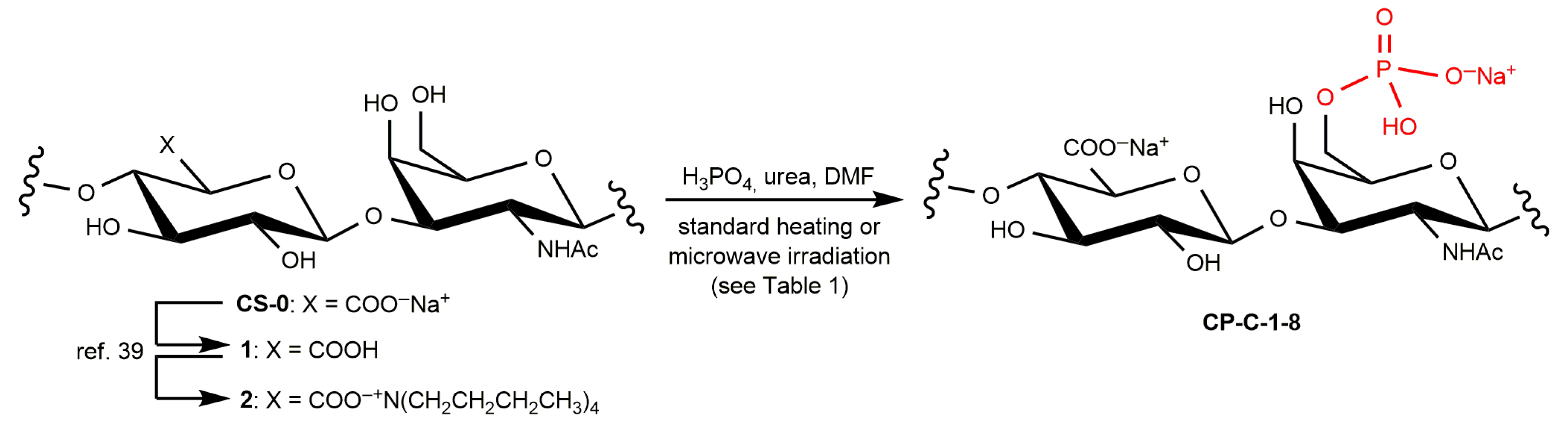
A screening of phosphorylation conditions performed on the n-tetrabutylammonium salt of E. coli sourced CS-0 (2 [39], Scheme 1) was very recently reported by us [27]. CP-C could be obtained by a conventional heating reaction either with PEP-K and TBAHS at 80 °C for 48 h, achieving a DP-6 (degree of phosphorylation at GalNAc O-6 site) of 0.36 or with H3PO4 in DMF at 120 °C for 3 h in the presence of urea to give a DP-6 of 0.49. The latter result (Table 1, entry 1) was selected as the best starting point to perform a screening of microwave irradiation conditions for the same phosphorylation method in order to increase the DP-6. Lower reaction time (2 h) and temperature (80 °C) with respect to conventional heating were initially tested to give CP-C-2 product (Table 1, entry 2), after n-tetrabutylammonium/Na+ exchange and purification by dialysis. No derivatization was achieved under these conditions, as detected by comparing 1H-NMR spectra of CP-C-1 and starting CS-0 (Figure S1 in Supplementary Materials).
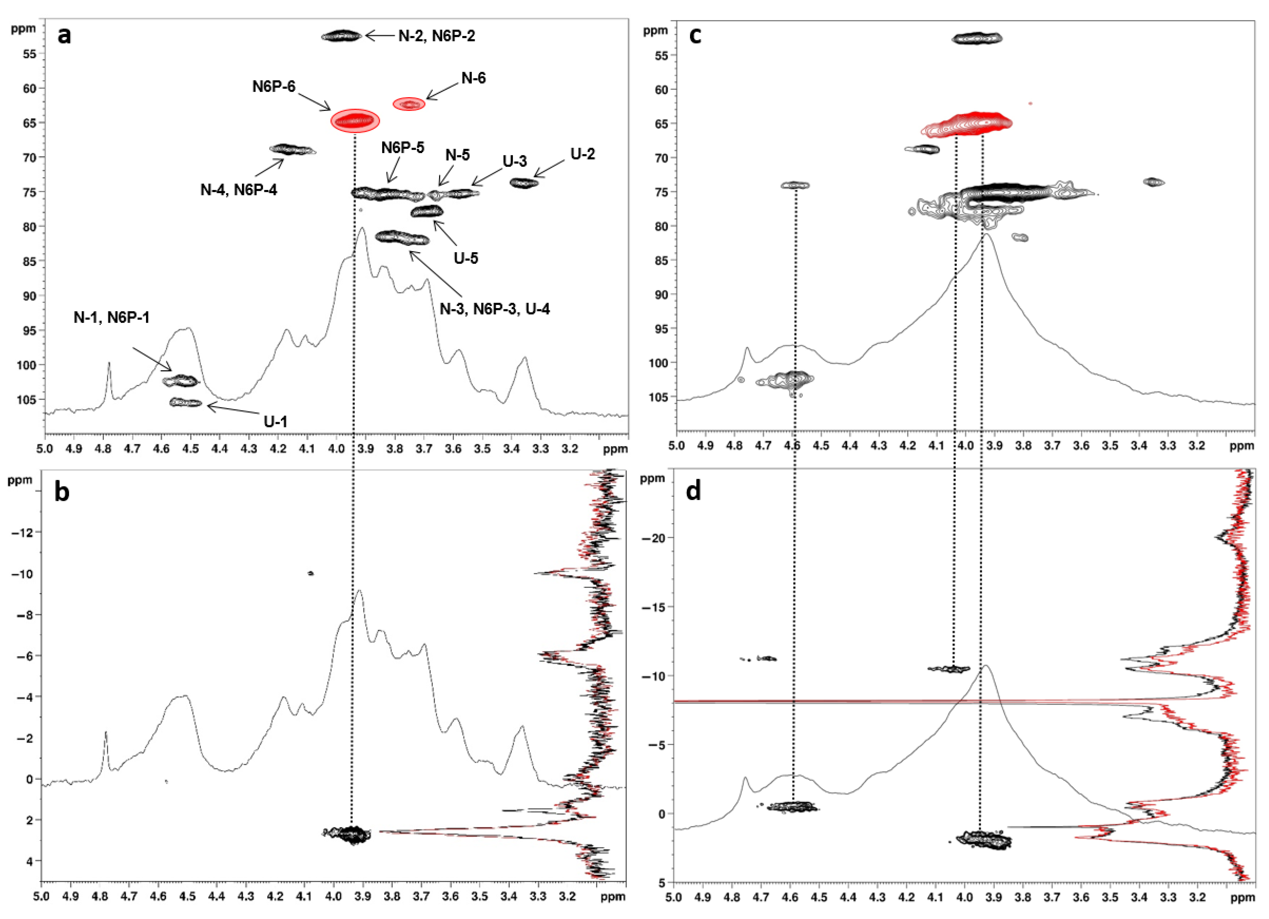
Under stronger microwave irradiating conditions (Table 1, entry 3), the regioselective installation of the phosphate group at GalNAc O-6 site could be achieved, as confirmed by a set of both 1D- and 2D-NMR experiments. The former set comprised 1H-NMR, 31P-NMR, and 31P-1D-DOSY spectra (the last one being fundamental to discriminate between phosphorus-containing moieties either covalently linked to the chondroitin polysaccharide or present in small molecule impurities such as inorganic phosphate or diphosphate), while the 1H,31P-HSQC and 1H,13C-DEPT-HSQC sequences were employed for 2D-NMR measurements. In particular, the latter 2D-NMR spectrum of CP-C-3 showed two small signals sensibly shifted to different chemical shift values with respect to starting CS-0, at δH/C 3.77/73.6 and 3.92/64.5 (Figure S2 in Supplementary Materials). These could be assigned to CH-5 and CH2-6 atoms of GalNAc-6-phosphate units, respectively, by comparison with values from the literature [27] and also by detecting the signal at δH/P 3.92/2.6 in the 1H,31P-HSQC 2D-NMR spectrum of CP-C-3 (Figure S3 in Supplementary Materials). By assuming that the signals at δH/C 3.92/64.5 and 3.76/62.4—assigned to CH2-6 atoms of GalNAc-6-phosphate and underivatized GalNAc units, respectively—displayed similar 1JC,H coupling constants and that a difference of around 5–8 Hz from the experimental set value could not cause a substantial variation in the integrated peak volumes [41], their relative integration returned a DP-6 equal to only 0.12 for CP-C-3. In order to increase it, the microwave phosphorylation was conducted under even stronger irradiating conditions (Table 1, entries 4–6). Finally, the 1H,13C-DEPT-HSQC 2D-NMR spectrum of the CP-C-6 sample (Figure 2a), which was obtained by reacting 2 for 3 h at 120 °C under 100 W microwave irradiation at 300 psi, showed a DP-6 equal to 0.83 without impairing the GalNAc O-6 regioselectivity. This value was considerably higher with respect to the DP-6 resulting from the reaction conducted under the same conditions but with standard heating (0.49 for CP-C-1) [27]. It bears noting that the attempt to further increase the latter by repeating the reaction twice gave a negligible enhancement of DP-6 (0.51; Table 1, entry 7), while an increase in temperature to 150 °C (Table 1, entry 8) afforded the CP-C-8 product with a quantitative DP-6 but a decreased regioselectivity. Indeed, its 1H,13C-DEPT-HSQC 2D-NMR spectrum showed a remarkably downfield-shifted CH signal at δH/C 4.60/73.9 (Figure 2c), which could be associated with the phosphorylation of secondary hydroxyls by the cross-peak at δH/P 4.60/−0.4 in the 1H,31P-HSQC 2D-NMR spectrum (Figure 2d). The order of reactivity of the secondary hydroxyls of chondroitin polysaccharide is known to have the OH at the C-2 atom of GlcA units as the most reactive one under sulfation conditions [42]. Unfortunately, the collected 2D-NMR spectra of CP-C-8 were not able to confirm or discard this trend, plausibly due to a too heterogeneous structure hampering a clear attribution of the signals.
Indeed, the higher structural heterogeneity of the CP-C-8 sample was related not only to the presence of multiple phosphorylation sites but also to the detection of peaks in different regions of its 31P and 31P-1D-DOSY spectra. Indeed, together with broad peaks at 2.0 and −0.4 ppm assigned to monophosphate groups at the two different derivatization positions, some additional signals of similar intensity were detected at −7.0, −10.5, and −11.2 ppm. These could be due to the presence of some diphosphate groups decorating the chondroitin backbone, as clearly evidenced by the 1H,31P-HSQC 2D-NMR spectrum of CP-C-8 (Figure 2d), showing cross-peaks at δH/P 4.06/−10.5 and 4.62/−11.2. In particular, these signals could be assigned to the phosphorus atoms of diphosphate groups directly attached at GalNAc O-6 and at a secondary hydroxyl site, respectively (α-phosphorus atoms), while the 31P-signal at −7.0 ppm, that was not correlated to any 1H signal in the 1H,31P-HSQC 2D-NMR spectrum, could be assigned to the distal phosphorus atoms of both diphosphate groups (β-phosphorus atoms) [43,44]. A minor amount of triphosphate groups decorating the chondroitin backbone could also be inferred by the less intense 31P signal at −20.0 ppm, which could be assigned to their middle β-phosphorus atoms, being the α- and γ-ones (closest and farthest to the attachment site on the polysaccharide backbone, respectively) overlapped with the α- and β-phosphorus signals of diphosphate groups [43]. Interestingly, the presence of signals associated with diphosphate groups could also be detected in the CP-C-6 product from microwave irradiated phosphorylation, although in a much lower amount with respect to CP-C-8. It bears noting that, even if the presence of cyclic 4,6-phosphate diesters on galacto-configured sugars was sometimes reported in natural polysaccharides from bacterial membranes [45,46], this structural feature could be ruled out in the CP-C-8 case. Indeed, it would have required a correlation in the 1H,31P-HSQC 2D-NMR spectrum between a single 31P-chemical shift with two different 1H-chemical shifts, assignable through the 1H,13C-DEPT-HSQC spectrum to a CH- and a CH2-type signal, respectively. This was not observed in the reported 2D-NMR spectra.
In parallel with the structural characterization by NMR, some hydrodynamic parameters were also determined for phosphorylated polysaccharides CP-C-3-8 by HP-SEC-TDA analysis (Table 2). For all the products obtained by phosphorylation under microwave irradiation (CP-C-3-6) a lower weight-average molecular weight was detected with respect to the starting polysaccharide CS-0 (Table 2, entry 1, 37 kDa), as expected for the acid character—although weak—of the employed phosphorylating agent (H3PO4), triggering the cleavage of some of the glycosidic bonds connecting the monose residues in the polysaccharide chain. Moreover, with the gradual increase in the strength of microwave irradiating conditions from CP-C-3 to CP-C-6, a gradually more pronounced depolymerization was detected with the weight-average molecular weight value lowering from 27 kDa (CP-C-3, Table 2, entry 2) to 11 kDa (CP-C-5, Table 2, entry 4) and the sample fraction exceeding 20 kDa lowering from 65% (CP-C-3) to 10% (CP-C-5). It bears noting that the product CP-C-6, which was obtained under the same microwave conditions of CP-C-5 but after a longer irradiation time (3 and 1 h, respectively), showed a slightly higher molecular weight (17 KDa, sample fraction with Mw > 20 kDa = 14%; Table 2, entry 5) with respect to the latter. This could be first ascribed to the much higher DP-6 of CP-C-6 with respect to CP-C-5 (0.83 vs. 0.27: see Table 1, entries 5 and 6), accounting for a much more frequent and therefore much “heavier” decoration of the chondroitin backbone with phosphate and—to a lower extent—even diphosphate groups.
A completely different result was observed for CP-C-8, as it showed a very large distribution of molecular weights, with four peaks of similar representativeness in the HP-SEC chromatogram spanning from 18 to hundreds of kDa and a fifth, minor one reaching even 4 MDa (Figures S12 and S13 in Supplementary Materials). The formation of such high-molecular-weight species could be ascribed to the presence of mono- and/or diphosphate diester moieties acting as cross-linkers between different polysaccharide chains. The cross-linking between polysaccharide chains through phosphodiester functionalities is not unprecedented, and includes also the case of a GAG polysaccharide, such as hyaluronic acid [47], although it has always been accomplished with the use of sodium trimetaphosphate rather than H3PO4 as the phosphorylation/cross-linking agent [48]. Cross-linking could occur through monophosphate and/or diphosphate diester. The formation of the latter in CP-C-8 is not in disagreement with its 31P-NMR data. Diphosphate diesters typically give a signal at approx. −10 ppm that can be overlapped with peaks related to α-phosphorous atoms of diphosphate monoester moieties [43]. The 31P-NMR spectrum of CP-C-8 indeed shows signals at such chemical shift values, as already discussed above (Figure 2d). Conversely, cross-linking through monophosphate diesters should be plausibly ruled out. Indeed, such moieties typically give 31P signals at approx. 0 ppm [43]. In spite of the 31P-NMR spectrum of CP-C-8 showing a signal in such a chemical shift region, the 1H,31P-HSQC 2D-NMR spectrum allows for correlating it only with a single 1H chemical shift (Figure 2d), corresponding in turn to a CH-tagged density in the 1H,13C-DEPT-HSQC 2D-NMR spectrum (Figure 2c). This would mean that the putative monophosphate diester cross-linking would bridge secondary hydroxyls placed at the same site in the two cross-linked chondroitin repeating units. This “symmetrical” cross-linking would be very unlikely not only from a statistical point of view, but also if one takes into consideration that primary hydroxyls are instead the most reactive sites towards phosphorylation, as theoretically expected and experimentally found in all the semi-synthesized derivatives, including CP-C-8.
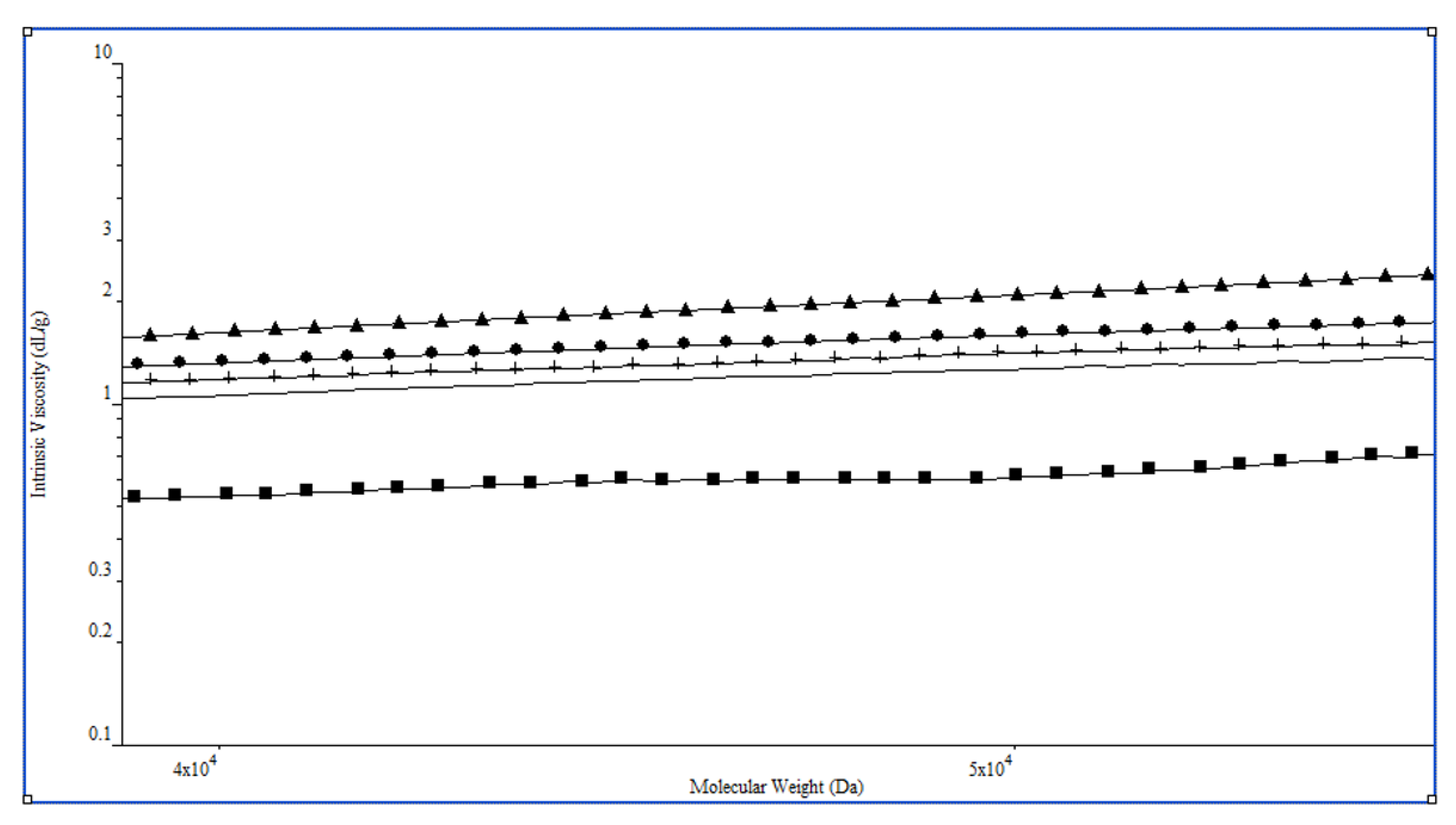
Therefore, NMR analysis of CP-C-8 supports the notion that the cross-linking reaction was really happening under the harshest phosphorylation conditions while employing a conventional heating protocol (150 °C, 3 h: Table 1, entry 8). It is important to note that any significant evidence of a cross-linking reaction could be found for all the other products (CP-C-3-7), neither by 31P-NMR spectroscopy (Figures S3–S7 in Supplementary Materials) nor by the presence of significantly represented peaks associated with high-molecular-weight species in the HP-SEC chromatogram (Figures S13 and S14 in Supplementary Materials). Nonetheless, a comparison of intrinsic viscosity vs. molecular weight relationships for the phosphorylated products obtained by microwave irradiation, or obtained by overlapping the Mark–Houwink–Sakurada plots for CP-C-3-6 (Figure 3), clearly revealed a gradual increase in the compactness of the polysaccharide conformation in solution with the increase in DP-6 (Table 1, entries 1, 3–6). These observations could be explained by polymer chain cross-linking, although the amount of phosphodiester moieties acting to this aim is too low to be detected by 31P-NMR spectroscopy in the case of CP-C-3-5, or can be ascribed only to minor peaks in CP-C-6. The higher dispersity (Mw/Mn) of the latter with respect to CP-C-3-5 derivatives concurred to define the same trend in the increasing degree of cross-linking. Moreover, an additional clue that a cross-linking reaction could happen, not only for CP-C-8 but also during the reactions giving products CP-C-3-7, comes from their lower water solubility with respect to the starting CS-0 polysaccharide. Indeed, the weight recovery after centrifugation of a 7.5 mg/mL suspension in pure water is quantitative for CS-0, while it spans from 47% to around 70% for the phosphorylated polysaccharides, the lower being associated with the CP-C-8 product, for which the HP-SEC-TDA analysis of the soluble fraction revealed an extensive cross-linking, as discussed above. The analysis of the structural features of the water-insoluble fractions could obviously be performed either by liquid-state NMR spectroscopy or by HP-SEC-TDA. It would require solid-state NMR techniques that are unfortunately not included among our current analytical facilities. Collaborative work with other research groups is being planned to tackle this issue. The results will be published as soon as possible elsewhere.
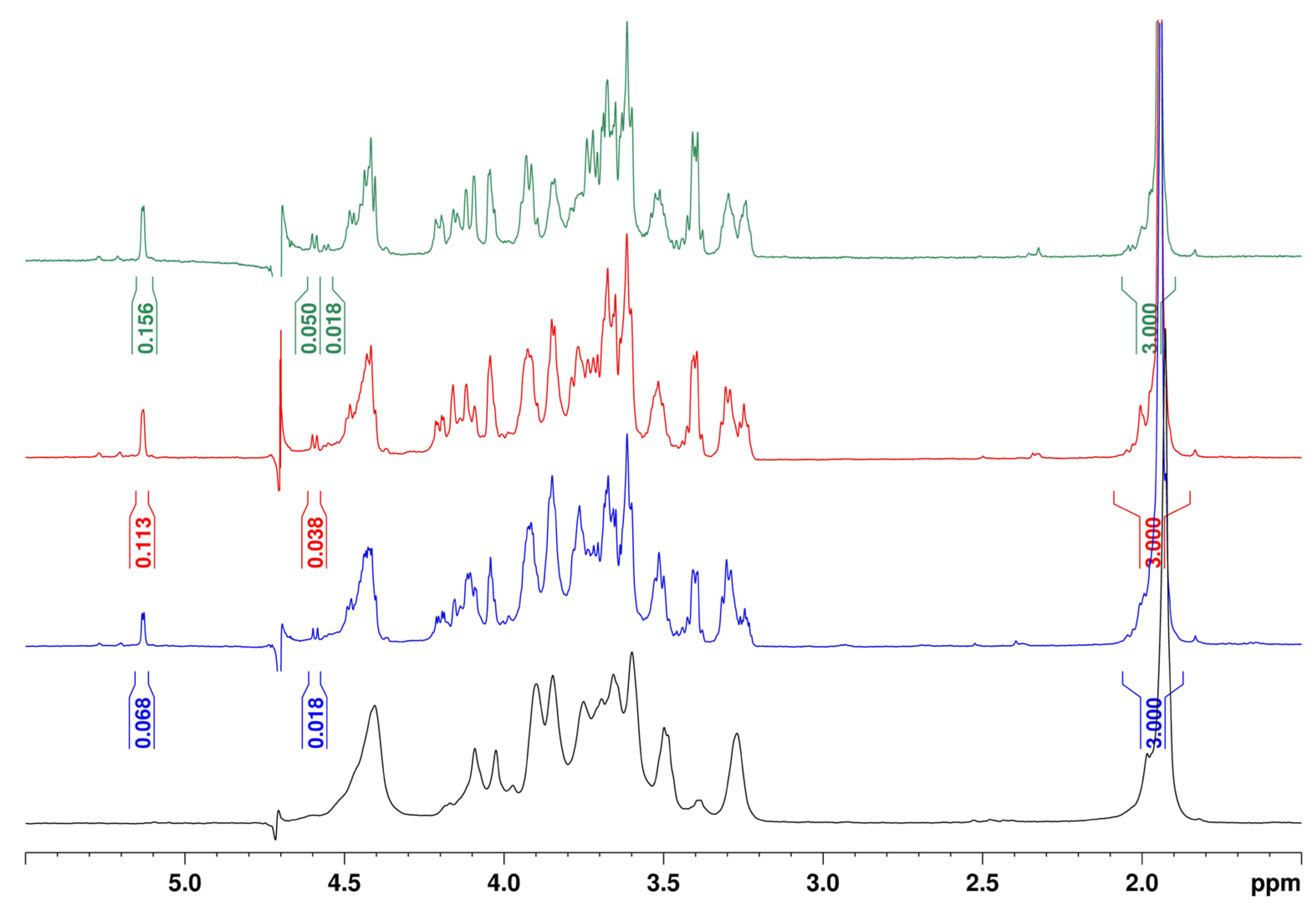
Successfully obtaining regioselectively phosphorylated CP-C derivatives prompted a preliminary study of their behavior in the presence of BTH, an endonuclease enzyme known for catalyzing the hydrolysis of hyaluronic acid, including some very recently reported, randomly phosphorylated derivatives thereof [49], and showing activity towards chondroitin and CS polysaccharides too [50]. The aim was not only to test the biodegradability of the obtained CP-C polysaccharides but also to open a direct access to low-molecular-weight species, i.e., CP oligosaccharides, that, to the best of our knowledge, are completely missing in the literature. It bears noting that several studies have demonstrated that low-molecular-weight chondroitin sulfate (LMW-CS) species have enhanced bioavailability and biological activities, including antioxidant, anticoagulation, anti-inflammatory, and neuroprotective effects, together with a safer profile in terms of absence of immunological responses [51,52]. Therefore, a future evaluation of LMW-CP-C biological properties would surely be worth pursuing. By incubating CP-C-7 with BTH in a NaOAc/AcOH buffer (pH = 5) at 37 °C [53], three low-molecular-weight chondroitin phosphate species (named LMW-CP-C-a, LMW-CP-C-b, and LMW-CP-C-c, respectively) could be obtained after purification of the crude reaction mixture by size-exclusion chromatography. Their characterization by 1H- and 31P-1D-NMR as well as 1H,13C-DEPT-HSQC- and 1H,31P-HSQC-2D-NMR confirmed a structure of LMW chondroitin species carrying phosphate groups at some of GalNAc O-6 sites. In particular, a comparison of CP-C-7 and LMW-CP-C-a-c 1H-NMR spectra clearly showed the presence of doublet signals at 5.13 and 4.59 ppm only in the latter case (Figure 4).
Since these signals corresponded in the 1H,13C-DEPT-HSQC spectra to cross-peaks at δC 92.3 and 96.2, respectively, they could be assigned to the anomeric H atom of α- and β-configured GalNAc-reducing end units, respectively. A comparison between the sum of their integral values and the area of the signal at 1.94 ppm related to the methyl group of (reducing and non-reducing) GalNAc units through Equation (1), gave an estimation of the average chain length in the three LMW-CP samples: LMW-CP-C-a length distribution could be centered on a 23-mer species, while LMW-CP-C-b and LMW-CP-C-c could be estimated to be shorter (13-mer and 9-mer, respectively).
average chain length = [I(CH3) / 3] / [I(α-reducing) + I(β-reducing)] ∗ 2
By using the same equation, we could also compare the kinetic rate of BTH depolymerization for the CP-C-7 sample and the starting chondroitin polysaccharide CS-0. From the 1H-NMR spectrum of the crude digest of the former (Figure S9 in Supplementary Materials), a length distribution centered on a 14-mer species was found, in agreement with the values indicated above for the fractions derived thereof (9-mer to 23-mer). Native CS-0 enzymatic degradation under the same conditions furnished a length distribution of the obtained oligomers in the crude reaction mixture centered on a 5-mer species. By considering that the starting Mw of CS-0 was approximately double with respect to CP-C-7 (42 vs. 23 kDa), it was possible to estimate an approx. 6-fold higher rate of degradation for the natural polysaccharide with respect to its phosphorylated derivative CP-C-7.
Finally, the presence of GalNAc-6-phosphate units within LMW-CP-C-a-c structures was confirmed by the presence of a signal at δH/P 3.84/3.9 in their 1H,31P-HSQC 2D-NMR spectra, corresponding to a 1H- and 13C-downfield-shifted CH2 signal at δH/C 3.84/63.0 in their 1H,13C-DEPT-HSQC spectra (Figures S10–S12 in Supplementary Materials).

4. Conclusions

The phosphorylation reaction of a microbial-sourced, unsulfated chondroitin polysaccharide with phosphoric acid was tested under different microwave irradiation and conventional heating conditions in order to semi-synthesize a phosphorylated mimic of one of the most commonly found chondroitin sulfate variants (CS-C), i.e., chondroitin-6-phosphate (CP-C). The collected products were subjected to a detailed characterization, in terms of chemical structure and hydrodynamic properties, by 1D- and 2D-NMR spectroscopy and HP-SEC-TDA analysis, respectively. The best result in terms of regioselectivity and degree of phosphorylation was achieved by microwave irradiation of the CS-0 starting material at 120 °C for 3 h. Under similar conditions, the application of a conventional heating protocol gave a lower DP-6, while the increase in the temperature enhanced the degree of derivatization but was detrimental for the regioselectivity and extensively triggered alternative reaction pathways, giving the formation of phosphate diester functionalities as cross-linkers between polysaccharide chains. The results from the screening presented in this work could be of a certain interest for any research devoted to the regioselective phosphorylation of a polysaccharide. Moreover, the obtained CP-C polysaccharides, as well as their enzymatically derived LMW counterparts, that were already obtained in this work from the former, will be subjected in the near future to a comparison of their structural features and, above all, of their biological properties with the sulfated CS-C counterparts. The results of such an investigation will be published as soon as possible elsewhere. Last but not least, the present study represents an additional reference—even if the generalizability of results across different microwave equipment is still to be proven—in the growing field of microwave-assisted derivatizations of polysaccharides.

Supplementary Materials

The following supporting information can be downloaded at: https://www.mdpi.com/article/10.3390/polysaccharides7010011/s1, Figure S1: 1H NMR spectra superimposition of CP-C-2 and CS-0; Figure S2: 1H, 1D-DOSY and 1H,13C-DEPT-HSQC NMR spectra superimposition of CP-C-3 and CS-0; Figures S3–S8: (a) 1H, 1D-DOSY, 1H,13C-DEPT-HSQC, and (b) 1H, 31P, 31P-1D-DOSY, 1H,31P-HSQC NMR spectra of CP-C-3-8; Figure S9: 1H NMR spectra superimposition of crude BTH digest of CS-0 and CP-C-7; Figures S10–S12: (a) 1H, 1H,13C-DEPT-HSQC and (b) 1H, 31P, 1H,31P-HSQC NMR spectra of LMW-CP-C-a-c; Figures S13,S14: HP-SEC profiles (RI signal) for CS-0 and CP-C-3-8.

Author Contributions

Conceptualization, F.E., S.T., A.I., and E.B.; methodology, F.E., A.L.G., and E.B.; investigation, F.E. and S.C.; data curation, F.E., S.C., A.L.G., and E.B.; writing—original draft preparation, A.L.G. and E.B.; writing—review and editing, F.E., S.T., A.I., D.C., and C.S.; supervision, S.T., D.C., A.L.G., and E.B.; funding acquisition, A.L.G., C.S., and E.B. All authors have read and agreed to the published version of the manuscript.

Funding

This research was funded by the Italian Ministry of Enterprises and Made in Italy (MIMIT, CROSSGAG project, CUP: B69J24001940005) and the Italian Ministry of University and Research (MUR, HealHyal project, CUP: E53D23020320001).

Data Availability Statement

The original contributions presented in this study are included in the article/Supplementary Materials. Further inquiries can be directed to the corresponding author.

Conflicts of Interest

The authors declare no conflicts of interest.

Abbreviations

The following abbreviations are used in this manuscript:
CSChondroitin sulfate
GAGGlycosaminoglycans
GalNAcN-acetyl-D-galactosamine
GlcAD-glucuronic acid
DPDegree of phosphorylation
DP-6Degree of phosphorylation at N-acetyl-D-galactosamine O-6 site
CS-0Unsulfated chondroitin
CP-CChondroitin 6-phosphate
NMRNuclear magnetic resonance
HP-SECHigh-performance size exclusion chromatography
TDATriple detector array
DMFN,N-dimethylformamide
HPLCHigh-performance liquid chromatography
NaOAcSodium acetate
AcOHAcetic acid
BTHBovine testicular hyaluronidase
TLCThin-layer chromatography
LMWLow molecular weight
DOSYDiffusion-ordered spectroscopy
DEPT-HSQCDistortionless enhancement by polarization transfer heteronuclear single-quantum correlation
RIRefractive index
RALSRight-angle light scattering
LALSLow-angle light scattering
MwWeight-average molecular weight
RhHydrodynamic radius
[η]Intrinsic viscosity
MnNumber-average molecular weight

References

  1. Bedini, E.; Corsaro, M.M.; Fernandez-Mayoralas, A.; Iadonisi, A. Chondroitin, dermatan, heparan, and keratan sulfate: Structure and functions. In Extracellular Sugar-Based Biopolymers Matrices; Cohen, E., Merzendorfer, H., Eds.; Springer Nature: Cham, Switzerland, 2019; pp. 187–223. [Google Scholar]
  2. Ricard-Blum, S.; Vivès, R.R.; Schaefer, L.; Götte, M.; Merline, R.; Passi, A.; Heldin, P.; Magalhães, A.; Reis, C.A.; Skandalis, S.S.; et al. A biological guide to glycosaminoglycans: Current perspectives and pending questions. FEBS J. 2024, 291, 3331–3366. [Google Scholar] [CrossRef]
  3. Collin, E.C.; Carroll, O.; Kilcoyne, M.; Peroglio, M.; See, E.; Hendig, D.; Alini, M.; Grad, S.; Pandit, A. Ageing affects chondroitin sulfates and their synthetic enzymes in the intervertebral disc. Sig. Transduct. Target. Ther. 2017, 2, 17049. [Google Scholar] [CrossRef] [PubMed]
  4. Perez, S.; Makshakova, O.; Angulo, J.; Bedini, E.; Bisio, A.; de Paz, J.L.; Fadda, E.; Guerrini, M.; Hricovini, M.; Hricovini, M.; et al. Glycosaminoglycans: What remains to be deciphered? J. Am. Chem. Soc. Au 2023, 3, 628–656. [Google Scholar] [CrossRef] [PubMed]
  5. Gama, C.I.; Tully, S.E.; Sotogaku, N.; Clark, P.M.; Rawat, M.; Vaidehi, N.; Goddard, W.A., III; Nishi, A.; Hsieh-Wilson, L.C. Sulfation patterns of glycosaminoglycans encode molecular recognition and activity. Nat. Chem. Biol. 2006, 2, 467–473. [Google Scholar] [CrossRef] [PubMed]
  6. Swarup, V.P.; Hsiao, T.W.; Zhang, J.; Prestwich, G.D.; Kuberan, B.; Hlady, V. Exploiting differential surface display of chondroitin sulfate variants for directing neuronal outgrowth. J. Am. Chem. Soc. 2013, 135, 13488–13494. [Google Scholar] [CrossRef]
  7. Miyata, S.; Komatsu, Y.; Yoshimura, Y.; Taya, C.; Kitagawa, H. Persistent cortical plasticity by upregulation of chondroitin 6-sulfation. Nat. Neurosci. 2012, 15, 414–422. [Google Scholar] [CrossRef]
  8. Sugiura, N.; Clausen, T.M.; Shioiri, T.; Gustavsson, T.; Watanabe, H.; Salanti, A. Molecular dissection of placental malaria protein VAR2CSA interaction with a chemo-enzymatically synthesized chondroitin sulfate library. Glycoconj. J. 2016, 33, 985–994. [Google Scholar] [CrossRef]
  9. Yu, Y.; Duan, J.; Leach, F.E., III; Toida, T.; Higashi, K.; Zhang, H.; Zhang, F.; Amster, I.J.; Linhardt, R.J. Sequencing the dermatan sulfate chain of decorin. J. Am. Chem. Soc. 2017, 139, 16986–16995. [Google Scholar] [CrossRef]
  10. Ly, M.; Leach, F.E., III; Laremore, T.N.; Toida, T.; Amster, I.J.; Linhardt, R.J. The proteoglycan bikunin has a defined sequence. Nat. Chem. Biol. 2011, 7, 827–833. [Google Scholar] [CrossRef]
  11. van Kuppevelt, T.H.; Oosterhof, A.; Versteeg, E.M.M.; Podhumljak, E.; van de Westerlo, E.M.A.; Daamen, W.F. Sequencing of glycosaminoglycans with potential to interrogate sequence-specific interactions. Sci. Rep. 2017, 7, 14785. [Google Scholar] [CrossRef]
  12. Mende, M.; Bednarek, C.; Wawryszyn, M.; Sauter, P.; Biskup, M.B.; Schepers, U.; Bräse, S. Chemical synthesis of glycosaminoglycans. Chem. Rev. 2016, 116, 8193–8255. [Google Scholar] [CrossRef] [PubMed]
  13. Zhang, X.; Lin, L.; Huang, H.; Linhardt, R.J. Chemoenzymatic synthesis of glycosaminoglycans. Acc. Chem. Res. 2020, 53, 335–346. [Google Scholar] [CrossRef] [PubMed]
  14. Gottschalk, J.; Elling, L. Current state on the enzymatic synthesis of glycosaminoglycans. Curr. Opin. Chem. Biol. 2021, 61, 71–80. [Google Scholar] [CrossRef] [PubMed]
  15. Badri, A.; Williams, A.; Awofiranye, A.; Datta, P.; Xia, K.; He, W.; Fraser, K.; Dordick, J.S.; Linhardt, R.J.; Koffas, M.A.G. Complete biosynthesis of a sulfated chondroitin in Escherichia coli. Nat. Commun. 2021, 12, 1389. [Google Scholar] [CrossRef]
  16. Liu, K.; Guo, L.; Chen, X.; Liu, L.; Gao, C. Microbial synthesis of glycosaminoglycans and their oligosaccharides. Trends Microbiol. 2023, 31, 369–383. [Google Scholar] [CrossRef]
  17. Restaino, O.F.; Schiraldi, C. Chondroitin sulfate: Are the purity and the structural features well assessed? A review on the analytical challenges. Carbohydr. Polym. 2022, 292, 119690. [Google Scholar] [CrossRef]
  18. Zappe, A.; Miller, R.L.; Struwe, W.B.; Pagel, K. State-of-the-art glycosaminoglycan characterization. Mass Spectrom. Rev. 2022, 41, 1040–1071. [Google Scholar] [CrossRef]
  19. Lane, R.S.; St. Ange, K.; Zolghadr, B.; Liu, X.; Schäffer, C.; Linhardt, R.J.; DeAngelis, P.L. Expanding glycosaminoglycan chemical space: Towards the creation of sulfated analogs, and novel and chimeric polymers. Glycobiology 2017, 27, 646–656. [Google Scholar] [CrossRef]
  20. Vessella, G.; Traboni, S.; Cimini, D.; Iadonisi, A.; Schiraldi, C.; Bedini, E. Development of semisynthetic, regioselective pathways for accessing the missing sulfation patterns of chondroitin sulfate. Biomacromolecules 2019, 20, 3021–3030. [Google Scholar] [CrossRef]
  21. Cimini, D.; Bedini, E.; Schiraldi, C. Biotechnological advances in the synthesis of modified chondroitin towards novel biomedical applications. Biotechnol. Adv. 2023, 67, 108185. [Google Scholar] [CrossRef]
  22. Liu, Q.; Chen, G.; Chen, H. Chemical synthesis of glycosaminoglycan-mimetic polymers. Polym. Chem. 2019, 10, 164–171. [Google Scholar] [CrossRef]
  23. Bojarski, K.K.; Becher, J.; Riemer, T.; Lemmnitzer, K.; Möller, S.; Schiller, J.; Schnabelrauch, M.; Samsonov, S.A. Synthesis and in silico characterization of artificially phosphorylated glycosaminoglycans. J. Mol. Struct. 2019, 1197, 401–416. [Google Scholar] [CrossRef]
  24. Lima, M.A.; Rudd, T.R.; Fernig, D.G.; Yates, E.A. Phosphorylation and sulfation share a common biosynthetic pathway, but extend biochemical and evolutionary diversity of biological macromolecules in distinct ways. J. R. Soc. Interface 2022, 19, 20220391. [Google Scholar] [CrossRef] [PubMed]
  25. Khaybrakhmanova, E.A.; Kozyrev, S.V.; Tyumkina, T.V.; Ponedel’kina, I.Y. Phosphorylation of hyaluronic acid. Chem. Proc. 2022, 12, 39. [Google Scholar] [CrossRef]
  26. Uchimura, K.; Nishitsuji, K.; Chiu, L.-T.; Ohgita, T.; Saito, H.; Allain, F.; Gannedi, V.; Wong, C.-H.; Hung, S.-C. Design and synthesis of 6-O-phosphorylated heparan sulfate oligosaccharides to inhibit amyloid β-aggregation. Chembiochem 2022, 23, e202200191. [Google Scholar] [CrossRef]
  27. Esposito, F.; Traboni, S.; Iadonisi, A.; Bedini, E. Towards the semi-synthesis of phosphorylated mimics of glycosaminoglycans: Screening of methods for the regioselective phosphorylation of chondroitin. Carbohydr. Polym. 2024, 324, 121517. [Google Scholar] [CrossRef]
  28. Illy, N.; Fache, M.; Ménard, R.; Negrell, C.; Caillol, S.; David, G. Phosphorylation of bio-based compounds: The state of the art. Polym. Chem. 2015, 6, 6257–6291. [Google Scholar] [CrossRef]
  29. Laffargue, T.; Moulis, C.; Remaud-Siméon, M. Phosphorylated polysaccharides: Applications, natural abundance, and new-to-nature structures generated by chemical and enzymatic functionalization. Biotech. Adv. 2023, 65, 108140. [Google Scholar] [CrossRef]
  30. Wang, N.; Kong, Y.; Li, J.; Hu, Y.; Li, X.; Jiang, S.; Dong, C. Synthesis and application of phosphorylated saccharides in researching carbohydrate-based drugs. Bioorg. Med. Chem. 2022, 68, 116806. [Google Scholar] [CrossRef]
  31. Zhou, S.; Huang, G. Preparation, structure and activity of polysaccharide phosphate esters. Biomed. Pharmacother. 2021, 144, 112332. [Google Scholar] [CrossRef]
  32. Dudley, G.B.; Richert, R.; Stiegman, A.E. On the existence of and mechanism for microwave-specific reaction rate enhancement. Chem. Sci. 2015, 6, 2144–2152. [Google Scholar] [CrossRef]
  33. Desbrières, J.; Petit, C.; Reynaud, S. Microwave-assisted modifications of polysaccharides. Pure Appl. Chem. 2014, 86, 1695–1706. [Google Scholar] [CrossRef]
  34. Chen, X.; Yang, J.; Shen, M.; Chen, Y.; Yu, Q.; Xie, J. Structure, function and advance application of microwave-treated polysaccharide: A review. Trends Food Sci. Technol. 2022, 123, 198–209. [Google Scholar] [CrossRef]
  35. Verdoliva, V.; Bedini, E.; De Luca, S. Sustainable chemical modification of natural polysaccharides: Mechanochemical, solvent-free conjugation of pectins and hyaluronic acid promoted by microwave radiations. Biomacromolecules 2024, 25, 6217–6228. [Google Scholar] [CrossRef]
  36. Gospodinova, N.; Grelard, A.; Jeannin, M.; Chitanu, G.C.; Carpov, A.; Thiéry, V.; Besson, T. Efficient solvent-free microwave phosphorylation of microcrystalline cellulose. Green Chem. 2002, 4, 220–222. [Google Scholar] [CrossRef]
  37. Huang, T.-Y.; Huang, M.-Y.; Tsai, C.-K.; Su, W.-T. Phosphorylation of levan by microwave-assisted synthesis enhanced anticancer ability. J. Biosci. Bioeng. 2021, 131, 98–106. [Google Scholar] [CrossRef]
  38. Cimini, D.; Restaino, O.F.; Schiraldi, C. Microbial production and metabolic engineering of chondroitin and chondroitin sulfate. Emerg. Top. Life Sci. 2018, 2, 349–361. [Google Scholar] [CrossRef]
  39. Vessella, G.; Vázquez, J.A.; Valcárcel, J.; Lagartera, L.; Monterrey, D.T.; Bastida, A.; García-Junceda, E.; Bedini, E.; Fernández-Mayoralas, A.; Revuelta, J. Deciphering structural determinants in chondroitin sulfate binding to FGF-2: Paving the way to enhanced predictability of their biological functions. Polymers 2021, 13, 313. [Google Scholar] [CrossRef]
  40. La Gatta, A.; De Rosa, M.; Marzaioli, I.; Busico, T.; Schiraldi, C. A complete hyaluronan hydrodynamic characterization using a size exclusion chromatography-triple detector array system during in vitro enzymatic degradation. Carbohydr. Polym. 2010, 404, 21–29. [Google Scholar] [CrossRef]
  41. Guerrini, M.; Naggi, A.; Guglieri, S.; Santarsiero, R.; Torri, G. Complex glycosaminoglycans: Profiling substitution patterns by two-dimensional nuclear magnetic resonance spectroscopy. Anal. Biochem. 2005, 337, 35–47. [Google Scholar] [CrossRef]
  42. Bedini, E.; Laezza, A.; Iadonisi, A. Chemical Derivatization of sulfated glycosaminoglycans. Eur. J. Org. Chem. 2016, 2016, 3018–3042. [Google Scholar] [CrossRef]
  43. Zhao, M.; Fujisawa, S.; Saito, T. Distribution and quantification of diverse functional groups on phosphorylated nanocellulose surfaces. Biomacromolecules 2021, 22, 5214–5222. [Google Scholar] [CrossRef] [PubMed]
  44. Ponedel’kina, I.Y.; Khaibrakhmanova, E.A.; Kozyrev, S.V.; Tyumkina, T.V. Solid-phase phosphorylation of polysaccharides by phosphorus pentoxide. Chem. Nat. Compd. 2023, 59, 625–628. [Google Scholar] [CrossRef]
  45. Cox, A.D.; Perry, M.B. Structural analysis of the O-antigen-core region of the lipopolysaccharide from Vibrio cholerae O139. Carbohydr. Res. 1996, 290, 59–65. [Google Scholar] [CrossRef]
  46. Senchenkova, S.N.; Zatonsky, G.V.; Shashkov, A.S.; Knirel, Y.A.; Jansson, P.E.; Weintraub, A.; Albert, M.J. Structure of the O-antigen of Vibrio cholerae O155 that shares a putative D-galactose 4,6-cyclophosphate-associated epitope with V. cholerae O139 Bengal. Eur. J. Biochem. 1998, 254, 58–62. [Google Scholar] [CrossRef]
  47. Dulong, V.; Lack, S.; Le Cerf, D.; Picton, L.; Vannier, J.P.; Miller, G. Hyaluronan-based hydrogels particles prepared by crosslinking with trisodium trimetaphosphate. Synthesis and characterization. Carbohydr. Polym. 2004, 57, 1–6. [Google Scholar] [CrossRef]
  48. Carvalho Alavarse, A.; Garcia Frachini, E.C.; Cruz Gomes da Silva, R.L.; Hashimoto Lima, V.; Shavandi, A.; Freitas Siqueira Petri, D. Crosslinkers for polysaccharides and proteins: Synthesis conditions, mechanisms, and crosslinking efficiency, a review. Int. J. Biol. Macromol. 2022, 202, 558–596. [Google Scholar] [CrossRef]
  49. Khaibrakhmanova, E.A.; Kozyrev, S.V.; Ponedel’kina, I.Y. In vitro biodegradability of phosphorylated hyaluronic acid by testicular hyaluronidase. Chem. Proc. 2023, 14, 75. [Google Scholar] [CrossRef]
  50. Wang, K.; Qi, L.; Zhao, L.; Liu, J.; Guo, Y.; Zhang, C. Degradation of chondroitin sulfate: Mechanism of degradation, influence factors, structure-bioactivity relationship and application. Carbohydr. Polym. 2023, 301, 120361. [Google Scholar] [CrossRef]
  51. Lan, R.; Li, Y.; Shen, R.; Yu, R.; Jing, L.; Guo, S. Preparation of low-molecular-weight chondroitin sulfates by complex enzyme hydrolysis and their antioxidant activities. Carbohydr. Polym. 2020, 241, 116302. [Google Scholar] [CrossRef]
  52. Li, H.; Yuan, Q.; Lv, K.; Ma, H.; Gao, C.; Liu, Y.; Zhang, S.; Zhao, L. Low-molecular-weight fucosylated glycosaminoglycan and its oligosaccharides from sea cucumber as novel anticoagulants: A review. Carbohydr. Polym. 2021, 251, 117034. [Google Scholar] [CrossRef]
  53. Kinoshita, A.; Yamada, S.; Haslam, S.M.; Morris, H.R.; Dell, A.; Sugahara, K. Isolation and structural determination of novel sulfated hexasaccharides from squid cartilage chondroitin sulfate E that exhibits neuroregulatory activities. Biochemistry 2001, 40, 12654–12665. [Google Scholar] [CrossRef]
Figure 1. Sulfation patterns of disaccharide repeating units of CSs (the ones that are most commonly found from animal sources are indicated in red).
Polysaccharides 07 00011 g001
Scheme 1. Regioselective phosphorylation of E. coli-sourced chondroitin to CP-C (phosphate group in red).
Polysaccharides 07 00011 sch001
Figure 2. Superimposed, zoomed 1H and 1H,13C-DEPT-HSQC (a,c) and 1H, 31P, 31P-1D-DOSY (in red) and 1H,31P-HSQC (b,d) NMR spectra (D2O, 400 MHz, 298 K) of CP-C-6 (a,b) and CP-C-8 (c,d) (N—GalNAc, N6P—GalNAc-6-phosphate, U—GlcA; densities enclosed in red circles were integrated for DP-6 estimation).
Polysaccharides 07 00011 g002
Figure 3. Overlap of the Mark–Houwink–Sakurada plots (intrinsic viscosity vs. Mw) for CS-0 (solid triangle), CP-C-3 (solid circle), CP-C-4 (cross), CP-C-5 (line, no style), and CP-C-6 (solid square).
Polysaccharides 07 00011 g003
Figure 4. 1H NMR spectra (D2O, 600 MHz, 298 K) of CP-C-7 (black), LMW-CP-C-a (blue), LMW-CP-C-b (red), and LMW-CP-C-c (green).
Polysaccharides 07 00011 g004
Table 1. Phosphorylation tests on 2 with H3PO4, urea, and DMF under different reaction conditions.
EntryProductConditionsMass YieldDP-6 a31P Signals b
1CP-C-1 cConventional heating: 120 °C, 3 h74%0.490.7
2CP-C-2Microwave irradiation: 80 °C, 1 h, 13 W, 20 psi56%0--
3CP-C-3Microwave irradiation: 100 °C, 1 h, 50 W, 50 psi51%0.122.6
4CP-C-4Microwave irradiation: 120 °C, 1 h, 100 W, 50 psi79%0.202.4
5CP-C-5Microwave irradiation: 120 °C, 1 h, 100 W, 300 psi66%0.272.5
6CP-C-6Microwave irradiation: 120 °C, 3 h, 100 W, 300 psi58%0.833.4, −5.9, −10.0
7CP-C-7Conventional heating: 120 °C, 3 h (twice)56%0.513.9
8CP-C-8Conventional heating: 150 °C, 3 h69%1.002.0, −0.4, −7.0,
−10.5, −11.2, −20.0
a Degree of phosphorylation at GalNAc O-6 site, measured by relative integration of CH2 signal at 3.92/64.5 and 3.75/62.3 ppm assigned to GalNAc-6 phosphate and GalNAc CH2-6, respectively, in the 1H,13C-DEPT-HSQC 2D-NMR spectrum (Figure 2a,c and Figures S3–S9). b Chemical shift values measured by 31P-DOSY 1D-NMR spectrum. c See [27].
Table 2. Hydrodynamic parameters for CS-0 starting material and phosphorylated polysaccharides CP-C-3,8.
EntryProductRecovery a
(wt%)
Peak Representativeness (wt%)Mw
(kDa)
Mw > 20 kDa
Fraction (wt%)
Mw/Mn[η]
(dL/g)
1CS-0103 ± 610037 ± 395 ± 81.12 ± 0.071.27 ± 0.06
2CP-C-367 ± 591 ± 327 ± 265 ± 31.39 ± 0.050.82 ± 0.05
3CP-C-468 ± 585 ± 613 ± 216 ± 11.40 ± 0.040.38 ± 0.05
4CP-C-564 ± 492 ± 511 ± 110 ± 11.39 ± 0.020.30 ± 0.03
5CP-C-651 ± 387 ± 618 ± 314 ± 13.3 ± 0.30.18 ± 0.02
6CP-C-77536 b23301.950.37
7CP-C-8472718372.080.088
23471001.090.093
26151001.160.21
185871001.240.49
640001001.261
a Weight recovery after centrifugation of a 7.5 mg/mL suspension of the product in pure water; b Data refer to only one of the two peaks detected by the RI detector; the second peak showed extremely low laser and viscometer signal intensity, thus failing to provide a molecular weight value and likely corresponding to inorganic phosphate species, as confirmed by comparison of 31P- and 31P-DOSY NMR spectra (see Figure S7 in Supplementary Materials).
Disclaimer/Publisher’s Note: The statements, opinions and data contained in all publications are solely those of the individual author(s) and contributor(s) and not of MDPI and/or the editor(s). MDPI and/or the editor(s) disclaim responsibility for any injury to people or property resulting from any ideas, methods, instructions or products referred to in the content.